Embed Size (px)
Citation preview
Atypical activation of dendritic cells byPlasmodium falciparumAnton Götza,b,1, Mei San Tanga, Maureen C. Tya, Charles Aramac, Aissata Ongoibac, Didier Doumtabec,Boubacar Traorec, Peter D. Cromptonb, P’ng Lokea, and Ana Rodrigueza,1
aDepartment of Microbiology, New York University School of Medicine, New York, NY 10016; bLaboratory of Immunogenetics, Malaria Infection Biologyand Immunity Section, National Institute of Allergy and Infectious Diseases, National Institutes of Health, Rockville, MD 20852; and cMalaria Researchand Training Centre, Department of Epidemiology of Parasitic Diseases, International Center of Excellence in Research, University of Sciences, Technique, andTechnology of Bamako, 91094 Bamako, Mali
Edited by Ira Mellman, Genentech, Inc., South San Francisco, CA, and approved October 31, 2017 (received for review May 22, 2017)
Dendritic cells (DCs) are activated by pathogens to initiate andshape immune responses. We found that the activation of DCs byPlasmodium falciparum, the main causative agent of humanmalaria, induces a highly unusual phenotype by which DCs up-regulate costimulatory molecules and secretion of chemokines,but not of cytokines typical of inflammatory responses (IL-1β, IL-6,IL-10, TNF). Similar results were obtained with DCs obtained frommalaria-naïve US donors and malaria-experienced donors fromMali. Contact-dependent cross-talk between the main DC subsets,plasmacytoid and myeloid DCs (mDCs) was necessary for increasedchemokine and IFN-α secretion in response to the parasite. Despitethe absence of inflammatory cytokine secretion, mDCs incubatedwith P. falciparum-infected erythrocytes activated antigen-specificnaïve CD4+ T cells to proliferate and secrete Th1-like cytokines. Thisunexpected response of human mDCs to P. falciparum exhibited atranscriptional program distinct from a classical LPS response, point-ing to unique P. falciparum-induced activation pathways that mayexplain the uncharacteristic immune response to malaria.
Plasmodium falciparum | dendritic cells | malaria | activation | cytokines
Malaria is still one of the most prevalent infectious diseasesworldwide, with more than 200 million cases per year and
more than 400,000 deaths. Most of these cases are caused by theprotozoan parasite Plasmodium falciparum (1). Malaria is char-acterized by cyclical fevers and high levels of inflammation, buteven though an early inflammatory response is crucial for parasiteclearance, excessive and persistent inflammation can contribute tosevere forms of the disease (2). At the same time, repeated epi-sodes of malaria fail to induce sterile immunity, indicating that theparasite is able to evade the host immune response (3). A moredetailed understanding of how P. falciparum interacts with thehuman immune system is needed to accelerate malaria vaccineresearch and the development of novel adjuvant therapies aimedat decreasing malaria morbidity and mortality.In particular, the role of dendritic cells (DCs) in initiating the
adaptive immune response to P. falciparum is still unclear. DCsrepresent a critical component of the immune system becausethey are not only important for early cytokine responses but alsoessential for bridging and regulating the innate and adaptiveimmune responses to vaccines and pathogens (4). DCs reside invirtually all tissues throughout the body, where they sample theirsurroundings for pathogens. Upon encountering pathogenicmaterial, they rapidly respond by undergoing a maturation pro-cess and migrating to secondary lymphoid organs to presentantigens to naïve T cells (5, 6). DC maturation is typically trig-gered through pattern recognition receptors and is characterizedby up-regulation of surface costimulatory molecules and secre-tion of immunomodulatory cytokines that are necessary for ef-fective T cell activation (5). The two major human DC subsets,plasmacytoid DCs (pDCs) and myeloid DCs (mDCs), have dis-tinct functions in initiating and coordinating the immune re-sponse. Whereas pDCs produce high levels of IFN-α uponactivation, mDCs up-regulate maturation markers and are effi-
cient antigen-presenting cells (7). Depending on their cytokinesecretion profile, mDCs can induce tolerance or an inflammatoryresponse by polarizing naïve T cells (8, 9).Studies in humans and mouse models have been contradictory
regarding the role of DCs in malaria (10, 11). Whereas somestudies propose that Plasmodium induces effective activation ofDCs (12–14), others point to inhibition of activation and/or ap-optosis induced by the parasite (15–17). Possible sources ofvariation between these studies are the different host species(mouse vs. human), the source of DCs (primary vs. monocyte-derived), the ratio of Plasmodium-infected erythrocytes to DCs,and the different procedures for parasite preparation.In the present study, we have used the most physiological in
vitro system available, freshly isolated human primary DCs in-cubated with clinically relevant ratios of whole infected eryth-rocytes to DCs (18). Circulating primary human DCs areimportant to replenish tissue-resident DCs (5) and have a similarphenotype to spleen-resident DCs and DCs found in skin-draining lymph nodes (19, 20). Although low in numbers, theycan be enriched from peripheral blood and used to analyze basicDC biology in vitro. We have performed a detailed analysis ofthe response of DCs to P. falciparum-infected red blood cells(RBCs; iRBCs) and found an atypical pattern of activation ofDCs characterized by increased expression of surface maturation
Significance
With more than 200 million cases per year worldwide and morethan 400,000 deaths, mostly affecting children in sub-SaharanAfrica, malaria is still one of the most prevalent infectiousdiseases. Infection with the malaria parasite Plasmodium fal-ciparum is characterized by high inflammation but also thefailure of the immune system to form efficient memory, lead-ing to recurring infections. No efficient vaccine is available todate. Here we have studied the response of dendritic cells(DCs), an essential cell type in the orchestration of immune andvaccine responses. We found that P. falciparum induces a dis-tinct transcriptional profile compared with a classical in-flammatory stimulus in primary human DCs, leading to a highlyatypical response, which may contribute to parasite immuneevasion during malaria.
Author contributions: A.G., C.A., A.O., D.D., B.T., P.D.C., and A.R. designed research; A.G.,M.C.T., and C.A. performed research; A.G., M.S.T., P.L., and A.R. analyzed data; and A.G.,M.S.T., P.L., and A.R. wrote the paper.
The authors declare no conflict of interest.
This article is a PNAS Direct Submission.
Published under the PNAS license.
Data deposition: The RNAseq data (fastq and count files) reported in this paper have beendeposited in the Gene Expression Omnibus (GEO) database, https://www.ncbi.nlm.nih.gov/geo (accession no. GSE89087).1To whom correspondence may be addressed. Email: [email protected] or [email protected].
This article contains supporting information online at www.pnas.org/lookup/suppl/doi:10.1073/pnas.1708383114/-/DCSupplemental.
E10568–E10577 | PNAS | Published online November 21, 2017 www.pnas.org/cgi/doi/10.1073/pnas.1708383114
Dow
nloa
ded
by g
uest
on
Janu
ary
27, 2
022

markers and chemokines with a near-complete absence of secre-tion of inflammatory cytokines. This unusual response of mDCs toP. falciparum was also demonstrated by genome-wide transcrip-tional profiling, which revealed a pattern distinct from LPS-induced activation and involved lipid synthesis-related pathways.
ResultsP. falciparum Induces an Atypical Activation Profile in Human PrimaryDCs. Primary human DCs more likely reflect DC function in vivocompared with murine or monocyte-derived human DCs gener-ated by culturing precursors for several days with cytokines (21,22). Therefore, we enriched lineage-negative and human leuko-cyte antigen-D related (HLA-DR)–positive primary human DCsfrom peripheral blood mononuclear cells (PBMCs) of healthymalaria-naïve donors by negative selection (Fig. 1A) to investigatethe effect of P. falciparum blood stage parasites on DC matura-tion. Enriched DCs were incubated with intact P. falciparum-iRBCs during the late stages of erythrocyte infection. UninfectedRBCs were used as a negative control and LPS as a positivecontrol. We then analyzed the ability of DCs to phagocytoseiRBCs and observed efficient uptake of iRBCs by DCs expressinghigh levels of surface HLA-DR (Fig. 1 B and C).Primary DCs obtained from seven donors were independently
assayed and showed that iRBCs, as well as LPS, induced significantup-regulation of the surface maturation markers CD80, CD86, CD40,and HLA-DR on DCs as determined by FACS compared withcontrol uninfected RBCs. Levels of marker up-regulation by P. falci-parum were comparable to LPS-induced maturation (Fig. 1D and E).Secretion of chemokines implicated in inflammation, such as
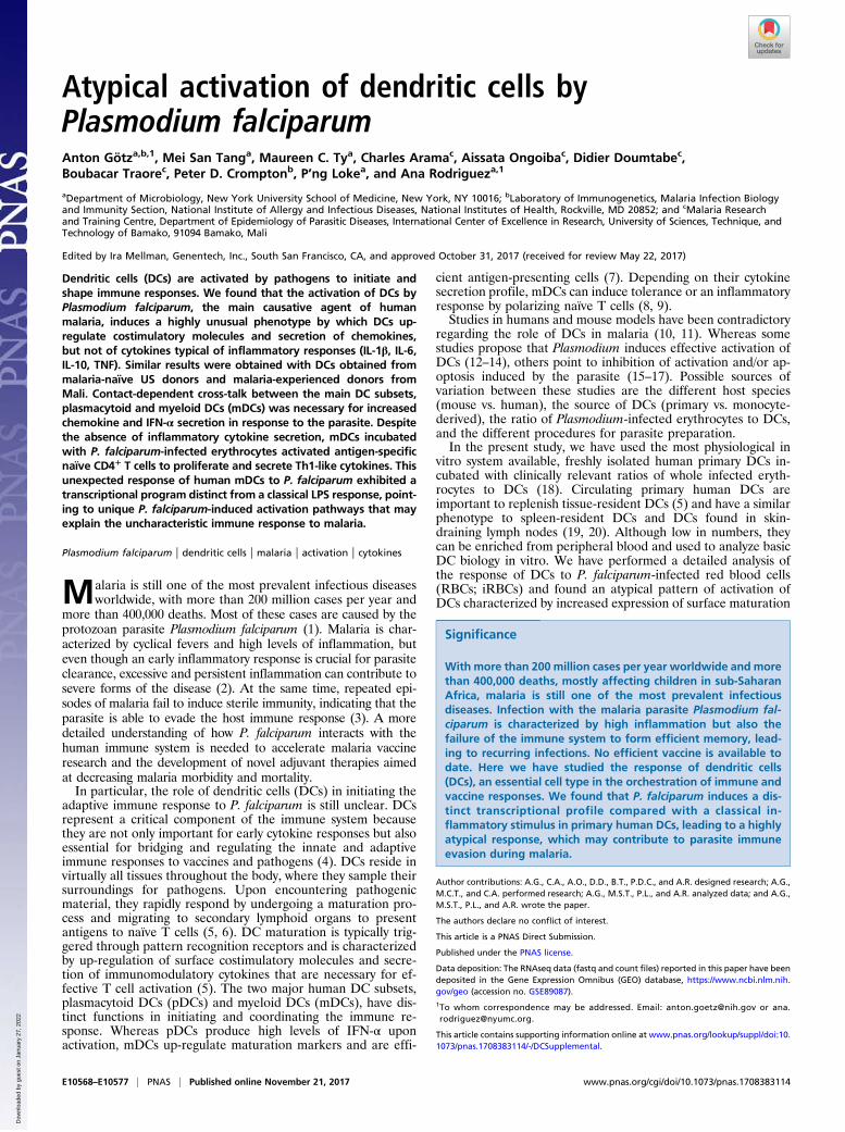
CCL2, CXCL9, and CXCL10, was also up-regulated after stim-ulation with iRBCs (Fig. 1F). However, iRBCs did not inducesecretion of cytokines typical of inflammatory responses (IL-1β,IL-6, IL-10, and TNF) that are induced by LPS (Fig. 1G). Thepercentage of apoptotic and dead DCs in the culture was com-parable to LPS control after 24 h incubation with iRBCs (SI
Appendix, Fig. S1). IL-12p70 was not detected in the supernatantof DCs after stimulation with iRBCs or LPS.To determine whether the lack of cytokine secretion could be
overcome by increased concentrations of iRBCs, we incubatedDCs with ratios as high as 1:30 DC:iRBC, but still did not ob-serve a notable cytokine response (Fig. 2A). We also determinedthat the DC response observed could not be interpreted as a low-intensity response induced by low concentrations of stimulus. Atitration of LPS concentrations showed that lower concentra-tions do not induce a pattern similar to iRBCs (SI Appendix, Fig.S2 A–C). Because molecules responsible for innate immune ac-tivation are found inside the iRBCs and are released upon egressduring late stages (23, 24), we also tested the capacity of iRBClysates to induce cytokine secretion in DCs. Although iRBC ly-sates induced the secretion of chemokines similarly to intactiRBCs, they did not induce considerable cytokine secretion (SIAppendix, Fig. S2 D and E). We also observed that, in contrast tohuman primary DCs, monocyte-derived DCs showed a completelack of maturation in response to iRBCs, that is, lack of cos-timulatory marker, cytokine, and chemokine up-regulation (SIAppendix, Fig. S3).P. falciparum-iRBCs in human patients express protein adhe-
sion knobs on their surface that mediate interactions with hostendothelial cells; however, these knobs are frequently lost duringin vitro culture (25). We next used gelatin flotation to selectiRBCs with knobs (26) and compared their capacity for stimula-tion of DCs with iRBCs lacking knobs. We observed that knobexpression did not increase cytokine secretion in DCs (Fig. 2B).Taken together, these results point to an atypical activation ofhuman primary DCs in response to P. falciparum-iRBCs, as clas-sical maturation of DCs typically involves up-regulation of surfacemarkers, as well as cytokine and chemokine secretion (27).To determine whether iRBCs can inhibit the capacity of pri-
mary DCs to mature, LPS was added to the cocultures of DCsand iRBCs after 24 h. DCs could still secrete cytokines in re-sponse to LPS (Fig. 2C) despite exposure to iRBCs, indicating
Fig. 1. DCs phagocytose P. falciparum-iRBCs and up-regulate maturation markers and secretion of chemokines, but not cytokines. DCs were incubated withlate-stage P. falciparum-iRBCs or uninfected RBCs at a ratio of 1:3 [DC:(i)RBC] for 24 h (A and D–G) or 1:3 [DC:carboxy-fluorescein succinimidyl ester (CFSE)-labeled (i)RBC] for 3 h (B and C) and analyzed for surface marker expression (A, D, and E), chemokine (F) and cytokine (G) secretion, or phagocytosis by FACS(B) and immunofluorescence microscopy (C). (A) DCs enriched by negative selection followed by positive selection for HLA-DR with magnetic beads fromPBMCs were gated first by using forward scatter (FSC) and SSC, followed by selection of HLA-DR+ cells, and used for further analysis in B and D–G. (B and C)Phagocytosis of iRBCs is observed as CFSE-positive DCs. Data from one representative experiment of three are shown. An example of surface marker ex-pression for one donor (D) and the analysis of seven (E and F) and eight (G) donors are shown (E–G), with each symbol representing results from one individualdonor and experiment (*P < 0.05, **P < 0.01, and ***P < 0.001 by Friedman test vs. RBCs or control; line depicts grand mean).
Götz et al. PNAS | Published online November 21, 2017 | E10569
IMMUNOLO
GYAND
INFLAMMATION
PNASPL
US
Dow
nloa
ded
by g
uest
on
Janu
ary
27, 2
022
that DC cytokine secretion in response to LPS was not perma-nently inhibited upon parasite encounter.Real-time PCR (RT-PCR) analysis of a panel of mRNAs re-
lated to DC activation confirmed our initial results showing in-creased expression of maturation markers and chemokines/chemokine receptors, but not cytokines (Fig. 2D), in response toiRBCs compared with uninfected RBCs.
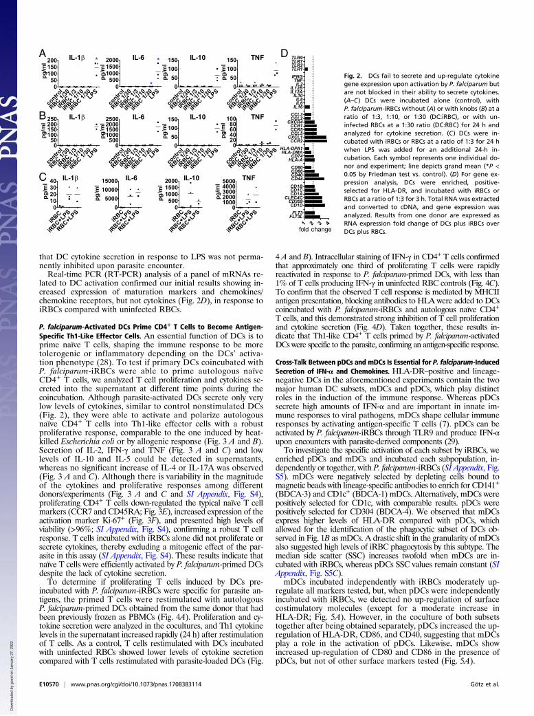
P. falciparum-Activated DCs Prime CD4+ T Cells to Become Antigen-Specific Th1-Like Effector Cells. An essential function of DCs is toprime naïve T cells, shaping the immune response to be moretolerogenic or inflammatory depending on the DCs’ activa-tion phenotype (28). To test if primary DCs coincubated withP. falciparum-iRBCs were able to prime autologous naïveCD4+ T cells, we analyzed T cell proliferation and cytokines se-creted into the supernatant at different time points during thecoincubation. Although parasite-activated DCs secrete only verylow levels of cytokines, similar to control nonstimulated DCs(Fig. 2), they were able to activate and polarize autologousnaïve CD4+ T cells into Th1-like effector cells with a robustproliferative response, comparable to the one induced by heat-killed Escherichia coli or by allogenic response (Fig. 3 A and B).Secretion of IL-2, IFN-γ and TNF (Fig. 3 A and C) and lowlevels of IL-10 and IL-5 could be detected in supernatants,whereas no significant increase of IL-4 or IL-17A was observed(Fig. 3 A and C). Although there is variability in the magnitudeof the cytokines and proliferative responses among differentdonors/experiments (Fig. 3 A and C and SI Appendix, Fig. S4),proliferating CD4+ T cells down-regulated the typical naïve T cellmarkers (CCR7 and CD45RA; Fig. 3E), increased expression of theactivation marker Ki-67+ (Fig. 3F), and presented high levels ofviability (>96%; SI Appendix, Fig. S4), confirming a robust T cellresponse. T cells incubated with iRBCs alone did not proliferate orsecrete cytokines, thereby excluding a mitogenic effect of the par-asite in this assay (SI Appendix, Fig. S4). These results indicate thatnaïve T cells were efficiently activated by P. falciparum-primed DCsdespite the lack of cytokine secretion.To determine if proliferating T cells induced by DCs pre-
incubated with P. falciparum-iRBCs were specific for parasite an-tigens, the primed T cells were restimulated with autologousP. falciparum-primed DCs obtained from the same donor that hadbeen previously frozen as PBMCs (Fig. 4A). Proliferation and cy-tokine secretion were analyzed in the cocultures, and Th1 cytokinelevels in the supernatant increased rapidly (24 h) after restimulationof T cells. As a control, T cells restimulated with DCs incubatedwith uninfected RBCs showed lower levels of cytokine secretioncompared with T cells restimulated with parasite-loaded DCs (Fig.
4 A and B). Intracellular staining of IFN-γ in CD4+ T cells confirmedthat approximately one third of proliferating T cells were rapidlyreactivated in response to P. falciparum-primed DCs, with less than1% of T cells producing IFN-γ in uninfected RBC controls (Fig. 4C).To confirm that the observed T cell response is mediated by MHCIIantigen presentation, blocking antibodies to HLA were added to DCscoincubated with P. falciparum-iRBCs and autologous naïve CD4+
T cells, and this demonstrated strong inhibition of T cell proliferationand cytokine secretion (Fig. 4D). Taken together, these results in-dicate that Th1-like CD4+ T cells primed by P. falciparum-activatedDCs were specific to the parasite, confirming an antigen-specific response.
Cross-Talk Between pDCs and mDCs Is Essential for P. falciparum-InducedSecretion of IFN-α and Chemokines. HLA-DR–positive and lineage-negative DCs in the aforementioned experiments contain the twomajor human DC subsets, mDCs and pDCs, which play distinctroles in the induction of the immune response. Whereas pDCssecrete high amounts of IFN-α and are important in innate im-mune responses to viral pathogens, mDCs shape cellular immuneresponses by activating antigen-specific T cells (7). pDCs can beactivated by P. falciparum-iRBCs through TLR9 and produce IFN-αupon encounters with parasite-derived components (29).To investigate the specific activation of each subset by iRBCs, we
enriched pDCs and mDCs and incubated each subpopulation, in-dependently or together, with P. falciparum-iRBCs (SI Appendix, Fig.S5). mDCs were negatively selected by depleting cells bound tomagnetic beads with lineage-specific antibodies to enrich for CD141+
(BDCA-3) and CD1c+ (BDCA-1) mDCs. Alternatively, mDCs werepositively selected for CD1c, with comparable results. pDCs werepositively selected for CD304 (BDCA-4). We observed that mDCsexpress higher levels of HLA-DR compared with pDCs, whichallowed for the identification of the phagocytic subset of DCs ob-served in Fig. 1B as mDCs. A drastic shift in the granularity of mDCsalso suggested high levels of iRBC phagocytosis by this subtype. Themedian side scatter (SSC) increases twofold when mDCs are in-cubated with iRBCs, whereas pDCs SSC values remain constant (SIAppendix, Fig. S5C).mDCs incubated independently with iRBCs moderately up-
regulate all markers tested, but, when pDCs were independentlyincubated with iRBCs, we detected no up-regulation of surfacecostimulatory molecules (except for a moderate increase inHLA-DR; Fig. 5A). However, in the coculture of both subsetstogether after being obtained separately, pDCs increased the up-regulation of HLA-DR, CD86, and CD40, suggesting that mDCsplay a role in the activation of pDCs. Likewise, mDCs showincreased up-regulation of CD80 and CD86 in the presence ofpDCs, but not of other surface markers tested (Fig. 5A).
A D
B
C
Fig. 2. DCs fail to secrete and up-regulate cytokinegene expression upon activation by P. falciparum butare not blocked in their ability to secrete cytokines.(A–C) DCs were incubated alone (control), withP. falciparum-iRBCs without (A) or with knobs (B) at aratio of 1:3, 1:10, or 1:30 (DC:iRBC), or with un-infected RBCs at a 1:30 ratio (DC:RBC) for 24 h andanalyzed for cytokine secretion. (C) DCs were in-cubated with iRBCs or RBCs at a ratio of 1:3 for 24 hwhen LPS was added for an additional 24-h in-cubation. Each symbol represents one individual do-nor and experiment; line depicts grand mean (*P <0.05 by Friedman test vs. control). (D) For gene ex-pression analysis, DCs were enriched, positive-selected for HLA-DR, and incubated with iRBCs orRBCs at a ratio of 1:3 for 3 h. Total RNA was extractedand converted to cDNA, and gene expression wasanalyzed. Results from one donor are expressed asRNA expression fold change of DCs plus iRBCs overDCs plus RBCs.
E10570 | www.pnas.org/cgi/doi/10.1073/pnas.1708383114 Götz et al.
Dow
nloa
ded
by g
uest
on
Janu
ary
27, 2
022

Analysis of IL-1β, IL-6, IL-10, and TNF in supernatants ofmDCs and pDCs cultured independently or together showed nosecretion of these factors in response to iRBCs (SI Appendix, Fig.S5D). In contrast, IFN-α that was secreted at low levels by pDCswas greatly up-regulated when mDCs were present in the cultures(Fig. 5B). Similar results were obtained for the chemokinesCXCL9 and CXCL10, with cocultures of mDCs and pDCsresulting in increased secretion of these factors (Fig. 5B), in-dicating that both subsets of DCs are required for these responses.These findings indicate that cross-talk between pDCs and mDCs isrequired for the efficient up-regulation of IFN-α and specificchemokine secretion upon stimulation with P. falciparum.To study whether the cross-talk between the two subpopulations
of DCs required direct contact between the cells, we separated eachsubpopulation by a semipermeable membrane that allows the flowof soluble factors, but not of cells. Under these conditions, weobserved that, although expression levels of surface costimulatorymolecules were similar, the secretion of CXCL9 and especiallyCXCL10 was greatly reduced (Fig. 5C), suggesting that cell-to-cellcontact is necessary to induce specific chemokine secretion.In human DC subsets, unlike in mice, only pDCs express TLR9
(30). Because pDCs can be activated by P. falciparum-iRBCsthrough TLR9 (29), we specifically inhibited the activation of thisreceptor with an antagonistic CpG oligodeoxynucleotide in pDC/mDC cocultures. A significant decrease of CXCL9 secretion wasobserved, suggesting that activation of pDCs through TLR9 con-
tributes to the chemokine response. To inhibit intracellular TLRsignaling more broadly, we used chloroquine, resulting in decreasedchemokine levels in the supernatants (Fig. 5D).
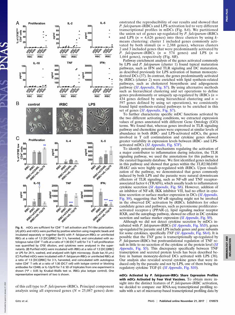
P. falciparum-Activated mDCs Are Sufficient to Prime Naïve CD4+ TCells to Become Th1-Like Effector Cells. To further characterize therole of DC subpopulation cross-talk, we analyzed its effects onT cell activation. We found that P. falciparum-activated mDCs,but not pDCs, efficiently drive proliferation and cytokine se-cretion in CD4+ T cells (Fig. 6A).As mDCs alone are able to effectively induce CD4+ T cell ac-
tivation, we focused next on these cells to further dissect the spe-cific requirements for P. falciparum-induced T cell activation. Wehad observed that mDCs alone do not secrete significant levels ofinflammatory cytokines (SI Appendix, Fig. S5) or the CXCR3-attractant chemokines CXCL9 and -10 in response to iRBCs(Fig. 5B); however, these cells respond by up-regulating cos-timulatory surface markers (Fig. 5A) and secreting CCL2 (Fig. 5B).First, we observed that the morphology of mDCs did not
change after incubation with iRBCs. Whereas mDCs activated byLPS were spread and exhibited typical protrusions, mDCs in-cubated with P. falciparum-iRBCs maintained a more roundedmorphology typical of immature DCs (Fig. 6B). In addition tothe lack of secretion of the main inflammatory cytokines bymDCs (SI Appendix, Fig. S5C), this finding further corroboratesthe atypical activation of mDCs by P. falciparum.
Fig. 3. P. falciparum-activated DCs prime autologous CD4+ T cells to become Th1-like effector cells in vitro. DCs were incubated with P. falciparum-iRBCs oruninfected RBCs at a ratio of 1:3 [DC:(i)RBC] for 3 h, harvested, and coincubated with autologous naïve CD4+ T cells at a ratio of 1:30 (DC:T cell). T cellproliferation was quantified by CFSE dilution, and cytokines were analyzed in the supernatants. (A) As a representative example, proliferation and cytokinelevels are shown for days 5, 7, and 11 for one individual donor. Mean with SD is shown for duplicates and triplicates. (B) DCs were additionally incubated withuninfected RBCs plus LPS, infected RBCs plus LPS, and heat-killed E. coli at a ratio of 1:10 (DC:E. coli) or allogenic DCs. Cytokine levels (levels below detectionlimit were set to the detection limit; C) and proliferation (D) of different donors and experiments are shown at day 11 (except for IL-2, shown at day 5).(E) Naïve T cell marker expression (CCR7 and CD45RA) was analyzed in T cells primed by P. falciparum-activated DCs at day 11. (F) DCs were incubated withinfected RBCs, infected RBCs plus LPS, and heat-killed E. coli at a ratio of 1:10 (DC:E. coli) or allogenic DCs, and CFSElow T cells were stained for the proliferationmarker Ki-67 at day 11. Each symbol represents one individual experiment from at least four different donors; line depicts grand mean (C–E). *P < 0.05 byWilcoxon test (C and D); *P < 0.05 by Student’s t test (E). Results from one donor and experiment are shown at day 11 in triplicates (except for allogenic; *P <0.05 and **P < 0.01 by one-way ANOVA vs. iRBCs in B and F).
Götz et al. PNAS | Published online November 21, 2017 | E10571
IMMUNOLO
GYAND
INFLAMMATION
PNASPL
US
Dow
nloa
ded
by g
uest
on
Janu
ary
27, 2
022
CD4+ T cell activation by mDCs in response to iRBCs wasfurther characterized with a panel of monoclonal antibodiesspecifically inhibiting essential components required for thisactivation. Blocking of CD40L resulted in inhibition of the se-cretion of typical Th1 cytokines (IFN-γ and TNF), but not T cellproliferation. Although we were not able to detect IL-12p70 insupernatants, blocking of IL-12p70 also led to lower IFN-γ butdid not affect TNF secretion (Fig. 6C), suggesting that levelsbelow the detection limit of this cytokine might promote Th1polarization in our system.Taken together, our findings indicate that the atypical activa-
tion of DCs by P. falciparum still leads to a strong Th1-like re-sponse in naïve CD4+ T cells, despite DCs immature morphologyand lack of IL-1β, IL-6, IL-10, and TNF cytokine production.
mDCs Respond Similarly to P. falciparum-iRBCs in Malaria-Naïve andMalaria-Experienced Adults. Adults living in areas of intensemalaria transmission are often infected with P. falciparum blood-stage parasites without symptoms (31, 32). This immunity, whichis gradually acquired over years of repeated infections, is knownto be antibody-mediated (33), but may also involve immuneregulation that limits parasitemia and inflammation (34, 35). Toinvestigate whether repeated malaria infections influence mDC
responses relative to responses observed in malaria-naïve do-nors, we analyzed mDCs from 11 adults residing in a rural villagein Mali with lifelong exposure to intense P. falciparum trans-mission (36). The donors were PCR-negative for Plasmodium atthe time of the blood draw, which occurred at the end of themalaria season. Freshly purified PBMCs were used to enrich formDCs, which were then incubated with RBCs or iRBCs. It hasbeen reported that spontaneous apoptosis is increased in DCs ofP. falciparum-infected individuals (16, 17). Although unexpectedin the uninfected subjects in this study, we investigated DC ap-optosis and subset percentages in freshly isolated PBMCs (SIAppendix, Fig. S6B). The subset percentages were similar tothose in healthy US adults and, except for two donors, DCsshowed very low annexin V binding. Similar to what we observedin malaria-naïve US donors (Fig. 5), mDCs from Malian donorsshowed moderately increased surface maturation marker ex-pression (Fig. 7A) and chemokine secretion (SI Appendix, Fig.S6C), but did not secrete significant amounts of cytokines (Fig.7B) when incubated with the parasite.
RNA Sequencing Analysis of mDC After Incubation with P. falciparum-iRBCs Reveals a Distinct Transcriptional Profile.A detailed analysis ofthe transcriptional profiles in mDCs, the DC subtype re-sponsible for T cell activation (Fig. 6A), was performed by usingRNA sequencing (RNA-seq) to better understand the response
A B
C D
Fig. 4. CD4+ T cells primed by P. falciparum are antigen-specific. DCs wereincubated with P. falciparum-iRBCs or uninfected RBCs at a ratio of 1:3 [DC:(i)RBC] for 3 h, harvested, and coincubated with autologous naïve CD4+
T cells at a ratio of 1:30 (DC:T cell). Approximately 11 d after the first stim-ulation, the primed T cells were stained with CFSE and restimulated withautologous DCs incubated with iRBCs or uninfected RBCs as depicted in A.T cell proliferation was quantified after 2–3 d, and cytokines were analyzedin the supernatants after 24 h (B). Each symbol represents one individualdonor and experiment; the line depicts the grand mean. Intracellular cyto-kine staining is shown for IFN-γ after 24 h of restimulation. One represen-tative experiment of two is shown (C). Inhibitory anti-MHCII antibodies (1,anti–HLA-DR/DP/DQ, clone Tü39; 2, anti-HLA-DR, clone L243, 25 μg/mL) wereadded to the coculture every second day, and proliferation and cytokineswere analyzed at day 11. One representative experiment of two is shown.Mean with SD is shown for triplicates (*P < 0.05, **P < 0.01, and ***P <0.001 by Kruskal–Wallis test for IFN-γ and by one-way ANOVA for all othersvs. iRBCs plus isotype control; D).
A
B
C
D
Fig. 5. Contact-mediated cross-talk between pDCs and mDCs is essential forP. falciparum-induced secretion of IFN-α and chemokines. Freshly purifiedpDCs and mDCs were incubated separately or together with P. falciparum-iRBCs or uninfected RBCs at a ratio of 1:3 [DC:(i)RBC] for 24 h and analyzedfor surface marker expression (A), IFN-α (B), and chemokine (C and D) se-cretion. For contact-independent coculture, each subtype was seeded withiRBCs or uninfected RBCs in the upper or lower compartment of a Transwellsystem, and supernatants were analyzed for chemokines. Contact-independent(−) and cocultures in contact (+) (C). A TLR9 antagonist and chloroquine wereused to inhibit TLR9 or intracellular TLR signaling, respectively (D). Each symbolrepresents results from one individual donor and experiment. Line depictsgrand mean [*P < 0.05 by Friedman test vs. RBC or control (B) or *P <0.05 and ***P < 0.001 by Student’s t test vs. iRBCs (D)].
E10572 | www.pnas.org/cgi/doi/10.1073/pnas.1708383114 Götz et al.
Dow
nloa
ded
by g
uest
on
Janu
ary
27, 2
022
of this cell type to P. falciparum-iRBCs. Principal componentanalysis using all expressed genes (N = 25,087 genes) dem-
onstrated the reproducibility of our results and showed thatP. falciparum-iRBCs and LPS activation led to very differenttranscriptional profiles in mDCs (Fig. 8A). We partitionedthe union set of genes up-regulated by P. falciparum-iRBCsand LPS (n = 4,626 genes) into three clusters by using k-means clustering: cluster 1 included genes commonly acti-vated by both stimuli (n = 2,388 genes), whereas clusters2 and 3 included genes that were predominantly activated byP. falciparum-iRBCs (n = 574 genes) and LPS (n =1,664 genes), respectively (Fig. 8B).Pathway enrichment analysis of the genes activated commonly
by LPS and P. falciparum (cluster 1) found typical maturationpathways, such as IFN and TLR signaling and DC maturation,as described previously for LPS activation of human monocyte-derived DCs (37). In contrast, the genes predominantly activatedby iRBCs (cluster 2) were enriched with lipid synthesis-relatedpathways, such as cholesterol biosynthesis and adipogenesispathway (SI Appendix, Fig. S7). By using alternative methodssuch as hierarchical clustering and set operations to definegenes predominantly or uniquely up-regulated by iRBCs (n =434 genes defined by using hierarchical clustering and n =597 genes defined by using set operations), we consistentlyfound lipid synthesis-related pathways to be enriched in thisset of genes (SI Appendix, Fig. S7).To further characterize specific mDC functions activated by
the two different activating conditions, we extracted expressionvalues of genes annotated with different Gene Ontology (GO)terms. We found that, whereas genes involved in TLR signalingpathway and chemokine genes were expressed at similar levels ofabundance in both iRBC- and LPS-activated mDCs, the genesinvolved in T cell costimulation and cytokine genes showedgreater variability in expression levels between iRBC- and LPS-activated mDCs (SI Appendix, Fig. S7F).To identify potential mechanisms regulating the activation of
a major contributor to inflammation during infection, the TLRsignaling pathway, we used the annotations for this pathway inthe curated Ingenuity database. We first identified genes includedin this pathway and showed that genes within the TLR2/MyD88/IRAK1 axis were highly up-regulated with iRBCs. Upon visuali-zation of the pathway, we demonstrated that genes commonlyinduced by both LPS and the parasite were natural downstreameffectors of TLR signaling, such as NF-κB and TNF receptor-associated factor 6 (TRAF6), which usually leads to inflammatorycytokine secretion (SI Appendix, Fig. S8). However, addition ofan inhibitor of NF-κB, IKK inhibitor VII, had no effect in cyto-kine secretion or surface marker expression in DCs (SI Appendix,Fig. S9), suggesting that NF-κB signaling might not be involvedin the observed DC activation by iRBCs. Inhibitors for othercandidate genes and pathways, such as peroxisome proliferator-activated receptor-γ (PPAR-γ), lipid signaling nuclear receptorRXR, and the autophagy pathway, showed no effect in DC cytokinesecretion and surface marker expression (SI Appendix, Fig. S9).Although we did not detect cytokine secretion by mDCs in-
cubated with P. falciparum-iRBCs (SI Appendix, Fig. S5D), genesup-regulated by parasite and LPS include genes and gene subunitsfor some cytokines, specifically TNF (SI Appendix, Fig. S8A). It ispossible that the TNF gene is transcriptionally up-regulated byP. falciparum-iRBCs but posttranslational regulation of TNF re-sult in little to no secretion of the cytokine at the protein level (SIAppendix, Fig. S5). This discrepancy specifically between TNFtranscription and secreted protein levels has been described be-fore in human monocyte-derived DCs activated with LPS (38).Our analysis also revealed several cytokine genes that were in-duced only by the parasite and not by LPS, one of them being theregulatory cytokine TGF-β1 (SI Appendix, Fig. S10).
mDCs Activated by P. falciparum-iRBCs Share Expression Profileswith mDCs Activated by Four Viral Vaccines. To obtain more in-sight into the distinct features of P. falciparum-iRBC activation,we decided to compare our RNA-seq transcriptional profiling re-sults with a set of microarray-based transcriptional profiling data by
Fig. 6. mDCs are sufficient for CD4+ T cell activation and Th1-like polarization.(A) pDCs and mDCs were purified by positive selection using magnetic beads andincubated separately or together (both) with P. falciparum-iRBCs or uninfectedRBCs at a ratio of 1:3 [DC:(i)RBC] for 3 h, harvested, and coincubated with au-tologous naïve CD4+ T cells at a ratio of 1:30 (DC:T cell) for 7 d. T cell proliferationwas quantified by CFSE dilution, and cytokines were analyzed in the super-natants. (B) Purified mDCs were incubated with iRBCs at a ratio of 1:3 [DC:(i)RBC]or LPS for 24 h, washed, and analyzed with light microscopy. (Scale bar,10 μm.)(C) Purified mDCs were incubated with P. falciparum-iRBCs or uninfected RBCs ata ratio of 1:3 [DC:(i)RBC] for 3 h, harvested, and coincubated with autologousnaïve CD4+ T cells at a ratio of 1:30 (DC:T cell) with isotype control or blockingantibodies for CD40L or IL-12p70 for 7 d. SD of triplicates from one experiment isshown (*P < 0.05 by Kruskal–Wallis test vs. iRBCs plus isotype control). Onerepresentative experiment of two is shown.
Götz et al. PNAS | Published online November 21, 2017 | E10573
IMMUNOLO
GYAND
INFLAMMATION
PNASPL
US
Dow
nloa
ded
by g
uest
on
Janu
ary
27, 2
022

Banchereau et al. (39), in which primary human CD1c+ DCs werestimulated with 13 different microbial vaccines, including inactivatedbacterial, inactivated viral, and live attenuated viral vaccines. By usinga similar strategy, we determined the “percentage expression” valuefor each gene of the gene cluster from our data set under each vaccinestimulation condition. We found the iRBC-specific gene cluster to bemost highly expressed in CD1c+ DCs stimulated with Ixiaro andHavrix, which are vaccines based on inactivated Japanese encephalitisand hepatitis A viruses, respectively; Gardasil, based on human pap-illoma virus-like particles; and Engerix-B, based on recombinantprotein hepatitis B surface antigen (Fig. 9A). This indicates that theactivation state of CD1c+ DCs stimulated with P. falciparum-iRBCsshares similarities with the activation state of CD1c+ DCs activated bythese four vaccines, suggesting common regulatory mechanisms inhow P. falciparum and these four vaccines activate CD1c+ DCs. Thecluster containing genes commonly up-regulated by LPS and iRBCswas most highly expressed with stimulation by Menomune andActHib, which are both LPS-containing vaccines (Fig. 9A).The similarities in activation profiles between iRBCs and the
four viral vaccines were contributed by 158 iRBC-specific genes,whereas the remaining iRBC-specific genes were largely un-changed across the data set (Fig. 9B). Most of these genes wereup-regulated upon stimulation with the vaccines (n = 132 genes),but a small subset of these iRBC-specific genes was down-regulated instead (n = 26 genes; SI Appendix, Fig. S11A). Thegenes commonly up-regulated by iRBCs and the vaccines areassociated with different metabolic pathways, such as L-serineand N-acetylglucosamine degradation, adipogenesis, and tri-acylglycerol degradation (SI Appendix, Fig. S11B). Additionally,the expression of several genes inferred to be regulated byTREM-1 (using the upstream regulator analysis in IngenuityPathway Analysis) in DCs stimulated with iRBCs, Ixiaro, Gardasil,Havrix, and Engerix-B suggests a shared pathway in DC activation (SIAppendix, Fig. S11 C and D and Table S1). Another set ofgenes with similar expression profiles during activation byiRBCs and the four vaccines were genes associated with mu-rine models of “alternatively activated macrophages.” Theseincluded ALDH1A2 (an enzyme that can produce retinoic acid),PPAR-γ, and EGR2 (40–42).We also compared the capabilities of the Ixiaro, Gardasil,
Havrix, and Engerix-B vaccines to produce cytokines by exam-ining the expression levels of genes annotated with the GO term“cytokine activity” when stimulated by each of these vaccines inan independent analysis. Although the four vaccines do not in-duce the expression of most cytokines, a small subset of cytokineswas transcriptionally induced (SI Appendix, Fig. S12A). Some of
these cytokines were also uniquely expressed during iRBC acti-vation, which include OSM, TNFSF14, and GREM1 (SI Ap-pendix, Fig. S10). This further suggests a similar DC activationstate by these vaccines and iRBCs.
DiscussionOur results indicate that human primary DCs undergo a dis-tinctive activation in response to P. falciparum-iRBCs, whichincludes up-regulation of HLA-DR and costimulatory moleculesand chemokines, but not secretion of significant amounts of cy-tokines. Previous studies had shown a direct inhibition of humanmonocyte-derived DC maturation induced by the parasite at highconcentrations (15) and a lack of typical maturation featuressuch as up-regulation of HLA-DR and costimulatory moleculesat lower concentrations (14). This discrepancy with the resultspresented here is most likely explained by the origin of DCs, asprevious studies had used monocyte-derived DCs, which we alsofound to lack up-regulation of HLA-DR and costimulatorymolecules at similar concentrations of parasite. Although work-ing with human primary DCs is an experimental hurdle becauseof their low frequency in peripheral blood, these results highlightthe importance of using primary cells in these studies.The low inflammatory cytokine response observed in DCs in
response to P. falciparum contrasts strongly with the high levelsof inflammation observed in patients with malaria (43). This lowinflammatory cytokine response to the parasite in vitro haspreviously been noticed in several in vitro studies that analyzedthe response of mice and human macrophages to Plasmodium-infected erythrocytes (44, 45) and suggests that additional in-flammatory stimuli may be generated by the host upon infection,which may explain the extreme differences in disease tolerancethat are observed in malaria-infected individuals (46). It is alsopossible that the lack of an inflammatory cytokine response fromindividual cell types, such as shown in the present work for DCs,or in macrophages (44, 45), is overcome in the presence of otherimmune cells, as cytokine responses tended to be higher whencultures of PBMCs were stimulated with iRBCs (47).The observed lack of an inflammatory cytokine response to
iRBCs is also unusual in the context of DC activation, in which,typically, HLA-DR and costimulatory molecules, chemokines,and cytokines are simultaneously up-regulated in response to astimulus (5). Similar to our results, a functionally distinct pathway of
A B
Fig. 8. CD1c+ mDCs show distinct gene expression upon stimulation withP. falciparum-iRBCs. CD1c+ mDCs were purified by positive selection usingmagnetic beads and incubated with P. falciparum-iRBCs or uninfected RBCsat a ratio of 1:3 [DC:(i)RBC] for 6 h before RNA extraction and whole-genomeRNA-seq of two individual donors and experiments. (A) Principal componentanalysis of all 25,087 genes showed that DCs stimulated with LPS or iRBCshave different gene expression profiles. Scatter diagram of the first twoprincipal components is shown. (B) K-means clustering partitioning theunion set of 4,626 up-regulated genes from LPS or iRBCs stimulations intothree distinct sets of genes. Cluster 1, genes commonly up-regulated by LPSand iRBCs; cluster 2, genes predominantly up-regulated by iRBCs; cluster 3,genes predominantly up-regulated by LPS.
Fig. 7. mDCs from individuals living in an endemic area up-regulatemarkers but fail to secrete cytokines upon stimulation with P. falciparum.mDCs were enriched from PBMCs from Malian adults and incubated withP. falciparum-iRBCs or uninfected RBC lysates at a ratio of 1:3 [DC:(i)RBC] for24 h and analyzed for surface marker expression (A) and cytokine secretion(B). Each symbol represents one individual donor and experiment. Line de-picts grand mean (*P < 0.05, **P < 0.01, and ***P < 0.001 by Friedman testvs. uninfected RBC control; n = 11).
E10574 | www.pnas.org/cgi/doi/10.1073/pnas.1708383114 Götz et al.
Dow
nloa
ded
by g
uest
on
Janu
ary
27, 2
022

DC activation has been described as a result of cluster disruption inhuman stem cell-derived DCs, which results in up-regulation ofHLA-DR and costimulatory molecules and certain chemokines, buta lack of secretion of inflammatory cytokines. However, these DCsalso induce a tolerogenic phenotype in T cells that is mediated byactivation of β-catenin in DCs (48, 49), which is not observed afterincubation of DCs with iRBCs. Ligation of CD47 also limits cyto-kine secretion by DCs, suggesting a possible mechanism, but it si-multaneously inhibits DC up-regulation of costimulatory moleculesand their capacity to stimulate T cell proliferation and IFN-γ pro-duction (50). We also identified cytokine genes specifically inducedonly by P. falciparum in DCs, i.e., not by LPS, with TGFB1 being aninteresting candidate that is secreted by DCs and suppresses liverstage CD8+ T cell responses in a malaria mouse model (51).A number of P. falciparum-derived molecules have been de-
scribed as TLR or inflammasome activators based on their ca-pacity to induce cytokine secretion in immune cells in vitro (23).In this context, it is intriguing that whole infected erythrocytes ortheir lysates do not induce potent cytokine responses in DCs invitro. It is possible that P. falciparum-infected RBCs also carryinhibitory molecules that could modulate the activation pheno-type of DCs. Indeed, we have observed that P. falciparum-iRBCsinduce increased transcription of genes, such as PPARA andPPARG, that are involved in the modulation of DC responses(52). Hemozoin has been shown to increase PPAR-γ expressionand inhibit DC maturation when human monocytes were loadedwith the pigment before differentiation (53). A possible mecha-nism for the suppression of cytokine responses by PPARs is theirinhibition of the transcription factor NF-κB (54), which regulatesinflammatory cytokine expression. However, inhibition of PPAR-γor NF-κB did not affect the DC cytokine secretion in response toiRBCs, suggesting that these factors may not be involved in theobserved low-cytokine DC response. Accordingly, it has beenshown that costimulatory marker up-regulation in the absence ofincreased cytokine secretion was independent of NF-κB in DCactivation (55). Costimulatory markers and chemokines can alsobe induced through IFN-α (56) or β-catenin signaling (48).We have observed that a large number of the specific signaling
pathways activated by P. falciparum are different from the onesinduced by a classical TLR activator such as LPS. We found ahigh number of lipid synthesis-related pathways, in particularcholesterol biosynthesis, which may contribute to the atypical acti-
vation phenotype of mDCs (57). In the list of pathways up-regulateduniquely by P. falciparum, we also found the TLR2 pathway that maybe induced by parasite glycosylphosphatidylinositol anchors, whichare known to activate TLR2 and induce an inflammatory response invitro (58) and in mice (59). TLR2 is also up-regulated in peripheralblood of patients with malaria (60), and a polymorphism in this geneis associated with protection from cerebral malaria (61), suggestingan important role for this receptor in the human response to malaria.Despite the low inflammatory cytokine secretion by DCs,
iRBC-primed DCs induced strong activation of T cells charac-terized by proliferation and secretion of Th1 cytokines such asIFN-γ and TNF. This response was found to be typically antigen-specific, as observed in the recall experiments, and mediated byCD40L and IL-12p70.In addition to the classical Th1 cytokines, T cells incubated
with iRBC-primed DCs also secrete IL-5 and IL-10, although itis unknown whether these cytokines are a reflection of differentsubtypes of T cells in the cultures or coexpression of multiplecytokines in a particular subtype. CD4+ T cells coproducing IL-10 and IFN-γ, or T regulatory-1 cells, correlate with exposure tomalaria and have been proposed to play a role in the modulationof immunity in vivo (35, 36, 63).It seems unusual that malaria-naïve donors present large
numbers of T cells specific for P. falciparum antigens. However,high frequencies of T cells specific for P. falciparum antigens innonexposed donors has been reported before (14, 64–66). Be-cause malaria is considered the strongest evolutionary-selectiveforce in recent human history (67), it is possible that having alarge number of T cells specific for this parasite may be underpositive selection.Distinct functional capabilities have been described for each of
the two main subtypes of DCs, pDCs and mDCs (7). Because thecharacteristics of these DC subpopulations are significantly dif-ferent in mice and humans (68), our analysis of the interactionsof primary DCs subtypes with iRBCs are particularly relevant. Asdescribed for other pathogens, mDCs are the subset that pre-dominantly phagocytoses iRBCs and the one solely responsiblefor the activation of T cells (7, 69). On the contrary, pDCs dif-ferentially secrete IFN-α, but not TNF, in response to iRBCs, aspreviously described (29), suggesting that P. falciparum activatesdifferent signaling pathways compared with classical activators ofpDCs that induce both IFN-α and TNF.Maturation of pDCs in response to bacterial stimulation or
TLR ligands requires cooperation from mDCs, whereby both cellcontact and soluble factors play essential roles (70, 71). On thecontrary, mDCs are not able to be activated by HIV but needbystander activation facilitated by the secretion of TNF and typeI IFNs by pDCs (72). We also observed that the presence ofmDCs increases the secretion of IFN-α and the up-regulation ofcostimulatory molecules by pDCs, which indicates a possible rolefor mDCs in the activation of pDCs. This is particularly relevantbecause recent studies have highlighted the importance of type IIFN in regulating the immune response to malaria (63, 73).Also, a significant increase in CXCL10 secretion that requires
direct contact between both subpopulations of DCs is observedin the cocultures. Because CXCL10 is a potent T cell recruiter,this probably leads to the recruitment of naïve T cells and ulti-mately heightens priming in vivo. A possible advantage for thiscooperation between pDCs and mDCs might be an increasedresponse even with inefficient activation of either one of thesubsets. mDCs are most likely not able to sense parasite DNAbecause of the lack of TLR9 expression, whereas pDCs do notrespond to TLR2 ligands as a result of the lack of expression ofthis receptor (70, 74). If parasites like Plasmodium evolved toescape the immune system by inhibiting DC activation, the cross-talk between pDCs being primed by parasite DNA throughTLR9 and mDCs being activated through TLR2 could still leadto a functional, although maybe not maximally efficient, re-sponse against the parasite. Indeed, CXCL10 has a detrimentaleffect on the development of immunity and promotes severe out-comes in murine (75, 76) and human (77) malaria. In agreement
A B
Fig. 9. The P. falciparum-iRBC–specific genes are most highly expressed inCD1c+ DCs activated with the vaccines Ixiaro, Gardasil, Havrix, and Engerix-B.(A) The iRBC-specific gene cluster is most highly induced with the vaccinesIxiaro, Gardasil, Havrix, and Engerix-B (names in red). Values shown here arepercentage expression, where a positive value indicates up-regulation and anegative value indicates down-regulation. (B) The high expression of theiRBC-specific gene cluster in the four vaccines is contributed by 158 genes(boxed in red). Values shown here represent statistical significance, as de-termined by the calculation of false discovery rate (FDR). Darker shades ofred represent a low FDR value and therefore statistical significance com-pared with expression levels under control medium.
Götz et al. PNAS | Published online November 21, 2017 | E10575
IMMUNOLO
GYAND
INFLAMMATION
PNASPL
US
Dow
nloa
ded
by g
uest
on
Janu
ary
27, 2
022

with our findings, a recent study investigating immune cell re-sponses in murine spleens during Plasmodium chabaudi infectionalso reported increased expression of CXCL9 and CXCL10 inclassical DCs but no increased cytokine gene expression whenanalyzing the transcriptomes of single DCs (78). Despite the dif-ferences between human and murine DC subsets, this suggests thata dominant chemokine response, specifically CXCL9 and -10,rather than inflammatory cytokines, might be a conserved responseto Plasmodium.To gain additional insight into possible pathways involved in DC
activation by P. falciparum-iRBCs, we compared our RNA-seqdata set with one of the only other high-quality transcriptionalprofiling experiments performed with primary human DCs thatwere incubated with different vaccines (39). We found that, al-though the majority of genes induced by the parasite were notdifferentially expressed in DCs stimulated with the different vac-cines, four viral vaccines share a subset of genes induced byP. falciparum-iRBCs. These four vaccines induce only low to in-termediate levels of DC maturation, while down-regulating CD40,confirming that crucial pathways in the response are not sharedbetween the vaccines and iRBCs. However, our analyses providedhints that certain pathways may be shared, including TREM-1 ac-tivation, as well as “alternative activation” pathways similar toM2 alternatively activated macrophages in mice. In malaria,TREM-1 has been described to have a role in severe diseaseoutcomes (79). We have also identified cytokines like OSM andTNFSF14 (LIGHT) that seem to be expressed in human primaryDC activation states that lack expression of typical inflammatorycytokines. Our findings provide insights into the complexity of DCactivation, showing that specific viral vaccines (which also actthrough unknown mechanisms) share distinctive features of iRBCactivation. The viral origin of the four vaccines together with thestrong induction of IFN-α secretion in pDCs by the parasite sug-gest an activation state similar to the one induced by viruses. In-terestingly, Zika virus activates primary human mDCs in a similarway as P. falciparum-iRBCs, namely the lack of cytokine secretionaccompanied by a relatively high CXCL9 and -10 response (80).These analyses highlight the complexity of DC response to dif-ferent microbial stimuli, suggesting that DC maturation should notbe cataloged as a yes-or-no response, but as a complex and nu-anced reaction that is tailored to each specific microbe.It is remarkable that a pathogen causing such a highly in-
flammatory disease as malaria fails to induce a potent cytokineresponse in DCs. The particular response of DCs we have ob-served may underlie some of the unconventional features of themalaria-adaptive immune response whereby DCs are expected tohave an important influence, such as the lack of sterilizing im-munity and its rapid decrease in the absence of exposure (81).
Materials and MethodsP. falciparum Culture and Isolation. Erythrocyte asexual stage cultures of theP. falciparum strain 3D7 were maintained as described in SI Appendix, SIMaterials and Methods.
US and Malian Donors. Peripheral venous blood from healthy donors in NewYork (United States) or Kambila (Mali) was obtained. Approval was obtainedfrom the institutional review board (IRB) at New York University School ofMedicine; the ethics committee of the Faculty of Medicine, Pharmacy andDentistry at the University of Sciences, Technique and Technology of Bamako;and theNational Institute ofAllergy and InfectiousDiseases/National Institutes ofHealth (NIH). Informed consent was obtained from each donor before donation.
DC Enrichment, Activation, and Phagocytosis. The different cell types wereenriched with magnetic beads. To analyze DC activation, DCs were culturedwith intact or lysed P. falciparum-iRBCs at a ratio of 1:3 (DC:iRBC) for 24 h.For phagocytosis, P. falciparum-infected RBCs were labeled and coincubatedwith the DCs at a ratio of 1:3 (DC:iRBC) for 3 h. Details are provided in SIAppendix, SI Materials and Methods.
Autologous CD4+ T Cell Activation and Restimulation. The naïve CD4+ T cellactivation was described before (82) and is detailed in SI Appendix, SI Ma-terials and Methods.
Cytokine and Chemokine Analysis. Cytokines and chemokines were quantifiedby using BD Cytometric Bead Arrays (BD Biosciences) and acquired on aFACSCalibur device (BD Biosciences).
Quantitative RT-PCR Array, RNA-Seq, and Comparison of Data with PublishedCD1c+ Vaccine Data Set. DCs were enriched with negative selection followedby HLA-DR positive selection and CD1c (BDCA-1)+ DC isolation kit, re-spectively. Cells were then incubated with P. falciparum-iRBCs at a ratio of1:3 (DC:iRBC). Analysis and comparison of RNA-seq data were performed asdetailed in SI Appendix, SI Materials and Methods.
Statistical Analysis. Statistical analysis is detailed in SI Appendix, SI Materialsand Methods.
ACKNOWLEDGMENTS. We thank the residents of Kambila, Mali, and thedonors at New York University for participating in this study; and Jeff Skinnerand Julio Gallego-Delgado for help with statistical analysis. New York Univer-sity School of Medicine Clinical and Translational Science Institute (CTSI) issupported by NIH Grant 3UL1 TR001445. The New York University School ofMedicine Genome Technology Center is supported by NIH Grant P30CA016087.Field studies in Mali are funded by the Division of Intramural Research, Na-tional Institutes of Allergy and Infectious Diseases, NIH. This work wassupported by a fellowship within the Postdoc-Programme of the GermanAcademic Exchange Service and the American Association of ImmunologyCareers in Immunology Fellowship Program (M.S.T.).
1. World Health Organization (2016) World Malaria Report 2016 (World Health Orga-
nization, Geneva).2. Storm J, Craig AG (2014) Pathogenesis of cerebral malaria–Inflammation and cy-
toadherence. Front Cell Infect Microbiol 4:100.3. Stanisic DI, Barry AE, Good MF (2013) Escaping the immune system: How the malaria
parasite makes vaccine development a challenge. Trends Parasitol 29:612–622.4. Banchereau J, Steinman RM (1998) Dendritic cells and the control of immunity. Nature
392:245–252.5. Banchereau J, et al. (2000) Immunobiology of dendritic cells. Annu Rev Immunol 18:767–811.6. Randolph GJ, Angeli V, Swartz MA (2005) Dendritic-cell trafficking to lymph nodes
through lymphatic vessels. Nat Rev Immunol 5:617–628.7. Collin M, McGovern N, Haniffa M (2013) Human dendritic cell subsets. Immunology
140:22–30.8. Lutz MB, Schuler G (2002) Immature, semi-mature and fully mature dendritic cells:
Which signals induce tolerance or immunity? Trends Immunol 23:445–449.9. van den Broek M (2007) Dendritic cells break bonds to tolerize. Immunity 27:544–546.10. Cockburn IA, Zavala F (2016) Dendritic cell function and antigen presentation in
malaria. Curr Opin Immunol 40:1–6.11. Wykes MN, Good MF (2008) What really happens to dendritic cells during malaria?
Nat Rev Microbiol 6:864–870.12. Perry JA, Rush A, Wilson RJ, Olver CS, Avery AC (2004) Dendritic cells from malaria-
infected mice are fully functional APC. J Immunol 172:475–482.13. Pouniotis DS, et al. (2004) Dendritic cells induce immunity and long-lasting protection
against blood-stage malaria despite an in vitro parasite-induced maturation defect.
Infect Immun 72:5331–5339.
14. Elliott SR, et al. (2007) Inhibition of dendritic cell maturation by malaria is dose de-
pendent and does not require Plasmodium falciparum erythrocyte membrane protein
1. Infect Immun 75:3621–3632.15. Urban BC, et al. (1999) Plasmodium falciparum-infected erythrocytes modulate the
maturation of dendritic cells. Nature 400:73–77.16. Pinzon-Charry A, et al. (2013) Apoptosis and dysfunction of blood dendritic cells in
patients with falciparum and vivax malaria. J Exp Med 210:1635–1646.17. Woodberry T, et al. (2012) Low-level Plasmodium falciparum blood-stage infection
causes dendritic cell apoptosis and dysfunction in healthy volunteers. J Infect Dis 206:
333–340.18. Scholzen A, Mittag D, Rogerson SJ, Cooke BM, Plebanski M (2009) Plasmodium
falciparum-mediated induction of human CD25Foxp3 CD4 T cells is independent of
direct TCR stimulation and requires IL-2, IL-10 and TGFbeta. PLoS Pathog 5:e1000543.19. Mittag D, et al. (2011) Human dendritic cell subsets from spleen and blood are similar
in phenotype and function but modified by donor health status. J Immunol 186:
6207–6217.20. Segura E, et al. (2012) Characterization of resident and migratory dendritic cells in
human lymph nodes. J Exp Med 209:653–660.21. Lundberg K, et al. (2013) Transcriptional profiling of human dendritic cell populations
and models–Unique profiles of in vitro dendritic cells and implications on function-
ality and applicability. PLoS One 8:e52875.
22. Robbins SH, et al. (2008) Novel insights into the relationships between dendritic cell
subsets in human and mouse revealed by genome-wide expression profiling. Genome
Biol 9:R17.
E10576 | www.pnas.org/cgi/doi/10.1073/pnas.1708383114 Götz et al.
Dow
nloa
ded
by g
uest
on
Janu
ary
27, 2
022
23. Gazzinelli RT, Kalantari P, Fitzgerald KA, Golenbock DT (2014) Innate sensing ofmalaria parasites. Nat Rev Immunol 14:744–757.
24. Ndungu FM, Urban BC, Marsh K, Langhorne J (2005) Regulation of immune responseby Plasmodium-infected red blood cells. Parasite Immunol 27:373–384.
25. Langreth SG, Reese RT, Motyl MR, Trager W (1979) Plasmodium falciparum: Loss ofknobs on the infected erythrocyte surface after long-term cultivation. Exp Parasitol48:213–219.
26. Goodyer ID, Johnson J, Eisenthal R, Hayes DJ (1994) Purification of mature-stagePlasmodium falciparum by gelatine flotation. Ann Trop Med Parasitol 88:209–211.
27. Blanco YC, et al. (2008) Hyperbaric oxygen prevents early death caused by experi-mental cerebral malaria. PLoS One 3:e3126.
28. Swafford D, Manicassamy S (2015) Wnt signaling in dendritic cells: Its role in regu-lation of immunity and tolerance. Discov Med 19:303–310.
29. Pichyangkul S, et al. (2004) Malaria blood stage parasites activate human plasmacy-toid dendritic cells and murine dendritic cells through a Toll-like receptor 9-de-pendent pathway. J Immunol 172:4926–4933.
30. Kastenmüller W, Kastenmüller K, Kurts C, Seder RA (2014) Dendritic cell-targetedvaccines–hope or hype? Nat Rev Immunol 14:705–711.
31. Langhorne J, Ndungu FM, Sponaas AM, Marsh K (2008) Immunity to malaria: Morequestions than answers. Nat Immunol 9:725–732.
32. Tran TM, et al. (2013) An intensive longitudinal cohort study of Malian children andadults reveals no evidence of acquired immunity to Plasmodium falciparum infection.Clin Infect Dis 57:40–47.
33. Cohen S, McGregor IA, Carrington S (1961) Gamma-globulin and acquired immunityto human malaria. Nature 192:733–737.
34. Jagannathan P, et al. (2014) Loss and dysfunction of Vδ2+γδ T cells are associated withclinical tolerance to malaria. Sci Transl Med 6:251ra117.
35. Portugal S, et al. (2014) Exposure-dependent control of malaria-induced inflammation inchildren. PLoS Pathog 10:e1004079.
36. Crompton PD, et al. (2008) Sickle cell trait is associated with a delayed onset of ma-laria: Implications for time-to-event analysis in clinical studies of malaria. J Infect Dis198:1265–1275.
37. Castiello L, et al. (2011) Monocyte-derived DC maturation strategies and relatedpathways: A transcriptional view. Cancer Immunol Immunother 60:457–466.
38. Messmer D, Messmer B, Chiorazzi N (2003) The global transcriptional maturationprogram and stimuli-specific gene expression profiles of human myeloid dendriticcells. Int Immunol 15:491–503.
39. Banchereau R, et al. (2014) Transcriptional specialization of human dendritic cellsubsets in response to microbial vaccines. Nat Commun 5:5283.
40. Chawla A (2010) Control of macrophage activation and function by PPARs. Circ Res106:1559–1569.
41. Gundra UM, et al. (2014) Alternatively activated macrophages derived from monocytesand tissuemacrophages are phenotypically and functionally distinct. Blood 123:e110–e122.
42. Jablonski KA, et al. (2015) Novel markers to delineate murine M1 and M2 macrophages.PLoS One 10:e0145342.
43. Crompton PD, et al. (2014) Malaria immunity in man and mosquito: Insights intounsolved mysteries of a deadly infectious disease. Annu Rev Immunol 32:157–187.
44. Couper KN, et al. (2010) Parasite-derived plasma microparticles contribute signifi-cantly to malaria infection-induced inflammation through potent macrophagestimulation. PLoS Pathog 6:e1000744.
45. Zhou J, Ludlow LE, Hasang W, Rogerson SJ, Jaworowski A (2012) Opsonization ofmalaria-infected erythrocytes activates the inflammasome and enhances in-flammatory cytokine secretion by human macrophages. Malar J 11:343.
46. Galatas B, Bassat Q, Mayor A (2016) Malaria parasites in the asymptomatic: lookingfor the hay in the haystack. Trends Parasitol 32:296–308.
47. Orengo JM, et al. (2009) Uric acid is a mediator of the Plasmodium falciparum-induced inflammatory response. PLoS One 4:e5194.
48. Jiang A, et al. (2007) Disruption of E-cadherin-mediated adhesion induces a func-tionally distinct pathway of dendritic cell maturation. Immunity 27:610–624.
49. Manicassamy S, et al. (2010) Activation of beta-catenin in dendritic cells regulatesimmunity versus tolerance in the intestine. Science 329:849–853.
50. Demeure CE, et al. (2000) CD47 engagement inhibits cytokine production and mat-uration of human dendritic cells. J Immunol 164:2193–2199.
51. Ocaña-Morgner C, et al. (2007) Role of TGF-beta and PGE2 in T cell responses duringPlasmodium yoelii infection. Eur J Immunol 37:1562–1574.
52. Jakobsen MA, Petersen RK, Kristiansen K, Lange M, Lillevang ST (2006) Peroxisomeproliferator-activated receptor alpha, delta, gamma1 and gamma2 expressions arepresent in human monocyte-derived dendritic cells and modulate dendritic cellmaturation by addition of subtype-specific ligands. Scand J Immunol 63:330–337.
53. Skorokhod OA, Alessio M, Mordmüller B, Arese P, Schwarzer E (2004) Hemozoin(malarial pigment) inhibits differentiation and maturation of human monocyte-derived dendritic cells: A peroxisome proliferator-activated receptor-gamma-mediated effect. J Immunol 173:4066–4074.
54. Laganà AS, et al. (2016) Pleiotropic actions of peroxisome proliferator-activated re-ceptors (PPARs) in dysregulated metabolic homeostasis, inflammation and cancer:Current evidence and future perspectives. Int J Mol Sci 17:E999.
55. Vander Lugt B, et al. (2017) Transcriptional determinants of tolerogenic and immu-nogenic states during dendritic cell maturation. J Cell Biol 216:779–792.
56. Parlato S, et al. (2001) Expression of CCR-7, MIP-3beta, and Th-1 chemokines in type IIFN-induced monocyte-derived dendritic cells: Importance for the rapid acquisition ofpotent migratory and functional activities. Blood 98:3022–3029.
57. Fessler MB (2015) Regulation of adaptive immunity in health and disease by choles-terol metabolism. Curr Allergy Asthma Rep 15:48.
58. Zhu J, Krishnegowda G, Li G, Gowda DC (2011) Proinflammatory responses by gly-cosylphosphatidylinositols (GPIs) of Plasmodium falciparum are mainly mediatedthrough the recognition of TLR2/TLR1. Exp Parasitol 128:205–211.
59. Krishnegowda G, et al. (2005) Induction of proinflammatory responses in macro-phages by the glycosylphosphatidylinositols of Plasmodium falciparum: Cell signalingreceptors, glycosylphosphatidylinositol (GPI) structural requirement, and regulationof GPI activity. J Biol Chem 280:8606–8616.
60. Yamagishi J, et al. (2014) Interactive transcriptome analysis of malaria patients andinfecting Plasmodium falciparum. Genome Res 24:1433–1444.
61. Greene JA, et al. (2012) Toll-like receptor polymorphisms and cerebral malaria: TLR2Δ22 polymorphism is associated with protection from cerebral malaria in a casecontrol study. Malar J 11:47.
62. Jagannathan P, et al. (2014) IFNγ/IL-10 co-producing cells dominate the CD4 responseto malaria in highly exposed children. PLoS Pathog 10:e1003864.
63. Montes de Oca M, et al. (2016) Type I interferons regulate immune responses inhumans with blood-stage Plasmodium falciparum infection. Cell Rep 17:399–412.
64. Fell AH, Silins SL, Baumgarth N, Good MF (1996) Plasmodium falciparum-specific T cellclones from non-exposed and exposed donors are highly diverse in TCR beta chain Vsegment usage. Int Immunol 8:1877–1887.
65. Geiger R, Duhen T, Lanzavecchia A, Sallusto F (2009) Human naive and memory CD4+T cell repertoires specific for naturally processed antigens analyzed using libraries ofamplified T cells. J Exp Med 206:1525–1534.
66. Zevering Y, et al. (1992) High frequency of malaria-specific T cells in non-exposedhumans. Eur J Immunol 22:689–696.
67. Kwiatkowski DP (2005) How malaria has affected the human genome and whathuman genetics can teach us about malaria. Am J Hum Genet 77:171–192.
68. Mestas J, Hughes CC (2004) Of mice and not men: Differences between mouse andhuman immunology. J Immunol 172:2731–2738.
69. Voisine C, Mastelic B, Sponaas AM, Langhorne J (2010) Classical CD11c+ dendriticcells, not plasmacytoid dendritic cells, induce T cell responses to Plasmodium chabaudimalaria. Int J Parasitol 40:711–719.
70. Piccioli D, et al. (2009) Human plasmacytoid dendritic cells are unresponsive to bac-terial stimulation and require a novel type of cooperation with myeloid dendritic cellsfor maturation. Blood 113:4232–4239.
71. van Beek JJ, et al. (2016) Human blood myeloid and plasmacytoid dendritic cells crossactivate each other and synergize in inducing NK cell cytotoxicity. OncoImmunology5:e1227902.
72. Fonteneau JF, et al. (2004) Human immunodeficiency virus type 1 activates plasma-cytoid dendritic cells and concomitantly induces the bystander maturation of myeloiddendritic cells. J Virol 78:5223–5232.
73. Mooney JP, Wassmer SC, Hafalla JC (2017) Type I interferon in malaria: A balancingact. Trends Parasitol 33:257–260.
74. Kadowaki N, et al. (2001) Subsets of human dendritic cell precursors express differenttoll-like receptors and respond to different microbial antigens. J Exp Med 194:863–869.
75. Ioannidis LJ, et al. (2016) Monocyte- and neutrophil-derived CXCL10 impairs efficientcontrol of blood-stage malaria infection and promotes severe disease. J Immunol 196:1227–1238.
76. Nie CQ, et al. (2009) IP-10-mediated T cell homing promotes cerebral inflammationover splenic immunity to malaria infection. PLoS Pathog 5:e1000369.
77. Wilson NO, et al. (2011) CXCL4 and CXCL10 predict risk of fatal cerebral malaria. DisMarkers 30:39–49.
78. Lönnberg T, et al. (2017) Single-cell RNA-seq and computational analysis using tem-poral mixture modelling resolves Th1/Tfh fate bifurcation in malaria. Sci Immunol 2:eaal2192.
79. Adukpo S, et al. (2016) Triggering receptor expressed on myeloid cells 1 (TREM-1) andcytokine gene variants in complicated and uncomplicated malaria. Trop Med IntHealth 21:1592–1601.
80. Bowen JR, et al. (2017) Zika virus antagonizes type I interferon responses during in-fection of human dendritic cells. PLoS Pathog 13:e1006164.
81. Wykes MN, Stephens R, Cockburn IA (2017) Adaptive immunity to Plasmodium bloodstages. Malaria: Immune Response to Infection and Vaccination, eds Mota MM,Rodriguez A (Springer, New York), pp 47–66.
82. Moser JM, et al. (2010) Optimization of a dendritic cell-based assay for the in vitropriming of naïve human CD4+ T cells. J Immunol Methods 353:8–19.
Götz et al. PNAS | Published online November 21, 2017 | E10577
IMMUNOLO
GYAND
INFLAMMATION
PNASPL
US
Dow
nloa
ded
by g
uest
on
Janu
ary
27, 2
022


![Life Sciences...76 3 Contribution of Natural Products to Drug Discovery in Tropical Diseases mosquito [2]. Plasmodium falciparum, Plasmodium vivax, Plasmodium ovale, Plasmodium malariae,andPlasmodium](https://img.pdfslide.us/doc/110x75/6049cbda4f3447749747f712/life-sciences-76-3-contribution-of-natural-products-to-drug-discovery-in-tropical.jpg)