Embed Size (px)
Citation preview
Dementia, First Edition. Edited by Joseph F. Quinn.
© 2014 John Wiley & Sons, Ltd. Published 2014 by John Wiley & Sons, Ltd.
1
Diagnosis and Differential Diagnosis
of Dementia
Richard Camicioli
Department of Medicine (Neurology), University of Alberta, Canada
1
Introduction
The burden of dementia, a substantial public health
concern, is felt in all societies. After defining
dementia, in the following chapter we discuss the
diagnosis and differential diagnosis. We outline an
approach to the general diagnostic work-up in this
chapter, with detailed recommendations for specific
situations (e.g. rapid progression, young onset,
prominent depression, question of normal pressure
hydrocephalus) in the chapters to follow.
Definitions
Dementia is a syndrome in which multiple-domain
cognitive impairment, generally including memory
impairment, is sufficiently severe to significantly affect
everyday function. Memory and one additional area
of cognitive impairment, including aphasia, apraxia,
agnosia, and executive dysfunction, are required to
be affected according to common criteria (DSM-IV).
There are other generic dementia criteria, including
the ICD-10 criteria,which require that several domains
are affected, and newer dementia criteria are being
developed (i.e. DSM-V) (Table 1.1). Some criteria
have not required memory impairment as a necessary
condition for dementia, since it might not be promi-
nently impaired in non-Alzheimer’s dementias, and
even occasional patients with Alzheimer’s disease can
exceptionally have relatively preserved memory.
There are specific criteria for patients with
cerebrovascular disease (vascular cognitive impair-
ment/ vascular dementia) and Parkinson’s disease
(Parkinson’s disease dementia - PDD), both of
which have a high risk of dementia. Recently, new
criteria for Alzheimer’s disease (AD) have been
proposed to take into account developments in
biomarkers and recognition of a prodomal state,
termed mild cognitive impairment, which often
leads to dementia. Dementia with Lewy bodies
(DLB) shares pathologies of Parkinson’s disease and
Alzheimer’s disease. Frontotemporal dementia also
has distinct features and varied pathology, and
typically presents with prominent behavioral fea-
tures (behavioral variant frontotemporal dementia)
or language impairment (non-fluent/agrammatic/
logopenic primary progressive aphasia or semantic
dementia). Some patients, particularly those
with logopenic progressive aphasia, actually have
Alzheimer’s disease pathology. Some frontotem-
poral dementia patients develop co-existent motor
neuron disease. Progressive supranuclear palsy (PSP),
corticobasal ganglionic degeneration (CBGD), and
Huntington’s disease are other neurodegenerative
disorders that usually have obvious and prominent
motor features; patients with these conditions often
have cognitive and behavioral problems and
develop dementia. Thus, while diagnostic criteria
for the dementias are in evolution, making a diag-
nosis and identifying the specific etiology remain
critical in the clinical setting.
Distinct pathologies can be successfully identified
by current clinical criteria, albeit with a rate of
misdiagnosis. The recognition of unusual presenta-
tions, atypical onset, and the prodromal phase
of dementias may be assisted by biomarkers
(which may differ in these settings). Clinicians must
Ta
ble
1.1
C
om
pa
riso
n o
f k
ey
gu
ide
lin
es
for
the
ass
ess
me
nt
of
de
me
nti
a
Gu
ide
lin
eM
en
tal s
tatu
sA
ctiv
itie
s o
f d
ail
y li
vin
gB
eh
avio
ral s
ymp
tom
sB
loo
d t
est
sB
rain
im
ag
ing
Oth
er
test
s
AA
N, 2
00
1Y
es
No
sp
eci
fic
reco
mm
en
da
tio
n
De
pre
ssio
n s
cre
en
CB
C, T
SH
, B1
2,
glu
cose
,
ele
ctro
lyte
s,
BU
N/C
r, li
ver
fun
ctio
n t
est
s
Str
uct
ura
l im
ag
ing
Se
lect
ive
Ca
na
dia
n
Co
nse
nsu
s,
20
04
Ye
sN
o s
pe
cifc
reco
mm
ed
ati
on
, bu
t
hig
hli
gh
ts d
em
en
tia
is a
cli
nic
al
dia
gn
osi
s
No
sp
eci
fic
reco
mm
en
da
tio
n,
bu
t h
igh
lig
hts
de
me
nti
a i
s a
clin
ica
l dia
gn
osi
s
B1
2, T
SH
,
ele
ctro
lyte
s,
calc
ium
, glu
cose
CT
or
MR
I: <
60
, ra
pid
on
set
(<2
mo
), s
ho
rt
du
rati
on
(<
2 y
rs),
he
ad
tra
um
a,
ne
uro
log
ica
l sig
ns
or
sym
pto
ms,
uri
na
ry
inco
nti
ne
nce
, ga
it
dis
ord
er,
ca
nce
r,
an
tico
ag
ula
nts
,
aty
pic
al c
og
nit
ive
fea
ture
s
Se
lect
ive
Eu
rop
ea
n
Fe
de
rati
on
of
Ne
uro
log
y,
20
10
Ye
s, a
nd
ass
ess
spe
cifi
c
do
ma
ins
Ye
sY
es
B1
2, f
ola
te, T
SH
,
calc
ium
,
glu
cose
, CB
C,
ren
al a
nd
live
r
test
s
CT
or
MR
I m
ay
be
use
d
Do
pa
min
e
SP
EC
T s
can
to
dif
fere
nti
ate
AD
an
d L
BD
AD
, Alz
he
ime
r’s
dis
ea
se; B
UN
, blo
od
ure
a n
itro
ge
n; C
BC
, co
mp
lete
blo
od
co
un
t; C
R, c
rea
tin
e; C
T, c
om
pu
ted
to
mo
gra
ph
y; L
BD
, Le
wy
bo
dy
de
me
nti
a; M
RI,
ma
gn
eti
c re
son
an
ce i
ma
gin
g; S
PE
CT
, sin
gle
ph
oto
n e
mis
sio
n c
om
pu
ted
to
mo
gra
ph
y; T
SH
, th
yro
id-s
tim
ula
tin
g h
orm
on
e.
Diagnosis and Differential Diagnosis of Dementia ∙ 3
nevertheless recognize these possibilities. Also,
it is important to keep in mind that overlapping
pathology often occurs in older patients with
cognitive impairment or dementia, which might
influence the clinical picture.
Other chapters consider young-onset and rapidly
progressive dementia. Here we consider dementia
in people 65 years of age and older.
Epidemiology
In 2010, dementia was estimated to affect 35.7 mil-
lion people world-wide. Alzheimer’s disease is the
most common dementia in people older than age 65
years, yet Alzheimer’s disease pathology is often
accompanied by vascular disease or Lewy bodies.
The latter two types of dementia can also occur in
“pure” form. The diagnosis of dementia increases
mortality risk, regardless of age or etiology of
dementia. It is important to recognize that dementia
may lead to a debilitated state and death in order to
direct interventions appropriately, including pallia-
tive approaches. Prediction of death can be chal-
lenging in patients with dementia, which may make
initiation of formal palliative care services difficult.
(Chapter 9 provides a detailed discussion of the role
of palliative care in dementia.)
Assessment
History
Obtaining an accurate medical history is central to
the diagnosis of dementia. This should identify and
qualify the nature of the symptoms as well as their
onset and progression. A critical challenge to
obtaining an accurate history is that patients them-
selves may not be able to self-monitor because of
their cognitive problems, so obtaining a collateral
history is necessary. Memory impairment is a
central feature of many dementias and can be
expected to interfere with recall of key historical
events. In addition, lack of insight can occur in
dementia and interfere with the acknowledgment of
symptoms. It is important to interview the infor-
mant and patient separately at some point in the
diagnostic process. While some standardized ques-
tionnaires are useful in identifying complaints,
these do not replace a thorough history, which
remains the gold standard. Instruments that can
complement the clinical history include the AD8
dementia screening questionnaire, the Informant
Questionnaire on Cognitive Decline in the Elderly
(IQCODE), the Deterioration Cognitive Observation
Scale (DECO), and the Alzheimer Questionnaire
(AQ). The General Practitioner Assessment of
Cognition (GPCog) includes both a cognitive screen
and questions regarding cognitive changes and
activities of daily living based on caregiver report,
which improves sensitivity and specificity for the
diagnosis of dementia.
While the initial focus of the history should be on
cognitive complaints and their functional implica-
tions, which allow for meeting dementia criteria,
psychiatric and behavioral changes need to be iden-
tified as they are often present in early dementia,
and can be prominent in some patients. In many
patients referred for cognitive decline, psychiatric
issues may predominate and may be the cause of the
so-called cognitive decline. Depression should rou-
tinely be assessed in patients with cognitive com-
plaints. It is key to screen for depressive symptoms,
and scales such as the Geriatric Depression Scale,
which has 15- and 30-item versions (www.stanford.
edu/~yesavage/GDS.html), and the Montgomery-
Asberg Depression Scale, can be helpful in this
regard, though the gold standard is a psychiatric
evaluation using a standardized interview schedule.
The Cornell Scale for Depression in Dementia is val-
idated for the dementia population. Depression can
be a risk factor for or coincident with the diagnosis
of dementia. Moreover, it can occur de novo in the
course of dementia. Although they are distinct
symptoms, depression and anxiety often co-exist.
Other mood symptoms, such as elation or euphoria,
also can occur in dementia, but primary psychiatric
disorders should be kept in the differential diag-
nosis if these are prominent.
While not absolute, the nature of cognitive deficits
can help in differentiating depression from dementia.
Patients with depression have long response latencies
science revisited
The clinical diagnosis of Alzheimer’s diease is
confirmed at brain autopsy in 90% of patients.
The clinical diagnosis of vascular dementia,
Lewy body dementia, and frontotemporal
dementia predicts the brain autopsy diagnosis,
but not as well as a clinical diagnosis of
Alzheimer’s.
4 ∙ Diagnosis and Differential Diagnosis of Dementia
whereas typical patients with Alzheimer’s disease
respond with normal latencies. Memory impair-
ment in depression is related to retrieval problems,
rather than problems with encoding, where cueing
does not improve recall. Alzheimer’s patients also
have additional cognitive deficits, particularly in
visuospatial and language domains, that would not
be seen in depression. As noted, depression can co-
exist with dementia and it is common in Alzheimer’s
disease as well as vascular dementia and dementia
with Lewy bodies.
Other neuropsychiatric problems that should be
considered include positive symptoms such as
disinhibition, irritability, agitation, aggression, or
abnormal motor behavior as well as negative symp-
toms such as apathy. Delusions and hallucinations
are also highly relevant. These symptoms can be
assessed using standardized instruments such as
the Neuropsychiatric Inventory (NPI). They can
occur early in the course of dementia and can evolve
over time. The Frontal Behavior Inventory can help
with the differentiation of Alzheimer’s disease from
frontotemporal dementia. Patients with frontotem-
poral dementia often lack emotional responsiveness
and can develop apathy, which can be mistaken
for depression but is characterized by lack of moti-
vation. Psychotic features, particularly visual hallu-
cinations, are characteristic of PDD and DLB.
Delusions are not as specific but can be equally
disturbing to family members.
It is critical to identify functional impairment. By
definition, patients with mild cognitive impairment
(MCI) do not have substantial functional impair-
ment, while patients with dementia do. Practically
speaking, at the time of diagnostic evaluation,
assessment of basic and instrumental activities of
daily living is performed by asking the patient and
their caregiver how the patient performs everyday
tasks. Basic activities of daily living such as getting in
and out of bed, dressing, walking, toileting, bathing,
and eating are not affected early in the course of
dementia. Instrumental activities of daily living
(IADL) such as answering the phone, taking pills,
handling money, shopping, cooking, and driving
are affected early in the course.
Typically a standardized questionnaire is used to
address activities of daily living. Examples include
the Functional Activities Questionnaire (FAQ) which
addresses IADLs, the Lawton and Brody IADL and
Physical Self-Maintenance Scale and the OARS
Functional Assessment Questionnaire. Assessment
is not as straightforward as it seems, as there is often
a mild degree of functional impairment in MCI,
where such impairments may predict future
cognitive decline. Moreover, a given patient’s living
situation might not tax their functional capacity.
Conversely, a patient who is working might have
some workplace impairment despite relatively well-
preserved cognitive assessment. In the setting of a
disorder that affects motor function, such as
Parkinson’s disease or after a stroke, it can be chal-
lenging to determine if a change in a patient’s
function is related to cognitive or motor function.
Prescribed and over-the-counter medications, as
well as substances of abuse (notably alcohol), are
important to identify as they might contribute to
cognitive impairment. If the patient is not able to
list these accurately, this suggests an important
area of functional impairment that requires inter-
vention. All co-morbid medical conditions need to
be identified. Vascular risk factors such as smoking,
diabetes, obesity, hyperlipidemia, hypertension,
atrial fibrillation, and non-central nervous system
(CNS) vascular disease (cardiac, renal, peripheral)
increase the risk for cerebrovascular events, which
can contribute to dementia, and can be covert.
These are risk factors for dementia in the absence of
identifiable stroke as well. Symptoms suggestive of
cancer, especially in patients with a rapid course,
raise the concern of direct or indirect central ner-
vous system involvement.
A detailed family history is critical. While familial
dementia commonly has a young onset, risk of
dementia in older people is also increased in the
setting of a family history. A third to half of people
with frontotemporal dementia have a family history
compared to roughly one in 10 patients with
Alzheimer’s disease. At a minimum, all first-degree
relatives should be identified and the presence of
neurological disorders determined. This history
should not be restricted to examining dementia risk,
since disorders such as Parkinson’s disease and
motor neuron disease may be associated with an
increased risk of dementia in family members. If the
family history is consistent with a hereditary
dementia, testing can be offered but this should be
done after appropriated counseling. At-risk family
members should only be tested after genetic
counseling. Huntington’s disease is a relatively
common cause of dementia in younger individuals,
Diagnosis and Differential Diagnosis of Dementia ∙ 5
but can occasionally be identified for the first time
in older patients without an obvious family history.
Physical examination
General examination
The general examination might identify specific
co-morbid conditions, such as atrial fibrillation,
congestive heart failure or chronic obstructive
pulmonary disease (COPD). An abdominal or rectal
mass, suggesting a neoplasm, might be uncovered.
These might directly or indirectly contribute to
cognitive dysfunction. Some findings on general
examinations, such as postural hypotension, may
suggest a specific diagnosis such as dementia with
Lewy bodies.
Cognitive evaluation
Cognitive assessment at the bedside is important for
both differential diagnosis and rating the severity of
cognitive impairment. Cognitive domains to be
assessed correspond to those involved in the diag-
nosis, including attention, orientation, memory,
executive function, language, praxis, and visuospa-
tial abilities.
Several standardized assessment instruments
have improved clinicians’ abilities to assess cogni-
tion. While these are helpful, the clinician needs to
be able go beyond such instruments at times, given
their limitations in scope and sensitivity. The Mini-
Mental Status Examination (MMSE) is the most
commonly used instrument and its advantages
include its widespread use and extensive validation.
Disadvantages include its relative insensitivity to
the diagnosis of dementia, which may be partly
related to exclusion of some cognitive domains,
such as executive function. Given the availability of
superior instruments, its use will likely decrease
over time. The MMSE may not be sensitive to
memory impairment since it only relies on the
immediate recall of three words. Expanded ver-
sions of the MMSE such as the 3MS might have
increased sensitivity. More comprehensive stan-
dardized instruments include the Short Portable
Mental Status Examination, the Montreal Cognitive
Assessment (MOCA) (www.mocatest.org), and the
Addenbrooke Cognitive Examination-Revised. The
Frontal Assessment Battery evaluates aspects of
frontal lobe function, and can be used as a
complement to tools such as the MMSE that do not
specifically address this cognitive domain. It can
assist in the differentiation of AD from frontotempo-
ral dementia and may be useful in assessing parkin-
sonian disorders.
Neuropsychological testing, which affords a com-
prehensive, objective, and standardized approach
to quantifying cognitive impairment, is helpful for
diagnosis and differential diagnosis, but is not avail-
able in all settings. It is particularly relevant in mild
or questionable cases, in cases where malingering is
suspected, or in subjects for whom ceiling or floor
effects might obscure interpretation of results on
simplified tests (for example, people with very high
or very low levels of education). Patients with clear
changes and obvious deficits on simpler tests may
not require neuropsychological testing. Moreover,
it should be borne in mind that subjects may have
test scores that are abnormal based on statistical
population-based comparison but that this may
represent a “normal” score or minimal change for
that individual. Neuropsychological tests can be help-
ful in following change over time. The shorter tests do
not necessarily validly assess cognitive subdomains as
can be done by neuropsychological batteries, which
may be important in differential diagnosis.
Neurological examination
A complete general neurological examination is
important. On cranial nerve testing, olfactory defi-
cits are common in Lewy body dementia. Visual
field defects or higher order visual defects may
suggest cerebrovascular disease affecting the visual
pathways or a posterior evolving dementia, such
as the visual variant of AD or the Heidenhain
variant of Creutzfeldt–Jakob disease (CJD). Other
cranial nerve examination clues to the etiology of
dementia can include vertical supranuclear gaze
difficulty suggesting progressive supranuclear palsy.
Nystagmus and restricted eye movement can be
seen in Wernicke’s encephalopathy, which can
tips and tricks
When should neuropsychological tests be
used?
In patients with worrisome history but good
performance on mental status exam.
In cases where malingering is suspected.
To distinguish depression with cognitive
symptoms from neurodegenerative disease.
6 ∙ Diagnosis and Differential Diagnosis of Dementia
evolve into alcoholic dementia. Gaze-evoked nys-
tagmus is non-specific and be seen with many
causes of cerebellar degeneration. Upper motor
neuron facial weakness suggests pyramidal involve-
ment. Lower motor neuron facial weakness and
involvement of other lower cranial nerves may
provide a clue to involvement of the subara-
chnoid space due to an inflammatory, neoplastic or
infectious process. Bulbar difficulties are seen in
processes involving the brainstem or upper motor
neuron lesions.
Focal weakness with other pyramidal signs can be
seen in patients with cerebrovascular disease.
Pyramidal signs can also be a clue to the presence of
motor neuron disease (amyotrophic lateral sclerosis –
ALS), in which features of lower motor neuron
dysfunction can also be found (fasciculations,
atrophy). Up to 10% of frontotemporal dementia
patients can develop features of ALS, which has a
substantial impact on prognosis and hence on
long-term planning.
Cerebellar dysfunction in dementia might indi-
cate a specific neurodegenerative disorder such as
multiple system atrophy, a paraneoplastic disorder,
CJD or celiac disease.
Neuropathy can be seen in renal failure, diabetes,
vitamin B12 deficiency, alcohol exposure or para-
neoplastic disorders. Neuropathy can also be seen
in HIV, Lyme disease or hepatitis C infections which
all can be associated with cognitive impairment
and dementia. In addition, some mitochondrial
disorders or disorders of central white matter (leu-
kodystrophies) can be associated with neuropathy.
Mitochondrial disorders are also classically associ-
ated with myopathy, myoclonus, and seizures.
Gait assessment is critical in patients with
dementia. While it is less often impaired in AD, it is
commonly affected in PDD, vascular dementia, and
dementia with Lewy bodies.
It is important to examine for adventitious
movements. The triad of tremor (rest tremor), bra-
dykinesia, and rigidity indicates parkinsonism, as
seen in dementia with Lewy bodies or Parkinson’s
disease. Chorea would suggest Huntington’s dis-
ease, neuroacanthocytosis or another Huntington-
like disorder. Dystonia in older adults may suggest a
cerebrovascular event, either in the basal ganglia or
thalamus. In addition to dystonia, unilateral chorea
or tremor in an older adult should lead to
consideration of a cerebrovascular event or other
lesion, generally involving frontostriatal circuits,
which can additionally be involved in cognition.
Myoclonus can be seen in degenerative disorders,
including advanced AD and DLB, and more rarely
Huntington’s disease and frontotemporal dementia.
Focal myoclonus as well as asymmetrical apraxia,
parkinsonism, and dystonia is characteristic of cor-
ticobasal ganglionic degeneration. Myoclonus is
included in the diagnostic criteria of CJD, though it
may not be seen early in the illness. When CJD is
considered, it is important to keep alternative
entities in mind. In terms of systemic illness, myoc-
lonus is most often a manifestation of any cause of
encephalopathy, including common disorders such
as renal or hepatic disease (asterixis), as well as rarer
autoimmune disorders such as steroid-responsive
encephalopathies associated with thyroid disease or
antibodies to the CNS. Rare disorders such as mito-
chondrial disease are characterized by myoclonus.
Laboratory studies
Blood and spinal fluid
Recommended tests for the assessment of dementia
include blood work-up, including complete blood
count (CBC), glucose, electrolytes (including
calcium), renal and hepatic tests and thyroid
function. Testing for vitamin B12 or folate deficiency
is recommended. In many areas grains are supple-
mented with folate, making folate deficiency
unlikely unless there are other reasons for malab-
sorption, in which case malabsorption of other
B-vitamins should be considered. If indicated,
based on the patient’s history, assessment for
chronic infections that can cause dementia, such as
HIV or syphilis, is indicated.
If the presentation suggests a delirium additional
testing should be done to rule out possible causes of
delirium such as acute infections (cultures, chest
x-ray, urinalysis). Work-up for inflammatory or
autoimmune disorders should also be considered.
The erythrocyte sedimentation rate (ESR), C-reactive
protein (CRP) or more specific tests such as rheu-
matoid factors and antinuclear antibodies can
provide clues to the presence of an autoimmune
disorder. Additional work-up is indicated if specific
disorders such as vasculitis, Sjögren’s syndrome,
sarcoidosis or a paraneoplastic or non-paraneoplastic
autoimmune encephalopathies are being considered.
A complete work-up may include cerebrospinal
fluid (CSF) examination, which can also be used to
Diagnosis and Differential Diagnosis of Dementia ∙ 7
seek evidence for acute or subacute infections,
autoimmune or inflammatory disorders or neo-
plastic involvement of the central nervous system.
While proteins in the CSF, such as beta-amyloid, tau
and phosphorylated tau, can be used to provide evi-
dence for AD, these tests are not currently used rou-
tinely. This may change as additional evidence
accumulates for their utility in treatment decisions,
and they become more readily available. The use of
CSF markers in the differential diagnosis of CJD is
controversial, because the markers (14-3-3, tau,
S100B) are non-specific. Nevertheless, CSF exami-
nation is important in rapidly progressive dementia
to rule out or rule in treatable dementia and the CSF
markers may improve diagnostic certainty in the
appropriate clinical context.
Imaging
Structural imaging (computed tomography [CT] or
magnetic resonance imaging [MRI]) scans can rule
out structural contributions to cognitive decline
(stroke, subdural hematoma, tumors) and are
increasingly being used to “rule in” specific diag-
noses, when atrophic or vascular changes are
observed that are consistent with a particular
dementia diagnosis. While MRI is more sensitive
than CT, CT scans can still provide meaningful
information. For example, vascular changes, sub-
dural hematomas and many tumors, while better
delineated on MRI, can be seen quite well on
modern CT scans. Similarly hydrocephalus can be
identified on CT in cases of suspected normal
pressure hydrocephalus (NPH). In NPH and other
forms of hydrocephalus, MRI allows examination of
the posterior fossa to search for lesions that might
lead to obstruction of CSF flow. An absence of ven-
tricular enlargement on CT can exclude NPH and
help to redirect further investigation and diagnostic
considerations. A caution in interpreting ventricular
enlargement is that it might be due to global atrophy.
In addition to better resolution, specific MRI
approaches (such as gradient echo MRI) can iden-
tify findings such as cerebral microbleeds, which
are not evident on CT. These suggest amyloid
angiopathy and are often seen in association with
white matter disease, which is found patients
with vascular cognitive impairment or dementia.
MRI imaging might help to discern an atrophy
pattern consistent with degenerative syndro-
mes such as progressive supranuclear palsy PSP,
multiple system atrophy (MSA) or corticobasal
ganglionic degeneration. Brain atrophy is less
prominent in DLB and PDD than in AD and it is not
prominent in PD until cognitive decline ensues. On
the other hand, atrophy is prominent in frontotem-
poral lobar degenerations.
Dopamine transporter imaging (123I-fluoropropyl-
2-beta-carbomethoxy-3-beta(4-iodophenyl)nortro-
pane single photon emission computed tomography:
FP-CIT SPECT) is useful in differentiating DLB from
dementias that do not affect the dopaminergic
system. Functional studies examining perfusion
(Tc-hexamethylpropyleneamine oxime [HMPAO]
SPECT) or glucose metabolism (fluorodeoxyglucose
[FDG] positron emission tomography [PET]) provide
additional information that can be useful in the
differential diagnosis of dementia. Building on
previous work with the C-11 compound Pittsburgh
Compound B (PIB), the availability of readily acces-
sible ligands, such as F-18-fluorbetapir, that can bind
amyloid will likely assist in differential diagnosis but,
as with all imaging modalities, will have to be inter-
preted in the clinical context. Newer techniques are
becoming available that examine brain networks
and their connectivity.
Types of dementia
Specific features of the history, examination, and
investigations can assist in the differential diag-
nosis of dementia (Figure 1.1, Table 1.2). After
reversible conditions have been ruled out, the cli-
nician should attempt to make a specific diagnosis
of dementia type. The most common causes of
dementia are Alzheimer’s disease, vascular
dementia, Lewy body dementia, and frontotempo-
ral dementia. Here some features of particular
importance are discussed.
caution
Be vigilant about the possibility of treatable
diagnoses, even though these are quite rare
with dementia. Other chapters include more
detailed discussion of treatable entities like
depression and NPH, or treatable
encephalopathies presenting as rapidly
progressive dementia or young-onset
dementia.
8 ∙ Diagnosis and Differential Diagnosis of Dementia
Alzheimer’s disease
Alzheimer’s disease typically presents with insidi-
ously progressive memory impairment, which
eventually involves executive function and visuo-
spatial function. Involvement of memory and at
least one other cognitive domain is necessary. In
addition, the cognitive deficits must be sufficient to
affect function, distinguishing it from mild cognitive
impairment. A key is that there should be no other
specific cause of the cognitive impairment, such as
a delirium or a psychiatric disorder, a principle that
holds for all primary degenerative dementias. The
AD criteria have been revised recently given the
realization that pathological changes likely precede
clinically manifest disease by years, if not decades.
In addition, the new revision acknowledges that
behavioral changes, in additional to cognitive
changes, may interfere with function. Among devel-
opments since the 1984 criteria were published is
refinement of the diagnosis of frontotemporal lobar
degeneration and dementia with Lewy bodies,
which now have their own criteria. It is acknowl-
edged that rare patients might meet criteria for a
specific dementia diagnosis and yet have positive
biomarkers for a different one.
While AD is most commonly a memory disorder,
focal cortical presentations are common, especially
in younger patients. The differential diagnosis of
this group of patients may be aided by biomarkers
that can be used to indicate Alzheimer pathology,
with the caveat that mixed pathology would not be
excluded.
Some patterns are highly suggestive of Alzheimer
pathology, while others suggest alternative pathology.
Posterior cortical atrophy is associated with promi-
nent visual impairment with visual agnosia and
Balint syndrome (asimulagnosia, visual ataxia,
and ocular apraxia) and is commonly associated
with AD, though DLB pathology may also lead to
impaired visuospatial function. The Heidenhain
variant of CJD is also characterized by visuospatial
impairment.
Vascular dementia
Vascular dementia is diagnosed when the presence
of strokes is confirmed and when the clinician
judges that the vascular events are responsible for
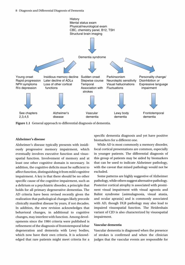
HistoryMental status examPhysical/neurological examCBC, chemistry panel, B12, TSHStructural brain imaging
Dementia syndrome
Young onsetRapid progressionNPH symptomsR/o depression
See chapters2,3,4,5
Alzheimer’sdisease
Vasculardementia
Lewy bodydementia
Frontotemporaldementia
Insidious memory declineLater decline of ADLsLoss of other cortical functions
Sudden onsetStepwise courseTemporalAssociation with strokes
ParkinsonismNeuroleptic sensitivityVisual hallucinationsFluctuations
Personality change/Disinhibition orExpressive language impairment
Figure 1.1 General approach to differential diagnosis of dementia.
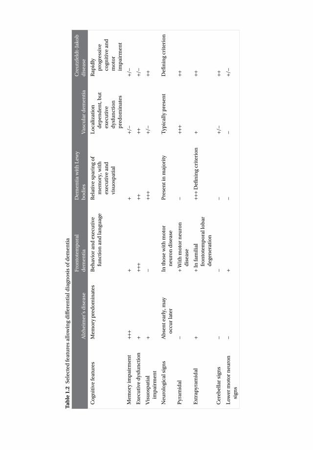
Ta
ble
1.2
S
ele
cte
d f
ea
ture
s a
llo
win
g d
iffe
ren
tia
l dia
gn
osi
s o
f d
em
en
tia
Alz
he
ime
r’s
dis
ea
se
Fro
nto
tem
po
ral
de
me
nti
a
De
me
nti
a w
ith
Le
wy
bo
die
sV
asc
ula
r d
em
en
tia
Cre
utz
feld
t–Ja
ko
b
dis
ea
se
Co
gn
itiv
e f
ea
ture
sM
em
ory
pre
do
min
ate
sB
eh
avio
r a
nd
exe
cuti
ve
fun
ctio
n a
nd
lan
gu
ag
e
Re
lati
ve s
pa
rin
g o
f
me
mo
ry, w
ith
exe
cuti
ve a
nd
visu
osp
ati
al
Lo
cali
zati
on
de
pe
nd
en
t, b
ut
exe
cuti
ve
dys
fun
ctio
n
pre
do
min
ate
s
Ra
pid
ly
pro
gre
ssiv
e
cog
nit
ive
an
d
mo
tor
imp
air
me
nt
Me
mo
ry i
mp
air
me
nt
+++
++
+/−
+/−
Exe
cuti
ve d
ysfu
nct
ion
+++
+++
+++/
−
Vis
uo
spa
tia
l
imp
air
me
nt
+−
+++
+/−
++
Ne
uro
log
ica
l sig
ns
Ab
sen
t e
arl
y, m
ay
occ
ur
late
r
In t
ho
se w
ith
mo
tor
ne
uro
n d
ise
ase
Pre
sen
t in
ma
jori
tyT
ypic
all
y p
rese
nt
De
fin
ing
cri
teri
on
Pyr
am
ida
l−
+ W
ith
mo
tor
ne
uro
n
dis
ea
se
−++
+++
Ext
rap
yra
mid
al
++
In f
am
ilia
l
fro
nto
tem
po
ral l
ob
ar
de
ge
ne
rati
on
+++
De
fin
ing
cri
teri
on
+++
Ce
reb
ell
ar
sig
ns
−−
−+/
−++
Lo
we
r m
oto
r n
eu
ron
sig
ns
−+
−−
+/−
10 ∙ Diagnosis and Differential Diagnosis of Dementia
cognitive decline. The prototype of vascular
dementia has an acute onset and stepwise decline,
with focal neurological signs and symptoms, but
this entity is rarely seen in clinical practice. It is
important to note that vascular risk factors or ath-
erosclerosis are not sufficient for a diagnosis of
vascular dementia; the brain parenchyma has to be
damaged by infarcts in order for this diagnosis to be
appropriately applied. Diffuse white matter changes
(leukoaraiosis) can also lead to vascular cognitive
impairment, often gradually progressive and associ-
ated with mood changes, gait impairment and
urinary frequency or incontinence.
Lewy body dementia
Lewy body dementia is distinguished by the
presence of parkinsonism, neuroleptic sensitivity,
fluctuations in consciousness, and spontaneous (i.e.
not drug induced) visual hallucinations, although
patients vary in the specific combinations of signs
and symptoms. In contrast to idiopathic Parkinson’s
disease, the parkinsonism in Lewy body dementia
tends to occur in the absence of rest tremor, is more
symmetrical, and does not respond as well to dopa-
minergic drugs. The diagnosis of Lewy body
dementia is also reserved for patients whose motor
symptoms have been present for less than 1 year
when dementia appears, in contrast to the 8–10
years of motor symptoms without dementia in idio-
pathic PD, which typically precedes PDD. Other fea-
tures suggestive of Lewy body dementia include
disproportionate visuospatial dysfunction and rapid
eye movement (REM) behavior disorder.
Frontotemporal dementia
Frontotemporal dementia may present as either a
language impairment or a behavioral variant.
Progressive aphasia due to frontotemporal pathology
is characterized by either progressive non-fluent
speech (progressive non-fluent aphasia) or loss of
knowledge of the meaning of items (semantic
dementia). They typically have a young onset, and can
develop behavioral features and asymmetrical focal
atrophy, which suggest frontotemporal dementia but
these features are insensitive. Criteria for possible
behavioral variant frontotemporal dementia (bvFTD)
include combinations of prominent behavioral
features such as disinhibition, apathy or inertia, loss of
sympathy or empathy, perseveration or compulsions,
hyperorality or executive dysfunction. Relative
sparing of episodic memory and visuospatial function
are cognitive features of bvFTD. It should be kept
in mind, however, that hippocampal pathology,
distinct from AD, can be found in bvFTD. Supportive
features include functional impairment sufficient to
indicate dementia (which allows differentiation
from primary psychiatric disorders), and imaging
showing frontal or anterior temporal atrophy or hypo-
metabolism in a pattern consistent with the diagnosis
increases the likelihood of probable bvFTD.
Problems with current classifications
The current diagnostic criteria are not absolute in
terms of specificity or sensitivity (see Table 1.1).
Some scenarios may suggest an acute CNS disorder,
yet be due to a degenerative dementia. Abrupt onset
suggests delirium, which needs to be ruled out to
make a diagnosis of dementia; however, delirium
may be a precursor to dementia and is more
common in the setting of dementia. Abrupt decline
can occur in DLB, where fluctuating cognition may
lead to a marked deterioration. In some, this might
be precipitated by an acute infection or medica-
tions, which can lead to diagnostic confusion.
Delirium increases the risk of mortality in older
people. Cerebrovascular disease also can present
acutely, and can lead to cognitive decline. A diffi-
culty with the diagnosis of vascular dementia is
establishing a temporal association between cere-
brovascular disease and dementia, given that
vascular events can be undetected without imaging
in some patients, despite their contribution to
cognitive decline.
A patient with incipient dementia of any kind is
susceptible to delirium. In such patients, a pro-
longed recovery, possible to a lower level of cognitive
function, may occur. While a typical patient with AD
has a decline of three points on the MMSE annually,
more rapid progression can be seen. When present,
like acute onset, this should prompt a search for
potentially treatable entities. Rapidly progressive
dementias are discussed in detail in Chapter 2.
Co-morbid conditions are also a reality in older
populations and may influence the course of
dementia but also compromise a confident diag-
nosis. Nevertheless, a progressive course despite
medical illness should not preclude the diagnosis of
degenerative dementia. Similar concerns apply in
psychiatric illness, in which a late-life degenerative
dementia can occur.
Diagnosis and Differential Diagnosis of Dementia ∙ 11
Focal presentations can also lead to diagnostic
confusion and uncertainty. Presentation with focal
symptoms is characteristic of frontotemporal dementia
and corticobasal ganglionic degeneration. Focal pre-
sentations can also occur in AD, in which language
impairment, characterized by paucity of speech
(which needs to be differentiated from primary
progressive aphasia), apraxia (which needs to be
differentiated from corticobasal ganglionic degen-
eration) and visuospatial impairment, which is
common in DLB, are not uncommon. Overlapping
pathologies, including AD, vascular changes and
Lewy bodies, present in a large proportion of patients
with dementia, and can confound the clinical picture.
Biomarkers
While biomarkers have been available for decades,
evidence is accumulating that they can be useful in
the differential diagnosis of dementia. Currently
most biomarkers require further validation in order
to be applied clinically beyond the research setting.
Beyond causal genes that are associated with auto-
somal dominant disease (APP, PSEN1, PSEN2),
genetic risk factors exist for AD and likely for
other dementias as well. Possession of an apolipo-
protein E4 (APOE 4) allele is a well-established
risk factor in AD. Recently identified risk alleles
include alterations in complement component
(3b/4b) receptor-1 (CR1), clusterin (CLU) and phos-
phatidylinositol binding clathrin assembly protein
(PICALM), each of which may provide a hint
regarding pathophysiology. For example, they may
point to inflammation, lipid metabolism or traffick-
ing of organelles. Other polymorphisms are being
identified but while these may be important scien-
tifically, they are not helpful in the clinic at present.
Blood markers are less strongly associated with
the development of dementia. Since the blood–brain
barrier impedes transmission of markers between
the brain and blood, it is not surprising that these
markers may not be as sensitive as spinal fluid
markers. Nevertheless, they may be helpful in identi-
fying potential risk factors, if not patterns related to
disease. Levels of plasma amyloid, particularly
beta-amyloid fragments 1-42 and 1-40, or their ratio,
have been inconsistently associated with increased
risk of dementia. Other markers include those for
oxidative stress, inflammation, glucose metabolism,
lipid metabolism, B-vitamin metabolism (i.e. homo-
cysteine), etc. In frontotemporal dementia, low
progranulin levels are found in some patients, in
whom they can predict the presence of mutations.
Developments in large-scale proteomic and metab-
olomic screening will likely identify marker patterns
associated with specific dementias. These may be
applicable to blood or cerebrospinal fluid but cur-
rently these approaches are not clinically available.
Cerebrospinal fluid is adjacent to the brain and
might be expected to better reflect CNS pathology.
As noted above, CSF examination is indicated if an
inflammatory, neoplastic or infectious etiology is
suspected in the evaluation of a patient. If NPH is
suspected, large-volume CSF drainage may help
diagnostically. CSF markers can be helpful in differ-
entiating AD from other forms of dementia such as
frontotemporal dementia or Lewy body dementia,
albeit with overlap. Specifically, low CSF amyloid
(1-42) concentrations are suggestive of Alzheimer
pathology. Increases in total tau (t-tau) and phos-
phorylated-tau (p-tau) are also present in AD but
these intraneuronal proteins can also reflect neu-
ronal damage and are therefore elevated in any
pathological processes that destroy brain tissue.
Current research is examining forms of beta-
amyloid and tau as prognostic markers in cognitively
normal individuals and people with mild cognitive
impairment. Additional markers have been exam-
ined and are likely to be developed. As with the
blood markers, these may provide insights into dis-
tinct pathologies and pathophysiological processes.
Imaging
As noted above, structural brain imaging is an
important part of the initial evaluation of patients
with suspected dementia. Assistance in differential
diagnosis and use as a marker in tracking disease
are also potential goals of brain imaging. MRI can
identify hippocampal atrophy which correlates with
the presence of AD pathology. Overall, the challenge
with use of biomarkers is their relative timing in
relation to clinical disease. For example, some
changes, like amyloid deposition, might occur pres-
ymptomatically with little change through the dis-
ease course, whereas others, such as global brain
atrophy, might be evident only after the disease is in
place. These concepts apply across dementias with
different markers or diagnostic signatures at play.
While positron emission tomography (PET) is not
routinely used, fluorodeoxyglucose (FDG) PET
scans can assist in differential diagnosis as can
12 ∙ Diagnosis and Differential Diagnosis of Dementia
technetium-HMPAO SPECT, which measures
cerebral blood flow. These approaches are particu-
larly helpful in differentiating AD, which shows pos-
terior hypoperfusion or decreased metabolism,
from frontotemporal dementia, which shows frontal
changes, and vascular dementia, which shows
patchy changes. Dementia with Lewy bodies is more
challenging to differentiate, likely because of over-
lapping AD pathology in many cases. Nevertheless,
the pattern of change in Lewy body dementia may
be distinct, with occipital hypometabolism or
decreased blood flow. In addition to assisting in the
differentiation of different types of dementia, meta-
bolic imaging may be helpful in identifying patients
with early cognitive decline at risk for transitioning
to dementia of the Alzheimer type. The recent
advent of practical ligands that allow imaging of
amyloid using PET will likely assist in identifying
patients with pathological changes of AD.
Parkinsonian disorders, related to loss of striatal
dopamine function, can be separated from other
dementing disorders such as AD. Currently
dopamine transporter imaging using SPECT or PET
imaging dopamine metabolism using 18-F-dopa
or presynaptic dopamine transporter using
11-C-dihydrotetrabenazine (DHTBZ) are helpful,
but have been most widely used in the research
setting. Readily available dopamine transporter
imaging with SPECT has lead to more widespread
use despite the caveat that such changes might not
separate different parkinsonian disorders. Cardiac
imaging using metaiodobenzylguanidine (MIBG)
shows loss of uptake in Lewy body-related disorders
but, like blood flow (HMPAO) and dopamine trans-
porter imaging using SPECT, this method might not
offer adequate sensitivity or specificity on its own.
Future of dementia diagnosis
The diagnosis of dementia is a clinical decision. The
increased availability of imaging and other modal-
ities will likely prove helpful, but they will not sup-
plant clinical decision making based on key aspects
of the history and physical examination.
Further reading
Carcaillon L, Berrut G, Sellal F, et al. Diagnosis
of Alzheimer’s disease patients with rapid
cognitive decline in clinical practice: interest of
the DECO questionnaire. J Nutr Health Aging
2011; 15(5): 361–6.
Clark CM, Schneider JA, Bedell BJ, et al, for the
AV45-A07 Study Group. Use of florbetapir-PET for
imaging beta-amyloid pathology. JAMA 2011;
305(3): 275–83.
Cummings JL, Mega M, Gray K, Rosenberg-
Thompson S, Carusi DA, Gornbein J. The
Neuropsychiatric Inventory: comprehensive
assessment of psychopathology in dementia.
Neurology 1994; 44(12): 2308–14.
Dubois B, Burn D, Goetz CG, et al. Diagnostic proce-
dures for Parkinson’s disease dementia: recom-
mendations from the Movement Disorder Society
Task Force. Mov Disord 2007; 22: 2314–24.
Dubois B, Slachevsky A, Litvan I, Pillon B. The FAB:
a Frontal Assessment Battery at bedside.
Neurology 2000; 55(11): 1621–6.
Feldman HH, Jacova C, Robillard A, et al. Diagnosis
and treatment of dementia: 2. Diagnosis. Can Med Assoc J 2008; 178(7): 825–36.
Galvin JE, Roe CM, Powlishta KK, et al. The AD8: a
brief informant interview to detect dementia.
Neurology 2005; 65(4): 559–64.
Goldman JS, Hahn SE, Catania JW, et al, for the
American College of Medical Genetics and the
National Society of Genetic Counselors. Genetic
counseling and testing for Alzheimer disease:
joint practice guidelines of the American College
of Medical Genetics and the National Society of
Genetic Counselors. Genet Med 2011; 13(6):
597–605.
Hort J, O’Brien JT, Gainotti G, Pirttila T, et al, for the
EFNS Scientist Panel on Dementia. EFNS guide-
lines for the diagnosis and management of
Alzheimer’s disease. Eur J Neurol 2010; 17(10):
1236–48.
Jack CR, Knopman DS, Jagust WJ, et al. Hypothetical
model of dynamic biomarkers of the Alzheimers
pathological cascade. Lancet Neurol 2010; 9(1):
119–28.
Jack CR Jr, Albert MS, Knopman DS, et al.
Introduction to the recommendations from the
National Institute on Aging-Alzheimer’s Associa-
tion workgroups on diagnostic guidelines for
Alzheimer’s disease. Alzheimers Dement 2011;
7(3): 257–62.
Kertesz A, Davidson W, Fox H. Frontal behavioral
inventory: diagnostic criteria for frontal lobe
dementia. Can J Neurol Sci 1997; 24(1): 29–36.
Knapskog AB, Barca ML, Engedal K. A comparison
of the validity of the Cornell Scale and the MADRS
Diagnosis and Differential Diagnosis of Dementia ∙ 13
in detecting depression among memory clinic
patients. Dement Geriatr Cogn Disord 2011; 32(4):
287–94.
McKeith IG, Dickson DW, Lowe J, et al, for the
Consortium on DLB. Diagnosis and management
of dementia with Lewy bodies: third report of
the DLB Consortium. Neurology 2005; 65(12):
1863–72.
McKhann GM, Knopman DS, Chertkow H, et al. The
diagnosis of dementia due to Alzheimer’s disease:
recommendations from the National Institute on
Aging-Alzheimer’s Association workgroups on
diagnostic guidelines for Alzheimer’s disease.
Alzheimers Dement 2011; 7(3): 263–9.
Rascovsky K, Hodges JR, Knopman D, et al.
Sensitivity of revised diagnostic criteria for the
behavioural variant of frontotemporal dementia.
Brain 2011; 134(Pt 9): 2456–77.
Román GC, Tatemichi TK, Erkinjuntti T, et al.
Vascular dementia: diagnostic criteria for
research studies. Report of the NINDS-AIREN
International Workshop. Neurology 1993; 43(2):
250–60.
Sabbagh MN, Malek-Ahmadi M, Kataria R, et al. The
Alzheimer’s questionnaire: a proof of concept
study for a new informant-based dementia
assessment. J Alzheimers Dis 2010; 22(3):
1015–21.
Sikkes SA, van den Berg MT, Knol DL, et al. How
useful is the IQCODE for discriminating between
Alzheimer’s disease, mild cognitive impairment
and subjective memory complaints? Dement Geriatr Cogn Disord 2010; 30(5): 411–16.
Teng EL, Chui HC. A Modified Mini-Mental State
(3MS) Examination. J Clin Psychiatry 1987; 48:
314–18.