Embed Size (px)
Citation preview
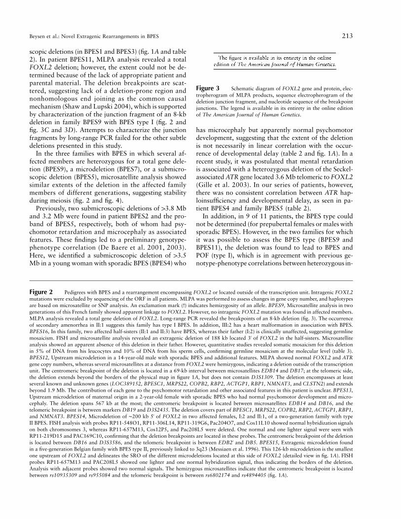
Am. J. Hum. Genet. 77:205–218, 2005
205
Deletions Involving Long-Range Conserved Nongenic Sequences Upstreamand Downstream of FOXL2 as a Novel Disease-Causing Mechanismin Blepharophimosis SyndromeD. Beysen,1 J. Raes,3 B. P. Leroy,1,2 A. Lucassen,4 J. R. W. Yates,5 J. Clayton-Smith,6H. Ilyina,7 S. Sklower Brooks,8 S. Christin-Maitre,9 M. Fellous,10 J. P. Fryns,11 J. R. Kim,12
P. Lapunzina,13 E. Lemyre,14 F. Meire,3 L. M. Messiaen,15 C. Oley,16 M. Splitt,17
J. Thomson,18 Y. Van de Peer,3 R. A. Veitia,10 A. De Paepe,1 and E. De Baere1
1Center for Medical Genetics and 2Department of Ophthalmology, Ghent University Hospital, and 3Research Group of Bioinformatics andEvolutionary Genomics, Department of Plant Systems Biology, Ghent University–Flemish Institute for Biotechnology, Ghent, Belgium; 4WessexClinical Genetics Service, University of Southampton, Southampton, United Kingdom; 5Department of Medical Genetics, University ofCambridge and Addenbrooke’s Hospital, Cambridge, United Kingdom; 6Academic Department of Medical Genetics, St. Mary’s Hospital,Manchester, United Kingdom; 7Belorussian Institute for Hereditary Disorders, Minsk, Belarus; 8New York State Institute for Basic Research inDevelopmental Disabilities, New York; 9Service d’Endocrinologie, Hopital Saint-Antoine, and 10INSERM U361 Reproduction etPhysiopathologie Obstetricale Hopital Cochin, Paris; 11Center for Human Genetics, Leuven, Belgium; 12Department of Biochemistry andMolecular Biology, Yeungnam University, Daegu, Republic of Korea; 13Servicio de Genetica, Hospital Universitario La Paz, Madrid; 14MedicalGenetics Service, Department of Pediatrics, Hopital Sainte-Justine, Universite de Montreal, Montreal; 15University of Alabama, Birmingham;16Birmingham Women’s Hospital, Birmingham, United Kingdom; 17Department of Clinical Genetics, Guy’s and St Thomas’ Hospital NationalHealth Service Trust, London; and 18Clinical Genetics, St. James’s University Hospital, Leeds, United Kingdom
The expression of a gene requires not only a normal coding sequence but also intact regulatory regions, which canbe located at large distances from the target genes, as demonstrated for an increasing number of developmentalgenes. In previous mutation studies of the role of FOXL2 in blepharophimosis syndrome (BPES), we identifiedintragenic mutations in 70% of our patients. Three translocation breakpoints upstream of FOXL2 in patients withBPES suggested a position effect. Here, we identified novel microdeletions outside of FOXL2 in cases of sporadicand familial BPES. Specifically, four rearrangements, with an overlap of 126 kb, are located 230 kb upstream ofFOXL2, telomeric to the reported translocation breakpoints. Moreover, the shortest region of deletion overlap(SRO) contains several conserved nongenic sequences (CNGs) harboring putative transcription-factor binding sitesand representing potential long-range cis-regulatory elements. Interestingly, the human region orthologous to the12-kb sequence deleted in the polled intersex syndrome in goat, which is an animal model for BPES, is containedin this SRO, providing evidence of human-goat conservation of FOXL2 expression and of the mutational mechanism.Surprisingly, in a fifth family with BPES, one rearrangement was found downstream of FOXL2. In addition, wereport nine novel rearrangements encompassing FOXL2 that range from partial gene deletions to submicroscopicdeletions. Overall, genomic rearrangements encompassing or outside of FOXL2 account for 16% of all moleculardefects found in our families with BPES. In summary, this is the first report of extragenic deletions in BPES,providing further evidence of potential long-range cis-regulatory elements regulating FOXL2 expression. It con-tributes to the enlarging group of developmental diseases caused by defective distant regulation of gene expression.Finally, we demonstrate that CNGs are candidate regions for genomic rearrangements in developmental genes.
Introduction
The expression pattern of many developmental genes isregulated at different levels. Usually, basal gene expres-sion is controlled by the core promoter, located imme-
Received April 22, 2005; accepted for publication May 19, 2005;electronically published June 16, 2005.
Address for correspondence and reprints: Dr. Elfride De Baere, Cen-ter for Medical Genetics, Ghent University Hospital, De Pintelaan 185,B-9000 Ghent, Belgium. E-mail: [email protected]
� 2005 by The American Society of Human Genetics. All rights reserved.0002-9297/2005/7702-0004$15.00
diately upstream of the ORF. However, spatiotemporallyand quantitatively correct gene expression is often con-trolled by long-range cis-acting regulatory elements asdistant as 1.1 Mb (Velagaleti et al. 2005). These elements(enhancers, insulators, and locus-control regions) can belocated upstream or downstream of the transcriptionunit of a gene. Interspecies comparative studies show astrong evolutionary conservation of these elements,which may reside in unrelated neighboring genes or in-tergenic regions. Genomic rearrangements involvingsuch elements outside of a transcription unit can giverise to human disease by a dissociation of the transcrip-
206 Am. J. Hum. Genet. 77:205–218, 2005
tion unit and its cis-acting regulatory elements, by analteration of the chromatin structure, or by both mech-anisms (reviewed by Kleinjan and van Heyningen [2005]).
Recently, it was shown that heterozygous mutationsof the FOXL2 gene (MIM 605597), which encodes aforkhead transcription factor, cause both types of ble-pharophimosis-ptosis-epicanthus inversus syndrome(BPES [MIM 110100]). BPES is a rare, autosomal dom-inant developmental disorder characterized by an eyelidmalformation. Two subtypes have been identified; typeI is associated with premature ovarian failure (POF),whereas type II is not (Zlotogora et al. 1983). It wasshown that haploinsufficiency of FOXL2 was causedby intragenic mutations, in most cases, and by submi-croscopic deletions encompassing FOXL2 and neigh-boring genes, in two cases (De Baere et al. 2001, 2003).A genotype-phenotype correlation was proposed mainlyon the basis of intragenic mutations distinguishing thetwo BPES types at the molecular level (De Baere et al.2003). Despite the ovarian phenotype in BPES, severalstudies have indicated that mutations in FOXL2 do notplay a major role in causing idiopathic POF (De Baereet al. 2001, 2002; Harris et al. 2002; Bodega et al.2004).
A spatiotemporally restricted expression pattern ofthe FOXL2 transcript and protein was demonstrated inseveral species (human, mouse, and goat). It was shownto be expressed in developing eyelids and in granulosacells of fetal and adult ovaries (Crisponi et al. 2001;Cocquet et al. 2002, 2003). Recently, two studies pre-senting homozygous knockout mice showed that gran-ulosa cell function is crucial not only for oocyte growthbut also for ovarian maintenance, thus providing a mo-lecular mechanism for POF in BPES in vivo (Schmidtet al. 2004; Uda et al. 2004).
During the positional cloning of FOXL2, three bal-anced translocations were characterized in patients witha classic BPES phenotype. It was shown that the break-points of these translocations disrupt two other genes(BPESC1 and MRPS22), both devoid of pathogenic mu-tations in other patients with BPES, suggesting a posi-tion effect (De Baere et al. 2000; Praphanphoj et al.2000; Crisponi et al. 2001, 2004).
Sequencing of the FOXL2 ORF, in combination withFISH analysis with a probe encompassing the FOXL2gene, revealed mutations in !70% of patients with BPES(De Baere et al. 2001, 2003). Here, we applied a novelmutation-detection strategy consisting of multiplex li-gation-dependent probe amplification (MLPA), geno-typing with 160 microsatellites and SNPs, and com-prehensive FISH analysis. This approach led to theidentification of nine novel deletions encompassingFOXL2, which ranged from partial and total gene de-letions to microdeletions and submicroscopic deletions.Notably, five novel deletions were found either up-
stream or downstream of the transcription unit ofFOXL2. These genic and extragenic (micro)deletionsaccount for 11% and 5%, respectively, of all moleculardefects found in our cohort with BPES. Finally, we re-port the first case of germline mosaicism in a familywith BPES that is confirmed at the molecular level.
Material and Methods
Patients and Diagnostic Criteria
The 37 consenting probands with BPES who wereincluded in this study presented with blepharophimo-sis, ptosis, epicanthus inversus, and telecanthus. Theclinical diagnosis was given by a clinical geneticist or anophthalmologist. When possible, karyotyping was per-formed in the referring laboratory. The karyotype wasnot confirmed by us. In each proband, intragenic mu-tations were excluded by sequencing of the FOXL2ORF, as described by De Baere et al. (2001).
Custom-Made MLPA
MLPA permits relative quantification of changes incopy number of specific chromosomal sequences (Schou-ten et al. 2002). A custom-made deletion-detection kitfor FOXL2 was developed by MRC-Holland (P054FOXL2) and contains three different probes for FOXL2(FOXL2-D01, FOXL2-D02, and FOXL2-D03), fourprobes for the ATR gene (located at 3q22-24), andprobes for other genes (TWIST, FOXC1, PITX2,FOXC2, and OA1). Probe FOXL2-D01 is located inthe forkhead domain (c.252–351; g.489–588), probeFOXL2-D02 is between the forkhead domain and thepolyalanine tract (c.575–641; g.812–878), and probeFOXL2-D03 is downstream of the polyalanine tract(c.1053–1113; g.1290–1350). MLPA was performed inaccordance with the manufacturer’s instructions.
FISH
A genomic BAC contig of region 3q23 was obtainedfrom the National Center for Biotechnology Informa-tion (NCBI) database. BACs were obtained from TheWellcome Trust Sanger Institute and BACPAC ResourcesCenter. PACs and cosmids had been previously obtainedfrom the UK–Medical Research Council and weremapped by FISH (De Baere et al. 1999). BAC, PAC, andcosmid DNA was prepared and labeled with the use ofstandard conditions. FISH analysis was performed asdescribed by De Baere et al. (1999).
Microsatellite Analysis
Fifteen polymorphic microsatellites (with prefix “D3S”)were selected from the NCBI on the basis of locationand heterozygosity. Primer sequences of markers with
Beysen et al.: Novel Extragenic Rearrangements in BPES 207
Table 1
Primer Sequences of Novel Microsatellites andSNaPshot Analysis
The table is available in its entirety in the online editionof The American Journal of Human Genetics.
prefixes “VY” and “NU” were obtained from Udar etal. (2003). A third series of microsatellites (Tel2 andmarkers with prefix “EDB”) was developed by a manualsearch for repeats in the BAC sequences derived fromNCBI. For a fourth series (with prefix “DB”), the dif-ferent BAC sequences were analyzed using the Find Sim-ple Repeats software, which selects repetitive sequencesthat are expected to have a very high degree of poly-morphism. The search was confined to sequences pre-senting repetitions with a maximum of 4 nt and havinga total length of at least 12 nt. Sequences with a score�30 were selected for primer design. Primer sequencesare given in table 1. Characterization of the polymorphicnature of the known and novel microsatellites was per-formed in a white population by use of an M13-systemdescribed by Schuelke (2000). For allele sizing, aliquotsof PCR reaction were mixed with ROX-500 or ROX-1000 size standard (Applied Biosystems) and formamideand were separated on an ABI PRISM 3100 GeneticAnalyzer (Applied Biosystems). Fragment sizing was per-formed automatically using GeneScan version 3.7. Thedifferent microsatellite alleles were numbered consecu-tively, in order of increasing size. The numbering is dif-ferent for each family.
SNaPshot Analysis
Twenty-seven SNP loci were selected from the Inter-national HapMap Project database, and amplificationprimers were designed so that PCR products of ∼300bp were obtained. Typing primers were designed to an-neal to the target DNA flanking the 3′ end of the SNP.A standard PCR reaction was performed, followed byexonuclease I and shrimp alkaline phosphatase (SAP)treatment (Amersham). A single-nucleotide primer ex-tension reaction was performed using an ABI PRISMSNaPshot Multiplex kit (Applied Biosystems), in accor-dance with the manufacturer’s instructions. After SAPtreatment, products were separated on an ABI PRISM3100 Genetic Analyzer (Applied Biosystems) and wereanalyzed with GeneScan version 3.7. Numbers were as-signed to alleles as follows: A p 1, C p 2, G p 3, andTp 4.
To assess the degree of germline mosaicism in anunaffected male, quantitative SNaPshot analysis wasperformed as described by Matyas et al. (2002). Fornormalization, genomic samples from four unrelated in-dividuals who have an identical SNP haplotype as thatof the test sample were used as controls.
Long-Range PCR
For long-range PCR, the Expand Long Template PCRSystem (Roche) was used. A master mix of 50 ml con-tained 300 ng of genomic DNA, 0.5 mM of each primer,5 ml of Expand Long Template PCR buffer 3 (containing10 # PCR buffer and 27.5 mM MgCl2), 3.75 U ofExpand Long Template enzyme mix (containing TaqDNA polymerase and Tgo DNA polymerase), 500 mMof each dNTP, and 3 mM additional MgCl2. Cyclingconditions included an initial denaturation cycle at 94�Cfor 2 min, followed by 10 cycles at 94�C for 20 s, 65�Cfor 30 s, and 68�C for 12 min; 25 cycles at 94�C for 30s and 65�C for 30 s and elongation for 12 min plus 20s for each successive cycle; and a final extension at 72�Cfor 7 min.
Construction of a High-Resolution Physical MapCovering Microdeletions
First, we generated a sequence contig of 16 over-lapping RPCI-11 BAC clones that span ∼1.8 Mb sur-rounding FOXL2 by a search of GenBank (NCBI). Thepolymorphic microsatellites selected from NCBI werepositioned on this BAC contig by use of the referencenumbering of build 34, version 3 (“the golden path”),and by use of BLAST (NCBI). The localization of theanonymous microsatellites and SNPs was verified byBLAST searches against the BAC sequences. The exactPAC and cosmid localization was determined by BLASTafter PAC and cosmid end sequencing, in accordancewith standard protocol. Finally, the different known andunassigned genes that are located in this region wereadded to the map according to NCBI data.
Comparative Sequence Analysis
To identify conserved nongenic sequences (CNGs),comparative sequence analysis of the regions of interestwas performed using the University of California–SantaCruz (UCSC) Genome Browser. The UCSC Multiz Align-ments and Conservation track contains a measure ofevolutionary conservation in human, chimp, mouse, rat,dog, chicken, pufferfish, and zebrafish based on a phy-logenetic hidden Markov model (phastCons). The over-all conservation score across all species, as well as pair-wise alignments of each species aligned to the humangenome, were retrieved, and gapped alignments wereincluded. The length of the alignment is based on thelengths of gaps in the human sequence at those alignmentpositions, relative to the longest nonhuman sequence.Alignments are based on the following genome assem-blies: human, May 2004 (hg17); chimp, November 2003(panTro1); mouse, May 2004 (mm5); rat, June 2003(rn3); dog, July 2004 (canFam1); chicken, February2004 (galGal2); Fugu, August 2002 (fr1); and zebrafish,
Beysen et al.: Novel Extragenic Rearrangements in BPES 209
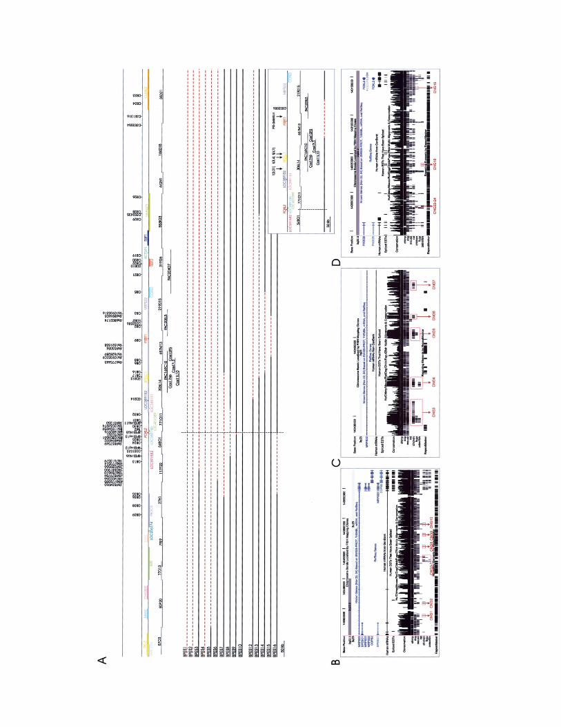
Figure 1 A, Physical map of a 1.8-Mb region flanking FOXL2 that shows the position of the BACs, PACs, cosmids, microsatellites, andSNPs that were used for identification and delineation of the deletions. The map is drawn to scale; the size equivalent to 50 kb is given at thebottom left corner. The location of FOXL2 is indicated by a vertical black line. The top line represents the localization of SNPs, and the secondline contains the known and anonymous microsatellites used in this study. In the third line, the location of the genes in this region is shown,whereas the BACs, PACs, and cosmids used for FISH are shown below the line. The lower part of A is an overview of all deletions identifiedin this study, except one. Every line represents a patient or family in whom a deletion was found. The dashed red line shows the maximalextent of the deletion. The first 10 lines represent the deletions that encompass FOXL2. The deletion identified in BPES11 is not shown, sincethe extent of the deletion could not be determined because of a lack of material for FISH and microsatellite analysis. The second group representsthe five extragenic deletions (four upstream and one downstream). Clinical details and molecular data on the patients with deletions are shownin table 2. In a box at the bottom right, a detailed view of the SRO of the four upstream extragenic deletions is shown. The position of FOXL2is marked by a vertical dashed black line, and the SRO is shown by the dashed red line. The positions of the known three translocationbreakpoints at 3q23 in BPES and of the orthologue of the PIS locus are indicated by vertical arrows. B and C, UCSC Genome Browser mapof the SRO of the four upstream extragenic rearrangements in families BPES12, BPES13, BPES14, and BPES15. CNGs are highlighted at thebottom with red boxes. Their names, based on our arbitrary numbering, are given below the boxes. Details on all CNGs can be found in table4. The CNGs shown in B are evolutionarily conserved up to chicken. C represents a more detailed view of CNG3–CNG7. CNG3–CNG5 areconserved up to zebrafish and/or pufferfish. They all represent potential cis-regulatory elements of the FOXL2 region. D, UCSC Genome Browsermap of the sequence contained in the downstream extragenic rearrangement in family BPES16. Five CNGs are indicated in this 3′ SRO, ofwhich several are conserved up to zebrafish and/or pufferfish (CNG14, CNG18, and CNG22–CNG24).
November 2003 (danRer1). Our selection criteria in-cluded conservation at least up to chicken, pufferfish,and/or zebrafish, independent of alignment length, per-centage of conservation, and presence of gaps.
The respective sequences of the selected elements andtheir corresponding mouse sequences were retrievedthrough the UCSC Genome Browser. Apart from thesesequences, an available 48-kb goat sequence from thepolled intersex syndrome (PIS) locus (GenBank accessionnumber AF404302) was retrieved for human-goat com-parison with the use of mVista. A pairwise alignmentwas performed between human and mouse, human andgoat, human and chicken, human and pufferfish, andhuman and zebrafish by use of ClustalW. To exclude thecoding potential of all putative CNGs, the human se-quence of the conserved region was compared with allavailable EST and protein sequences in GenBank, as wellas with all gene predictions for the selected region avail-able from the UCSC Genome Browser and the BiomaxHuman Genome Database (blastn, blastx, tblastx, andnrprot). In addition, sequences were scanned for repet-itive elements using RepeatMasker. Sequences that didnot show significant similarity to known proteins, genesand/or transcripts, or transposable elements were se-lected as CNGs. These were searched against the Trans-fac database (version 8.3) of known vertebrate cis-actingregulatory DNA elements and trans-acting factors by useof the Match tool included in that database (Matys etal. 2003). Only highly significant matches with tran-scription-factor binding sites were considered, by use ofthe built-in “minimize false positive matches” setting.
Results and Discussion
The mutation detection rate of !70% in previous mu-tation studies of FOXL2 in BPES could be explained byclinical misdiagnosis; the sensitivity of the strategy used,
which might not allow detection of changes in gene copynumber or in distant sequences outside of the transcrip-tion unit of FOXL2 (De Baere et al. 2001, 2003); orthe presence of a second BPES locus, although there isno evidence to support the existence of a second locus.Here, we developed a novel combined approach con-sisting of (1) custom-made MLPA for detection ofchanges in gene copy number of FOXL2 and othergenes; (2) a genotyping panel, comprising 15 known and32 anonymous microsatellites and 26 SNPs; and (3)comprehensive FISH analysis whenever possible.
Genomic Rearrangements Encompassing FOXL2
A clue to subtle FOXL2 deletions was found in similarobservations for FOXC1, which encodes a forkheadtranscription factor associated with ocular anterior seg-ment dysgenesis (IRID1 [MIM 601631]). Irrespective ofwhether the gene is duplicated or deleted, a nearly iden-tical ocular phenotype is observed, showing that exactgene dosage of FOXC1 is critical for normal eye devel-opment (Lehmann et al. 2003).
Our approach allowed identification of 11 deletionsencompassing FOXL2, of which 9 are novel. These de-letions contribute to 11% (11/100) of all molecular de-fects found in our series of patients with BPES and arefound in 30% (11/37) of mutation-negative patients.They occur in sporadic cases and familial cases and areabsent in all unaffected family members analyzed. Agraphical overview of these deletions is given in figure1A. Clinical data for the 11 families and molecular de-tails on the deletions are summarized in table 2.
The rearrangements include one partial FOXL2 de-letion (in BPES10), an 8-kb deletion removing the wholeFOXL2 ORF (in BPES9), two microdeletions of up to420 kb, encompassing FOXL2 and neighboring genes(in BPES7 and BPES8), four submicroscopic deletions(in BPES2, BPES4, BPES5, and BPES6), and two micro-
210
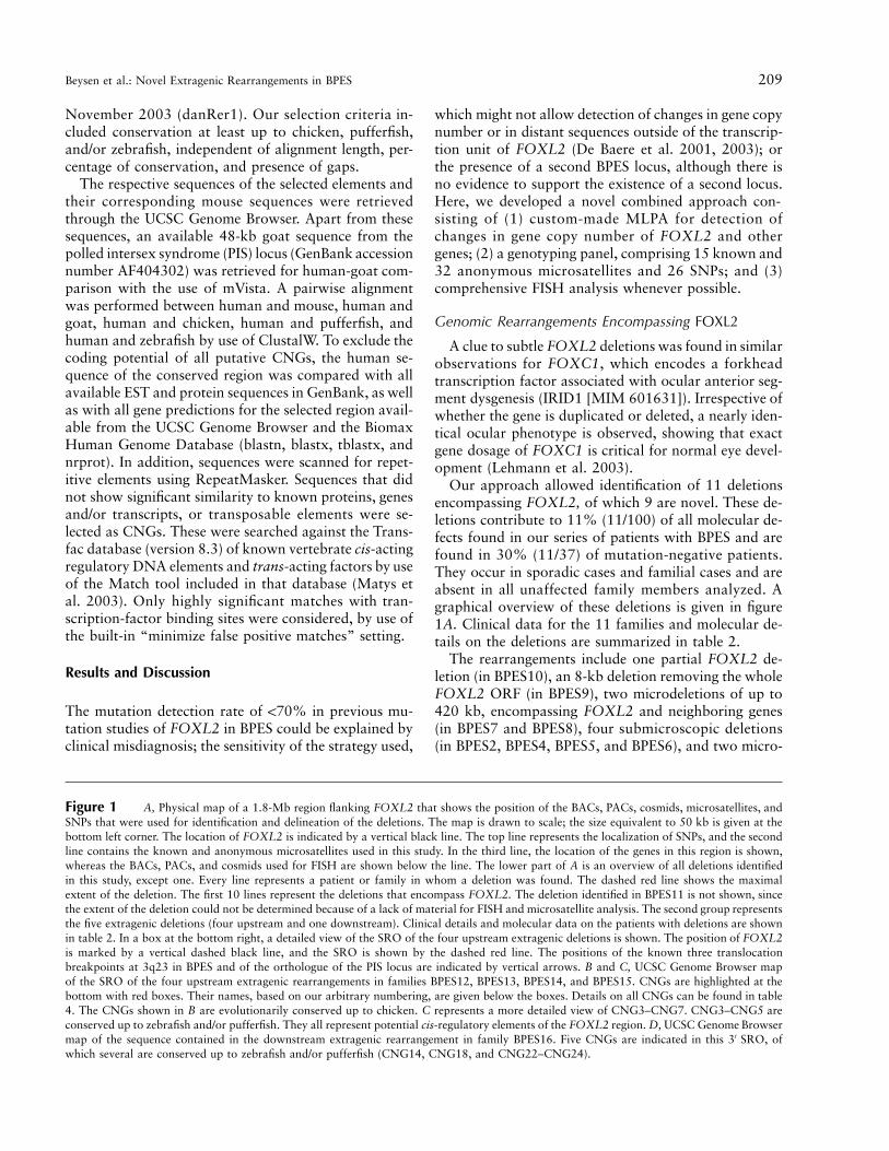
Table 2
Summary of Clinical and Molecular Findings in Patients with BPES Who Have Deletions Encompassing FOXL2 or Outside of the Transcription Unit of FOXL2
Type of GenomicRearrangementand Family
Typeof
BPESa Originb Clinical Datac Type of Deletiond Karyotype MLPA Findingd Extent of Deletiond
Delineation ofCentromericBreakpointe
Delineation ofTelomeric
Breakpointe
Confirmedby FISH
ParentalOrigin
Encompassing FOXL2:BPES1 F2 KR 6-y-old male with psychomotor re-
tardation and VSD (Cha et al.2003)
Microscopic delincl. FOXL2 in II:2
… Total FOXL2 andATR del
16.1 Mb in II:2(137606245–143780323)
D3S1238–D3S3528(2.20 Mb)
Telomeric to ATR No Maternal
BPES2 S NO 11-y-old male with psychomotor re-tardation, widely spaced nipples,scoliosis, high arched palate, andumbilical hernia (De Baere et al.2003)
Submicroscopic delincl. FOXL2
Normal Total FOXL2 andATR del
13.8 Mb(139895393–143780323)
D3S3617–DB39(1.81 Mb)
Telomeric to ATR Yes Maternal
BPES3 S GB 4-y-old female with postnatalgrowth retardation andmicrocephaly
Microscopic delincl. FOXL2
46,XX,del(3)(q22q23)
Total FOXL2 del(3 probes); ATRnormal
Max. 3.6 Mb(140031707–143650777)
rs2881900–DB13(26.99 kb)
D3S1309–ATR(1.44 Mb)
No Maternal
BPES4 S CA 3-y-old female with normal psycho-motor development and no intra-uterine and postnatal growth re-tardation; ultrasound of uterusand ovaries was normal at age 13mo
Submicroscopic delincl. FOXL2
Normal Total FOXL2 del(3 probes); par-tial ATR del (1probe)
13.5 Mb(140058657–143651142)
D3S3617–DB13(1.97 Mb)
ATR gene Yes Maternal
BPES5 F DK II:1 had psychomotor and growthretardation and high arched pal-ate; I:1 had growth retardation,with no clinical data about psy-chomotor development (De Baereet al. 2001)
Submicroscopic delincl. FOXL2
Normal Total FOXL2 del(3 probes)
Max. 3.2 Mb(139548627–142750847)
In BAC RP11-80P20(147 kb)
D3S3554–D3S3711(1.66 Mb)
Yes Unknown
BPES6 S ES 1.5-y-old female with abnormalnose, normal psychomotor devel-opment, and no growth retarda-tion; ultrasound of uterus andovaries was normal
Submicroscopic delincl. FOXL2
Normal Total FOXL2 del(3 probes); ATRnormal
Max. 2.7 Mb(140036995–142750847)
rs9289566–rs6776585(36.77 kb)
D3S1309–D3S3711(541.72 kb)
No Paternal
BPES7 F ES Father and son with normal psycho-motor development
Microdel incl. FOXL2 Normal Total FOXL2 del(3 probes)
Max. 412 kb(139949789–140359539)
DB36–DB35 (9.4 kb) DB16–DB6 (28.21kb)
Yes Paternal?
BPES8 S CD-BE 3-y-old female with normal psycho-motor development
Microdel incl. FOXL2 … Total FOXL2 del(3 probes)
Max. 243 kb(140075997–140319005)
NU6–D3S1535 (15.8kb)
DB 31–DB17(45.11 kb)
Yes Paternal
BPES9 F1 FR II:1 had secondary amenorrhea; III:2had heart malformation (mild in-terauricular communication)
Total FOXL2 del … Total FOXL2 del(3 probes)
8,226 nt(140141021–140149217)
In BAC RP11-548O1,nt 60868
BAC RP11-548O1,nt 69094
No Unknown
BPES10 S GB 4-y-old female with normal psycho-motor development
Partial FOXL2 del Normal Partial FOXL2del (2/3 probes)
Max. 6 kb(140147210–140150682)
In FOXL2, betweenMLPA probes 2and 3 (416 nt)
FOXL2–Tel2(2,440 nt)
No Unknown
BPES11 S1 AU 36-y-old female with normal psycho-motor development, menarche atage 18 y, only two periods, andvery high FSH levels; IVF failed
(Micro)del incl. FOXL2 Normal Total FOXL2 del(3 probes)
… … … No Unknown
211
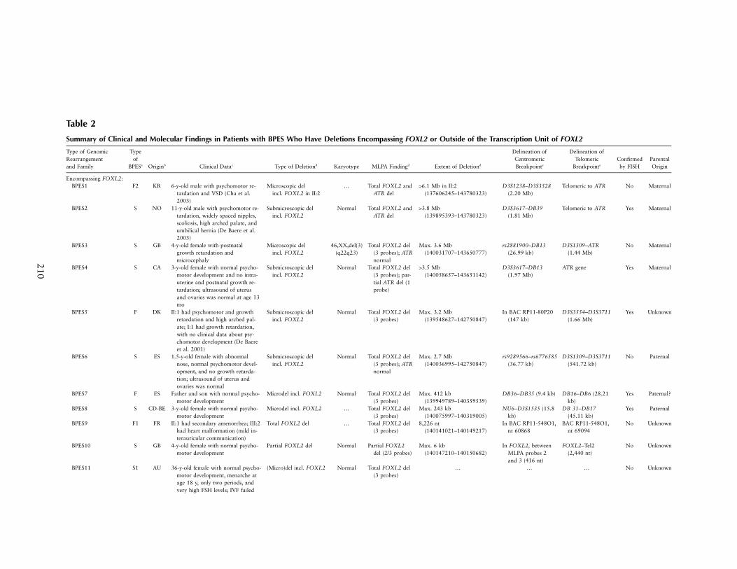
Outside transcriptionunit of FOXL2:BPES12 S BY 15-y-old male with normal psycho-
motor development, intrauterineand postnatal growth retardation,microcephaly, ventricular septumdefect, small mouth, contractureof interphalangeal joints, and rightiris heterochromia
Microdel upstream ofFOXL2 (at 101 kb)
Normal Two copies ofFOXL2 andATR (D3S1309is heterozygous)
Max. 1.9 Mb(140249840–142209012)
EDB14–DB17 (69.40kb)
DB33–D3S1309(1.03 Mb)
No Paternal
BPES13 S PL 2-y-old female with stenosis of lacri-mal duct, normal psychomotor de-velopment, microcephaly, and re-nal reflux
Microdel upstream ofFOXL2 (at 101 kb)
Normal Two copies ofFOXL2
Max. 567 kb(140249840–140816803)
EDB14–DB16 (81.64kb)
DB19–D3S2435(129.11 kb)
No Maternal
BPES14 F2 GB 2-generation family. I:2 had astigma-tism, three pregnancies, oligome-norrhea at age 40 y, and amenor-rhea at age 47 y. II:1 hadstrabismus, normal menarche atage 12 y, and oligomenorrheawith elevated FSH and LH at age38 y. Both I:2 and II:1 had nor-mal growth and psychomotordevelopment.
Microdel upstream ofFOXL2 (at 183 kb)
… Two copies ofFOXL2 andATR
Max. 244 kb(140331480–140575742)
DB16–D3S3586(144.53 kb)
rs4894405–DB5(71.90 kb)
Yes Unknown
BPES15 F2 BE 5-generation family with typicalBEPS. Affected members, normalpsychomotor development
Microdel upstream ofFOXL2 (at 231 kb)
Normal Two copies ofFOXL2
Max. 126 kb(140377649–140503837)
rs10935309–rs955084(27.14 kb)
rs6802174–rs4894405 (14.47kb)
Yes Unknown
BPES16 F GB Half-sisters II:1 and II:3 were af-fected; father I:2 was not affected,suggestive of germline mosaicism.II:1 had a premature birth (32wk) with respiratory distress syn-drome, intrauterine and postnatalgrowth retardation, microcephaly,moderate learning difficulties ap-parent from age 7 y, and strabis-mus; II:3 had normal psychomo-tor development
Microdel downstreamof FOXL2 (at 28.7kb)
Normal Two copies ofFOXL2
Max. 188 kb(139959089–140147132)
DB35–rs9834963(38.27 kb)
rs7627822–FOXL2(28.7 kb)
Yes Paternal
a F1 p familial, type I; F2 p familial, type II; F p familial, type undetermined; S p sporadic; S1 p sporadic, type I.b AU p Australia; BE p Belgium; BY p Belarus; CA p Canada; CD p Congo; DK p Denmark; ES p Spain; FR p France; GB p Great Britain; KR p South Korea; NO p Norway; PL p Poland.c FSH p follicle-stimulating hormone; IVF p in vitro fertilization; LH p luteinizing hormone; VSD p ventricle septum defect; y p year.d del p deletion; incl. p including; Max. p at maximum.e In parentheses is the length of the interval in which the centromeric and the telomeric breakpoints are located.
Beysen et al.: Novel Extragenic Rearrangements in BPES 213
Figure 3 Schematic diagram of FOXL2 gene and protein, elec-tropherogram of MLPA products, sequence electropherogram of thedeletion junction fragment, and nucleotide sequence of the breakpointjunctions. The legend is available in its entirety in the online editionof The American Journal of Human Genetics.
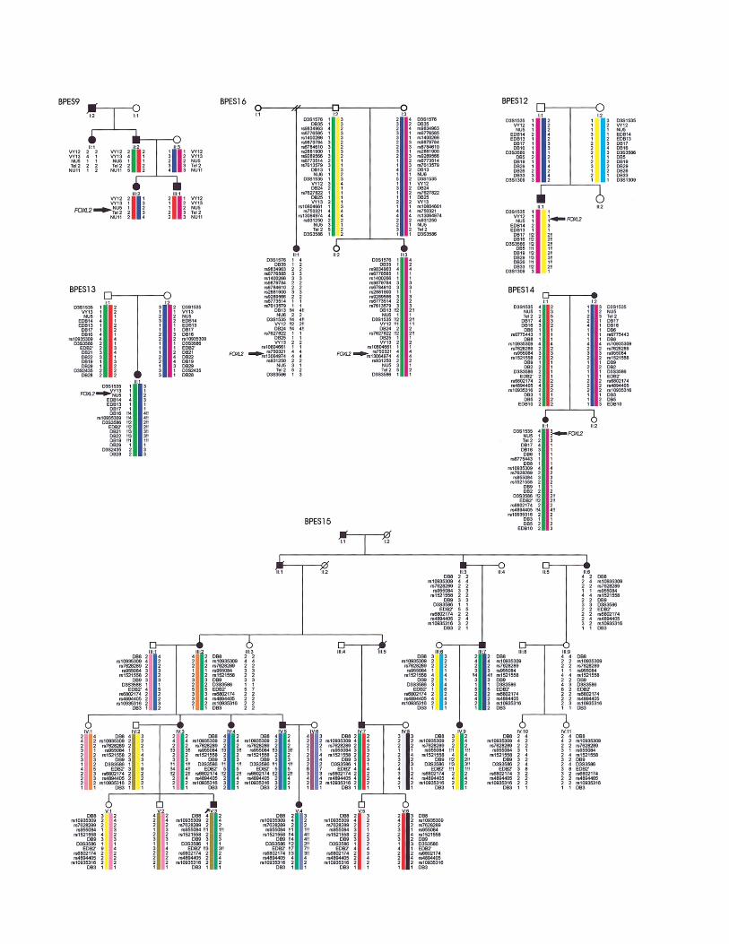
Figure 2 Pedigrees with BPES and a rearrangement encompassing FOXL2 or located outside of the transcription unit. Intragenic FOXL2mutations were excluded by sequencing of the ORF in all patients. MLPA was performed to assess changes in gene copy number, and haplotypesare based on microsatellite or SNP analysis. An exclamation mark (!) indicates hemizygosity of an allele. BPES9, Microsatellite analysis in twogenerations of this French family showed apparent linkage to FOXL2. However, no intragenic FOXL2 mutation was found in affected members.MLPA analysis revealed a total gene deletion of FOXL2. Long-range PCR revealed the breakpoints of an 8-kb deletion (fig. 3). The occurrenceof secondary amenorrhea in II:1 suggests this family has type I BPES. In addition, III:2 has a heart malformation in association with BPES.BPES16, In this family, two affected half-sisters (II:1 and II:3) have BPES, whereas their father (I:2) is clinically unaffected, suggesting germlinemosaicism. FISH and microsatellite analysis revealed an extragenic deletion of 188 kb located 3′ of FOXL2 in the half-sisters. Microsatelliteanalysis showed an apparent absence of this deletion in their father. However, quantitative studies revealed somatic mosaicism for this deletionin 5% of DNA from his leucocytes and 10% of DNA from his sperm cells, confirming germline mosaicism at the molecular level (table 3).BPES12, Upstream microdeletion in a 14-year-old male with sporadic BPES and additional features. MLPA showed normal FOXL2 and ATRgene copy numbers, whereas several microsatellites at a distance from FOXL2 were hemizygous, indicating a deletion outside of the transcriptionunit. The centromeric breakpoint of the deletion is located in a 69-kb interval between microsatellites EDB14 and DB17; at the telomeric side,the deletion extends beyond the borders of the physical map in figure 1A, but does not contain D3S1309. The deletion encompasses at leastseveral known and unknown genes (LOC389152, BPESC1, MRPS22, COPB2, RBP2, ACTGP1, RBP1, NMNAT3, and CLSTN2) and extendsbeyond 1.9 Mb. The contribution of each gene to the psychomotor retardation and other associated features in this patient is unclear. BPES13,Upstream microdeletion of maternal origin in a 2-year-old female with sporadic BPES who had normal psychomotor development and micro-cephaly. The deletion spans 567 kb at the most; the centromeric breakpoint is located between microsatellites EDB14 and DB16, and thetelomeric breakpoint is between markers DB19 and D3S2435. The deletion covers part of BPESC1, MRPS22, COPB2, RBP2, ACTGP1, RBP1,and NMNAT3. BPES14, Microdeletion of ∼200 kb 5′ of FOXL2 in two affected females, I:2 and II:1, of a two-generation family with typeII BPES. FISH analysis with probes RP11-548O1, RP11-306L14, RP11-319G6, Pac204O7, and Cos11L10 showed normal hybridization signalson both chromosomes 3, whereas RP11-657M13, Cos12P5, and Pac208L5 were deleted. One normal and one lighter signal were seen withRP11-219D15 and PAC169C10, confirming that the deletion breakpoints are located in these probes. The centromeric breakpoint of the deletionis located between DB16 and D3S3586, and the telomeric breakpoint is between EDB2′ and DB5. BPES15, Extragenic microdeletion foundin a five-generation Belgian family with BPES type II, previously linked to 3q23 (Messiaen et al. 1996). This 126-kb microdeletion is the smallestone upstream of FOXL2 and delineates the SRO of the different microdeletions located at this side of FOXL2 (detailed view in fig. 1A). FISHprobes RP11-657M13 and PAC208L5 showed one lighter and one normal hybridization signal, thus indicating the borders of the deletion.Analysis with adjacent probes showed two normal signals. The hemizygous microsatellites indicate that the centromeric breakpoint is locatedbetween rs10935309 and rs955084 and the telomeric breakpoint is between rs6802174 and rs4894405 (fig. 1A).
scopic deletions (in BPES1 and BPES3) (fig. 1A and table2). In patient BPES11, MLPA analysis revealed a totalFOXL2 deletion; however, the extent could not be de-termined because of the lack of appropriate patient andparental material. The deletion breakpoints are scat-tered, suggesting lack of a deletion-prone region andnonhomologous end joining as the common causalmechanism (Shaw and Lupski 2004), which is supportedby characterization of the junction fragment of an 8-kbdeletion in family BPES9 with BPES type I (fig. 2 andfig. 3C and 3D). Attempts to characterize the junctionfragments by long-range PCR failed for the other subtledeletions presented in this study.
In the three families with BPES in which several af-fected members are heterozygous for a total gene dele-tion (BPES9), a microdeletion (BPES7), or a submicro-scopic deletion (BPES5), microsatellite analysis showedsimilar extents of the deletion in the affected familymembers of different generations, suggesting stabilityduring meiosis (fig. 2 and fig. 4).
Previously, two submicroscopic deletions of 13.8 Mband 3.2 Mb were found in patient BPES2 and the pro-band of BPES5, respectively, both of whom had psy-chomotor retardation and microcephaly as associatedfeatures. These findings led to a preliminary genotype-phenotype correlation (De Baere et al. 2001, 2003).Here, we identified a submicroscopic deletion of 13.5Mb in a young woman with sporadic BPES (BPES4) who
has microcephaly but apparently normal psychomotordevelopment, suggesting that the extent of the deletionis not necessarily in linear correlation with the occur-rence of developmental delay (table 2 and fig. 1A). In arecent study, it was postulated that mental retardationis associated with a heterozygous deletion of the Seckel-associated ATR gene located 3.6 Mb telomeric to FOXL2(Gille et al. 2003). In our series of patients, however,there was no consistent correlation between ATR hap-loinsufficiency and developmental delay, as seen in pa-tient BPES4 and family BPES5 (table 2).
In addition, in 9 of 11 patients, the BPES type couldnot be determined (for prepubertal females or males withsporadic BPES). However, in the two families for whichit was possible to assess the BPES type (BPES9 andBPES11), the deletion was found to lead to BPES andPOF (type I), which is in agreement with previous ge-notype-phenotype correlations between heterozygous in-
214 Am. J. Hum. Genet. 77:205–218, 2005
Figure 4 Haplotypes of families BPES5 and BPES7, in whichaffected members carry a deletion encompassing FOXL2, illustratingthe stability of deletions in the different generations. The legend isavailable in its entirety in the online edition of The American Journalof Human Genetics.
Table 3
Results of Quantitative SNaPshot Analysis with SNPsrs6773514, rs9289566, and rs9879784
The table is available in its entirety in the online editionof The American Journal of Human Genetics.
tragenic mutations leading to a FOXL2 null allele andBPES type I (De Baere et al. 2003). Because of the smallsample size, this hypothesis needs to be evaluated inadditional studies of patients carrying (micro)deletions.To allow reliable genotype-phenotype correlations forpsychomotor development and POF risk, an accuratedelineation of deletions is a prerequisite.
In some informative families, it was possible to de-termine the parental origin of the deletion, but no par-ent-of-origin effect was observed (maternal :paternaldeletion ratio was 4:3) (table 2). Thus, BPES does notbelong to the group of conditions in which sex-depen-dent mechanisms may play a role in the origin of theunderlying genomic rearrangement (e.g., Charcot-MarieTooth 1A and hereditary neuropathy with liability topressure palsies) (Lopes et al. 1997).
Genomic Rearrangements Outside of the TranscriptionUnit of FOXL2
A complex regulation of gene expression has beensuggested for several forkhead transcription factorsby the occurrence of chromosomal rearrangements(translocations) upstream (FOXC1) and downstream(FOXC2) of their respective transcription units (Davieset al. 1999; Fang et al. 2000). For the FOXL2 locus,specifically, three translocation breakpoints located 170kb upstream of the disease gene cause exactly the samephenotype as loss-of-function mutations (De Baere et al.2000; Praphanphoj et al. 2000; Crisponi et al. 2001,2004).
Here, we report five novel deletions mapping up-stream and downstream of the transcription unit ofFOXL2 in patients with BPES with a phenotype iden-tical to that caused by intragenic mutations. The pres-ence of an intact transcription unit (coding sequence andcopy number) was confirmed by sequencing and MLPAin all affected patients. The extragenic genomic rear-rangements contribute to 5% (5/100) of all the molec-ular defects identified in our series of patients with BPESand are found in 14% (5/37) of our mutation-negativepatients with BPES. They are found in patients withsporadic BPES and patients with familial BPES and areabsent in unaffected individuals. An overview of the lo-cation and extent of these rearrangements is given in
figure 1A, and relevant clinical data for the patients aregiven in table 2.
First, we identified a genomic rearrangement—pre-sumed to be a microdeletion of ∼188 kb—downstreamof FOXL2 in family BPES16, in which two affected half-sisters have typical BPES, whereas their father is clini-cally unaffected, which is suggestive of germline mosa-icism (fig. 2 and table 2). To confirm paternal germ-line mosaicism at a molecular level, quantitative SNaP-shot analysis was performed for three informativeSNPs located in the deletion (rs9879784, rs9289566,and rs6773514) (table 3). We estimated that ∼10% ofthe paternal germ cells and 5% of somatic peripheralblood lymphocytes carry the deletion (table 3). The es-timated recurrence risk in this family is 10%, being sig-nificantly higher than the risk of a de novo event (!1%).This is the first case of germline mosaicism in BPES thatis documented at the molecular level. This study indi-cates that this is an important issue to be considered ingenetic counseling.
Second, we identified four overlapping microdele-tions, which ranged from 126 kb to 1.9 Mb, upstreamof the transcription unit of FOXL2. These extragenicdeletions were found in two patients with a sporadicform of BPES (BPES12 and BPES13) and in two families(BPES14 and BPES15) (fig. 1A and fig. 2). In two ofthese families, assessment of the BPES type was possible;the absence of female infertility or overt POF in affectedfemales of families BPES14 and BPES15 suggests thatthese 5′-located microdeletions do not significantly per-turb ovarian expression of FOXL2 and thus lead toBPES type II.
Of the two affected half-sisters in family BPES16 withthe downstream microdeletion, II:3 has typical BPESwith apparently normal psychomotor development atage 4 years, whereas II:1 has BPES and also presentsmoderate learning difficulties that were apparent fromage 6 years, intrauterine and postnatal growth retar-dation, and microcephaly (fig. 2 and table 2). She wasborn at a gestational age of 32 weeks because of pre-eclampsia. There is no indication of a different extentof the deletion in the two patients. Another patient(BPES12), heterozygous for a 1.9-Mb upstream deletion,has BPES and presents additional features, including mi-crocephaly, intrauterine and postnatal growth retarda-tion, and a ventricular septum defect (fig. 2 and table2). These additional features might represent a contig-uous gene syndrome that can be explained by the extent
Beysen et al.: Novel Extragenic Rearrangements in BPES 215
Table 4
Details of CNGs Found in the Respective SROs of theUpstream and Downstream Genomic Rearrangements
The table is available in its entirety in the online editionof The American Journal of Human Genetics.
of the deletion. In the other three families (BPES13,BPES14, and BPES15), no associated features were pre-sent. For the extragenic rearrangements, we can con-clude that there is no association with psychomotor re-tardation and that there are as yet no arguments for acorrelation with POF risk.
In familial cases BPES14, BPES15, and BPES16, mi-crosatellite and FISH analysis indicated that the extentof the respective extragenic deletion is equal in each gen-eration, suggesting stability during meiosis in these fam-ilies (fig. 2). These clinical and molecular observationsunderscore the value of using complementary tools todelineate the extent of microdeletions together with as-sessment of the gene copy number of FOXL2.
The shortest region of deletion overlap (SRO) of the5′ microdeletions involves a 126-kb region. Interestingly,it contains the human orthologue of the PIS locus in thegoat. PIS, characterized by polledness (absence of horns)and XX intersexuality, is an animal model for BPES.Pailhoux et al. (2001) showed that it was caused by a12-kb deletion located 280 kb upstream of FOXL2. Thisdeletion does not contain any coding sequences and af-fects transcription of at least two genes in the PIS goat:the noncoding PISRT1 and FOXL2, located at 20 kband 280 kb from the deletion, respectively. It was sug-gested that the PIS locus contains elements involved inlong-range cis-regulation of goat FOXL2 (Pailhoux etal. 2001). Our current study lends further support tothis hypothesis.
Moreover, this SRO is located telomeric to threeknown translocation breakpoints in BPES (fig. 1A). Cris-poni et al. (2004) noted that these breakpoints are alllocated in intron 6 of the MRPS22 gene. Using human-mouse comparative sequence analysis, they showed thepresence of three highly conserved sequence blocks up-stream of the most distal breakpoint, in introns 6, 11,and 12 of MRPS22. The PIS locus was shown to belocated in the conserved sequence block in intron 11 ofMRPS22. It was postulated that these conserved se-quence blocks are candidate regions that might functionas distant enhancers or have an influence on higher-orderchromatin structure, both of which affect long-range cis-regulation of FOXL2 expression.
CNGs in the SRO of Extragenic Rearrangements
Recent experimental approaches demonstrated that afraction of CNGs in the genome act as cis-transcriptionalregulators, although little is known about their function
(Dermitzakis et al. 2002, 2003). Furthermore, it has beenshown that conserved sequences are clustered arounddevelopmental genes and function as regulatory elementsthat affect embryonic development (Bejerano et al. 2004;Plessy et al. 2005). In addition, genomic variation inCNGs is presumed to be associated with phenotypicvariability and even with human monogenic conditions(reviewed by Dermitzakis et al. [2005]). To identify po-tential CNGs, a comparative sequence analysis was un-dertaken in the SRO of the extragenic deletions. Wemade use of publicly available, gapped and ungappedalignments of the UCSC Genome Browser. Although ahuman-mouse alignment is generally used, we made aselection of conserved sequences across more distant spe-cies to enhance the functional significance of the selectedCNGs. We performed human-chicken, human-puffer-fish, human-zebrafish, and human-goat comparisons(the latter only for an available 48-kb goat sequence[GenBank accession number AF404302]). In the tworegions of interest, we identified 25 putative CNGs com-mon to human and chicken, pufferfish, and/or zebrafishand 1 common to human and goat. Seventeen of thosewere eventually selected as CNGs, after we filtered forcoding and transposable elements (table 4). Some ofthem (CNG3–CNG5) are conserved up to pufferfish orzebrafish, strongly suggesting that they function as aregulatory element (table 4). CNG3–CNG7 are all con-tained in the human orthologue of the PIS locus. TheseCNGs were found to be located in the 9-kb conservedsequence block previously described by Crisponi et al.(2004).
Analysis of these sequences with a Transfac databaserevealed several interesting putative transcription-factorbinding sites for FOXP3 in CNG5 and CNG9; for HNF-1 in CNG3, CNG9, CNG22, and CNG24; for HNF-4in CNG1 and CNG2; and for embryonic gonadal reg-ulator SOX9 in CNG3. Interestingly, a regulatory linkbetween SOX9 and the PIS locus has already been sug-gested by Nikic and Vaiman (2004). We found that abinding site for FOXL1 (ATTCAATAAAGAAGTA),previously identified by Crisponi et al. (2004) in the 9-kb conserved sequence block, was situated in CNG3.The conserved nature of the elements located in the hu-man orthologue of the PIS locus, together with the pre-diction of potentially relevant binding sites, providearguments that they are cis-acting elements (e.g., an en-hancer) regulating correct spatiotemporal FOXL2 ex-pression during embryonic development.
It is nearly impossible to obtain human samples thatwould allow meaningful analysis of regulation ofFOXL2 expression in developing eyelids and ovary. Suchanalysis currently relies on genotype-phenotype corre-lations of genomic rearrangements and BPES, compar-ative sequence analysis, and experimental validation—for instance, in animal models. In the two families in
216 Am. J. Hum. Genet. 77:205–218, 2005
which the BPES type could be assessed, BPES14 andBPES15, we found that the 5′ extragenic rearrangementsled to BPES type II. This “mild” phenotype can be ex-plained either (1) by a selective regulation of craniofacialFOXL2 expression by the 5′ distant elements or (2) byregulation of both craniofacial and ovarian expressionbut with the absence of an ovarian phenotype in the caseof a heterozygous deletion. The latter situation wouldbe identical to the regulation of FOXL2 expression inthe goat, in which both the craniofacial and ovarianexpression appear to be regulated by the PIS locus, butin which phenotypic expression of polledness and XXintersexuality depend on the heterozygous and homo-zygous states of the deletion, respectively.
There are other diseases known to be caused by ge-nomic rearrangements lying at considerable distancesfrom the disease gene and leading to phenotypes iden-tical to those resulting from intragenic mutations. How-ever, the majority is caused by chromosomal rearrange-ments (translocations), and only a minority results fromdeletions presumably affecting a regulatory element. Thefirst reported example was that of the locus of controlregion (LCR) of the b-globin gene cluster. Translocationbreakpoints and deletions in this conserved elementcause autosomal recessive b-thalassemia due to silencingof the b-globin gene, which maps 50 kb downstream ofthe LCR (Driscoll et al. 1989). A hotspot for microde-letions has been identified, in patients with X-linkeddeafness type 3, that is 900 kb proximal to the POU3F4disease gene. The 8-kb SRO of these deletions containsa 2-kb sequence that is 80% conserved between humanand mouse (de Kok et al. 1996). More recently, the au-tosomal recessive van Buchem disease was found to becaused by a homozygous deletion that is 52 kb down-stream of the disease gene SOST and contains two CNGs(Balemans et al. 2002). Similarly, the upstream anddownstream genomic rearrangements described here dis-sociate putative regulatory CNGs from their target geneFOXL2 and lead to BPES, one of the first autosomaldominant conditions for which this mechanism has beendemonstrated.
Our approach led to the identification of a moleculardefect in 16 (43%) of 37 mutation-negative patients withBPES analyzed in this study. The lack of a sequencevariation or a rearrangement in 21 patients, representing17% (21/121) of the total patient group, might be ex-plained by the fact that assessment of the four diagnosticcriteria of BPES was done by different clinicians. In ad-dition, the current strategy does not allow for detectionof inversions and subtle extragenic rearrangements, suchas deletions, duplications, and point mutations in reg-ulatory elements. The latter mutational mechanism wasdemonstrated in autosomal dominant preaxial polydac-tyly syndrome in humans, for which four different path-ogenic point mutations were found in a regulatory ele-
ment of 750 bp named “ZRS,” apparently regulatingexpression of SHH that maps 1 Mb away (Lettice et al.2003).
General Conclusions
In summary, this is the first study, to our knowledge,to show that deletions mapping downstream and up-stream of the FOXL2 transcription unit cause BPES.Several potential upstream and downstream cis-actingregulatory elements were identified by delineation ofthese genomic rearrangements and by comparative gen-omics. The identification of the upstream deletions con-taining the human orthologue of the PIS locus in fourpatients with BPES provides evidence of human-goatconservation of the regulation of FOXL2 expression andof the mutational mechanism. This study provides in-sight into the regulation of expression of FOXL2, oneof the earliest markers of ovarian development. Moregenerally, it contributes to the enlarging group of humandevelopmental diseases caused by defective long-rangeregulation of gene expression. It further demonstratesone of the functions of conserved noncoding regions—that is, to act as a cis-regulatory element. Finally, ourresults emphasize that CNGs may be candidate regionsfor harboring genomic rearrangements in monogenic de-velopmental disorders.
Acknowledgments
We thank the families with BPES for their cooperation. Weacknowledge Sarah De Jaegere, Alicia Bordere, and EvelienVanoverschelde, for their technical assistance. This study wassupported by Bijzonder Onderzoeksfonds grant BOF2002/DRMAN/047 from Ghent University (to D.B.) and by the Fundfor Scientific Research-Flanders grant 1.5.244.05 (to E.D.B.).
Web Resources
Accession numbers and URLs for data presented herein areas follows:
BACPAC Resources Center, http://bacpac.chori.org/Biomax Human Genome Database, http://www.biomax.de/
products/bhgdb/bhgdb.htmClustalW, http://www.ebi.ac.uk/clustalw/Find Simple Repeats software, http://zeon.well.ox.ac.uk/
git-bin/microsatellite.cgiGenBank, http://www.ncbi.nlm.nih.gov/Genbank/ (for PIS lo-
cus [accession number AF404302] and 48-kb goat sequence[accession number AF404302])
International HapMap Project, http://www.hapmap.org/index.html.en
MRC-Holland, http://www.mrc-holland.commVista, http://genome.lbl.gov/vista/index.shtmlNCBI, http://www.ncbi.nlm.nih.gov/Online Mendelian Inheritance in Man (OMIM), http://www
.ncbi.nlm.nih.gov/Omim/ (for FOXL2, BPES, and IRID1)
Beysen et al.: Novel Extragenic Rearrangements in BPES 217
RepeatMasker, http://www.repeatmasker.org/UCSC Genome Browser, http://www.genome.ucsc.eduThe Wellcome Trust Sanger Institute, http://www.sanger.ac.uk/
References
Balemans W, Patel N, Ebeling M, Van Hul E, Wuyts W, LaczaC, Dioszegi M, Dikkers FG, Hildering P, Willems PJ, VerheijJB, Lindpaintner K, Vickery B, Foernzler D, Van Hul W(2002) Identification of a 52 kb deletion downstream of theSOST gene in patients with van Buchem disease. J MedGenet 39:91–97
Bejerano G, Pheasant M, Makunin I, Stephen S, Kent WJ,Mattick JS, Haussler D (2004) Ultraconserved elements inthe human genome. Science 304:1321–1325
Bodega B, Porta C, Crosignani PG, Ginelli E, Marozzi A (2004)Mutations in the coding region of the FOXL2 gene are nota major cause of idiopathic premature ovarian failure. MolHum Reprod 10:555–557
Cha SC, Jang YS, Lee JH, Kim HK, Kim SC, Kim S, Baek SH,Jung WS, Kim JR (2003) Mutational analysis of forkheadtranscriptional factor 2 (FOXL2) in Korean patients withblepharophimosis-ptosis-epicanthus inversus syndrome.Clin Genet 64:485–490
Cocquet J, De Baere E, Gareil M, Pannetier M, Xia X, FellousM, Veitia RA (2003) Structure, evolution and expression ofthe FOXL2 transcription unit. Cytogenet Genome Res 101:206–211
Cocquet J, Pailhoux E, Jaubert F, Servel N, Xia X, PannetierM, De Baere E, Messiaen L, Cotinot C, Fellous M, VeitiaRA (2002) Evolution and expression of FOXL2. J Med Ge-net 39:916–921
Crisponi L, Deiana M, Loi A, Chiappe F, Uda M, Amati P,Bisceglia L, Zelante L, Nagaraja R, Porcu S, Ristaldi MS,Marzella R, Rocchi M, Nicolino M, Lienhardt-Roussie A,Nivelon A, Verloes A, Schlessinger D, Gasparini P, BonneauD, Cao A, Pilia G (2001) The putative forkhead transcrip-tion factor FOXL2 is mutated in blepharophimosis/ptosis/epicanthus inversus syndrome. Nat Genet 27:159–166
Crisponi L, Uda M, Deiana M, Loi A, Nagaraja R, ChiappeF, Schlessinger D, Cao A, Pilia G (2004) FOXL2 inactivationby a translocation 171 kb away: analysis of 500 kb of chro-mosome 3 for candidate long-range regulatory sequences.Genomics 83:757–764
Davies AF, Mirza G, Flinter F, Ragoussis J (1999) An inter-stitial deletion of 6p24-p25 proximal to the FKHL7 locusand including AP-2a that affects anterior eye chamber de-velopment. J Med Genet 36:708–710
De Baere E, Beysen D, Oley C, Lorenz B, Cocquet J, De SutterP, Devriendt K, Dixon M, Fellous M, Fryns JP, Garza A,Jonsrud C, Koivisto PA, Krause A, Leroy BP, Meire F, PlompA, Van Maldergem L, De Paepe A, Veitia R, Messiaen L(2003) FOXL2 and BPES: mutational hotspots, phenotypicvariability, and revision of the genotype-phenotype corre-lation. Am J Hum Genet 72:478–487
De Baere E, Dixon MJ, Small KW, Jabs EW, Leroy BP,Devriendt K, Gillerot Y, et al (2001) Spectrum of FOXL2gene mutations in blepharophimosis-ptosis-epicanthus in-
versus (BPES) families demonstrates a genotype-phenotypecorrelation. Hum Mol Genet 10:1591–1600
De Baere E, Fukushima Y, Small K, Udar N, Van Camp G,Verhoeven K, Palotie A, De Paepe A, Messiaen L (2000)Identification of BPESC1, a novel gene disrupted by a bal-anced chromosomal translocation, t(3;4)(q23;p15.2), in apatient with BPES. Genomics 68:296–304
De Baere E, Lemercier B, Christin-Maitre S, Durval D, Mes-siaen L, Fellous M, Veitia R (2002) FOXL2 mutation screen-ing in a large panel of POF patients and XX males. J MedGenet 39:e43
De Baere E, Van Roy N, Speleman F, Fukushima Y, De PaepeA, Messiaen L (1999) Closing in on the BPES gene on3q23: mapping of a de novo reciprocal translocationt(3;4)(q23;p15.2) breakpoint within a 45-kb cosmid andmapping of three candidate genes, RBP1, RBP2, and b′-COP, distal to the breakpoint. Genomics 57:70–78
de Kok YJ, Vossenaar ER, Cremers CW, Dahl N, Laporte J,Hu LJ, Lacombe D, Fischel-Ghodsian N, Friedman RA, Par-nes LS, Thorpe P, Bitner-Glindzicz M, Pander HJ, Heil-bronner H, Graveline J, den Dunnen JT, Brunner HG, Rop-ers HH, Cremers FP (1996) Identification of a hot spot formicrodeletions in patients with X-linked deafness type 3(DFN3) 900 kb proximal to the DFN3 gene POU3F4. HumMol Genet 5:1229–1235
Dermitzakis ET, Reymond A, Antonarakis SE (2005) Con-served non-genic sequences—an unexpected feature of mam-malian genomes. Nat Rev Genet 6:151–157
Dermitzakis ET, Reymond A, Lyle R, Scamuffa N, Ucla C,Deutsch S, Stevenson BJ, Flegel V, Bucher P, Jongeneel CV,Antonarakis SE (2002) Numerous potentially functional butnon-genic conserved sequences on human chromosome 21.Nature 420:578–582
Dermitzakis ET, Reymond A, Scamuffa N, Ucla C, KirknessE, Rossier C, Antonarakis SE (2003) Evolutionary discrim-ination of mammalian conserved non-genic sequences(CNGs). Science 302:1033–1035
Driscoll MC, Dobkin CS, Alter BP (1989) gdb-Thalassemiadue to a de novo mutation deleting the 5′ b-globin geneactivation-region hypersensitive sites. Proc Natl Acad SciUSA 86:7470–7474
Fang J, Dagenais SL, Erickson RP, Arlt MF, Glynn MW, GorskiJL, Seaver LH, Glover TW (2000) Mutations in FOXC2(MFH-1), a forkhead family transcription factor, are re-sponsible for the hereditary lymphedema-distichiasis syn-drome. Am J Hum Genet 67:1382–1388
Gille JJP, de Ru MH, Nieuwint AWM, van Hagen JM (2003)A patient with a genomic deletion encompassing FOXL2and ATR. Am J Hum Genet Suppl 73:287
Harris SE, Chand AL, Winship IM, Gersak K, Aittomaki K,Shelling AN (2002) Identification of novel mutations inFOXL2 associated with premature ovarian failure. MolHum Reprod 8:729–733
Kleinjan DA, van Heyningen V (2005) Long-range control ofgene expression: emerging mechanisms and disruption indisease. Am J Hum Genet 76:8–32
Lehmann OJ, Sowden JC, Carlsson P, Jordan T, BhattacharyaSS (2003) Fox’s in development and disease. Trends Genet19:339–344
Lettice LA, Heaney SJ, Purdie LA, Li L, de Beer P, Oostra BA,
218 Am. J. Hum. Genet. 77:205–218, 2005
Goode D, Elgar G, Hill RE, de Graaff E (2003) A long-range Shh enhancer regulates expression in the developinglimb and fin and is associated with preaxial polydactyly.Hum Mol Genet 12:1725–1735
Lopes J, Vandenberghe A, Tardieu S, Ionasescu V, Levy N,Wood N, Tachi N, Bouche P, Latour P, Brice A, LeGuern E(1997) Sex-dependent rearrangements resulting in CMT1Aand HNPP. Nat Genet 17:136–137
Matyas G, Giunta C, Steinmann B, Hossle JP, Hellwig R (2002)Quantification of single nucleotide polymorphisms: a novelmethod that combines primer extension assay and capillaryelectrophoresis. Hum Mutat 19:58–68
Matys V, Fricke E, Geffers R, Gossling E, Haubrock M, HehlR, Hornischer K, Karas D, Kel AE, Kel-Margoulis OV, KloosDU, Land S, Lewicki-Potapov B, Michael H, Munch R, Reu-ter I, Rotert S, Saxel H, Scheer M, Thiele S, Wingender E(2003) TRANSFAC: transcriptional regulation, from pat-terns to profiles. Nucleic Acids Res 31:374–378
Messiaen L, Leroy BP, De Bie S, De Pauw K, Van Roy N,Speleman F, Van Camp G, De Paepe A (1996) Refined ge-netic and physical mapping of BPES type II. Eur J HumGenet 4:34–38
Nikic S, Vaiman D (2004) Conserved patterns of gene ex-pression in mice and goats in the vicinity of the polled in-tersex syndrome (PIS) locus. Chromosome Res 12:465–474
Pailhoux E, Vigier B, Chaffaux S, Servel N, Taourit S, FuretJP, Fellous M, Grosclaude F, Cribiu EP, Cotinot C, VaimanD (2001) A 11.7-kb deletion triggers intersexuality and pol-ledness in goats. Nat Genet 29:453–458
Plessy C, Dickmeis T, Chalmel F, Strahle U (2005) Enhancersequence conservation between vertebrates is favoured indevelopmental regulator genes. Trends Genet 21:207–210
Praphanphoj V, Goodman BK, Thomas GH, Niel KM, ToomesC, Dixon MJ, Geraghty MT (2000) Molecular cytogenetic
evaluation in a patient with a translocation (3;21) associatedwith blepharophimosis, ptosis, epicanthus inversus syn-drome (BPES). Genomics 65:67–69
Schmidt D, Ovitt CE, Anlag K, Fehsenfeld S, Gredsted L, TreierAC, Treier M (2004) The murine winged-helix transcriptionfactor Foxl2 is required for granulosa cell differentiation andovary maintenance. Development 131:933–942
Schouten JP, McElgunn CJ, Waaijer R, Zwijnenburg D,Diepvens F, Pals G (2002) Relative quantification of 40 nu-cleic acid sequences by multiplex ligation-dependent probeamplification. Nucleic Acids Res 30:e57
Schuelke M (2000) An economic method for the fluorescentlabeling of PCR fragments. Nat Biotechnol 18:233–234
Shaw CJ, Lupski JR (2004) Implications of human genomearchitecture for rearrangement-based disorders: the genomicbasis of disease. Hum Mol Genet Suppl 13:R57–R64
Uda M, Ottolenghi C, Crisponi L, Garcia JE, Deiana M, Kim-ber W, Forabosco A, Cao A, Schlessinger D, Pilia G (2004)Foxl2 disruption causes mouse ovarian failure by pervasiveblockage of follicle development. Hum Mol Genet 13:1171–1181
Udar N, Yellore V, Chalukya M, Yelchits S, Silva-Garcia R,Small K (2003) Comparative analysis of the FOXL2 geneand characterization of mutations in BPES patients. HumMutat 22:222–228
Velagaleti GV, Bien-Willner GA, Northup JK, Lockhart LH,Hawkins JC, Jalal SM, Withers M, Lupski JR, StankiewiczP (2005) Position effects due to chromosome breakpointsthat map ∼900 kb upstream and ∼1.3 Mb downstream ofSOX9 in two patients with campomelic dysplasia. Am JHum Genet 76:652–662
Zlotogora J, Sagi M, Cohen T (1983) The blepharophimosis,ptosis, and epicanthus inversus syndrome: delineation of twotypes. Am J Hum Genet 35:1020–1027