Embed Size (px)
Citation preview

Deformable Medical Image Registration: A Survey
Aristeidis Sotiras [Member, IEEE],Section of Biomedical Image Analysis, Center for Biomedical Image Computing and Analytics,Department of Radiology, University of Pennsylvania, Philadelphia, PA 19104 USA
Christos Davatzikos [Senior Member, IEEE], andSection of Biomedical Image Analysis, Center for Biomedical Image Computing and Analytics,Department of Radiology, University of Pennsylvania, Philadelphia, PA 19104 USA
Nikos Paragios [Fellow, IEEE]Center for Visual Computing, Department of Applied Mathematics, Ecole Centrale de Paris,Chatenay-Malabry, 92 295 FRANCE, the Equipe Galen, INRIA Saclay - Ile-de-France, Orsay,91893 FRANCE and the Universite Paris-Est, LIGM (UMR CNRS), Center for Visual Computing,Ecole des Ponts ParisTech, Champs-sur-Marne, 77455 FRANCEAristeidis Sotiras: [email protected]; Christos Davatzikos: [email protected]
AbstractDeformable image registration is a fundamental task in medical image processing. Among its mostimportant applications, one may cite: i) multi-modality fusion, where information acquired bydifferent imaging devices or protocols is fused to facilitate diagnosis and treatment planning; ii)longitudinal studies, where temporal structural or anatomical changes are investigated; and iii)population modeling and statistical atlases used to study normal anatomical variability. In thispaper, we attempt to give an overview of deformable registration methods, putting emphasis onthe most recent advances in the domain. Additional emphasis has been given to techniques appliedto medical images. In order to study image registration methods in depth, their main componentsare identified and studied independently. The most recent techniques are presented in a systematicfashion. The contribution of this paper is to provide an extensive account of registrationtechniques in a systematic manner.
Index TermsDeformable registration; medical image analysis; bibliographical review
I. IntroductionDeformable registration [1]–[10] has been, along with organ segmentation, one of the mainchallenges in modern medical image analysis. The process consists of establishing spatialcorrespondences between different image acquisitions. The term deformable (as opposed tolinear or global) is used to denote the fact that the observed signals are associated through anon-linear dense transformation, or a spatially varying deformation model.
In general, registration can be performed on two or more images. In this paper, we focus onregistration methods that involve two images. One is usually referred to as the source or
Copyright (c) 2010 IEEE.
Personal use of this material is permitted. However, permission to use this material for any other purposes must be obtained from theIEEE by sending a request to [email protected].
NIH Public AccessAuthor ManuscriptIEEE Trans Med Imaging. Author manuscript; available in PMC 2014 January 01.
Published in final edited form as:IEEE Trans Med Imaging. 2013 July ; 32(7): 1153–1190. doi:10.1109/TMI.2013.2265603.
NIH
-PA Author Manuscript
NIH
-PA Author Manuscript
NIH
-PA Author Manuscript
moving image, while the other is referred to as the target or fixed image. In this paper, thesource image is denoted by S, while the target image is denoted by T. The two images aredefined in the image domain Ω and are related by a transformation W.
The goal of registration is to estimate the optimal transformation that optimizes an energy ofthe form:
(1)
The previous objective function (1) comprises two terms. The first term, , quantifies thelevel of alignment between a target image T and a source image S. Throughout this paper,we interchangeably refer to this term as matching criterion, (dis)similarity criterion ordistance measure. The optimization problem consists of either maximizing or minimizingthe objective function depending on how the matching term is chosen.
The images get aligned under the influence of transformation W. The transformation is amapping function of the domain Ω to itself, that maps point locations to other locations. Ingeneral, the transformation is assumed to map homologous locations from the targetphysiology to the source physiology. The transformation at every position x ∈ Ω is given asthe addition of an identity transformation with the displacement field u, or W(x) = x+u(x).The second term, , regularizes the transformation aiming to favor any specific propertiesin the solution that the user requires, and seeks to tackle the difficulty associated with the ill-posedness of the problem.
Regularization and deformation models are closely related. Two main aspects of this relationmay be distinguished. First, in the case that the transformation is parametrized by a smallnumber of variables θ and is inherently smooth, regularization may serve to introduce priorknowledge regarding the solution that we seek by imposing task-specific constraints on thetransformation. Second, in the case that we seek the displacement of every image element(i.e., non-parametric deformation model), regularization dictates the nature of thetransformation.
Thus, an image registration algorithm involves three main components: (i) a deformationmodel, (ii) an objective function, and (iii) an optimization method. The result of theregistration algorithm naturally depends on the deformation model and the objectivefunction. The dependency of the registration result on the optimization strategy follows fromthe fact that image registration is inherently ill-posed. Devising each component so that therequirements of the registration algorithm are met is a demanding process.
Depending on the deformation model and the input data, the problem may be ill-posedaccording to Hadamard’s definition of well-posed problems [11]. In probably all realisticscenarios, registration is ill-posed. To further elaborate, let us consider some specific cases.In a deformable registration scenario, one seeks to estimate a vector for every positiongiven, in general, scalar information conveyed by image intensity. In this case, the numberof unknowns is greater than the number of constraints. In a rigid 2D setting, let us consider aconsider a scenario where two images of a disk (white background, gray foreground) areregistered. Despite the fact that the number of parameters is only 6, the problem is ill-posed.The problem has no unique solution since a translation that aligns the centers of the disksfollowed by any rotation results in a meaningful solution.
Given non-linear and non-convex objective functions, in general, no closed-form solutionsexist to estimate the registration parameters. In this setting, the search methods reach only alocal minimum in the parameter space. Moreover, the problem itself has an enormous
Sotiras et al. Page 2
IEEE Trans Med Imaging. Author manuscript; available in PMC 2014 January 01.
NIH
-PA Author Manuscript
NIH
-PA Author Manuscript
NIH
-PA Author Manuscript
number of different facets. The approach that one should take depends on the anatomicalproperties of the organ (for example, the heart and liver do not adhere to the same degree ofdeformation), the nature of observations to be registered (same modality versus multimodalfusion), the clinical setting in which registration is to be used (e.g., off-line interpretationversus computer assisted surgery).
An enormous amount of research has been dedicated to deformable registration towardstackling these challenges due to its potential clinical impact. During the past few decades,many innovative ideas regarding the three main algorithmic registration aspects have beenproposed. General reviews of the field may be found in [1]–[7], [9]. However due to therapid progress of the field such reviews are to a certain extent outdated.
The aim of this paper is to provide a thorough overview of the advances of the past decadein deformable registration. Nevertheless, some classic papers that have greatly advanced theideas in the field are mentioned. Even though our primary interest is deformable registration,for the completeness of the presentation, references to linear methods are included as manyproblems have been treated in this low-degree-of-freedom setting before being extended tothe deformable case.
The main scope of this paper is focused on applications that seek to establish spatialcorrespondences between medical images. Nonetheless, we have extended the scope tocover applications where the interest is to recover the apparent motion of objects betweensequences of successive images (optical flow estimation) [12], [13]. Deformable registrationand optical flow estimation are closely related problems. Both problems aim to establishcorrespondences between images. In the deformable registration case, spatialcorrespondences are sought, while in the optical flow case, spatial correspondences, that areassociated with different time points, are looked for. Given data with a good temporalresolution, one may assume that the magnitude of the motion is limited and that imageintensity is preserved in time, optical flow estimation can be regarded as a small deformationmono-modal deformable registration problem.
The remainder of the paper is organized by loosely following the structural separation ofregistration algorithms to three components: 1) deformation model, 2) matching criteria, and3) optimization method. In Sec. II, different approaches regarding the deformation model arepresented. Moreover, we also chose to cover in this section the second term of the objectivefunction, the regularization term. This choice was motivated by the close relation betweenthe two parts. In Sec. III, the first term of the objective function, the matching term, isdiscussed. The optimization methods are presented in Sec. IV. In every section, particularemphasis was put on further deepening the taxonomy of registration method by grouping thepresented methods in a systematic manner. Sec. V concludes the paper.
II. Deformation ModelsThe choice of deformation model is of great importance for the registration process as itentails an important compromise between computational efficiency and richness ofdescription. It also reflects the class of transformations that are desirable or acceptable, andtherefore limits the solution to a large extent. The parameters that registration estimatesthrough the optimization strategy correspond to the degrees of freedom of the deformationmodel1. Their number varies greatly, from 6 in the case of global rigid transformations, tomillions when non-parametric dense transformations are considered. Increasing thedimensionality of the state space results in enriching the descriptive power of the model.
1Variational approaches in general attempt to determine a function, not just a set of parameters.
Sotiras et al. Page 3
IEEE Trans Med Imaging. Author manuscript; available in PMC 2014 January 01.
NIH
-PA Author Manuscript
NIH
-PA Author Manuscript
NIH
-PA Author Manuscript
This model enrichment may be accompanied by an increase in the model’s complexitywhich, in turns, results in a more challenging and computationally demanding inference.Furthermore, the choice of the deformation model implies an assumption regarding thenature of the deformation to be recovered.
Before continuing, let us clarify an important, from implementation point of view, aspectrelated to the transformation mapping and the deformation of the source image. In theintroduction, we stated that the transformation is assumed to map homologous locationsfrom the target physiology to the source physiology (backward mapping). While from atheoretical point of view, the mapping from the source physiology to the target physiology ispossible (forward mapping), from an implementation point of view, this mapping is lessadvantageous.
In order to better understand the previous statement, let us consider how the direction of themapping influences the estimation of the deformed image. In both cases, the source image iswarped to the target domain through interpolation resulting to a deformed image. When theforward mapping is estimated, every voxel of the source image is pushed forward to itsestimated position in the deformed image. On the other hand, when the backward mappingis estimated, the pixel value of a voxel in the deformed image is pulled from the sourceimage.
The difference between the two schemes is in the difficulty of the interpolation problem thathas to be solved. In the first case, a scattered data interpolation problem needs to be solvedbecause the voxel locations of the source image are usually mapped to non-voxel locations,and the intensity values of the voxels of the deformed image have to be calculated. In thesecond case, when voxel locations of the deformed image are mapped to non-voxel locationsin the source image, their intensities can be easily calculated by interpolating the intensityvalues of the neighboring voxels.
The rest of the section is organized by following coarsely and extending the classification ofdeformation models given by Holden [14]. More emphasis is put on aspects that were notcovered by that review.
Geometric transformations can be classified into three main categories (see Fig. 1): i) thosethat are inspired by physical models, ii) those inspired by interpolation and approximationtheory, iii) knowledge-based deformation models that opt to introduce specific priorinformation regarding the sought deformation, and iv) models that satisfy a task-specificconstraint.
Of great importance for biomedical applications are the constraints that may be applied tothe transformation such that it exhibits special properties. Such properties include, but arenot limited to, inverse consistency, symmetry, topology preservation, diffeomorphism. Thevalue of these properties was made apparent to the research community and were graduallyintroduced as extra constraints.
Despite common intuition, the majority of the existing registration algorithms areasymmetric. As a consequence, when interchanging the order of input images, theregistration algorithm does not estimate the inverse transformation. As a consequence, thestatistical analysis that follows registration is biased on the choice of the target domain.
Inverse consistency—Inverse consistent methods aim to tackle this shortcoming bysimultaneously estimating both the forward and the backward transformation. The datamatching term quantifies how well the images are aligned when one image is deformed by
Sotiras et al. Page 4
IEEE Trans Med Imaging. Author manuscript; available in PMC 2014 January 01.
NIH
-PA Author Manuscript
NIH
-PA Author Manuscript
NIH
-PA Author Manuscript
the forward transformation, and the other image by the backward transformation.Additionally, inverse consistent algorithms constrain the forward and backwardtransformations to be inverse mappings of one another. This is achieved by introducingterms that penalize the difference between the forward and backward transformations fromthe respective inverse mappings. Inverse consistent methods can preserve topology but areonly asymptotically symmetric. Inverse-consistency can be violated if another term of theobjective function is weighted more importantly.
Symmetry—Symmetric algorithms also aim to cope with asymmetry. These methods donot explicitly penalize asymmetry, but instead employ one of the following two strategies. Inthe first case, they employ objective functions that are by construction symmetric to estimatethe transformation from one image to another. In the second case, two transformationfunctions are estimated by optimizing a standard objective function. Each transformationfunction map an image to a common domain. The final mapping from one image to anotheris calculated by inverting one transformation function and composing it with the other.
Topology preservation—The transformation that is estimated by registration algorithmsis not always one-to-one and crossings may appear in the deformation field. Topologypreserving/homeomorphic algorithms produce a mapping that is continuous, onto, andlocally one-to-one and has a continuous inverse. The Jacobian determinant containsinformation regarding the injectivity of the mapping and is greater than zero for topologypreserving mappings. The differentiability of the transformation needs to be ensured in orderto calculate the Jacobian determinant. Let us note that Jacobian determinant and Jacobianare interchangeably used in this paper and should not be confounded with the Jacobianmatrix.
Diffeomorphism—Diffeomoprhic transformations also preserve topology. Atransformation function is a diffeomorphism, if it is invertible and both the function and itsinverse are differentiable. A diffeomorphism maps a differentiable manifold to another.
In the following four subsections, the most important methods of the four classes arepresented with emphasis on the approaches that endow the model under consideration withthe above desirable properties.
A. Geometric Transformations Derived From Physical ModelsFollowing [5], currently employed physical models can be further separated in fivecategories (see Fig. 1): i) elastic body models, ii) viscous fluid flow models, iii) diffusionmodels, iv) curvature registration, and v) flows of diffeomorphisms.
1) Elastic Body Modelsa) Linear Models: In this case, the image under deformation is modeled as an elastic body.The Navier-Cauchy Partial Differential Equation (PDE) describes the deformation, or
(2)
where F(x) is the force field that drives the registration based on an image matchingcriterion, μ refers to the rigidity that quantifies the stiffness of the material and λ is Lamé’sfirst coefficient.
Broit [15] first proposed to model an image grid as an elastic membrane that is deformedunder the influence of two forces that compete until equilibrium is reached. An external
Sotiras et al. Page 5
IEEE Trans Med Imaging. Author manuscript; available in PMC 2014 January 01.
NIH
-PA Author Manuscript
NIH
-PA Author Manuscript
NIH
-PA Author Manuscript
force tries to deform the image such that matching is achieved while an internal one enforcesthe elastic properties of the material.
Bajcsy and Kovacic [16] extended this approach in a hierarchical fashion where the solutionof the coarsest scale is up-sampled and used to initialize the finer one. Linear registrationwas used at the lowest resolution.
Gee and Bajscy [17] formulated the elastostatic problem in a variational setting. Theproblem was solved under the Bayesian paradigm allowing for the computation of theuncertainty of the solution as well as for confidence intervals. The Finite Element Method(FEM) was used to infer the displacements for the element nodes, while an interpolationstrategy was employed to estimate displacements elsewhere. The order of the interpolatingor shape functions, determines the smoothness of the obtained result.
Linear elastic models have also been used when registering brain images based on sparsecorrespondences. Davatzikos [18] first used geometric characteristics to establish a mappingbetween the cortical surfaces. Then, a global transformation was estimated by modeling theimages as inhomogeneous elastic objects. Spatially-varying elasticity parameters were usedto compensate for the fact that certain structures tend to deform more than others. Inaddition, a non-zero initial strain was considered so that some structures expand or contractnaturally.
In general, an important drawback of registration is that when source and target volumes areinterchanged, the obtained transformation is not the inverse of the previous solution. In orderto tackle this shortcoming, Christensen and Johnson [19] proposed to simultaneouslyestimate both forward and backward transformations, while penalizing inconsistenttransformations by adding a constraint to the objective function. Linear elasticity was usedas regularization constraint and 3D Fourier series were used to parametrize thetransformation.
Leow et al. [20] took a different approach to tackle the inconsistency problem. Instead ofadding a constraint that penalizes the inconsistency error, they proposed a unidirectionalapproach that couples the forward and backward transformation and provides inverseconsistent transformations by construction. The coupling was performed by modeling thebackward transformation as the inverse of the forward. This fact was also exploited duringthe optimization of the symmetric energy by only following the gradient direction of theforward mapping.
He and Christensen [21] proposed to tackle large deformations in an inverse consistentframework by considering a sequence of small deformation transformations, each modeledby a linear elastic model. The problem was symmetrized by considering a periodic sequenceof images where the first (or last) and middle image are the source and target respectively.The symmetric objective function thus comprised terms that quantify the difference betweenany two successive pairs of images. The inferred incremental transformation maps wereconcatenated to map one input image to another.
b) Nonlinear Models: An important limitation of linear elastic models lies in their inabilityto cope with large deformations. In order to account for large deformations, nonlinear elasticmodels have been proposed. These models also guarantee the preservation of topology.
Rabbitt et al. [22] modeled the deformable image based on hyperelastic material properties.The solution of the nonlinear equations was achieved by local linearization and the use ofthe Finite Element method.
Sotiras et al. Page 6
IEEE Trans Med Imaging. Author manuscript; available in PMC 2014 January 01.
NIH
-PA Author Manuscript
NIH
-PA Author Manuscript
NIH
-PA Author Manuscript
Pennec et al. [23] dropped the linearity assumption by modeling the deformation processthrough the St Venant-Kirchoff elasticity energy that extends the linear elastic model to thenonlinear regime. Moreover, the use of log-Euclidean metrics instead of Euclidean onesresulted in a Riemannian elasticity energy which is inverse consistent. Yanovsky et al. [24]proposed a symmetric registration framework based on the St Venant-Kirchoff elasticity. Anauxiliary variable was added to decouple the regularization and the matching term.Symmetry was imposed by assuming that the Jacobian determinants of the deformationfollow a zero mean, after log-transformation, log-normal distribution [25].
Droske and Rumpf [26] used an hyperelastic, polyconvex regularization term that takes intoaccount the length, area and volume deformations. Le Guyader and Vese [27] presented anapproach that combines segmentation and registration that is based on nonlinear elasticity.The authors used a poly-convex regularization energy based on the modeling of the imagesunder deformation as Ciarlet-Geymonat materials [28]. Burger et al. [29] also used apolyconvex regularization term. The authors focused on the numerical implementation ofthe registration framework. They employed a discretize-then-optimize approach [9] thatinvolved the partitioning voxels to 24 tetrahedra.
2) Viscous Fluid Flow Models—In this case, the image under deformation is modeledas a viscous fluid. The transformation is governed by the Navier-Stokes equation that issimplified by assuming a very low Reynold’s number flow:
(3)
These models do not assume small deformations, and thus are able to recover largedeformations [30]. The first term of the Navier-Stokes equation (3), constrains neighboringpoints to deform similarly by spatially smoothing the velocity field. The velocity field isrelated to the displacement field as v(x; t) = ∂tu(x; t) + (∇u(x; t)v(x; t)). The velocity field isintegrated in order to estimate the displacement field. The second term allows structures tochange in mass while μf and λf are the viscosity coefficients.
Christensen et al. [30] modeled the image under deformation as a viscous fluid allowing forlarge magnitude non-linear deformations. The PDE was solved for small time intervals andthe complete solution was given by an integration over time. For each time interval asuccessive over-relaxation (SOR) scheme was used. To guarantee the preservation oftopology, the Jacobian was monitored and each time its value fell under 0.5, the deformedimage was re-gridded and a new one was generated to estimate a transformation. The finalsolution was the concatenation of all successive transformations occurring for each re-gridding step. In a subsequent work, Christensen et al. [31] presented a hierarchical way torecover the transformations for brain anatomy. Initially, global affine transformation wasperformed followed by a landmark transformation model. The result was refined by fluidtransformation preceded by an elastic registration step.
An important drawback of the earliest implementations of the viscous fluid models, thatemployed SOR to solve the equations, was computational inefficiency. To circumvent thisshortcoming, Christensen et al. employed a massive parallel computer implementation in[30]. Bro-Nielsen and Gramkow [32] proposed a technique based on a convolution filter inscale-space. The filter was designed as the impulse response of the linear operator L = μf Δu+(μf + λf) ∇ (∇ · v) defined in its eigen-function basis. Crun et al. [33] proposed a multi-gridapproach towards handling anisotropic data along with a multi-resolution scheme opting forfirst recovering coarse velocity estimations and refining them in a subsequent step. Cahill etal. [34] showed how to use Fourier methods to efficiently solve the linear PDE system that
Sotiras et al. Page 7
IEEE Trans Med Imaging. Author manuscript; available in PMC 2014 January 01.
NIH
-PA Author Manuscript
NIH
-PA Author Manuscript
NIH
-PA Author Manuscript
arises from equation (3) for any boundary condition. Furthermore, Cahill et al. extendedtheir analysis to show how these methods can be applied in the case of other regularizers(diffusion, curvature and elastic) under Dirichlet, Neumann or periodic boundary conditions.
Wang and Staib [35] used fluid deformation models in an atlas-enhanced registration settingwhile D’Agostino et al. tackled multi-modal registration with the use of such models in [36].More recently, Chiang et al. [37] proposed an inverse consistent variant of fluid registrationto register Diffusion Tensor images. Symmetrized Kullback-Leibler (KL) divergence wasused as the matching criterion. Inverse consistency was achieved by evaluating the matchingand regularization criteria towards both directions.
3) Diffusion Models—In this case, the deformation is modeled by the diffusion equation:
(4)
Let us note that most of the algorithms, based on this transformation model and described inthis section, do not explicitly state the equation (4) in their objective function. Nonetheless,they exploit the fact that the Gaussian kernel is the Green’s function of the diffusionequation (4) (under appropriate initial and boundary conditions) to provide an efficientregularization step. Regularization is efficiently performed through convolutions with aGaussian kernel.
Thirion, inspired by Maxwell’s Demons, proposed to perform image matching as a diffusionprocess [38]. The proposed algorithm iterated between two steps: i) estimation of the demonforces for every demon (more precisely, the result of the application of a force during oneiteration step, that is a displacement), and ii) update of the transformation based on thecalculated forces. Depending on the way the demon positions are selected, the way the spaceof deformations is defined, the interpolation method that is used, and the way the demonforces are calculated, different variants can be obtained. The most suitable version formedical image analysis involved 1) selecting all image elements as demons, 2) calculatingdemon forces by considering the optical flow constraint, 3) assuming a non-parametricdeformation model that was regularized by applying a Gaussian filter after each iteration,and 4) a trilinear interpolation scheme. The Gaussian filter can be applied either to thedisplacement field estimated at an iteration or the updated total displacement field. Thebijectivity of the transformation was ensured by calculating for every point the differencebetween its initial position and the one that is reached after composing the forward with thebackward deformation field, and redistributing the difference to each field. The bijectivity ofthe transformation can also be enforced by limiting the maximum length of the updatedisplacement to half the voxel size and using composition to update the transformation.Variants for the contour-based registration and the registration between segmented imageswere also described in [38].
Most of the algorithms described in this section were inspired by the work of Thirion [38]and thus could alternatively be classified as “Demons approaches”. These methods share theiterative approach that was presented in [38] that is, iterating between estimating thedisplacements and regularizing to obtain the transformation. This iterative approach resultsin increased computational efficiency. As it will be discussed later in this section, thisfeature led researchers to explore such strategies for different PDEs.
The use of Demons, as initially introduced, was an efficient algorithm able to provide densecorrespondences but lacked a sound theoretical justification. Due to the success of thealgorithm, a number of papers tried to give theoretical insight into its workings. Fischer andModersitzki [39] provided a fast algorithm for image registration. The result was given as
Sotiras et al. Page 8
IEEE Trans Med Imaging. Author manuscript; available in PMC 2014 January 01.
NIH
-PA Author Manuscript
NIH
-PA Author Manuscript
NIH
-PA Author Manuscript
the solution of linear system that results from the linearization of the diffusion PDE. Anefficient scheme for its solution was proposed while a connection to the Thirion’s Demonsalgorithm [38] was drawn.
Pennec et al. [40] studied image registration as an energy minimization problem and drewthe connection of the Demons algorithm with gradient descent schemes. Thirion’s imageforce based on optical flow was shown to be equivalent with a second order gradient descenton the Sum of Square Differences (SSD) matching criterion. As for the regularization, it wasshown that the convolution of the global transformation with a Gaussian kernel correspondsto a single step of a first order gradient descent of a functional that penalizes the remainderof the transformation after convolving it with a high-pass filter.
Vercauteren et al. [41] adopted the alternate optimization framework that Cachier et al. [42]proposed, to relate symmetric Demons forces with the Efficient Second-order Minimization(ESM) [43]. In this framework, an auxiliary variable was used to decouple the matching andregularization terms. Matching was performed by minimizing the data term through ESMoptimization while regularization was achieved by Gaussian smoothing.
In [44], Vercauteren et al. proposed a variant of Thirion’s algorithm endowed with thediffeomorphic property. In contrast to classical Demons approaches, in every iteration of thealgorithm an update field is estimated. In order to estimate the current transformation, acompositional update rule is used between the previous estimate and the exponential map ofthe update field. The exponential map is efficiently calculated by using the scaling andsquaring method [45], [46] and the composition of displacement fields. The exponentiationof the displacement field ensures the diffeomorphism of the mapping.
To further facilitate the use of the Demons algorithm in anatomical computational studies,Vercauteren et al. [47] extended Demons to be symmetric. Initially, it was shown how thecomplete spatial transformation can be represented in the log-domain. Subsequently, asymmetric extension was provided by averaging the forward and backward forces that werecomputed separately.
The efficiency of this two-step iterative strategy spurred research interest in seeking amathematical justification of the smoothing step to allow for deformations bearing differentphysical properties [32], [48]–[50].
Stefanescu et al. presented a way to perform adaptive smoothing by taking into accountknowledge regarding the elasticity of tissues in [51]. A non-stationary diffusion filter wasused to smooth less inside areas where greater deformations were expected and smooth moreinside objects where coherence should be preserved. The authors also proposed to take intoaccount the local image gradient content during smoothing. In areas with large imagegradients where the local confidence for the established correspondences is higher,smoothing is scaled down. On the contrary, smoothing is scaled up in homogeneous areas.
Cahill et al. [48] showed that curvature and fluid registration can be formulated as twocoupled diffusion equations. Their stationary solution may be approached via successiveGaussian convolutions, thus yielding a Demons algorithm for these cases. In a subsequentwork, Cahill et al. [49] showed how to extend the curvature regularization to consider localimage gradient content. The authors proposed a coupled PDE system whose stationarysolution can be attained by consecutive convolutions with the Green’s function of thediffusion equation.
In another example, Mansi et al. [50] introduced a physical constraint in the registrationprocess to estimate the myocardium strain from Cine-MRI. The logDemons algorithm [47]
Sotiras et al. Page 9
IEEE Trans Med Imaging. Author manuscript; available in PMC 2014 January 01.
NIH
-PA Author Manuscript
NIH
-PA Author Manuscript
NIH
-PA Author Manuscript
was endowed with the incompressibility constraint by making the velocity field divergence-free. This was achieved by solving the Poisson equation under 0-Dirichlet boundaryconditions within a subdomain of the image showing the myocardium.
The earliest registration methods of this family of models used an SSD criterion to drive thematching. As a consequence, they were appropriate for mono-modal image registration.Subsequent approaches coped with the multi-modal registration problem. Guimond et al.[52] proposed a method that alternates between Demons based registration and intensitycorrection. Other efforts include the encoding of similarity metrics such as NormalizedMutual Information by Tristán-Vega et al. [53] and Modat et al. [54].
The application of the Demons algorithm is not limited to scalar images and has beenextended to multi-channel images [55], diffusion tensor ones [56], as well as differentgeometries [57]. Peyrat et al. used multi-channel Demons to register 4D time-series ofcardiac images by enforcing trajectory constraints in [55]. Each time instance wasconsidered as a different channel while the estimated transformation between successivechannels was considered as constraint. Yeo et al. [56] derived Demons forces from thesquared difference between each element of the Log-Euclidean transformed tensors whiletaking into account the reorientation introduced by the transformation. Finally, the Demonsframework was employed to register cortical surfaces parametrized as spheres by Yeo et al.[56]. To generalize Demons on the sphere, a method was introduced to measure the distancebetween two transformations and to regularize the transformation.
4) Curvature Registration—In this case, the deformation is modeled by the followingequilibrium equation:
(5)
This regularization scheme does not penalize affine linear transformations. As aconsequence, unless an initial significant miss-alignment in space is present, theseregistration frameworks do not necessarily require an additional affine linear pre-registrationstep.
Fischer and Modersitzki used this constraint in [58], [59]. To solve equation (5), the Gâteauxderivatives with respect to the data and regularization terms were calculated and a finitedifference scheme was employed to solve the resulting PDE. Neumann boundary conditionswere used since they result in a highly structured matrix problem that can be solvedefficiently. Despite this fact, the resulting underlying function space penalizes the affinelinear displacements as pointed out by Henn in [60]. Thus, Henn proposed to includesecond-order terms as boundary conditions in the energy and applied a semi-implicit timediscretization scheme to solve the full curvature registration problem.
Glocker et al. [61] used an approximation of the curvature penalty in the case of parametricgrid-based deformation models. The approximation was derived by simultaneouslyexamining the displacements of two neighboring grid nodes while the third was assumed tobe fixed. Beuthien et al. [62], inspired by the approach presented in [32] for the viscous fluidregistration scenario, proposed another way to solve the curvature based registrationproblem. Instead of devising a numerical scheme to solve the PDE that results from theequilibrium equation (5), recursive convolutions with an appropriate Green’s function wereused.
5) Flows of Diffeomorphisms—Flows of diffeomorphisms have also been proposed tomodel the deformation. In this case, the deformation is modeled by considering its velocity
Sotiras et al. Page 10
IEEE Trans Med Imaging. Author manuscript; available in PMC 2014 January 01.
NIH
-PA Author Manuscript
NIH
-PA Author Manuscript
NIH
-PA Author Manuscript
over time according to the Lagrange transport equation [30], [63], [64]. The regularizationterm constrains the velocity field to be smooth:
(6)
||·||V is a norm on the space V of smooth velocity vector fields defined as ||f||V = ||Df||L2,where D is a differential operator and ||·||L2 is the L2 norm of square integrable functions.Choosing a kernel associated with V allows for the modeling of different types of spatialregularization [63]. While most often a single Gaussian kernel is used [65], it is possible touse multiple kernels and smooth the deformations adaptively at different scales [65], [66].Lastly, the fact that the velocity field varies over time allows for the estimation of largedeformations [67].
This framework, known as Large Deformation Diffeomorphic Metric Mapping (LDDMM),allows for the definition of a distance between images or sets of points [68], [69]. Thedistance between these elements is defined as a geodesic, according to a metric, thatconnects them and can be used for studies of anatomical variability [70]. A number oftheoretical aspects of this framework and especially the ones related with computationalanalysis were further developed in [71]–[75]. The interested reader is referred to [76] for anoverview of its evolution and the corresponding equations.
The LDDMM framework has been extended to solve a number of problems. Among itsextensions, one may cite volume registration for scalar [67], [77]–[79] vector-[80] andtensor-valued data [81], point-matching [68], point-matching on spheres [82], matching setsof unlabeled points [83]–[85], shape-matching [65], [86], curve-mapping [87]–[90] andhybrid registration [91], [92].
Even though the LDDMM framework provides diffeomorphic transformations, it is notsymmetric. To encode the symmetric property a number of approaches have been proposed[77], [78], [93]. Beg and Khan [77] focused on providing symmetric data terms. Younes[93] also discussed ways to render the alignment process symmetric while Avants et al. [78]presented a symmetric LDDMM registration process driven by cross-correlation
The mathematical rigor of the LDDMM framework comes at an important cost. The fact thatthe velocity field has to be integrated over time results in high computational and memorydemands. Moreover, the gradient descent scheme that is usually employed to solve theoptimization problem of the geodesic path estimation converges slowly [79]. More efficientoptimization techniques for the LDDMM have been investigated in [79], [94], [95].
Cotter and Holm presented an approach that involves a particle mesh method in [95].Marsland and McLachlan [94] formulated the problem in a PDE framework and used aparticle method to solve for the diffeomorphism. More recently, Ashburner and Friston [79]gave a Gauss-Newton implementation of the algorithm in [95]. These approaches werebased on the fact that the initial velocity field is sufficient to calculate the intermediate andfinal deformations. In other words, the diffeomorphism is parametrized by the initialvelocity field. These calculations are possible by reformulating the initial boundary problemto an initial value one. The initial conditions comprise the initial velocity and the startingposition. The optimization opts to estimate the initial velocity field that best aligns theimages. This approach is known as geodesic shooting.
An alternative way to efficiently calculate diffeomorphisms involves the simplification ofthe problem by decreasing its degrees of freedom. Stationary velocity fields [96] have been
Sotiras et al. Page 11
IEEE Trans Med Imaging. Author manuscript; available in PMC 2014 January 01.
NIH
-PA Author Manuscript
NIH
-PA Author Manuscript
NIH
-PA Author Manuscript
used towards this direction. Despite being limited with respect to the diffeomorphisms thatthey can capture, stationary velocity fields are a common choice among many researchers[97]–[100].
Hernandez et al. followed this approach and used stationary Ordinary Differential Equations(ODEs) in the LDDMM framework [101]. Ashburner [97] assumed the velocity field to beconstant over time in order to propose a fast diffeomorphic image registration that was basedon either membrane, bending or linear elastic energy. The solution was estimated throughintegration over time by composing successive solutions. Given an even number of steps,this was performed efficiently by a scaling and squaring approach [45], [46]. Furthermore,the exponential of the flow field was used to guarantee that the inferred mapping isdiffeomorphic. The energy was optimized using the Levenberg-Marquardt algorithmcoupled with a full multi-grid approach to efficiently compute its update step.
B. Geometric Transformations Derived From Interpolation TheoryRather than being motivated by a physical model, the models of this class are derived fromeither interpolation theory or approximation theory. In interpolation theory, displacements,considered known in a restricted set of locations in the image, are interpolated for the rest ofthe image domain. In approximation theory, we assume that there is an error in theestimation of displacements. Thus, the transformation smoothly approximates the knowndisplacements rather than taking the exact same values. These models are rich enough todescribe the transformations that are present in image registration problems, while havinglow degrees of freedom and thus facilitating the inference of the parameters. Among themost important families of interpolation strategies, one may cite (see Fig. 1): i) Radial BasisFunctions, ii) Elastic Body Splines, iii) Free-Form Deformations, iv) basis functions fromsignal processing, and v) piecewise affine models.
1) Radial Basis Functions—One of the most important families of interpolationstrategies is that of Radial Basis Functions (RBFs), where the value at an interpolation pointx is given as function of its distance r from the known sample p, or
(7)
Zagorchev and Goshtasby presented an evaluation study comparing RBFs used astransformation functions in non-rigid image registration in [102]. More recently, Yang et al.[103] presented an analysis with respect to the ability of RBFs to preserve topology. Animportant property of RBFs is that they are able to interpolate a deformation field fromirregularly placed known values. A common property of most RBFs, that are described inthis section, is their global support. Knowing the displacement at one point influences thevalues of points in the whole image domain. As a consequence, interpolation in sparselypopulated areas is feasible. On the other hand, this behavior is undesirable when seekinglocal transformations. In order to counter it, sufficient landmarks are required in the regionsof interest.
Bookstein proposed the use of Thin-Plate Splines (TPS) for image registration in [104],[105]. TPS minimize the bending energy assuming infinite boundary conditions. Thesolution is given in a closed-form and its uniqueness is guaranteed in most cases.Nonetheless, TPS, as proposed by Bookstein, are known to exhibit certain shortcomings.The transformation from one image domain to another is not inverse consistent [106].Moreover, their support is global, which hinders the recovery of local image warpings
Sotiras et al. Page 12
IEEE Trans Med Imaging. Author manuscript; available in PMC 2014 January 01.
NIH
-PA Author Manuscript
NIH
-PA Author Manuscript
NIH
-PA Author Manuscript
[107]–[109]. Furthermore, TPS do not take into consideration possible errors in theestimation of the displacements in the landmark positions [110]. Lastly, as the number ofpoints increases, the interpolation becomes computationally demanding [111]. A number ofresearchers have worked to lessen the importance of these shortcomings [106]–[111].
In [106], Johnson and Christensen tackled the inverse inconsistency problem. Theyconsidered the minimization of the bending energy under cyclic boundary conditions in aneffort to account for the great consistency error that they observed in the boundary of theimages. Additionally, a term that penalizes the consistency error was introduced in theobjective function to render the registration inverse consistent.
Li et al. coped with the problem of the global nature of TPS in [107]. TPS were constructedin such a way that their support is restricted locally. In a subsequent work, Yang et al. [108]defined the support of each point in an adaptive way by taking into consideration thedistribution of the points in the image domain. These approaches [107], [108] were based onheuristics and a truncation of the original basis, to limit the influence of the control points.Rohr and Wörz [109] introduced a variant of TPS which assumes that the forces that act atthe landmarks, also influence the region around them. These forces are described by aGaussian function of the radial distance from the landmark instead of a Dirac delta functionas in the classical TPS. The parametrization of the forces by the standard deviation of theGaussian function allows for the control of the locality of the transformation.
Rohr et al. [110] proposed to take into consideration the landmark localization error whenestimating the dense deformation field through the use of approximating Thin-Plate Splines.The authors proposed to weaken the interpolation constraint and estimate the transformationby minimizing a functional that weights the approximation error according to the (isotropicor anisotropic) landmark position estimation error. The approximation problem admits ananalytical solution that consists of the same basis functions as the interpolation problem.
Three ways to address the computational problems related with the presence of a greatnumber of landmarks were studied by Donato and Belongie [111]. The straightforwardapproach of sub-sampling the points was compared to two more elaborated ones that useeither a subset of the basis functions or a matrix approximation technique based on theNyström method. The more sophisticated methods were shown to outperform the naiveapproach in terms of mean squared error. The matrix approximation method was also shownto be useful when principal warp analysis was taken into account.
Marsland and Twining [69], [112] employed Clamped-Plate Splines for groupwiseregistration and groupwise analysis of deformable registrations. Clamped-Plate Splinesminimize the same energy as TPS though under specific boundary conditions. Camion andYounes introduced Goedesic Interpolating Splines (GIS) following the LDDMM framework[113]. The dense deformation field that results from the interpolation with these splines isdiffeomorphic. Younes extended this method to combine GIS with affine transformations in[114] while two ways to calculate them were presented by Mills et al. [115].
Ruprecht et al. have proposed another family of RBFs, that of multi-quadratics, that hasglobal support [116]. Little et al. extended this approach to cope with the presence of a rigidobject [117].
Arad et al. [118] suggested the use of Gaussian functions to parametrize the deformation.The choice of an appropriate Gaussian kernel allows for the control of their spatialinfluence. By choosing a small size for the Gaussian kernel, their influence can be greatlyrestricted and thus local displacements may be recovered. A recent example of the use ofthis deformation model in brain registration can be found in [119].
Sotiras et al. Page 13
IEEE Trans Med Imaging. Author manuscript; available in PMC 2014 January 01.
NIH
-PA Author Manuscript
NIH
-PA Author Manuscript
NIH
-PA Author Manuscript
Zagorchev and Goshtasby [102] investigated the use of the normalized weighted average ofsparse displacements to create dense deformation fields. Despite the global support of thecontrol points, the locality of the transformation can be adapted by choosing an appropriateweighting function.
In medical image analysis, the presence of different anatomical structures characterized bydifferent properties and the subsequent need to recover local deformations render theprevious models not well suited. To cope successfully with such cases, interpolationmethods where control points have spatially limited influence are appropriate.
Fornefett et al. [120] investigated the use of Wendland functions [121], [122] that exhibit thedesired locality property, for deformable registration. Other local support radial basisfunctions include the C2 smooth Wu functions [123] and the functions proposed byBuhmann [124]. Rohde et al. [125] applied the Wu functions in image registration andderived bounds for the basis function’s coefficients so that the Jacobian of the computedtransformation remains positive.
More recently, Siddiqui et al. [126] defined a new model based on the cosine function.Contrary to what is claimed in the paper, the new model is not positive definite [127]. Areal-valued, continuously differentiable function is called positive definite on aneighborhood of the origin, if it is zero for the origin and greater than zero for the rest of thepoints in the neighborhood. The positive definiteness of the functions is important because itguarantees that the system of linear equations, that arises when estimating the coefficients ofthe interpolation problem, is solvable for all possible sets of pairs of correspondinglandmarks in the two image domains, which are not colinear in 2D and coplanar in 3D[120]. Lowitzsch [128] introduced a class of RBFs that are vector-valued analogues of theWendland functions [121], [122]. This class of RBFs provide interpolated displacementfields that are divergence free.
Yang et al. [103] compared the previous locally constrained radial basis functions by usingtransformations on random point sets, artificial images and medical images.
2) Elastic Body Splines—Splines, though mainly inspired by interpolation andapproximation theory, may also be inspired by physical models. Such is the case of ElasticBody Splines (EBS), which were introduced by Davis et al. [129]. These splines aresolutions of the Navier-Cauchy equilibrium equation for a homogeneous isotropic elasticbody subjected to forces. When the force field that drives the registration based on thelandmark correspondences is given as a radial symmetric function of the distance from thelandmark, one can solve the equation analytically.
Kohlrausch et al. [130] extended the previous work by considering forces that are given as aGaussian function of the distance from the landmark (Gaussian EBS). The size of the kernelof the Gaussian can be used to parametrize the compactness of the model’s support. As aresult, the transformation model can cope better with local deformations. An analyticsolution for the equilibrium equation also exists for this type of force field.
Wörz and Rohr extended Gaussian EBS in [131]. Instead of opting for an exactinterpolation, an approximation strategy was employed to account for errors in the landmarkdisplacements. The PDE was extended to incorporate Gaussian forces that were weighted bythe localization uncertainty. The uncertainties, depending on their isotropic or anisotropicnature, were represented as either scalar weights or matrices. An analytic solution wasobtained for the extended equation.
Sotiras et al. Page 14
IEEE Trans Med Imaging. Author manuscript; available in PMC 2014 January 01.
NIH
-PA Author Manuscript
NIH
-PA Author Manuscript
NIH
-PA Author Manuscript
3) Free Form Deformations—Free-Form Deformations (FFDs) is one of the mostcommon types of transformation models in medical image registration. A rectangular grid G= Kx × Ky × Kz is superimposed on the image (size Nx × Ny × Nz, Kx ≪ Nx, Ky ≪ Ny, Kz≪ Nz) that gets deformed under the influence of the control points. The dense deformation isgiven as a summation of tensor products of univariate splines. FFDs were first popularizedin the computer graphics community [132], [133] but gained wide acceptance in the medicalimage analysis community when coupled with cubic-B splines [134]–[137].
The displacement field is given as
(8)
where i = ⌊x/Nx⌋ − 1, j = ⌊y/Ny⌋ −1, k = ⌊z/Nz⌋ − 1, μx = x/Nx − ⌊x/Nx⌋, μy = y/Ny − ⌊y/Ny⌋and μz = z/Nz − ⌊z/Nz⌋. Bl represents the lth basis function of the B-spline and d denotesdisplacement. This transformation model is simple and can efficiently provide smoothdeformations. Moreover, it requires few degrees of freedom to describe local deformations.
While in general the transformations that result from cubic B-spline FFDs are smooth, thepreservation of topology is not guaranteed. Rueckert et al. [138] imposed the hardconstraints proven in [139] to produce diffeomorphic deformation fields. The requiredcondition is that the maximum displacement should not be greater than four tenths of thegrid spacing. Preservation of topology may also be ensured through the use of softconstraints (see Sec. II-D1).
Many extensions of FFDs have been proposed in the literature. While FFDs are usuallyuniform, non-uniform approaches have been proposed. Schnabel et al. [140] proposed to usemulti-level B-splines. In this case, the transformation was given as a summation of theindividual transformations of each level. The authors proposed to assign to every controlpoint a status, either active or passive, in order to simulate a non-uniform control pointdistribution. Active control points were allowed to move, while passive control pointsremained fixed. Wang and Jiang [141] employed non-uniform rational B-splines (NURBS)to perform medical image registration in an adaptive focus manner. Shi et al. [142] used themulti-level B-splines model of [140] while imposing that only a sparse subset of the controlpoints is active.
Noblet et al. [143] presented a symmetric extension of FFDs. The authors assumed that bothimages deform toward a common domain under the influence of two isomorphic grids. Thecommon domain was assumed to be in an equal distance from the source and the target.Given the parametric nature of the transformation, this results in constraining thedisplacements of the corresponding nodes in the two grids to sum to zero. Moreover, inorder to calculate the mapping from one image domain to the other, the respective estimatedmappings toward the common domain should be invertible. Feng et al. [144] proposed aninverse consistent method based on FFDs. The proposed method did not require theinversion of the deformation field. It examined how well the composition of the twotransformations mapped back to the image domain. Sotiras and Paragios [145] used a similarmodel to [143]. The two models differed in the way the invertibility of the mappings wasguaranteed, and the fact that in [145], the registration problem was formulated as a discretelabeling one.
FFDs have been extended to tackle multiple-image registration where hard constraints areemployed to define a reference domain [146]–[149]. Moreover, the transformation model
Sotiras et al. Page 15
IEEE Trans Med Imaging. Author manuscript; available in PMC 2014 January 01.
NIH
-PA Author Manuscript
NIH
-PA Author Manuscript
NIH
-PA Author Manuscript
has been extended to the spatio-temporal domain where B-splines are also used for thetemporal axis [150]–[152].
4) Basis Functions from Signal Representation—Inspired by the mathematical toolsthat are available to represent and analyze signals, many researchers have used Fourier andWavelet analysis to model transformations. An important reason to use them is the fact thatthey can naturally provide a multi-resolution decomposition of the displacement field. Thisis a useful property for the coarse-to-fine schemes that are commonly applied in medicalimage registration to ease the computations and handle large deformations.
Christensen and Johnson employed a Fourier-based transformation scheme in theirconsistent registration framework [19]. The Fourier series representation of thetransformation simplifies the linear elasticity constraint, thus allowing an efficient numericalimplementation. Ashburner and Friston [153] tackled nonlinear registration by employing atransformation model that was parametrized as a linear combination of Discrete CosineTransform basis functions. The separable nature of the basis functions was exploited by theauthors to accelerate calculations.
Fourier basis functions are well localized in the frequency domain. On the contrary, they arenot localized at all in the spatial domain. Wavelet basis functions, being localized in bothdomains, can model local deformations more efficiently than Fourier basis [154].
Amit [154] presented two variational approaches for 2D image matching. In the first case,the transformation was parametrized with respect to the Fourier basis, while in the secondcase, it was parametrized with respect to a wavelet basis. The reported experimental resultsindicated that the second method was able to capture local deformations with more accuracythan the Fourier method. Wu et al. [155] used a wavelet-based deformation model. The Cai-Wang wavelet was employed to generate a multi-resolution description in Sobolev spaceyielding intrinsically smooth deformations. Based on this model, the authors were able totreat global and local information simultaneously in a coarse-to-fine approach. Gefen et al.[156] modeled the deformation field with a finite-supported, semi-orthogonal wavelettoward tackling the problem of aligning rat brain histological images. In order to ease theoptimization burden, the authors exploited the natural multi-resolution and multi-banddecomposition of the wavelet coefficients. The transformation parameters were first inferredfor low resolution levels, separately for each subband, before proceeding to finer resolutionlevels.
Musse et al. [157] presented a topology-preserving multi-resolution approach for 2D images.The authors used nonorthogonal Riesz basis of polynomial splines due to their compactness.The topology was preserved by controlling the Jacobian through hard linear constraints.Noblet et al. extended this approach to the 3D domain in [158] and further validated it in[159]. In the 3D case, the same multi-resolution framework was used, though the topologycould not be preserved by satisfying linear constraints. This was made possible by solving aconstrained optimization problem where the Jacobian was enclosed between two userspecified bounds. Cathier [160] used the same wavelet basis as in [155] to decompose thetransformation in a multi-resolution fashion. An L1 penalty on the wavelet coefficients wasused to regularize the registration problem. This regularization led to sparse transformationswith respect to the wavelet basis and thus facilitated their storage in memory.
5) Locally Affine Models—Locally affine models parametrize the transformation bylocally linear deformations. One may discern two different cases: i) piecewise affine models,and ii) poly-affine ones. In the first case, the image is mosaicked by a set of triangles ortetrahedra whose nodes parametrize the deformation. Inside each region, affine interpolation
Sotiras et al. Page 16
IEEE Trans Med Imaging. Author manuscript; available in PMC 2014 January 01.
NIH
-PA Author Manuscript
NIH
-PA Author Manuscript
NIH
-PA Author Manuscript
takes place. Efficiency and invertibility are the main strengths of this method, while lack ofsmoothness in the region boundaries is its main limitation. In the second case, fuzzy regionsare used in order to tackle the aforementioned drawback and produce a smoothtransformation.
a) Piecewise Affine Models: Some of the most recent approaches using a piecewise affinemodel include, but are not limited to, the following. Hellier et al. [161] proposed a multi-resolution and multi-grid approach. The image was partitioned adaptively into cubes and anaffine transformation was inferred for each one. A regularization energy term encouragedneighboring pairs to deform similarly. In a similar fashion, Zhang et al. [162] tackleddiffusion tensor registration by taking into consideration tensor reorientation. The imageswere separated into contiguous blocks and an affine transformation was recovered for eachone of them. Regularization on the interface of regions ensured the global smoothness of thetransformation.
Pitiot et al. [163] reconstructed 3D volumes of histological images by employing apiecewise affine transformation model. The images were separated into independentcomponents through hierarchical clustering. In a subsequent step, affine registration wasperformed for each pair of regions. The final transformation was estimated by calculatingthe affine transformation for each region and applying a non-linear interpolation in betweenthe regions. Commowick et al. presented similar approach was presented in [164]. The maindifference between the two methods lies in the fact that a regularization step followed toimprove the smoothness in the interpolated areas. The regularization was based on the Log-Euclidean framework using Euclidean differences between the logarithms of the affinetransformations.
Two more recent applications of piecewise affine models were presented in [165], [166].Cootes et al. [165] favored the use of piecewise affine transformations as they can be easilyinverted. Buerger et al. [166] proposed a hierarchical framework to adaptively separate theimages into regions. Splitting was formulated as an energy minimization problem and threecriteria were used. The first criterion tried to group regions with rich structural information.The second criterion grouped regions with significant residual error in large blocks, whilethe last criterion encouraged regions with similar motion to be considered together. Thesecond was found to perform best.
Most approaches that employ piecewise linear strategies consider the affine transformationsindependently. As a result, singularities may occur and the transformation is not globallyinvertible. To account for this drawback, sophisticated methods have been introduced.Narayanan et al. proposed a transformation model that is affine at the center of a region andreduces to identity as the distance from the center increases [167]. This novel transformationmodel has a closed form and can be computed efficiently. Moreover, constraints were givenin the form of bounds on the translation so that invertibility is ensured.
b) Poly-Affine Models: Arsigny et al. [168] presented a poly-rigid/affine transformationmodel. the transformation is parametrized by a set of anchor points ai, a parameter pi thatdefines the importance of every point and a distance σi. Fuzzy regions are defined bycalculating the influence of an anchor point at each position x of the image as pi * Gai, σi (x),Gai,σi denotes a Gaussian function parametrized by a mean value ai and a standard deviationσi. Given the transformation of the set of anchor points, the global transformation at eachpoint is given by a distance-weighted sum of infinitesimal velocities at the known points,integrated over time. No closed form exists and a computationally expensive integration ofODEs is necessary. Arsigny et al. [169] extended the poly-affine transformation so that its
Sotiras et al. Page 17
IEEE Trans Med Imaging. Author manuscript; available in PMC 2014 January 01.
NIH
-PA Author Manuscript
NIH
-PA Author Manuscript
NIH
-PA Author Manuscript
inverse is also poly-affine. Moreover, the fusion of affine transformations was renderedinvariant to affine changes of the coordinate system.
C. Knowledge-based Geometric TransformationsIn medical image analysis, there are registration scenarios that involve a specific well-defined task. More specifically, registration is either performed between any image and aspecific target image or involves image acquisitions of specific anatomical organs. In thesecases, it is possible to introduce knowledge about the deformations one tries to recover.
Introducing knowledge regarding the deformation may be achieved in two ways. In the casethat the target domain is fixed in registration because it exhibits desired properties (e.g., it ismanually annotated), one can learn a high dimensional statistical model of deformations byperforming pairwise registrations between the target image and the data that one has at theirdisposition. Subsequently, when a new image is to be registered to the target image, thelearned model can be used to penalize configurations that diverge from it. The secondmethod consists of exploiting our knowledge about the deformability of the tissues andconstructing biomechanical/biophysical deformation models that mimic their properties.
The main motivation behind creating more informed priors is to render the registrationmethod more robust and stable. A registration method is characterized as robust, when itsperformance does not drastically degrade for small deviations of the input images from thenominal assumptions. In other words, the presence of a small fraction of artifacts or outliersresults in small changes in the result. Robustness is, for example, important whenencountering images of pathology (e.g., images characterized by the presence of tumors thatcan be regarded as outliers). A registration method is characterized as stable, when smallchanges in the input data result in small changes in the result. The stability of the method is,for example, important in longitudinal studies when temporal smoothness, or stable results,can be associated to normality and differences are attributed to temporal anatomicalchanges. On the other hand, the quality of the solution is conditioned on the quality of thelearned model. Learning a high dimensional model is a challenging task that is furtherimpeded by the limited number of training samples.
1) Statistically-Constrained Geometric Transformations—Statistical deformationmodels (SDMs) capture statistical information about deformation fields across a populationof subjects. These methods are able to reduce the number of degrees of freedom, andconsequently the computational demands of the problem, while achieving robustperformance. Nonetheless, the use of SDMs implies important assumptions. First, oneshould be able to train the high dimensional statistical model from an often limited numberof subjects. Second, it is assumed that the set of images used during the learning step isrepresentative of the population that will be analyzed. Hence, a statistically-constrainedregistration framework is limited by previously-observed deformations. Subsequentrefinement by conventional registration has been proposed to cope with this limitation.
Statistical models of variability have been applied successfully to many problems in medicalimage analysis. One of the most prominent examples concerns statistical models of shapevariability applied to segmentation [170]. Cootes et al. [170] studied shape variability byperforming Principal Component Analysis (PCA) on point correspondences. Wang andStaib [35] combined a statistical shape model over boundary points and a physics-basedregularization term in a Bayesian approach to solve the atlas-based registration problem.
PCA has also been applied in the case of dense deformation fields to derive priors that canbe used to constrain registration. Gee and Bajcsy [17] described a recursive way to updatethe model given new observations while accounting for the limited number of samples.
Sotiras et al. Page 18
IEEE Trans Med Imaging. Author manuscript; available in PMC 2014 January 01.
NIH
-PA Author Manuscript
NIH
-PA Author Manuscript
NIH
-PA Author Manuscript
Wouters et al. [171] used PCA to model the deformation and registration was performed byadjusting the coefficients of the principal components while maximizing Mutual Information(MI).
Tang et al. [172] also used PCA to learn an SDM to accelerate image alignment. Once themodel was learned, the authors created a set of intermediate target images by sampling alongeach dimension of the estimated multidimensional Gaussian distribution. The registration ofa new image was performed by projecting it to the intermediate target image that is closestin intensity similarity, and by refining the result with a conventional registration method. Ina similar approach, Kim et al. [173] used support vector regression models to predict theintermediate target image. The regression models had learned the correlations betweendeformations and image appearances.
Rueckert et al. [174] performed statistical analysis on the displacement of the control pointsof the FFD grid that deforms the image. Loeckx et al. [175] used a similar model to tacklelung radiograph registration. The statistical model was augmented by incorporatingtranslation and scaling, to account for the fact that the training set was created by manualalignment of image pairs without prior global spatial normalization. Pszczolkowski et al.demonstrated that the model in [174] can encode landmark position information [176].
Glocker et al. [177] also proposed a model that captures variations in the displacements ofthe control points of the FFD grid. In the first place, a clustering step was performed toreveal the co-dependencies between node displacements. Then, Gaussian mixture modelswere used to represent the probability density function (PDF) of the relative displacement oftwo cluster centers and thus capture information about the global nature of the desireddeformations. Similarly, PDFs were learned over the relative displacements of the clusterand its cluster members capturing the local information of the desired deformations. Thelearned priors were introduced as soft constraints in a discrete Markov Random Fieldregistration framework through the consideration of appropriate pairwise interactions.
Xue et al. [178] tackled the problem of training a high dimensional SDM from a limitednumber of samples by employing wavelet-based decompositions and estimating the PDF ofeach band by applying PCA to each one. Two SDMs were trained, one captured variationsabout the deformation fields while the second encoded information about the Jacobiandeterminant of the deformation fields. The registration result was constrained by the thesemodels as well as a nested Markov random field (MRF) regularization scheme. In asubsequent work, Xue and Shen [179] proposed the use of conventional registration to refinethe result of the statistically-constrained method.
Pennec et al. [23] presented a statistical framework for nonlinear registration that takes intoaccount the means and the covariances of the deformation tensors by computing theirMahalanobis distance. Brun et al. [180] further developed this framework by computingstatistical priors on both the deformation tensors and the displacement vector fields in anonconservative Lagrangian fluid registration algorithm. In both approaches, statisticalpriors were used to guide registration, instead of constraining it to follow the learneddistribution.
Lester et al. [181] presented a modified version of the viscous fluid registration algorithmthat incorporated tissue information by letting the viscosity vary according to the tissue. In asimilar context, Commowick et al. [182] proposed to introduce prior knowledge regardingthe stiffness of the deformability of different structures by weighting an elastic-typeregularization term by a space-varying scalar or tensor field. The computation of the scalarmap of deformability was based on the mean of the absolute value of the logarithm of the
Sotiras et al. Page 19
IEEE Trans Med Imaging. Author manuscript; available in PMC 2014 January 01.
NIH
-PA Author Manuscript
NIH
-PA Author Manuscript
NIH
-PA Author Manuscript
determinant of the Jacobian while the stiffness tensor map was based on the mean of theabsolute value of the logarithm of the deformation tensor.
Yeo et al. [183] presented a conceptually complementary approach. Instead of learning theset of admissible deformations, the weights for a weighted SSD similarity criterion wereinferred by optimizing the cross-validation error of a specific task. One could argue thatestimating these weights is implicitly equivalent to estimating a stiffness map.
2) Geometric Transformations Inspired by Biomechanical/Biophysical Models—Biomechanical/Biophysical models are also inspired by physical properties. Theirdifference with respect to the models presented in Sec. II-A is that they relate closely toanatomy and physiology. Usually, Finite Element Methods (FEMs) are employed to modelthe biomechanical/biophysical properties of the tissues under consideration.
The main motivation behind using the methods of this category is the surmise that moreinformed priors regarding the biomechanical properties of the tissues will allow the reliableestimation of complex deformation fields with the use of few degrees of freedom. What ismore, the limited search space results in improved efficiency when compared to the standardapproaches. Moreover, one assumes that by creating models of deforming organs that areconsistent to their physical properties, the plausibility of the estimated deformation willimprove and registration will be able to better cope with challenges due to the presence ofoutliers or large deformations. These models are more suitable for intra-individualregistration since the biophysical model is no longer valid in inter-individual settings.Nonetheless, one may advocate in favor of their use in inter-individual settings on the basisthat, depending on the application, it may be meaningful to let an anatomical structurebehave realistically.
On the downside, when opting for models that aim to faithfully represent anatomicalstructures, one needs to accurately define the material properties as well as the necessarygeometry and boundary conditions. This is a challenging procedure that is emphasized byour limited understanding of the material properties. As a consequence, the choice of theparameter values is approximately determined, while at the same time is general and notcase-specific. The definition of the geometry requires an accurate segmentation ofanatomical structures as well as appropriately meshing the image domain. Suitable boundaryconditions can be specified by providing displacement constraints for the segmented organsurfaces. Uncertainty in the specification of these parameters may lead to undesirable bias.
a) Tumor growth models: Registration between normal atlas and pathological brain imagesin the presence of tumors is a problem that may profit from the existence of brain-tumorinteraction models [184]–[187]. One approach to tackle such cases is to correct for thetopological difference between the pair of images by accounting for the tumor and its effectsin neighboring structures in the normal subject.
Kyriacou et al. [188] used a simple uniform expansion model for the tumor. The authorssimulated a tumor-free anatomy that was subsequently used in a normal-to-normal atlasregistration. The tumor influence was taken into account in order to produce the finaldeformation field. Cuadra et al. used a radial expansion model of the lesion in two cases[189], [190]. In the first case [189], the authors combined the model of lesion growth withthe Demons registration algorithm [38]. In the second case [190], they used a variationalmethod based on mutual information [191]. Ganser et al. also employed a simple radialgrowth model in order to perform registration between the Talairach atlas and a subject[192]. The matching process was driven by establishing point correspondences betweensegmented structures and the atlas. An RBF deformation model was used to estimate the
Sotiras et al. Page 20
IEEE Trans Med Imaging. Author manuscript; available in PMC 2014 January 01.
NIH
-PA Author Manuscript
NIH
-PA Author Manuscript
NIH
-PA Author Manuscript
dense deformation field. Nowinski and Belov [193] refined the result of a Talairachlandmark registration by assuming a radial mass-effect tumor model.
Richer models have also been considered. Clatz et al. [184] refined the result of an affineregistration between a normal atlas and a patient’s image by using a coupled model thatpredicts the anisotropic evolution of the tumor as well as its mass-effect. Methods thatcombine sophisticated brain-tumor interaction with deformable registration have beenproposed in [194]–[197]. Mohamed et al. [194] trained a statistical model of the tumor-induced deformation based on a great number of tumor model simulations. This model wasused to estimate the mass-effect in the atlas domain before applying deformable registration.Zacharaki et al. [195], [196] also trained a statistical model based on simulations of thetumor effect [185]. The parameters of the learned model were inferred through optimizationthat considered both deformation field information and image similarity. Gooya et al. [197]addressed the registration between a normal subject and a subject with Glioblastomamultiforme brain tumors. The tumor was modeled by [186]. An expectation-maximizationsetting was used to jointly estimate the parameters of the model and the warping.
b) Biomechanical models of the breast: Another field for application of biomechanicalmodels is breast imaging. Biomechanical modeling is important in tackling largedeformations which are typical in breast imaging applications such as image-guidedinterventions [198], cancer diagnosis [199] and surgical planning [200]. The ability of FEMsto realistically simulate breast deformations has led to their use for the validation ofregistration methods [201]–[203].
There are two main causes of breast deformation, gravity and plate compression. Whenpatients are imaged under different positions (typically prone-supine), the breast is deformedgreatly under the influence of gravity. FEMs have been used either to register the images[200], [204] or to provide a more appropriate initialization for standard intensity-basednonrigid registration methods [205]–[207].
The breast is typically compressed in mammography under the pressure of two plates inorder to flatten and spread the tissue. As a consequence, alignment between 2Dmammograms and images from other, (typically 3D), modalities is a challenging problem.FEMs have been used to tackle this problem [198], [208]–[212]. While these methods aim toalign different images, they do not opt to optimize an image-similarity criterion. Instead,alignment is determined by the modeling assumptions and boundary conditions. Imagedriven approaches have been proposed toward estimating subject-specific tissue properties[213]–[215].
c) Biomechanical models of the prostate: Biomechanical models have also been used tomodel the prostate and its surrounding organs with applications in preoperative-intraoperative image registration problems [216] and treatment planning [217]. Mohamed etal. [218] and Hu et al. used a biomechanical model of the prostate to simulate training datato learn a statistical model that was subsequently used to constrain the registration.Alterovitz et al. [219] presented a 2D biomechanical model whose material properties andexternal forces were optimized by maximizing the overlap between the segmented prostatein both images. Crouch et al. [220] used medial shape models to facilitate meshing andboundary condition calculation.
d) Miscellaneous: Biomechanical models span a great range of applications. Detailing themall here is both out of scope and impossible. Nonetheless, let us note that they have beenapplied in the estimation of: cardiac movement [221]–[226], brain shift during surgicaloperations [227] and lung movement [228], [229].
Sotiras et al. Page 21
IEEE Trans Med Imaging. Author manuscript; available in PMC 2014 January 01.
NIH
-PA Author Manuscript
NIH
-PA Author Manuscript
NIH
-PA Author Manuscript
D. Task-Specific ConstraintsAccording to Hadamard’s definition of well-posed problems [11], unregularizedoptimization of similarity measures for high-dimensional deformable transformation modelsis, in general, an ill-posed problem. In order to cope with the difficulty associated with theill-posedness of the problem, regularization is necessary. Moreover, regularization allows usto introduce any prior knowledge we may have regarding the physical properties of theunderlying anatomical structure and helps optimization avoid local minima.
There are two possible ways to regularize the problem: implicitly and explicitly. Implicitregularization may be achieved by parameterizing the deformation field with smoothfunctions. Explicit regularization may be achieved through the use of either hard constraintsor soft constraints. Hard constraints are the constraints that the solution must satisfy in orderfor the registration to be successful. Soft constraints are introduced as additional terms in theenergy function that penalize non-regular configurations. Soft constraints encode ourpreference regarding specific configurations, but deviations from the preferredconfigurations are allowed if driven by the other term(s) of the energy function. Physics-based deformation models are typical examples of explicit regularization. Moreover, explicitregularization may be used to achieve specific goals that are tailored to the problem at hand.Such goals include (see Fig. 1): i) topology preservation, ii) volume preservation, and iii)rigidity constraints. Task-specific constraints can be, and often are, used in conjunction withphysics-based models (Sec. II-A) and interpolation-based models (Sec. II-B).
1) Topology Preservation—One of the most important properties that a registrationalgorithm should exhibit is the preservation of topology. The preservation of topology isequivalent to the invertibility of the deformation field. The Jacobian of the deformation fieldis very informative regarding the local properties of the deformation field. In order to avoidsingularities in the deformation field, Christensen et al. [30] proposed to track the values ofthe Jacobian. When its value dropped below a threshold, an intermediate deformed imagewas created and the registration process was reinitialized.
Another way to enforce the preservation of topology is through the use of constraints, i.e.,by including in the objective function an appropriate term that acts upon the Jacobian.Christensen and Johnson [19] added to the objective function a term that penalizes small andlarge Jacobian values for both the forward and backward transformation. Similarly, Rueckertet al. [138] introduced a term in the objective function that penalizes values of the Jacobiandeterminant that are close to zero.
A different strategy is to formulate registration as a inequality constraint optimizationproblem. Musse et al. derived linear inequality constraints so that the topology is preserved[157]. The optimization was solved by employing a fast method that bears a resemblance tosequential linear programming. Noblet et al. extended the previous framework in the 3Dcase [158]. The authors optimized the energy under the constraint that the Jacobian will staybetween user specified bounds. Interval analysis techniques were used in order to solve theoptimization problem. Haber and Modersitzki [230] also used inequality constraints. Theyused a variant of a log-barrier method to solve the optimization problem. Instead of solvingthe initial constrained problem, a sequence of unconstrained ones was employed. The weightfor the barrier terms increased gradually for each unconstrained problem that was optimizedby applying a variant of the Gauss-Newton’s method.
Sdika [137] also proposed a constrained optimization framework to ensure that thetransformation is invertible. Two constraints were investigated for the case of atransformation model parametrized by cubic B-splines. The first constrained the Jacobian ofevery pixel to be greater than a threshold. This constraint did not control the value of the
Sotiras et al. Page 22
IEEE Trans Med Imaging. Author manuscript; available in PMC 2014 January 01.
NIH
-PA Author Manuscript
NIH
-PA Author Manuscript
NIH
-PA Author Manuscript
Jacobian between the voxels. To account for that, the author proposed a second constraintthat relates the Jacobian with its derivative. In that way, the Jacobian was restricted to bewithin a range of values. Moreover, its derivatives were constrained to be close to zero whenapproaching values close to the bounds. Chun and Fessler devised a simpler penalty for thecase of B-splines [231]. The penalty takes into account the difference between two adjacentnodes and is memory efficient.
2) Volume Preservation—In many applications, volume preservation is also important.Such a constraint is of particular interest when we know that the imaged anatomicalstructure is not compressible and that all changes are due to either motion or intensitychanges provoked by the action of a contrast agent. A simple example is a rigid part of thebody such as a bone structure. More complicated cases include deformable structures thatpreserve their volume such as breast, myocardium and liver.
Tanner et al. [232] proposed a sequential approach for volume preserving deformableregistration using an FFD model. First, a standard registration was performed. Based on itsresult, the areas whose volume should be preserved were identified. Then, the control pointsof the FFD that influenced these areas were grouped and restricted to move by a constantdisplacement that is equal to the mean value of their displacements during the initialregistration step. Finally, the registration was solved again for the rest of the variables.Greene et al. also presented a sequential approach for image-guided adaptive radiotherapyusing an FFD model [233]. First, the organs of interest and the bones were segmented andindependently registered. Then, a constrained framework was used to estimate the FFDtransformation that maps from one image to another. The displacements of the control pointsthat influence the segmented objects were constrained to be close to the displacements thatwere calculated during the individual object registrations.
Rohlfing et al. employed a volume preserving strategy to register contrast-enhanced MRbreast images [234]. The objective function comprised an image matching term and a termthat penalized volume changes. The penalty integrated the absolute logarithm of theJacobian determinant and was zero only when local volume was preserved. Haber andModersitzki [235] presented a constrained optimization approach for volume preservation.The proposed energy function, consisting of a matching and regularization term, wasminimized under the constraint that the determinant of the transformation is equal to one(det(I + ∇u) − 1 = 0).
The myocardium is known to be a nearly incompressible material. Therefore, applicationsinvolving the deformation of the myocardium may profit from including anincompressibility constraint. Bistoquet et al. [236] approximated the previous constraintwith ∇ · u = 0. This constraint was enforced by the use of divergence-free radial basisfunctions as deformation model [128]. In addition, a hard constraint was introduced in theobjective function to penalize deviations from incompressibility. Dauguet et al. [237]constrained the determinant of the Jacobian to be close to one in a predefined region byusing Lagrange multipliers. Mansi et al. took a different approach in [50]. They constrainedthe velocity field v to be divergence-free. This method was based on the fact that theintegration over time of divergence-free velocities results in incompressible deformations.
3) Rigidity Constraints—The presence of rigid anatomical structures in medical imagesmotivates the incorporation of rigidity constraints in image registration. Loeckx et al. [238]locally constrained a non-rigid FFD registration method by penalizing deviations of theJacobian from orthogonality. Staring et al. [239] imposed rigidity by introducing threeconditions. The first condition required the second derivatives of the transformation to bezero. The second condition forced the orthonormality of the rotation matrix, while the third
Sotiras et al. Page 23
IEEE Trans Med Imaging. Author manuscript; available in PMC 2014 January 01.
NIH
-PA Author Manuscript
NIH
-PA Author Manuscript
NIH
-PA Author Manuscript
condition required the determinant of the Jacobian to be equal to one. Modersitzki [240] hasalso investigated local rigidity in a variational setting. Modersitzki introduced a third term inan objective function comprising a matching and a regularization term. The additional termcontrolled the rigidity of the transformation by forcing its Jacobian to be linear, orthogonaland orientation preserving.
III. Matching CriteriaWe can distinguish three groups of registration methods according to how they exploit theavailable information to drive the matching process (see Fig. 2).
On one hand, geometric methods opt for the establishment of correspondences betweenlandmarks. The landmarks are assumed to be placed in salient image locations which areconsidered to correspond to meaningful anatomical locations. The underlying assumption isthat saliency in the image level is equivalent to anatomical regions of interest. Geometricregistration is robust with respect to the initial conditions and the existence of largedeformations. The solution of the registration problem is obtained in a relativelystraightforward way once landmarks have been extracted. However, locating reliablelandmarks is an open problem and an active topic of research. Most importantly, the sparseset of directly obtained correspondences gives rise to the need for extrapolation.Interpolation results in a decrease in accuracy as the distance from the landmarks increases.The interest regarding geometric methods has decreased during the past decade.Nevertheless, geometric methods constitute a reliable approach for specific applications.They are of interest when intensity information is undermined due to the presence ofpathologies while geometric structures remain stable (e.g., retina registration [241]).Geometric registration has also important applications in image-guided interventions [242],[243].
On the other hand, iconic methods, often referred to as either voxel-based or intensity-basedmethods, quantify the alignment of the images by evaluating an intensity-based criterionover the whole image domain. When compared to the geometric methods, this approach hasthe potential to better quantify and represent the accuracy of the estimated densedeformation field. Nonetheless, it comes at the cost of increased computational expense.Where geometric methods use a small subset of image voxels to evaluate the matchingcriterion, iconic methods may use them all. Moreover, due to the fact that salient points arenot explicitly taken into account by the matching criterion, the important information theycontain is not fully exploited to drive the registration. In addition, initial conditions greatlyinfluence the quality of the obtained result due to the non-convexity of the problem.
Hybrid methods combine both types of information in an effort to get the best of bothworlds.
A. Geometric MethodsGeometric methods aim to register two images by minimizing a criterion that takes intoaccount landmark information. Before describing any methods, let us introduce the knownand unknown variables of the problem.
The known variables consist of two sets of landmarks (K = {κ1, ···, κn} and Λ = {λ1, ···,λm}). These sets of landmarks can be created using a key-point detector strategy. The firstset of landmarks contains points belonging to the source domain ΩS, while the secondcontains points that belong to the target one ΩT. The set of unknown variables comprises: i)the correspondence, and ii) the transformation.
Sotiras et al. Page 24
IEEE Trans Med Imaging. Author manuscript; available in PMC 2014 January 01.
NIH
-PA Author Manuscript
NIH
-PA Author Manuscript
NIH
-PA Author Manuscript
Three classes of methods can be separated based on which unknown variable is estimated[244] (see also Fig. 2): i) methods that infer only the correspondence, ii) methods that inferonly the spatial transformation, and iii) methods that infer both variables. Let us emphasizethat these are not the different components of geometric methods. Indeed, methods that inferonly the correspondence can be used in conjunction with an interpolation to establish densecorrespondences between two images. Nonetheless, these are different methods that exploitgeometric information in order to solve distinct problems.
In the remainder of this section, we are going to first give a brief presentation of strategiesfor detecting point of interests. Then, we are going to continue with the presentation ofmethods based on the previous classification. In this section, we interchangeably use theterms landmarks, points of interest and key-points.
1) Detecting points of interest—The first step in geometric registration is to detectpoints of interest. Images that contain sufficient details facilitate point detection. Medicalimages are not as rich in details as natural images [4]. That is why, point detection hasmainly drawn the interest of the computer vision community. Landmark extraction has beenstudied more in the case of 2D images and less in the case of 3D images. Before continuing,let us refer the interested reader to a recent book by Goshtasby [10] where point-detectorsand descriptors are more extensively studied.
The detection and the matching of points of interest are inherently coupled with the way thelandmarks are described. The richness of the description is important in order to detectsalient points and better disambiguate between close potential candidates during matching.Moreover, as the imaged objects undergo deformations, the appearance of the points ofinterest will vary between images. Therefore, descriptors should be invariant to suchchanges in order to allow robust detection and matching under deformations.
A detailed overview of the point detectors that have been proposed in the computer visionliterature is out of the scope of this review. Nonetheless, let us give a brief description ofsome important key-point detector methods. Harris et al. proposed to identify corners byexploiting the information conveyed by the structure tensor A [245]. Specifically, points ofinterest are determined by considering the following quantity: det(A) − α Tr(A)2. In similarlines, Shi and Tomasi [246] proposed to use the minimal eigenvalue of the structure tensorin order to track points of interest.
Many extensions to the Harris detector have been proposed in the literature. Their main aimwas to impose a certain invariance. One may cite the approach proposed by Triggs [247] andaffine-invariant Harris and Hessian [248]. Affine invariance is important as it enables thedetection of points under affine transformations and a lot of efforts have been concentratedin defining such detectors. An evaluation study comparing the most important affineinvariant detectors was presented by Mikolajczyk et al. [249]. For review of point detectionmethods, the interested reader is referred to the works of Schmid et al. [250] and Triggs[247]. Evaluation studies of point and corner detectors have been performed by Schmid etal. [250] and Mokhtarian and Mohanna [251].
An alternative way to determine point of interests is by performing scale-space analysis anddetecting blob-like regions. The use of the Laplacian of Gaussian has been investigated toperform this task. The image is convolved with different scales of a Gaussian kernel and ateach level the Laplacian operator is applied. Lindeberg proposed to track across scales thelocal maxima/minima of the response of the Laplacian operator in order to detect key-points
2http://www.mindboggle.info/papers/evaluationNeuroImage2009/data/
Sotiras et al. Page 25
IEEE Trans Med Imaging. Author manuscript; available in PMC 2014 January 01.
NIH
-PA Author Manuscript
NIH
-PA Author Manuscript
NIH
-PA Author Manuscript
[252]. Kadir and Brady [253] proposed a multiscale approach for the detection of salientregions. The algorithm was based on the use of local entropy to quantify saliency. Matas etal. proposed a technique for blob detection [254]. A multiscale representation of imageregions was created by thresholding for different values in the intensity domain. Theseregions were tracked and selected based on their area’s stability to change of the thresholdvalue.
Lowe [255] proposed to use the Difference of Gaussians, that is an approximation of theLaplacian, to create a scale-space representation. Feature points were detected by extractingthe local minima/maxima of the this scale-space representation. The local Hessianinformation was used to reject spurious points. Lowe’s Scale Invariant Feature Transform(SIFT) algorithm to describe key-points was based on the gradient information at the scale apoint of interest was detected. For every pixel in a neighborhood of the key-point thegradient magnitude was computed. Its value was weighted depending on its distance fromthe key-point. From these values, gradient orientation histograms were computed andnormalized to account for photometric variations. Many variants of SIFT have beenproposed.
Ke and Sukthankar proposed PCA-SIFT [256] where the gradient image of the local patch isprojected to lower dimensional space constructed by Principal Component Analysis (PCA).Mikolajczyk and Schmid proposed the use of Gradient Location and Orientation Histogram(GLOH) [257]. The authors proposed to use a log-polar pattern for the spatial sampling andPCA to decrease the dimensionality of the descriptor. Bay et al. [258] proposed theSpeeded-Up Robust Features (SURF) that are based on the application of the Haar waveletin the region of the point of the interest. Morel and Guoshen proposed an affine invariantversion of SIFT [259]. Invariance was introduced by simulating the latitude and longitudeangles.
For a comparison between the original SIFT and its variants see [260]. For a comparison ofthe performance of different feature descriptors, the interested reader is referred to [257],[261].
The development of such generic approaches to extract points of interest is less investigatedin medical image analysis. Nonetheless, a number of extensions of SIFT in higherdimensions have been proposed. Cheung and Hamarneh extended SIFT in the nD domain[262] and reported results for the 3D and 4D case. Ni et al. also presented an extension ofSIFT to the 3D image domain [263]. These approaches ignored the tilt-orientationinformation. Allaire et al. [264] proposed another extension of SIFT in the 3D domain that isfully orientation invariant. The authors proposed to create an additional histogram todetermine the tilt angle. Flitton et al. [265] proposed the use of a tilt histogram for the fulldefinition of the 3D orientation when extending SIFT in the 3D domain.
Cheung and Hamarneh [262] validated their extension of SIFT by matching landmarksbetween 3D MR images and 3D + t CT images. Ni et al. used 3D SIFT to 3D ultrasoundvolume stitching toward panorama creation [263]. Allaire et al. used 3D SIFT to registerplanning CT data to cone bean CT data [264]. Niemeijer et al. used matched SIFT points inorder to tackle rigid registration between Optical Coherence Tomography (OCT) images[266]. Han extended the SURF descriptor [258] to 3D and used it in a hybrid registrationframework [267]. Yang et al. used salient the scale invariant features [253] to tacklegeometric registration that infers both the correspondence and the spatial transformation[268]. Toews and Wells used 3D scale invariant features for rigid model-to-imageregistration [269]. We are going to discuss in more detail these methods in the section that ismore related to each one of them.
Sotiras et al. Page 26
IEEE Trans Med Imaging. Author manuscript; available in PMC 2014 January 01.
NIH
-PA Author Manuscript
NIH
-PA Author Manuscript
NIH
-PA Author Manuscript
To the best of our knowledge, feature detection in medical image analysis is performed in atask specific manner, usually as product of a segmentation preprocessing step. Pennec et al.extracted points and lines in surfaces through the use of differential geometry for rigid brainregistration [270]. In brain image registration, sulci information has been used in [89],[271]–[273]. The cortical surface information has also served as feature in [273]–[275].
Retina image registration is another application where extracting geometric cues has beeninvestigated. The intensities in the nonvascular part of the image are homogeneous, whileimportant information is conveyed by the vasculature. Can et al. used the branching andcrossover points of the blood vessel structure as feature points [276]. Stewart et al.additionally used the centerlines of the segmented vasculature [241]. For each centerlinepoint, its location, tangent direction and width were retained. Vascular structures are alsoimportant in brain sift correction [277], pulmonary CT images [278] and liver registration[279]. That is why a number of task-tailored detectors have been devised [280]–[283].
Lastly, fiducial markers are also used to guide image registration. Some resent studiesregarding the errors in the process are given in [284]–[286].
2) Methods that infer only the correspondences—Methods that belong to this classaim to solve only the correspondence problem. In other words, these methods aim to assignevery point κi ∈ K to its corresponding point λj ∈ Λ. Establishing solely correspondencescan be useful when they are used in combination with an interpolation-based transformationmodel to estimate dense displacements between the two images. Hybrid registration (seeSec. III-C) is another case where such methods are of interest. One uses the sparse geometriccorrespondences along with an iconic criterion to improve the estimation of the spatialtransformation.
Having established a discriminative and ideally deformation invariant description of the key-points, correspondences may be established either by i) relying solely on the closeness of thedescriptions, or ii) by incorporating structural constraints. For a different classification aswell as the presentation of some earlier works in the field, the interested reader is referred to[244].
a) Matching by descriptor distance: In the first case, the information contained by thedescriptor is used to determine the correspondences. There is an implicit assumption that thedescriptors are constructed so that the use of the Euclidean distance is sufficient to rankpotential matches. This construction can be achieved by appropriate rescaling of the featurevector values. Based on an established ranking, different matching strategies may beconsidered.
The simplest strategy is thresholding; points that exhibit a similarity higher than a thresholdare matched. The definition of the threshold can be achieved by studying the ReceiverOperating Characteristic (ROC) curve. A different strategy is to assign each point to itsclosest candidate. Closeness is de-fined based on the Euclidean distance in the descriptorspace. As the probability of detecting a false positive is significant, a threshold is stillneeded to control it. The third strategy is to take into account the ratio between the distancewith the nearest and the second nearest neighbor in the feature space. For an evaluation ofthese strategies, the interested reader is referred to [257]. A fourth strategy consists ofverifying the uniqueness of the matching by evaluating the third criterion in both theforward and backward direction [262], [266], [267]. A point a is matched to b if and only ifa is the best match for b, and b is the best match for a.
Sotiras et al. Page 27
IEEE Trans Med Imaging. Author manuscript; available in PMC 2014 January 01.
NIH
-PA Author Manuscript
NIH
-PA Author Manuscript
NIH
-PA Author Manuscript
While being intuitive and efficient, these matching approaches discard any informationregarding the spatial location of the key-points in the image. The incorporation of suchknowledge aims to better constrain the matching problem and further reduce the number oferroneous correspondences.
b) Matching through geometric constraints: A popular way to introduce structuralconstraints is by formulating the problem as graph matching. Leordeanu and Hebert [287]proposed a spectral technique to solve the matching problem. Pairwise constraints were usedto preserve pairwise geometry. Berg et al. [288] formulated the problem of recoveringfeature correspondences as an integer quadratic programming problem. Changes in thelength and the direction of vectors defined by pairs of features were penalized. Torresani etal. also employed pairwise constraints to model local spatial coherence [289]. Moreover, theauthors showed that is possible to handle outliers during the optimization.
Despite the success pairwise constraints have had in many applications, they are limitedwith respect to the relations they can model. Recently, a number of researchers have tried totackle the graph matching problem with higher order constraints. Duchenne et al. [290]generalized the spectral matching method [287] to higher order constraints. A tensor poweriteration method was employed to solve the matching problem. Zass and Shashua in [291]proposed a similar formulation, while using a different optimization method. Wang et al.[292] proposed a higher-order graph matching formulation that incorporates learnedstructural constraints in a segmentation framework. The inference was performed by a dualdecomposition based method [293].
3) Methods that infer only the spatial transformation—The aim of these methods isto estimate the spatial transformation that, when applied, will align the two sets oflandmarks K and Λ. These methods do not aim to explicitly establish correspondencesbetween the two landmark sets. The output of the algorithm is the spatial transformation thatrelates the two point sets and not an explicit assignment of the every point κi ∈ K to a pointλj ∈ Λ.
Two different classes of methods can be distinguished according to whether thecorrespondences are known or not. The case of known correspondences is briefly presentedfor completeness reasons. We focus more on the case of unknown correspondences becauseit is more challenging and there is a number of recent algorithms that have been proposed totackle the problem.
a) Known correspondences: Two categories of methods should be considered (see Fig. 2).The first one assumes that the correspondences are known in an exact or inexact way. Thisproblem is known as exact or inexact landmark matching. In the exact case, a smoothtransformation is sought so that the correspondences are respected exactly or aregularization energy is optimized under correspondence constraints. In the inexact case, acompromise between matching and smoothing the deformation is preferred.
Procrustes analysis is a popular method for shape analysis and is useful when homologiesbetween point-sets are given [165], [294]–[296]. In Procrustes analysis, a least-squaresdistance is minimized. Given the correspondences, a solution that consists of translating,rotating and scaling can be analytically calculated [295].
Given the correspondences, one may estimate non-rigid transformations by adopting aninterpolation strategy (Sec. II-B). Radial basis functions are able to produce densedeformation fields for any spatial distribution of points. Moreover, approximating splinesare able to account for the uncertainty in the estimated correspondences [110], [131]. Guo et
Sotiras et al. Page 28
IEEE Trans Med Imaging. Author manuscript; available in PMC 2014 January 01.
NIH
-PA Author Manuscript
NIH
-PA Author Manuscript
NIH
-PA Author Manuscript
al. has presented a solution for both the exact and inexact landmark matching problems forthe case of diffeomorphic deformations [297]. Glaunes et al. have extended this method tothe case where the domain is a sphere [82].
b) Unknown correspondences: The second subclass opts to estimate the transformationwithout concerning itself with the establishment of correspondences. These methods aremore robust to missing correspondences and outliers. One may distinguish two differentsubclasses depending on the nature of the transformation that is estimated. The methods thatbelong in the first category estimate a global linear transformation, while the methods in thesecond category estimate a non-rigid transformation. For the description of some of themethods that belong to the first class, we refer the reader to [244]. Because the aim of thisreview is to describe the recent advances in deformable registration, we are going to focushere on the second class of methods.
The estimation of non-rigid transformations was achieved through the use of alternativerepresentations of the geometric information. One possibility is to represent the point sets asprobability distributions. In this case, the non-rigid transformation is estimated byminimizing a distance measure between the two distributions. Glaunes et al. [84] extendedthe large diffeomorphic deformation framework in the case of distributions and unlabeledpoint sets. Point sets were modeled as a weighted sum of Dirac measures and a kernel-basederror measure was used. Tsin and Kanade [298] proposed to register point sets based on ameasure called kernel correlation. The proposed measure is proportional to the correlation oftwo kernel density estimates. Singh et al. presented a similar approach based on kerneldensity correlation [299].
Gaussian Mixture Models (GMMs) are a common way to model distributions. Jian et al.[300] modeled each point set using GMMs and used a L2 distance to compare them.Myronenko and Song [301] recast registration as a probability density estimation problem.The points of the first set were considered as the centroids of the GMMs which were fittedto the data (or points of the second set) by likelihood maximization. Special care was takenso that the centroids move in a coherent way. Roy et al. [302] modeled each feature of eachshape as GMM. A mixture model was used to represent the shape by assuming that featuresare independent and identically distributed. A closed-form distance between the twodistributions was used. Wang et al. used a similar model to tackle the problem of thesimultaneous registration of multiple point sets [303]. Jensen-Shannon divergence was usedas the similarity metric. The drawback of this approach was that the problem could not besolved in closed-form. Instead, a computationally and memory demanding estimation basedon the law of large numbers was required. In a subsequent work, Wang et al. [304]alleviated this shortcoming by using the generalized L2-divergence that allows for a closed-form solution. Tustison et al. also used a GMM with the difference that the Gaussians werenot isotropic [305]. The Havrda-Charvat-Tsallis (HCT) divergence was used to compare thetwo distributions.
Another way to perform non-rigid registration of shapes and points without caring toestablish correspondences is by adopting a representation of the geometric informationbased on the use of signed distance functions. In this case, the geometric primitives (e.g.,landmarks or shapes) are assigned to zero distance, while the rest of the image elements areassigned a signed value based on their euclidean distance from the geometric primitives.Based on this representation, the optimal transformation can be estimated by performingstandard intensity-based registration.
Paragios et al. embedded shapes to the higher dimensional space defined by the signeddistance transform and register them by evaluating the sum of squared differences criterion
Sotiras et al. Page 29
IEEE Trans Med Imaging. Author manuscript; available in PMC 2014 January 01.
NIH
-PA Author Manuscript
NIH
-PA Author Manuscript
NIH
-PA Author Manuscript
over a narrow band around the shapes [306]. Huang et al. used the same shaperepresentation and investigated the use of Mutual Information to globally align them [307].The sum of squared differences was used to non-rigidly register the shapes. Savinaud et al.represented both silhouettes and landmarks using the Euclidean distance transform [308].Leow et al. used implicit representation to tackle brain warping [83], [85]. Leow et al.formulated the energy minimization problem as a curve evolution problem motivated by thegeodesic active contours [309].
4) Methods that infer both the correspondences and the transformation—Thelast class of methods aims to estimate the correspondences and the transformation at thesame time. This is usually performed in an iterative way. First, one component is estimated,and then the other component is refined based on this estimation.
One of the most well known approaches is the Iterative Closest Point (ICP) methodproposed by Besl and McKay [310]. Simplicity and speed are the main characteristics of thismethod. Correspondences are defined based on a closest (in a geometric sense) neighborprinciple. Based on this estimation, the transformation is calculated. Then, a new closestneighbor is assigned to each key-point and the process continues until convergence. ICP hasdrawn a lot of attention and a number of researchers have tried to improve the method overthe years. Rusinkiewicz and Levoy reviewed different variants of ICP [311]. Liu hasreported an overview of the improvements over ICP [312]. Pottmann et al. presented a studyof the convergence properties of the ICP algorithm [313].
Penney et al. [314] proposed to add Gaussian noise to the positions of the points in one setbefore each iteration of the original ICP. The magnitude of the noise was decreased as theprocess advanced. The motivation behind this strategy was to improve the precision androbustness of the algorithm. Granger and Pennec [315] proposed an approach named multi-scale EM-ICP. The method is similar to standard ICP with a Mahalanobis distance. Theprincipal difference lies in the estimation of the transformation step where multiple matchesweighted by Gaussian weights were considered. The problem was solved in an Expectation-Maximization fashion. Sharp et al. [316] investigated the use of shape features in addition tothe positional information when estimating the correspondences.
Stewart et al. [241] proposed a dual-bootstrap ICP method to register retinal images. Themethod operated initially on small regions where accurate correspondences could beobtained. Based on these correspondences low order transformations were estimated. In thesubsequent steps, the size of the regions as well as the order of the transformation modelwere refined. The region refinement was based on the uncertainty of the transformation. Liu[312] used collinearity and closeness constraints in order to increase the robustness andaccuracy of the algorithm for free form deformation. Estepar et al. [317] allowed foranisotropic noise in both target and source point sets in order to render the algorithm morerobust. The problem was cast in the form of a Generalized Total Least Square problem.Maier-Hein et al. [318] recently proposed a related work that accounts for localization error.
Let us note that ICP method, as well as its variants presented here, estimate a global lineartransformation. An important extension to non-rigid scenarios was proposed by Chui et al.[244]. The proposed Thin-Plate Spline Robust Point Matching (TPS-RPM) algorithmiterates between estimating the correspondence with the softassign method and computingthe transformation with a TPS model. Chui et al. [273] further refined the latter approach byiteratively solving a clustering and matching problem.
Sotiras et al. Page 30
IEEE Trans Med Imaging. Author manuscript; available in PMC 2014 January 01.
NIH
-PA Author Manuscript
NIH
-PA Author Manuscript
NIH
-PA Author Manuscript
B. Iconic MethodsIn iconic methods, the matching term integrates the evaluation of a dissimilarity criterionthat takes into account the intensity information of the image elements. Devising anappropriate criterion is an important and difficult task. The criterion should be able toaccount for the different physical principles behind the acquisition of the two images andthus for the intensity relation between them. Moreover, the properties of the similarityfunction (e.g., its convexity) may influence the difficulty of the inference and thus thequality of the obtained result.
An ideal dissimilarity criterion would take low values when points belonging to the sametissue class are examined and high values when points from different tissue classes arecompared. Moreover, an ideal criterion should be convex, allowing for accurate inference.There is an important balance that should be struck between the convexity and ability todistinguish between points belonging to different tissues. On the one hand, convexifying theobjective function will facilitate the solution of the problem. On the other hand, it may leadto a less realistic problem because the problem is non-convex in its nature.
At this point, two cases should be distinguished regarding the iconic methods (see also Fig.2): i) the mono-modal case, involving images from one modality, and ii) the multi-modalone, involving images from multiple modalities.
1) Mono-Modal Registration—In the mono-modal case, the same imaging device isused to capture the same type of information for both volumes.
a) Intensity-based methods: Different matching criteria can be devised depending on theassumptions about the intensity relationship between the images. In the case that the sameanatomical structures are assumed to correspond to similar intensity values, the Sum ofSquared or Absolute Differences (SSD and SAD respectively) can be used as a matchingcriterion. The choice between the two depends on the assumption regarding the noise thatcorrupts the image intensities. In the case that a linear relation is assumed between the signalintensities, the optimal criterion is Cross Correlation (CCor) and Correlation Coefficient(CCoef) [1], [78], [319].
b) Attribute-based methods: Intensity information may lead to ambiguous matching andlocal minima in the objective function when pixels of the same anatomical structure takesimilar intensity values [119]. A number of researchers have proposed to increase thedimensionality of the feature space in order to cope with this shortcoming. A way toaugment the feature space is by introducing local information through the use of attributesthat represent the geometric structure of the underlying anatomy. These approaches arereferred to as feature- or attribute-based ones. These approaches focus on a different way torepresent image information, while they use standard similarity measures.
Shen and Davatzikos [119] proposed the use of an attribute vector including GeometricMoment Invariants in an attempt to capture local anatomical information at different spatialscales. The motivation was that a rich enough attribute vector would be able to differentiatevoxels that would be considered the same based only on their intensity information. Thus,fewer local minima would be present and better accuracy may be achieved. To furtherreduce the effect of the local minima, they proposed a hierarchical scheme that successivelyapproximated the objective function by progressively increasing the number of voxels wherethe matching criterion was evaluated.
The previous method requires a pre-segmentation step in order to introduce local spatialinformation. Xue et al. proposed the use of Daubechies wavelets to populate the attribute
Sotiras et al. Page 31
IEEE Trans Med Imaging. Author manuscript; available in PMC 2014 January 01.
NIH
-PA Author Manuscript
NIH
-PA Author Manuscript
NIH
-PA Author Manuscript
vector in order to remove the requirement for segmentation [320]. The attribute vector wasconstructed in a multiscale fashion to be translation and rotation invariant. Shen proposed totackle the above shortcoming by using local histograms and boundary information asattributes [321]. Wu et al. [322] proposed to use a learning approach in two ways. First, theauthors proposed to learn the optimal scale for the geometric features for each voxel.Second, they proposed to learn which voxels should be used to drive the registrationprocess. They proposed to take into account the saliency and the consistency of thedescription of the voxels across the training data.
Local information may also be incorporated by exploiting the local frequencyrepresentations obtained as response to Gabor filters [323], [324]. Gabor features haveproven successful for both mono-modal and multi-modal image registration as they are ableto capture information across different scales and orientations. Ou et al. [323] optimized theGabor features to be more distinctive and employed the notion of mutual saliency to let themost reliable points drive the registration process. However, Liao and Chung [325] arguedthat frequency spectra of MRI brain images often exhibit non-Gaussian behavior and thusthe choice of Gabor filters is not optimal. They proposed the use of symmetric alpha stablefilters and showed experimentally that they outperform Gabor features in non-rigid MRIbrain registration. Liao and Chung proposed a new feature for non-rigid registration in[326]. The feature is a uniform spherical region descriptor and is invariant with respect torotation as well as monotonic gray-level transformation. Thus, it is able to account for thepresence of a bias field. Myronenko and Song proposed to use Residual Complexity (RC) toaccount for complex spatially-varying intensity distortions [327]. This method attempts toregister two images by minimizing the number of basis functions that are required to codethe residual image.
2) Multi-Modal Registration—Multi-modal registration is more challenging as thechoice of an appropriate matching criterion is a harder task. Two main approaches have beenproposed to solve the problem (see also Fig. 2): i) use of information theoretic measures, andii) reduction of the multimodal problem to a mono-modal problem. The latter can beachieved by either a) simulating one modality from another, or b) mapping both modalitiesto a common domain. Here, we are going to focus primarily on information theoreticapproaches as they constitute the most frequently used way to tackle the challenges posed bymulti-modal registration. Reduction techniques will also be briefly discussed.
a) Information theoretic approaches: Information theoretic approaches were popularizedby two different groups, one in US [328], [329], and one in Belgium [330], [331]. Bothteams investigated the use of Mutual Information (MI) in multi-modal image registration.The difference between their approaches is the way entropy is estimated. Wells et al. [328]and Viola and Wells [329] used a non-parametric estimator. Collignon et al. [330] and Maeset al. [331] used histograms instead. An important property of MI is its generality. MI doesnot assume any relationship between the image intensities. For a survey on MI-basedregistration methods, the interested reader is referred to the review by Pluim et al. [332].
The widespread use and study of MI has revealed some of its shortcomings. Primarily, it isnot overlap invariant. Thus, in certain cases it may be possible for mutual information to bemaximized when the images get misaligned. Studholme et al. proposed a Normalizedversion of Mutual Information (NMI) in order to remedy this shortcoming [333]. Recently,Cahill and co-workers elaborated upon the idea of overlap invariance and showed thatneither NMI, MI, CR, CCor nor CCoef are invariant to changes of overlap and proposedappropriate invariant versions of the previous similarity measures in [334].
Sotiras et al. Page 32
IEEE Trans Med Imaging. Author manuscript; available in PMC 2014 January 01.
NIH
-PA Author Manuscript
NIH
-PA Author Manuscript
NIH
-PA Author Manuscript
The success of MI paved the way for the introduction of an important number of statisticalcriteria in image registration. Roche et al. [335] argued that the generality of mutualinformation can be a drawback when a reasonable hypothesis can be made regarding therelationship between the intensities. They proposed to use the Correlation Ratio (CR) as theappropriate similarity measure when the assumption of functional dependence between theimage intensities is valid.
Pluim et al. [336] compared the performance of a number of f-information measures(including MI) in medical image registration. In the context of registration, f-measuresquantify the difference between the joint distribution of the intensities and the jointdistribution that would arise if images were independent. The most important finding of thestudy was that there are f-measures that were able to perform better than MI at the cost ofmore difficult inference.
The idea to use divergence measures to compare joint intensity distributions has attractedsignificant attention and different divergence measures have been proposed for multimodalimage registration. Chung et al. [337] and Guetter et al. [338] used Kullback-LeiblerDivergence (KLD) to register multi-modal images. The joint intensity distribution was eitherlearned from aligned pairs of images or by segmenting corresponding anatomical structures.Images got aligned by minimizing the divergence between the observed and estimateddistributions. Liao et al. used Jensen-Shannon Divergence (JSD) to compare learneddistributions in [339]. JSD is symmetric, bounded and true metric.
Another family of information theoretic approaches is built upon Renyi Entropy (RE) [340].Let p be a random variable with n possible outcomes, then RE is defined as
, α ≥ 0 and α ≠ 1. pi denotes the probability of the outcome i. Basedon this entropy, the Jensen-Renyi divergence can be defined. It is symmetric, convex for α∈ (0, 1) and is maximum when the distributions are degenerate. He et al. proposed its use forimage registration [341]. Neemuchwala et al. [342] used a Minimum Spanning Tree (MST)to estimate the RE. Spanning graphs were also used by Sabuncu and Ramadge [343]. Martinand Durrani introduced a generalization of KLD in [344]. The new divergence measure wasbased on modified Bessel functions of the second kind and allowed for an efficient recursivecomputation. The generalization of KLD was shown to perform better than the standardmeasures of divergence.
Most of the aforementioned approaches share a common drawback; they are based on asingle pixel joint probability model. As a consequence, by changing the positions of thepixels in a random way and evaluating the statistical criterion, the same similarity isobtained [345]. This extreme case demonstrates that when spatial information is ignored,registration may fail because the matching criterion is not able to quantify the differencebetween the two images. Shading artifacts pose a more reasonable challenge whereinformation theoretic measures may fail [345]. To rectify this shortcoming, local context canbe introduced in the used criterion.
One way to relax the way statistical criteria are globally taken into account consists ofcomputing them locally, thus coping with the fact that the relation between the two imageintensities is non stationary. This approach was investigated by Hermosillo et al. [191] andKaraçali [346]. Hermossilo et al. derived the Euler-Lagrange equations for MI, CR andCCoef based on locally estimated probability distribution functions. Karaçali followed adeterministic rationale to express the mutual information, joint entropy and the sum ofmarginal entropies over small spherical regions in closed form.
Sotiras et al. Page 33
IEEE Trans Med Imaging. Author manuscript; available in PMC 2014 January 01.
NIH
-PA Author Manuscript
NIH
-PA Author Manuscript
NIH
-PA Author Manuscript
Local evaluation of mutual information has also been proposed by other researchers. Forinstance, Studholme et al. [347] investigated the use of Regional Mutual Information (RMI).The proposed similarity function is a linear weighted sum of local evaluations of MI andaims to reduce the error caused by local intensity changes. Sundar et al. proposed a robustway to compute MI [348]. The authors proposed to adaptively parcel late the image usingoctrees. The size of the octants is proportional to the homogeneity of the underlying image.These octant define the sampling strategy that is used to estimate the entropy. More samplesare taken in regions where the density of octants is higher. In this case, the octree parcelationchanges when objects move in the image and thus the estimation of the entropy changes.The method was applied in rigid registration. Loeckx et al. [349] proposed to condition theevaluation of MI upon the position. More recently, Zhuang et al. used locally evaluated MIin combination with standard global MI [350]. Under their approach, the local evaluation ofthe probability distribution function assesses pixels relatively to their distance from the FFDcontrol points.
An alternative way to introduce local context is by inserting spatial information. This hasbeen mainly achieved by incorporating additional features that capture local geometricinformation, which results in higher order entropic measures.
Pluim et al. [351] used the intensity image gradient as an additional cue. The proposedalgorithm sought not only to maximize NMI but also intensity gradient information. Thiswas simply achieved by multiplying NMI with a measure that takes into consideration boththe intensity gradient magnitude and its orientation. This measure encourages the alignmentof strong intensity gradients.
Rueckert et al. [345] proposed to use second-order MI to encode local information byconsidering co-occurrences of intensities between neighboring voxels. That approachrequires a 4D-histogram to estimate the information measures. To account for the highdimension of the histogram and the curse of dimensionality, the number of bins was keptreasonably small.
Russakoff et al. [352] proposed Regional Mutual Information that pushed forward theprevious idea by taking into account co-occurrences between regions. Moreover, an efficientway to deal with the curse of dimensionality was presented. Assuming a high-dimensionaldistribution, the data points were transformed so that they were independent in eachdimension. Then, the entropy was estimated by summing the distributed 1D entropies.Bardera et al. [353] investigated NMI between blocks of image elements. The high-dimensional NMI was estimated using random lines and a reduced number of bins.Recently, a similar approach was presented by Yi and Soatto [354]. Their approach is basedupon learning a dictionary of image patches. Each image patch is represented by the label ofits closest dictionary element. Then, higher-order mutual information can be estimated byusing this label representation while accounting for the euclidean transformation that mapsthe patch to the label.
Instead of explicitly taking into account neighboring voxels, another way to consider localinformation is by extracting features that concisely describe regional characteristics. Holdenet al. [355] employed Gaussian scale space derivatives and incorporated them as anadditional information channel in a higher dimensional MI criterion. Gan et al. alsoemployed a multi-dimensional NMI criterion [356]. The authors proposed to construct a newfeature field by considering the average rate of intensity change between any two points inthe images. The proposed feature, named Maximum Distance-Gradient, is calculated for aset of special points placed in important gradient areas by finding for which point theaverage rate of intensity change is maximized. This feature contains information regarding
Sotiras et al. Page 34
IEEE Trans Med Imaging. Author manuscript; available in PMC 2014 January 01.
NIH
-PA Author Manuscript
NIH
-PA Author Manuscript
NIH
-PA Author Manuscript
the local edge content, the maximum rate of intensity change as well as the direction inwhich this change happens. The magnitude of the MDG vector field formed thesupplementary channel, while its orientation was used as a second element in the similarityfunction.
The approaches that employ a higher dimensional statistical criterion are troubled by thecurse of dimensionality. There are not enough samples to accurately calculate higherdimensional statistical criteria. To be able to handle such calculations, most researchersresort to crude implementation approximations such as limiting the number of histogrambins. Nevertheless, ways to estimate high dimensional entropies have been proposed andused to perform image registration.
Sabuncu and Ramadge [357] introduced spatial information through the construction offeature vectors. The resulting high dimensional entropy was estimated with the use of theMST estimator. Neemuchwala et al. used entropic graphs to tackle high dimensional α-MIregistration of ultrasound images [358]. Both approaches coped with global linearregistration. Staring et al. tackled deformable registration of Cervical MRI using high-dimensional MI in [359]. Features were used to describe local geometric information and ak-nearest neighbor graph was used to estimate the multi-dimensional MI.
Spatial information is not the only type of information that can be used to endow registrationwith increased robustness and accuracy. Assuming that a prior step of segmentation hasbeen performed, tissue classification information may also help disambiguate betweenvoxels that belong to different tissues but share common appearance properties.
Studholme et al. [360] segmented regions by thresholding and labeling connectedcomponents. The labels were used as an additional image and the matching criterion tookinto account the difference between the entropies of each image and the joint entropy.
Knops et al. [361] performed a k-means clustering before registration. Based on thisclustering, voxels that shared similar intensity profiles but belonged to different anatomicalstructures were mapped to different intensity bins during the construction of the histogram.The new remapped intensities along with the initial one contributed to an NMI-basedsimilarity criterion.
D’Agostino et al. [362] took into account voxel class probabilities in the matching criterionin order to tackle the labeled-to-labeled and intensity-to-labeled image registration. For thelabeled-to-labeled case, KLD was used to compare the distribution of the joint classes. Forthe intensity-to-labeled registration, a version of MI was used with the difference that one ofthe features is a class probability and not intensity.
b) Reduction to mono-modal registration: An alternative way to proceed with multi-modal registration is to reduce the problem to a mono-modal one. By this reduction, oneaims to simplify the problem and facilitate its solution. There are two possible ways toperform such a task. First, one can simulate one modality from another so that at the endboth images come from the same modality. Second, one can map both images to a thirddomain where the registration will take place.
Simulating one modality from another can be achieved by taking advantage of the availableknowledge regarding the physical properties of the imaging device. In this case, the goal isto model the imaging process. An alternative way is to exploit available co-registered pairsof images. In this case, machine learning techniques can be used to capture the relationbetween the intensities.
Sotiras et al. Page 35
IEEE Trans Med Imaging. Author manuscript; available in PMC 2014 January 01.
NIH
-PA Author Manuscript
NIH
-PA Author Manuscript
NIH
-PA Author Manuscript
Roche et al. [363] tackled ultrasound (US) to MR rigid registration by simulating an USimage from the MR one. The authors exploited MR intensities and MR gradient magnitudeinformation in order to predict US intensities. Complex phenomena such as US signalattenuation and speckle were neglected. As a consequence, the simulated images roughlyresembled actual US images. Wein et al. [364] simulated an US image in order to tackle theproblem of CT-to-US rigid/affine registration. In order to simulate the US image, the authorsemployed a model that was based on the physical principles of ultrasound. A locallyevaluated statistical criterion was used to drive the registration.
Michel and Paragios [365] used the mixture of experts methods to learn the conditionalprobability of the target intensity given a source patch. The conditional probability was thenused to drive a Markov Random Field to regularize the simulated image.
In the second case, both modalities are mapped to a common space. As both modalitiesimage the same anatomical structure, the assumption can be made that the local geometrywould be helpful to establish meaningful correspondences. Thus, in principle, most methodsapply filters to extract geometrical information. This information is subsequently used in amono-modal registration setting.
Maintz et al. [366] tackled rigid multi-modal registration by using morphological tools tocreate new gray-value intensity images. The proposed method applied morphologicalopening and closing to extract edge information and then cross-correlation to align theimages. It resembles a surface registration with the difference that instead of having binaryvalues, real ones were used.
Droske and Rumpf [26] proposed an approach that was motivated by the mathematicalmorphology theory that states that an image can be characterized uniquely by the entity ofits level sets. The common space was defined by mapping every point to its normalizedintensity gradient. The registration was formulated in a variational framework where amorphological, contrast invariant, matching criterion was minimized under the influence ofan appropriate regularization term. Haber and Modersitzki [367] assumed that borders ofanatomical structures correspond to intensity changes and thus opted to exploit intensitygradient information. An intermediate image domain was created by taking into account thenormalized intensity gradient field. This field conveys purely geometric information andaccounts for the fact that the gradient magnitude may vary among different modalities. Thesimilarity function was based on the difference in angles between the normalized gradientvectors.
Butz and Thiran [368] investigated the use of edge related information to cope with affinemulti-modal registration. They used an edgeness operator that takes into account the localedge variance to map both images to a common space. Mutual information drivenregistration was then performed coupled with a multi-scale genetic optimization. Dependingon the nature of the images, other operators may be applied. Penney et al. used theprobability of vessel presence along with normalized cross-correlation to rigidly registerMRI with ultrasound images [369].
Gabor filtering has also been used to map the images to a common domain because of theirability to capture local edge and texture information [370]. Liu et al. used local frequencyrepresentations to tackle rigid/affine multi-modal registration [324]. These representationsare robust to edge strength and contrast differences. They were estimated by calculating thelocal phase gradient of the most significant Gabor filter response. Then, the integral squarederror was chosen as the matching criterion. Jian et al. used local frequency maps fordeformable registration [371]. The authors used the Riesz transform to estimate the localfrequency information. Ou et al. used Gabor filters in deformable image registration [323].
Sotiras et al. Page 36
IEEE Trans Med Imaging. Author manuscript; available in PMC 2014 January 01.
NIH
-PA Author Manuscript
NIH
-PA Author Manuscript
NIH
-PA Author Manuscript
Specifically, the responses of the filters were used to construct a rich vector descriptor. Theimages were aligned by minimizing a weighted sum of the vector differences.
Andronache et al. tackled the problems related to the estimation of MI in small patches in[372]. Their strategy consisted of identifying the patches where the estimation of MIbecomes unreliable and then mapping them to a common pseudo-modality. The pseudo-modality depicted only common structures in both images and was constructed bydecreasing the variance of the mapped intensities. In the intermediate domain, simplercriteria may be used to drive registration.
Recently, Heinrich et al. [373] presented a new descriptor for multi-modal registration. Thedriving idea behind the new descriptor is the use of similarities between neighboring patchesas features. This idea is borrowed from the image de-noising literature. Once the descriptoris constructed a vector-difference can be used as a matching criterion. Wachinger et al. [374]proposed two techniques that derive from information theory and manifold learning to createthe intermediate structural representation. The first one used the entropy of a patch centeredaround the voxel to assign a new intensity value. The second method used LaplacianEigenmaps to embed the patches in a lower-dimensional manifold that preserves localdistances.
Lee et al. presented a supervised technique to learn the similarity measure for multi-modalimage registration [375]. The approach was formulated in a discriminative setting where thegoal is to optimize a similarity function so that correct correspondences are assigned highvalues and erroneous ones low. Support vector machine regression was employed to learnthe metric.
Bronstein et al. presented a supervised technique whose aim was to learn a similarity metricthat discerns between corresponding and non-corresponding points [376]. This techniquemaps both modalities to a Hamming metric space where true correspondences are likely tohave the same code, while wrong ones are not. The embedding was constructed by usingAdaBoost. Michel et al. investigated the application of the previous method to the problemof 3D deformable registration [377].
It should also be noted that some of the techniques that were previously presented under theinformation theoretic class of methods learn a similarity measure. The difference is that agenerative framework is employed. Given co-registered data, the joint distribution of theintensities is learned. Then, either a maximum likelihood approach [378] or a divergencecriterion [337]–[339], [343] is used to compare the estimated and learned distributions.
C. Hybrid MethodsIconic and geometric registration methods each bear certain advantages while suffering fromshortcomings. Hybrid methods try to capitalize on the advantages of each by usingcomplementary information in an effort to get the best of both worlds. Among hybridmethods, the following subclasses may be distinguished based on the way the geometricinformation is exploited, that is (see also Fig. 2): i) as initialization, ii) as constraint, or iii) ina coupled fashion.
1) Additional information used independently—In the first subclass, each type ofinformation is taken into account in a separate and sequential way. Registration isdecomposed into two independent steps, each one acting on a different type of information.Typically, geometric registration precedes, resulting in a rough alignment of the two images.Subsequently, iconic registration is performed to refine the result.
Sotiras et al. Page 37
IEEE Trans Med Imaging. Author manuscript; available in PMC 2014 January 01.
NIH
-PA Author Manuscript
NIH
-PA Author Manuscript
NIH
-PA Author Manuscript
a) Exploiting landmarks information: Johnson and Christensen initialized their consistentintensity algorithm with the result of a consistent landmark approach in [379]. The landmarkand intensity registration were solved independently in an iterative way until a criterion onthe number of iterations was met. Paquin et al. proposed a multiscale approach for hybridimage registration [380]. The authors identified bony structure landmarks and used them tocoarsely align the images. In the finer levels, intensity-based deformable registration wasperformed. Yin et al. [381] inferred the optimal displacements of a cubic B-spline FFDmodel by alternately minimizing the sum of squared intensity differences and the distancebetween corresponding landmarks.
b) Exploiting surface information: Liu et al. [274] proposed a hybrid algorithm thatcombined surface and volume information to register cortical structures. The algorithm wasinitialized with the result of a volumetric approach [119] and was subsequently refined usinga surface warping method. Postelnicu et al. [382] started from the geometric registration,propagated the result to the whole volume using a biophysical model of the brain and refinedit with a non-linear optical flow registration algorithm. Gibson et al. [383] presented anapproach where brain images are initially registered using an intensity-based method [67]and the result is refined by cortical surface registration [57]. Auzias et al. investigated theuse of the diffeomorphic sulcal-based cortical registration (DISCO) [92] in collaborationwith an intensity method (DARTEL [97]). The methods were used in a sequential manner.
c) Exploiting segmented structures information: Camara et al. [384] presented anapproach where the result of the registration of segmented structures is refined by iconicregistration.
2) Additional information used as constraint—Using one type of informationindependently of the other to initialize the following step usually results in an increase of therobustness of the registration procedure. However, there is no guarantee that thecorrespondences that were established during the previous step will be preserved. Toovercome this limitation, a number of researchers have proposed to use the correspondencesthat were estimated during the first step to constrain the estimation of the correspondencesduring the following step. The spatial influence of these constraints varies from point-wiseto global.
a) Additional information used as soft constraint: The Hellier and Barillot proposed tocouple dense and landmark-based approaches for non-rigid brain registration in [272]. In afirst step, sulci were extracted and modeled as active ribbons. Then, a matching pointalgorithm was used to establish geometric correspondences. These correspondences weresubsequently used in a robust function as constraints with local spatial support. Hartkens etal. combined normalized mutual information with geometric cues to tackle brain registrationin [385]. Two kinds of geometric cues were employed, landmarks and surfaces. Thecorrespondences for the landmarks were fixed while the surface correspondences wereestimated in an ICP fashion. The ratio between the iconic and geometric terms wascalculated automatically based on their derivatives. Papademetris et al. [386] used sulcalconstraints to constrain iconic registration. A robust point matching method was used toestablish correspondences between the sulcal landmarks while accounting for outliers. Thematching criterion comprised an intensity similarity term and a term ensuring that theestimated deformation field adhered to the point correspondences.
Rohr et al. [387] used the local correlation coefficient as intensity similarity criterion alongwith the adherence to point correspondences to register 2D electrophoresis images. Avantset al. [91] added a landmark inexact matching term in the LDDMM framework in order to
Sotiras et al. Page 38
IEEE Trans Med Imaging. Author manuscript; available in PMC 2014 January 01.
NIH
-PA Author Manuscript
NIH
-PA Author Manuscript
NIH
-PA Author Manuscript
compare human and chimpanzee cortices. Landmarks were provided manually to establisheither anatomical or functional correspondences between the two species.
Azar et al. proposed a user-guided registration method [388]. The proposed algorithmiterated between estimating the transformation W that maps one image to another and theestimation of two dense deformation fields. The landmark-based deformation field wasinitialized by using the user provided landmark correspondences and TPS interpolation. Insubsequent iterations it was estimated by taking into account the landmark correspondencesand the transformation W of the previous iteration. The intensity-based deformation fieldwas estimated by minimizing an intensity based similarity criterion while taking intoaccount the transformation W of the previous iteration. The transformation W was given asan adaptive combination of the intensity- and landmark-based deformation fields. Landmarkinformation was weighted more in their vicinity of landmarks. This method was able toincorporate any intensity-based algorithm though it could not guarantee convergence.
Wörz and Rohr proposed a spline-based registration framework that uses both intensity andlandmark information [389]. The authors proposed to estimate a dense deformation field byusing a set of corresponding landmarks and their localization uncertainties. The solution ofthe registration problem was a compromise between matching the image data, being regularand being close to the landmark-based deformation field. Biesdorf et al. presented a similarapproach in [390]. The difference was that a local measure of mutual information was usedas an intensity criterion. Lu et al. [391] incorporated landmark information in thediffeomorphic Demons registration algorithm [99]. The authors proposed to include the sumof squared landmark Euclidean distances in the matching criterion along with point-wisemutual information [392].
b) Additional information used as hard constraint: While most methods establishgeometric correspondences and then encourage the intensity driven deformation field tocomply with them without guaranteeing their preservation, Joshi et al. [393] imposedgeometric correspondences as hard constraints. First correspondences were establishedbetween the cortical gray/white matter and gray/CSF surfaces using sulcal constraints. Thecorrespondences were then propagated to the whole cortical volume with the use of anharmonic map. Following that, the dense deformation field was refined by consideringimage intensity information under the hard constraint that the deformation is zero for thepreviously registered surfaces.
3) Coupled approaches—In the previous approaches, the information flows in onedirection. By formulating the problems in a decoupled way, iconic registration may profitfrom geometric methods either by being initialized closer to the solution or by being drivenby an extra force of adherence to correspondences. However, geometric registration does notbenefit from iconic registration because its solution is independently obtained. In this classof methods, the two problems are unified and solved by minimizing a single objectivefunction simultaneously. As a consequence, the solution of each problem takes advantage ofthe information coming from the other problem, and the final solution of the registration isconsistent with both types of information.
Cachier et al. [271] proposed such a universal energy function for the problem ofdeformable registration. The coupling of the two problems was performed through theintroduction of an auxiliary smooth deformation field. The authors proposed to extract sulcimodeled as point distributions and use them in the coupled formulation to accomplish brainregistration. The problem was solved by iterating between three steps: i) solving for thedeformation that minimizes the iconic criterion, ii) solving the geometric problem byestablishing correspondences between the closest points of the geometrical structures, iii)
Sotiras et al. Page 39
IEEE Trans Med Imaging. Author manuscript; available in PMC 2014 January 01.
NIH
-PA Author Manuscript
NIH
-PA Author Manuscript
NIH
-PA Author Manuscript
and finally opting for a smooth deformation that respects both iconic and geometricconstraints.
Joshi et al. [394] proposed an approach to couple surface-and intensity-based registration.An approach to tackle surface registration is to map both surfaces to a sphere and performregistration there. Joshi et al. proposed to additionally map the interior brain volumes to theinterior of the spheres through harmonic maps. Then, correspondences can be established byinvolving the complete sphere domain, or both the surface and iconic information at thesame time.
Sotiras et al. [148] presented a coupled approach that aims to simultaneously estimate thecorrespondences between two landmark sets, and a dense displacement field parametrizedby cubic B-splines that maps one image space to another. The problem was formulated as afirst-order Markov Random Field described by a two-layer undirected graph. The graphnodes represent the latent variables (displacement parameters and landmarkcorrespondences), while the edges represent the relationships between the variables. Thefirst layer of the graph modeled the iconic registration problem, while the second layermodeled the geometric correspondence problem. Inter-layer edges imposed the consistencybetween the two problems by approximating a coupling constraint.
Some of the limitations of this work were addressed in subsequent attempts. Honnorat et al.[395] used the exact L2 distance to couple the geometric and iconic information for theproblem of guide-wire tracking. The inner product was developed to allow its modeling bypairwise relations. Kurkure et al. [396] used learned higher-order potential for the layer ofthe graph that models the geometric problem. As a consequence, the requirement for aglobal linear registration was reduced.
Siless et al. [397] proposed a coupled approach based on the diffeomorphic Demonsalgorithm [99]. The authors proposed to define the update field as the addition of anintensity-based update field and a geometric-based update field. The intensity-based updatefield was calculated as in [99]. The geometric-based update field was estimated byminimizing the squared Euclidean distance between each point and its closest one. In asubsequent work, Siless et al. extended log-domain diffeomorphic demons [47] to take intoaccount geometric information represented in the space of currents [398]. Cifor et al. [399]also extended [47] to take into account geometric information.
IV. Optimization MethodsThe aim of optimization is to infer the optimal transformation (see Sec. II) that best alignstwo images according to an objective function comprising a matching term (see Sec. III) anda regularization term (see Eq. 1). As a consequence, the choice of the optimization methodsimpacts the quality of the obtained result.
Optimization methods may be separated into two categories based on the nature of thevariables that they try to infer (see Fig. 3): i) continuous, and ii) discrete. The first class ofmethods solves optimization problems where the variables assume real values. On thecontrary, methods in the second class solve problem the variables take values from a discreteset. Both classes of methods are constrained with respect to the nature of the objectivefunction as well as the structure to be optimized. Heuristic and metaheuristic methods do notbear the previous constraints. However, they do not enjoy theoretical guarantees regardingthe optimality of the solution.
Sotiras et al. Page 40
IEEE Trans Med Imaging. Author manuscript; available in PMC 2014 January 01.
NIH
-PA Author Manuscript
NIH
-PA Author Manuscript
NIH
-PA Author Manuscript
A. Continuous OptimizationContinuous optimization methods are constrained to problems where the variables take realvalues and the objective function is differentiable. Image registration is a problem where theapplication of continuous optimization methods has been studied. Continuous optimizationmethods estimate the optimal parameters following an update rule of the following form:
(9)
where θ denotes the vector of parameters of the transformation, t indexes the number ofiteration, αt is the step size or gain factor, and g defines the search direction. The searchdirection is calculated by taking into account both the matching and the regularization term.Therefore, it should be written as gt( (θt) + (θt)). Nonetheless, we prefer the use ofgt(θt) in order to reduce the clutter of unnecessary notation.
There are various ways to define the previous parameters. For example, the step size may beconstant, decrease with each iteration or such that it minimizes the objective function alongthe search direction (exact or inexact line search). The search direction can be specified byexploiting only first-order information or, for example, by also taking into considerationsecond-order information. It is the choice of these parameters that distinguishes differentmethods.
Commonly used methods include (see also Fig. 3): i) Gradient Descent (GD), ii) ConjugateGradient (CG), iii) Powell’s conjugate directions, iv) Quasi-Newton (QN), v) Levenberg-Marquardt (LM), and vi) Stochastic gradient descent. Klein et al. [400] reported a studycomparing optimization strategies in image registration using mutual information assimilarity metric and cubic B-spline FFDs as deformation model.
1) Gradient descent methods—An approach to optimize the objective function is byfollowing the direction that decreases the energy, or its negative gradient. In other words, thedirection is given as g = −∇θ(θ). Klein et al. [400] studied two variants of gradient descent.The first employed a function of the step size that decayed with each iteration, while thesecond was based on the inexact line search algorithm of Moré and Thuente [401]. Otherline strategies include keeping the step size fixed, monotone line search [402], line searchand golden section search [403].
Gradient descent has been used to solve various registration problems. In the LDDMMframework, usually posed in a variational setting, gradient descent is often used to solve theproblem [67], [72], [80]. Johnson and Christensen’s consistent registration approach [379]as well as Rueckert et al.’s FFD registration algorithm [135] were also based on a gradientdescent optimization scheme. Without trying to give a full account of all registrationmethods that employ gradient descent, let us also cite two more variational approaches [26],[307].
2) Conjugate gradient methods—Techniques that have better convergence rates thangradient descent have also been tested. Conjugate gradient descent methods try to exploit theknowledge conveyed by the previous gradients and propose a search direction that does notfollow the new gradient but is conjugate to the previous direction. Thus, the direction now isgiven as gt = f(∇θ(θt), gt−1), where f usually denotes a linear combination, gt = −∇θ(θt) +βtgt−1. Different ways to define the weighting factor βt. Among the well-known formulas forβt, one may cite the Fletcher-Reeves [404], the Polak-Ribière [405], the Polak-Ribière-Polyak [406] and the Hestenes-Stiefel [407]. For a review on CG methods, the interestedreader is referred to the work of Hager and Zhang [408].
Sotiras et al. Page 41
IEEE Trans Med Imaging. Author manuscript; available in PMC 2014 January 01.
NIH
-PA Author Manuscript
NIH
-PA Author Manuscript
NIH
-PA Author Manuscript
Some examples of registration methods that use conjugate gradient descent as an optimizerare [71], [82], [382], [393]. An interesting approach tailored for FFD image registrationusing a preconditioned gradient scheme was presented in [409]. Tustison et al. [409]observed that problematic energy topologies appear in the standard gradient schemes forFFD image registration. This is caused by the nature of the uniform B-splines that leads todisproportionate weighting of the control points. The authors proposed an approach toaccount for this fact by normalizing the gradient based on the spline basis functions.
3) Powell’s conjugate directions method—Powell’s optimization approach or theDirection Set method [403] is another method that has been used in image registration.Powell’s method aims to minimize the objective function by following conjugate directions.Contrary to the CG methods, the conjugate directions are calculated without the use ofgradient information. The basic procedure that Powell proposed sets the initial direction tothe basis vectors gi = ei, i = 1, ···, N; optimizes along each parameter axis independently
from the rest; performs the replacement while adding and iterates untilconvergence.
Powell’s method is gradient free and has been applied in low degrees of freedom registrationtasks e.g., [331], [336], [337], [351], [356]. A drawback of Powell’s method is that it tendsto find search directions that are linearly dependent [403]. As a consequence, theoptimization fails even for moderate scale problems.
4) Quasi-Newton methods—Another class of optimization methods that has been testedin registration applications is that of Quasi-Newton (QN) methods [403]. This class ofmethods aims to accumulate information from the previous iterations and take advantage ofit in order to achieve better convergence. More specifically, these methods aim to estimatethe inverse Hessian matrix H−1(θ) and use it to define the search direction. Thus, the searchdirection is defined as g = −Ĥ−1(θ) ∇θ(θ), where the ^ denotes that an approximation is used(the true Hessian is used in the case of Newton’s or the Newton-Raphson method). Twomain algorithms exist in this category, the Davidon-Fletcher-Powell (DFP) and the Broyden-Fletcher-Goldfarb-Shanno (BFGS). BFGS is considered to be more efficient than DFP[410], [411].
A version of BFGS that uses less memory (L-BFGS) was tested in [400]. Other effortswhere researchers have investigated the use of Quasi-Newton methods in image registrationcan be found in [100], [152], [303], [349].
5) Gauss-Newton method—An optimization method of the same family is the Gauss-Newton (GN) algorithm. It is devised to solve optimization problems involving sum ofsquared function values. This is of particular interest for image registration as this type ofobjective function is common when aligning images of the same modality. This algorithmdoes not require the computation of second derivatives. Instead, the Hessian is approximatedby ignoring derivatives higher than first order with Ĥ = 2JTJ where J denotes the Jacobian.The search direction is now given as g = −(JT(θ)J(θ))−1∇θ(θ).
The Gauss-Newton optimizer has been used in [79], [230], [240]. The Gauss-Newtonalgorithm is frequently used in the Demons registration framework to optimize the similaritymeasure when tackling mono-modal registration [41], [44], [56], [57], [99]. In the demonsregistration setting, an extension of Gauss-Newton by Malis [43] was employed to derivethe symmetric demons forces [47], [50]. This algorithm exploits more knowledge withrespect to the problem at hand. More specifically, it takes advantage of the fact that whenthe images are aligned, the gradient of the source can be approximated by the gradient of thetarget. Recently, Zikic et al. [412] proposed a preconditioning scheme that improves the
Sotiras et al. Page 42
IEEE Trans Med Imaging. Author manuscript; available in PMC 2014 January 01.
NIH
-PA Author Manuscript
NIH
-PA Author Manuscript
NIH
-PA Author Manuscript
convergence speed of registration algorithms. The scheme is based on normalizing thelength of the point force vectors.
6) Levenberg-Marquardt algorithm—A method related to the previous one that hasbeen applied to the problem of image registration is the Levenberg-Marquardt algorithm.The search direction in this case is given by: g = −(Ĥ−1(θ) + ζI) ∇θ(θ). I is the identitymatrix and ζ is a weighting factor that regulates the performance of the optimizer withrespect to its speed and stability. By decreasing its value, greater speed may be achieved. Atthe limit, when ζ equals to zero, we fall to the previous algorithm. On the contrary, when itsvalue increases, the stability increases as well.
For some applications of the LM approach the interested reader is referred to [97], [136],[155], [156]. Based on the LM algorithm, Thevanez and Unser [413] proposed an efficientoptimizer for mutual information driven registration. Kybic and Unser [136] compared theLM algorithm with GD, GD with a quadratic step size estimation and CG to find that itperforms the best for an FFD registration task.
7) Stochastic gradient descent methods—The aforementioned techniques cover thedeterministic gradient methods that are used most often to solve the optimization problemsthat arise when tackling image registration. In medical image registration, the computationof the derivative information can be computationally demanding because of the greatdimensionality of both the data and the search space. Thus, to alleviate the computationalburden, researchers have investigated the use of stochastic gradient approaches. Their updaterule is based on an approximation of the gradient, or θt+1 = θt + αtĝt(θt).
The variants of the stochastic gradient approach differ with respect to how the gradient isapproximated. In [400], three approaches were discussed. The first one, referred to asKiefer-Wolfowitz (KW), approximates the gradient by a finite difference scheme. Thesecond one, known as Simultaneous Perturbation (SP), estimates the gradient by perturbingit not along the basis axis but instead along a random perturbation vector Δ whose elementsare independent and symmetrically Bernoulli distributed. The last method was proposed byRobbins and Monro (RM). It is more general, in the sense that it only assumes that anapproximation of the gradient exists. This method uses a step-size that decreases with timein order to decrease the inaccuracy. [400] estimated the gradient by using a subset of theimage voxels sampled uniformly. The conclusion of [400] is that the RM method performsbest.
The RM method was extended in two subsequent works by employing adaptive image-driven strategies. Klein et al. [414] presented an adaptive step mechanism, while Bhagalia etal. [415] proposed an edge-driven importance sampling to improve the gradientapproximation. Stochastic gradient descent schemes have been applied in image registrationsettings that employ lower degrees of freedom deformation models (e.g., global linear orcubic B-spline FFDs). For some applications of stochastic gradient see [147], [328], [329],[359].
8) Constrained optimization methods—All the previous approaches aim to solve anunconstrained optimization problem. As discussed in Sec. II-D, constrained optimizationproblems arise when trying to impose task-specific conditions on the deformation field. Thesolution of such optimization problems is more challenging. The optimization strategies thatare usually employed transform the constrained to an unconstrained one that can be solvedefficiently. For example, a log-barrier method was used in [230]. Another way to solve theproblem is by augmenting the dimensionality of the problem using the method of Lagrangemultipliers [235], [237].
Sotiras et al. Page 43
IEEE Trans Med Imaging. Author manuscript; available in PMC 2014 January 01.
NIH
-PA Author Manuscript
NIH
-PA Author Manuscript
NIH
-PA Author Manuscript
B. Discrete OptimizationDiscrete optimization methods are constrained to problems where the variables take discretevalues. Recently, discrete Markov Random Field (MRF) formulations have beeninvestigated to tackle image registration.
An MRF is a probabilistic graphical model represented by an undirected graph , consistingof set of vertices and a set of edges ( = { , }). The set of nodes encodes the randomvariables, while the set of edges represents the relationships between the variables. Therandom variables take values in a discrete label set . The corresponding energy is the sumof all unary potentials of the nodes p ∈ (i.e., data cost) along with the pairwisepotentials (i.e., regularization cost) modeled by the edges connecting nodes p and q (i.e,pq ∈ ). Minimizing the previous energy results in an assigning to each random variable p
an optimal label .
Discrete optimization methods can be classified according to the techniques they employinto three categories (see also Fig. 3): i) graph-based methods, ii) message passing methods,and iii) Linear-Programming (LP) approaches.
1) Graph-based methods—The first class of methods is based on the max-flow min-cutprinciple [416] that states that the maximum amount of flow that can pass from the source tothe sink is equal to the minimum cut that separates the two terminal nodes. The two terminalnodes are defined as source and sink depending on the direction of their edges. The cost of acut is given by the sum of the weights of the edges that were removed.
Greig et al. [417] showed how to calculate the exact maximum a posteriori estimation forthe case of the Ising model through a single graph cut computation. Boykov et al. [418]proposed the α-expansion algorithm that extended [417] to the multi-label case. Thisalgorithm starts from an initial labeling and then checks every label to see if the energy maybe decreased by allowing any set of nodes to change their label to the one under study. Theoptimal labeling at each iteration is estimated by performing a single graph cut.
In medical image registration, α-expansion is the optimizer used by Tang and Chung [419],So et al. [420]–[422] and Liao and Chung [326]. The authors constructed a graph the size ofthe image assuming a 6-connectivity scheme and densely sampled the solution spaceresulting in a large set of candidate solutions. The size of the graph as well as the large labelset resulted in important computational times.
2) Belief propagation methods—Belief Propagation (BP) methods [423] constitute thesecond class of methods. These methods are based on local message exchange between thenodes of the graph and then backtracking to recover the best solution to the problem. Beliefpropagation methods can provide an exact inference for chain and tree-structured graphs. Inthe case of graphs that contain loops, Loopy Belief propagation methods have been shown toconverge to satisfactory solutions [424], [425].
A drawback related to the messages is the large storage requirement when a large set ofsolutions is involved. Yang et al. [426] proposed a constant space O(1) BP method that doesnot depend on the number of labels. The basic idea of the method was to a apply a coarse-to-fine strategy to the solution space so that the overall complexity remains constant. Thenumbers of labels decreased from coarser to finer levels by keeping only the ones for whosethe cost was the smallest. Heinrich et al. [427] applied this technique in a discreteregistration setting to recover respiratory motion.
Sotiras et al. Page 44
IEEE Trans Med Imaging. Author manuscript; available in PMC 2014 January 01.
NIH
-PA Author Manuscript
NIH
-PA Author Manuscript
NIH
-PA Author Manuscript
Shekhovtsov et al. [428] proposed an efficient MRF deformation model for non-rigid 2Dimage matching by decomposing the original grid graph into two isomorphic layers. Thenodes of each layer modeled the displacement along each axis. Nodes placed atcorresponding positions in each layer were connected with an edge that modeled the datamatching term. Intra-layer edges encoded the regularization term. This decompositionreduced the number of operations required to update the messages. Lee et al. extended thismodel to the 3D case [429]. The graph was decomposed into three layers and ternaryinteractions were used to model the data cost.
Liu et al. [430] used the 2D decomposed model [428] along with loopy BP to match SIFT-descriptors along the flow vectors. Kwon et al. proposed a similar approach that matchesdense local descriptors using a higher-order smoothness prior [431].
3) Linear-Programming approaches—The last class of methods comprises techniquesthat are based on Linear Programming. These approaches aim to solve an LP relaxation ofthe original problem that is in general NP-hard. Komodakis et al. [432], [433] cast theoriginal problem as a linear integer program and proposed a method (FastPD) that takes intoaccount the primal and dual LP relaxations.
Glocker et al. [434], [435] used FastPD to infer the displacements of a grid-baseddeformation model in image registration. Hard constraints on the set of solutions, imposedthrough the construction of the label set, enforced the diffeomorphic property on thedeformation field despite the use of a simple first-order regularization term. Glocker et al.extended this method to tackle atlas-based registration in [436], and to knowledge-basedregistration with the use of learned pairwise relations in [177]. Ou et al. [323] used it tosolve feature-based registration, while Sotiras et al. to solve diffusion tensor registration[437] and symmetric iconic registration [145].
Sotiras et al. used this optimizer to tackle group-wise registration [438], [439]. Savinaud etal. extended this method to multi-channel images [440]. These methods modeled theregistration problem with the use of an n-layer graph where intra-layer edges encoded asmoothing term and inter-layer edges encoded the data matching term. Zikic et al. [441]tackled linear registration by using FastPD to perform inference in a graph where each nodeencoded a different parameter of the transformation, while the edges relating them modeledtheir interactions.
TRW-S or sequential tree-reweighted message passing is also based on an LP relaxation.The algorithm aims to solve the dual of the relaxation that provides a lower bound of theoptimal MRF energy. The goal is to maximize the lower bound that is given by a convexcombination of trees.
Shekhovtsov et al. [428] used it to optimize their efficient decomposed MRF deformationmodel. Kwon et al. [442] used TRW-S to perform inference in a factor graph that modelshigher-order spatial smoothness constraints for image registration. Sotiras et al. used it toperform hybrid registration [148]. Lee et al. [443] used TRW-S to solve the optical flowestimation problem based on an adaptive convolution kernel prior.
C. MiscellaneousThe continuous and discrete methods are limited regarding what objective functions andstructures they can optimize. Heuristic and metaheuristic methods, on the contrary, canhandle a wide range of problems and explore large solution spaces. Nevertheless, they arenot able to provide any guarantee with respect to the optimality of the solution.
Sotiras et al. Page 45
IEEE Trans Med Imaging. Author manuscript; available in PMC 2014 January 01.
NIH
-PA Author Manuscript
NIH
-PA Author Manuscript
NIH
-PA Author Manuscript
1) Greedy approaches—Making at each step the locally optimal choice is an approachthat has been used in image registration. This greedy strategy requires the definition of a setof plausible solutions and a score function. Being gradient free and intuitive, it was appliedto tackle the problem of feature-driven image registration. The candidate sets wereconstructed in a multi-resolution fashion while a standard similarity measure was used.More information about the practical implementation of this strategy can be found in [119],[274], [320]–[322].
2) Evolutionary algorithms—Evolutionary algorithms have been used in medical imageregistration to mainly tackle linear registration [444]. These algorithms derive from thetheory of evolution and natural selection. They start from an initial set of solutions that areranked according to a fitness measure and a subset of them is chosen in a stochastic fashionto generate a new set of solutions. The new set of solutions is generated by adapting thecurrent set following a nature-motivated strategy such as mutation. In [400], the covariancematrix adaptation method was investigated [445] and found to converge slowly. For a moreelaborated presentation and comparison of state-of-the-art evolutionary methods for imageregistration the interested reader is referred to the work of Santamaria et al. [444].
V. DiscussionDeformable registration is a mature field that has been extensively studied. As aconsequence, an important body of research work has been devoted to its improvement andapplication in clinical settings. In this review, we have made an effort to provide acomprehensive survey of the recent developments in the field of deformable medical imageregistration. Our approach was structured around the three core registration components, i)deformation models, ii) matching criteria, and iii) optimization methods. For everycomponent, particular emphasis was placed on classifying the methods appertaining to itaccording to their theoretical foundations. We focused our presentation on giving an accountof recent approaches that have not be be covered in previous reviews. Let us now summarizethe contents of this paper.
In Sec. II we have presented the transformation models for deformable image registration.We discussed physics-based models (see Sec. II-A) which provide transformations thatcomply with a physical model. The transformation is estimated through the solution of aPDE that can be computationally demanding. Interpolation-based methods were presented inSec. II-B). These models do not assume, in general, that the deformed object behavesaccording to a natural law. Instead, they exploit interpolation and approximation theory toconstruct the deformation field. In Sec. II-C we discussed knowledge-based approaches thatexploit our knowledge regarding the problem through the use of more informed priors at thecost of being constrained to well-defined settings. We concluded this section by presentingconstraints (see Sec. II-D) that have been devised to enforce certain properties on theresulting transformation.
In Sec. III we have classified similarity criteria based on the type of information theyexploit. We have presented intensity-based matching criteria in Sec. III-B according towhether they tackle mono-modal or multi-modal registration problems. In the mono-modalcase, the use of standard similarity criteria (e.g., SSD or SAD) involving either intensities ormulti-channel data extracted from the image through the application of filters is well-accepted by the community. In the multimodal case, the use of information theoreticmeasures has become the prevalent solution. In Sec. III-A, we presented registrationapproaches that exploit geometric information. The presentation was organized according tothe unknown variables the methods estimate. We concluded the section by presenting
Sotiras et al. Page 46
IEEE Trans Med Imaging. Author manuscript; available in PMC 2014 January 01.
NIH
-PA Author Manuscript
NIH
-PA Author Manuscript
NIH
-PA Author Manuscript
coupled approaches that opt to bridge the gap between the iconic and the geometric methods(see Sec. III-C).
The third component of registration, optimization, is discussed in Sec. IV. Registration is aninherently continuous problem. As a consequence, continuous optimization methods havebeen the main driving force behind registration algorithms. These methods are presented inSec. IV-A. Recently, discrete optimization techniques have been proposed to tackledeformable registration. We discuss these approaches in Sec. IV-B. Heuristic andmetaheuristic approaches are briefly introduced in Sec. IV-C.
Image registration is a particularly active field of research. New methods, spanning allaspects of registration, are devised that tackle the shortcomings of the existing ones resultingin a fluid research domain. In this review, we opted to map the research field by reportingthe recent advances related to the methodological aspects of registration.
An important topic that was not covered is the evaluation of registration methods.Evaluation of registration methods is a particularly difficult problem because of the lack of a“ground truth”. The absence of knowledge of correspondences between images makes thequantitative validation of the registration performance a challenging task. Moreover,because of the different requirements of the applications that are based on deformableregistration, the notion of correspondence should vary according to application context,aiming to properly characterize error [446].
Nonetheless, the increasing availability of annotated data sets (e.g., the LONI ProbabilisticBrain Atlas [447], the Internet Brain Segmentation Repository - IBSR [448], the CUMC12dataset2 acquired at the Columbia University Medical Center, the MGH10 dataset2 scannedat the MGH/MIT/HMS Athinoula A. Martinos Center for Biomedical Imaging) has madepossible evaluation studies like the one by Klein et al. [449]. Moreover, the development ofevaluation projects for image registration (i.e., Non-rigid Image Registration EvaluationProject - NIREP [450]) and the increasing understanding regarding the use of surrogatemeasures for the measurement of the accuracy of registration [451] will further facilitate thecomparison between different algorithms.
Landmark correspondences can also be used for the evaluation of registration accuracy.Manual identification and matching of landmarks across scans is a tedious task. As aconsequence few datasets are available providing such reference standards. One may cite thePOPI model [452] containing 40 landmarks in every frame of a 4D lung CT acquisition, orthe 4D CT dataset made available by Castillo et al. [453] with landmarks in the maximuminhale and exhale phase. The development of dedicated methods for reference standardconstruction [454] and the organization of registration challenges [455] create the necessaryconditions for objective comparison of registration methods.
The increased availability of data along with the publication of the source code of themethods will lead to evaluation studies that will allow us to quantify the performance of theregistration components and draw conclusions regarding their applicability in specificregistration settings.
AcknowledgmentsThe authors would like to thank the anonymous reviewers for their helpful and constructive comments that greatlyimproved the paper.
Sotiras et al. Page 47
IEEE Trans Med Imaging. Author manuscript; available in PMC 2014 January 01.
NIH
-PA Author Manuscript
NIH
-PA Author Manuscript
NIH
-PA Author Manuscript
References1. Brown LG. A survey of image registration techniques. ACM Computing Surveys. 1992; 24(4):325–
376.
2. Maintz JA, Viergever MA. A survey of medical image registration. Medical Image Analysis. 1998;2(1):1–36. [PubMed: 10638851]
3. Hajnal, JV.; Hill, DL.; Hawkes, DJ., editors. Medical image registration. Boca Raton, FL: CRCPress; 2001.
4. Zitova B, Flusser J. Image registration methods: a survey. Image and Vision Computing. 2003;21(11):977–1000.
5. Modersitzki, J. Numerical methods for image registration. New York: Oxford University Press;2004.
6. Goshtasby, A. 2-D and 3-D image registration for medical, remote sensing, and industrialapplications. Hoboken, NJ: John Wiley & Sons, Inc; 2005.
7. Szeliski R. Image alignment and stitching: A tutorial. Foundations and Trends® in ComputerGraphics and Vision. 2006; 2(1):1–104.
8. Fischer B, Modersitzki J. Ill-posed medicine–an introduction to image registration. InverseProblems. 2008; 24(3):034008.
9. Modersitzki, J. FAIR: Flexible algorithms for image registration. Philadelphia: SIAM; 2009.
10. Goshtasby, AA. Image registration: Principles, tools and methods. Springer; 2012.
11. Hadamard, J. Lectures on the Cauchy’s Problem in Linear Partial Differential Equations. NewHaven, CT: Yale University Press; 1923.
12. Fleet, D.; Weiss, Y. Handbook of Mathematical Models in Computer Vision. Springer Verlag;2006. Optical flow estimation; p. 237-257.
13. Baker S, Scharstein D, Lewis JP, Roth S, Black MJ, Szeliski R. A Database and EvaluationMethodology for Optical Flow. International Journal of Computer Vision. 2010; 92(1):1–31.
14. Holden M. A review of geometric transformations for nonrigid body registration. IEEETransactions on Medical Imaging. 2008; 27(1):111–128. [PubMed: 18270067]
15. Broit, C. PhD dissertation. University of Pennsylvania; 1981. Optimal registration of deformedimages.
16. Bajcsy R, Kovačič S. Multiresolution elastic matching. Computer Vision, Graphics, and ImageProcessing. 1989; 46(1):1–21.
17. Gee JC, Bajcsy R. Elastic matching: Continuum mechanical and probabilistic analysis. BrainWarping. 1999:183–197.
18. Davatzikos C. Spatial transformation and registration of brain images using elastically deformablemodels. Computer Vision and Image Understanding. 1997; 66(2):207–222. [PubMed: 11543561]
19. Christensen GE, Johnson HJ. Consistent image registration. IEEE Transactions on MedicalImaging. 2001; 20(7):568–582. [PubMed: 11465464]
20. Leow A, Huang S-C, Geng A, Becker J, Davis S, Toga A, Thompson P. Inverse consistentmapping in 3D deformable image registration: its construction and statistical properties.International Conference on Information Processing in Medical Imaging. 2005:493–503.
21. He J, Christensen GE. Large deformation inverse consistent elastic image registration.International Conference on Information Processing in Medical Imaging. 2003:438–449.
22. Rabbitt RD, Weiss JA, Christensen GE, Miller MI. Mapping of hyperelastic deformable templatesusing the finite element method. Proceedings SPIE Vision Geometry. 1995:252–265.
23. Pennec X, Stefanescu R, Arsigny V, Fillard P, Ayache N. Riemannian elasticity: A statisticalregularization framework for nonlinear registration. International Conference on Medical ImageComputing and Computer-Assisted Intervention. 2005:943–950.
24. Yanovsky I, Le Guyader C, Leow A, Toga AW, Thompson PM, Vese L, et al. Unbiasedvolumetric registration via nonlinear elastic regularization. Workshop on MathematicalFoundations of Computational Anatomy: Medical Image Computing and Computer-AssistedIntervention. 2008
Sotiras et al. Page 48
IEEE Trans Med Imaging. Author manuscript; available in PMC 2014 January 01.
NIH
-PA Author Manuscript
NIH
-PA Author Manuscript
NIH
-PA Author Manuscript
25. Leow AD, Yanovsky I, Chiang MC, Lee AD, Klunder AD, Lu A, Becker JT, Davis SW, TogaAW, Thompson PM. Statistical properties of Jacobian maps and the realization of unbiased large-deformation nonlinear image registration. IEEE Transactions on Medical Imaging. 2007; 26(6):822–832. [PubMed: 17679333]
26. Droske M, Rumpf M. A variational approach to nonrigid morphological image registration. SIAMJournal on Applied Mathematics. 2004; 64(2):668–687.
27. Le Guyader C, Vese LA. A combined segmentation and registration framework with a nonlinearelasticity smoother. Computer Vision and Image Understanding. 2011; 115(12):1689–1709.
28. Ciarlet PG, Geymonat G. Sur les lois de comportement en élasticité non linéaire compressible. C RAcad Sci Paris Sér II. 1982; 295:423–426.
29. Burger M, Modersitzki J, Ruthotto L. A hyperelastic regularization energy for image registration.SIAM Journal on Scientific Computing. 2013; 35(1):B132–B148.
30. Christensen GE, Rabbitt RD, Miller MI. Deformable templates using large deformationkinematics. IEEE Transactions on Image Processing. 1996; 5(10):1435–1447. [PubMed:18290061]
31. Christensen GE, Joshi SC, Miller MI. Volumetric transformation of brain anatomy. IEEETransactions on Medical Imaging. 1997; 16(6):864–877. [PubMed: 9533586]
32. Bro-Nielsen M, Gramkow C. Fast fluid registration of medical images. Visualization inBiomedical Computing. 1996:265–276.
33. Crum WR, Tanner C, Hawkes DJ. Anisotropic multi-scale fluid registration: evaluation inmagnetic resonance breast imaging. Physics in Medicine and Biology. 2005; 50(21):5153–5174.[PubMed: 16237247]
34. Cahill ND, Noble JA, Hawkes DJ. Fourier methods for nonparametric image registration.International Conference on Computer Vision and Pattern Recognition. 2007:1–8.
35. Wang Y, Staib LH. Physical model-based non-rigid registration incorporating statistical shapeinformation. Medical Image Analysis. 2000; 4(1):7–20. [PubMed: 10972317]
36. D’Agostino E, Maes F, Vandermeulen D, Suetens P. A viscous fluid model for multimodal non-rigid image registration using mutual information. Medical Image Analysis. 2003; 7(4):565–575.[PubMed: 14561559]
37. Chiang MC, Leow AD, Klunder AD, Dutton RA, Barysheva M, Rose SE, McMahon KL, DeZubicaray GI, Toga AW, Thompson PM. Fluid registration of diffusion tensor images usinginformation theory. IEEE Transactions on Medical Imaging. 2008; 27(4):442–456. [PubMed:18390342]
38. Thirion JP. Image matching as a diffusion process: an analogy with Maxwell’s demons. MedicalImage Analysis. 1998; 2(3):243–260. [PubMed: 9873902]
39. Fischer B, Modersitzki J. Fast diffusion registration. AMS Contemporary Mathematics, InverseProblems, Image Analysis, and Medical Imaging. 2002; 313:117–127.
40. Pennec X, Cachier P, Ayache N. Understanding the “Demon’s algorithm”: 3D non-rigidregistration by gradient descent. International Conference on Medical Image Computing andComputer-Assisted Intervention. 1999:597–606.
41. Vercauteren T, Pennec X, Malis E, Perchant A, Ayache N. Insight into efficient image registrationtechniques and the Demons algorithm. International Conference on Information Processing inMedical Imaging. 2007:495–506.
42. Cachier P, Bardinet E, Dormont D, Pennec X, Ayache N. Iconic feature based nonrigidregistration: the PASHA algorithm. Computer Vision and Image Understanding. 2003; 89(2–3):272–298.
43. Malis E. Improving vision-based control using efficient second-order minimization techniques.International Conference on Robotics and Automation. 2004:1843–1848.
44. Vercauteren T, Pennec X, Perchant A, Ayache N. Non-parametric diffeomorphic imageregistration with the demons algorithm. International Conference on Medical Image Computingand Computer-Assisted Intervention. 2007:319–326.
45. Higham NJ. The scaling and squaring method for the matrix exponential revisited. SIAM Journalon Matrix Analysis and Applications. 2005; 26(4):1179–1193.
Sotiras et al. Page 49
IEEE Trans Med Imaging. Author manuscript; available in PMC 2014 January 01.
NIH
-PA Author Manuscript
NIH
-PA Author Manuscript
NIH
-PA Author Manuscript
46. Moler C, Van Loan C. Nineteen dubious ways to compute the exponential of a matrix, twenty-fiveyears later. SIAM Review. 2003; 45(1):3–49.
47. Vercauteren T, Pennec X, Perchant A, Ayache N. Symmetric log-domain diffeomorphicregistration: A demons-based approach. International Conference on Medical Image Computingand Computer-Assisted Intervention. 2008:754–761.
48. Cahill ND, Noble JA, Hawkes DJ. Demons algorithms for fluid and curvature registration.International Symposium on Biomedical Imaging. 2009:730–733.
49. Cahill ND, Noble JA, Hawkes DJ. A Demons algorithm for image registration with locallyadaptive regularization. International Conference on Medical Image Computing and Computer-Assisted Intervention. 2009:574–581.
50. Mansi T, Pennec X, Sermesant M, Delingette H, Ayache N. ilogdemons: A demons-basedregistration algorithm for tracking incompressible elastic biological tissues. International Journalof Computer Vision. 2011; 92(1):92–111.
51. Stefanescu R, Pennec X, Ayache N. Grid powered nonlinear image registration with locallyadaptive regularization. Medical Image Analysis. 2004; 8(3):325–342. [PubMed: 15450226]
52. Guimond A, Roche A, Ayache N, Meunier J. Three-dimensional multimodal brain warping usingthe demons algorithm and adaptive intensity corrections. IEEE Transactions on Medical Imaging.2001; 20(1):58–69. [PubMed: 11293692]
53. Tristán-Vega A, Vegas-Sánchez-Ferrero G, Santiago A-F. Local similarity measures for demons-like registration algorithms. International Symposium on Biomedical Imaging. 2008:1087–1090.
54. Modat M, Vercauteren T, Ridgway GR, Hawkes DJ, Fox NC, Ourselin S. Diffeomorphic Demonsusing normalized mutual information, evaluation on multimodal brain MR images. ProceedingsSPIE Medical Imaging: Image Processing. 2010:76, 232K–1–76, 232K–8.
55. Peyrat J-M, Delingette H, Sermesant M, Pennec X, Xu C, Ayache N. Registration of 4D time-series of cardiac images with multichannel diffeomorphic Demons. International Conference onMedical Image Computing and Computer-Assisted Intervention. 2008:972–979.
56. Yeo BTT, Vercauteren T, Fillard P, Peyrat JM, Pennec X, Golland P, Ayache N, Clatz O. DT-REFinD: Diffusion tensor registration with exact finite-strain differential. IEEE Transactions onMedical Imaging. 2009; 28(12):1914–1928. [PubMed: 19556193]
57. Yeo BTT, Sabuncu MR, Vercauteren T, Ayache N, Fischl B, Golland P. Spherical Demons: fastdiffeomorphic landmark-free surface registration. IEEE Transactions on Medical Imaging. 2010;29(3):650–668. [PubMed: 19709963]
58. Fischer B, Modersitzki J. Curvature based image registration. Journal of Mathematical Imagingand Vision. 2003; 18(1):81–85.
59. Fischer B, Modersitzki J. A unified approach to fast image registration and a new curvature basedregistration technique. Linear Algebra and its Applications. 2004; 380:107–124.
60. Henn S. A full curvature based algorithm for image registration. Journal of Mathematical Imagingand Vision. 2006; 24(2):195–208.
61. Glocker B, Komodakis N, Paragios N, Navab N. Approximated curvature penalty in non-rigidregistration using pairwise mrfs. Advances in Visual Computing. 2009:1101–1109.
62. Beuthien B, Kamen A, Fischer B. Recursive greens function registration. International Conferenceon Medical Image Computing and Computer-Assisted Intervention. 2010:546–553.
63. Dupuis P, Grenander U, Miller MI. Variational problems on flows of diffeomorphisms for imagematching. Quarterly of Applied Mathematics. 1998; 56(3):587–600.
64. Trouvé A. Diffeomorphisms groups and pattern matching in image analysis. International Journalof Computer Vision. 1998; 28(3):213–221.
65. Risser L, Vialard F, Wolz R, Murgasova M, Holm DD, Rueckert D. Simultaneous multi-scaleregistration using large deformation diffeomorphic metric mapping. IEEE Transactions onMedical Imaging. 2011; 30(10):1746–1759. [PubMed: 21521665]
66. Sommer S, Nielsen M, Lauze F, Pennec X. A multi-scale kernel bundle for lddmm: towards sparsedeformation description across space and scales. International Conference on InformationProcessing in Medical Imaging. 2011:624–635.
Sotiras et al. Page 50
IEEE Trans Med Imaging. Author manuscript; available in PMC 2014 January 01.
NIH
-PA Author Manuscript
NIH
-PA Author Manuscript
NIH
-PA Author Manuscript
67. Beg MF, Miller MI, Trouvé A, Younes L. Computing large deformation metric mappings viageodesic flows of diffeomorphisms. International Journal of Computer Vision. 2005; 61(2):139–157.
68. Joshi SC, Miller MI. Landmark matching via large deformation diffeomorphisms. IEEETransactions on Image Processing. 2000; 9(8):1357–1370. [PubMed: 18262973]
69. Marsland S, Twining CJ. Constructing diffeomorphic representations for the groupwise analysis ofnonrigid registrations of medical images. IEEE Transactions on Medical Imaging. 2004; 23(8):1006–1020. [PubMed: 15338734]
70. Miller MI, Trouvé A, Younes L. On the metrics and Euler-Lagrange equations of computationalanatomy. Annual Review of Biomedical Engineering. 2002; 4(1):375–405.
71. Miller MI, Younes L. Group actions, homeomorphisms, and matching: A general framework.International Journal of Computer Vision. 2001; 41(1–2):61–84.
72. Vaillant M, Miller MI, Younes L, Trouvé A, et al. Statistics on diffeomorphisms via tangent spacerepresentations. NeuroImage. 2004; 23(Supplement 1):S161–S169. [PubMed: 15501085]
73. Trouvé A, Younes L. Metamorphoses through lie group action. Foundations of ComputationalMathematics. 2005; 5(2):173–198.
74. Miller MI, Trouvé A, Younes L. Geodesic shooting for computational anatomy. Journal ofMathematical Imaging and Vision. 2006; 24(2):209–228. [PubMed: 20613972]
75. Younes L, Qiu A, Winslow RL, Miller MI. Transport of relational structures in groups ofdiffeomorphisms. Journal of Mathematical Imaging and Vision. 2008; 32(1):41–56. [PubMed:19809583]
76. Younes L, Arrate F, Miller MI. Evolutions equations in computational anatomy. NeuroImage.2009; 45(1, Supplement 1):S40–S50. [PubMed: 19059343]
77. Beg MF, Khan A. Symmetric data attachment terms for large deformation image registration. IEEETransactions on Medical Imaging. 2007; 26(9):1179–1189. [PubMed: 17896591]
78. Avants BB, Epstein CL, Grossman M, Gee JC. Symmetric diffeomorphic image registration withcross-correlation: Evaluating automated labeling of elderly and neurodegenerative brain. MedicalImage Analysis. 2008; 12(1):26–41. [PubMed: 17659998]
79. Ashburner J, Friston KJ. Diffeomorphic registration using geodesic shooting and gauss–newtonoptimisation. NeuroImage. 2011; 55(3):954–967. [PubMed: 21216294]
80. Cao Y, Miller MI, Winslow RL, Younes L. Large deformation diffeomorphic metric mapping ofvector fields. IEEE Transactions on Medical Imaging. 2005; 24(9):1216–1230. [PubMed:16156359]
81. Cao Y, Miller MI, Mori S, Winslow RL, Younes L. Diffeomorphic matching of diffusion tensorimages. Computer Vision and Pattern Recognition Workshop. 2006:67.
82. Glaunès J, Vaillant M, Miller MI. Landmark matching via large deformation diffeomorphisms onthe sphere. Journal of Mathematical Imaging and Vision. 2004; 20(1–2):179–200.
83. Leow A, Thompson R, Protas H, Huang S-C. Brain warping with implicit representations.International Symposium on Biomedical Imaging. 2004:603–606.
84. Glaunès J, Trouvé A, Younes L. Diffeomorphic matching of distributions: A new approach forunlabelled point-sets and sub-manifolds matching. International Conference on Computer Visionand Pattern Recognition. 2004:712–718.
85. Leow A, Yu CL, Lee SJ, Huang SC, Protas H, Nicolson R, Hayashi KM, Toga AW, ThompsonPM. Brain structural mapping using a novel hybrid implicit/explicit framework based on the level-set method. NeuroImage. 2005; 24(3):910–927. [PubMed: 15652325]
86. Vaillant M, Glaunès J. Surface matching via currents. International Conference on InformationProcessing in Medical Imaging. 2005:381–392.
87. Qiu A, Younes L, Wang L, Ratnanather JT, Gillepsie SK, Kaplan G, Csernansky J, Miller MI.Combining anatomical manifold information via diffeomorphic metric mappings for studyingcortical thinning of the cingulate gyrus in schizophrenia. NeuroImage. 2007; 37(3):821–833.[PubMed: 17613251]
88. Glaunès J, Qiu A, Miller MI, Younes L. Large deformation diffeomorphic metric curve mapping.International Journal of Computer Vision. 2008; 80(3):317–336. [PubMed: 20419045]
Sotiras et al. Page 51
IEEE Trans Med Imaging. Author manuscript; available in PMC 2014 January 01.
NIH
-PA Author Manuscript
NIH
-PA Author Manuscript
NIH
-PA Author Manuscript
89. Durrleman S, Pennec X, Trouvé A, Thompson P, Ayache N. Inferring brain variability fromdiffeomorphic deformations of currents: an integrative approach. Medical Image Analysis. 2008;12(5):626–637. [PubMed: 18658005]
90. Durrleman S, Pennec X, Trouvé A, Ayache N. Statistical models of sets of curves and surfacesbased on currents. Medical Image Analysis. 2009; 13(5):793–808. [PubMed: 19679507]
91. Avants BB, Schoenemann PT, Gee JC. Lagrangian frame diffeomorphic image registration:Morphometric comparison of human and chimpanzee cortex. Medical Image Analysis. 2006;10(3):397–412. [PubMed: 15948659]
92. Auzias G, Colliot O, Glaunès JA, Perrot M, Mangin JF, Trouvé A, Baillet S. Diffeomorphic brainregistration under exhaustive sulcal constraints. IEEE Transactions on Medical Imaging. 2011;30(6):1214–1227. [PubMed: 21278014]
93. Younes L. Jacobi fields in groups of diffeomorphisms and applications. Quarterly of AppliedMathematics. 2007; 65(1):113–134.
94. Marsland S, McLachlan R. A Hamiltonian particle method for diffeomorphic image registration.International Conference on Information Processing in Medical Imaging. 2007:396–407.
95. Cotter CJ, Holm DD. Singular solutions, momentum maps and computational anatomy. Arxivpreprint nlin/0605020. 2006
96. Arsigny V, Commowick O, Pennec X, Ayache N. A log-Euclidean framework for statistics ondiffeomorphisms. International Conference on Medical Image Computing and Computer-AssistedIntervention. 2006:924–931.
97. Ashburner J. A fast diffeomorphic image registration algorithm. NeuroImage. 2007; 38(1):95–113.[PubMed: 17761438]
98. Bossa M, Hernandez M, Olmos S. Contributions to 3D diffeomorphic atlas estimation: applicationto brain images. International Conference on Medical Image Computing and Computer-AssistedIntervention. 2007:667–674.
99. Vercauteren T, Pennec X, Perchant A, Ayache N. Diffeomorphic Demons: Efficient non-parametric image registration. NeuroImage. 2009; 45(1, Supplement 1):S61–S72. [PubMed:19041946]
100. De Craene M, Camara O, Bijnens BH, Frangi AF. Large diffeomorphic FFD registration formotion and strain quantification from 3D-US sequences. International Conference on FunctionalImaging and Modeling of the Heart. 2009:437–446.
101. Hernandez M, Bossa MN, Olmos S. Registration of anatomical images using paths ofdiffeomorphisms parameterized with stationary vector field flows. International Journal ofComputer Vision. 2009; 85(3):291–306.
102. Zagorchev L, Goshtasby A. A comparative study of transformation functions for nonrigid imageregistration. IEEE Transactions on Image Processing. 2006; 15(3):529–538. [PubMed:16519341]
103. Yang X, Xue Z, Liu X, Xiong D. Topology preservation evaluation of compact-support radialbasis functions for image registration. Pattern Recognition Letters. 2011; 32(8):1162–1177.
104. Bookstein FL. Principal warps: Thin-plate splines and the decomposition of deformations. IEEETransactions on Pattern Analysis and Machine Intelligence. 1989; 11(6):567–585.
105. Bookstein FL. Thin-plate splines and the atlas problem for biomedical images. InternationalConference on Information Processing in Medical Imaging. 1991:326–342.
106. Johnson HJ, Christensen GE. Landmark and intensity-based, consistent thin-plate spline imageregistration. International Conference on Information Processing in Medical Imaging. 2001:329–343.
107. Li J, Yang X, Yu J. Compact support thin plate spline algorithm. Journal of Electronics (China).2007; 24(4):515–522.
108. Yang X, Zhang Z, Pei J. Elastic image deformation using adaptive support radial basic function.International Conference on Wavelet Analysis and Pattern Recognition. 2008:158–162.
109. Rohr K, Wörz S. An extension of thin-plate splines for image registration with radial basisfunctions. International Symposium on Biomedical Imaging. 2012:442–445.
Sotiras et al. Page 52
IEEE Trans Med Imaging. Author manuscript; available in PMC 2014 January 01.
NIH
-PA Author Manuscript
NIH
-PA Author Manuscript
NIH
-PA Author Manuscript
110. Rohr K, Stiehl HS, Sprengel R, Buzug TM, Weese J, Kuhn M. Landmark-based elasticregistration using approximating thin-plate splines. IEEE Transactions on Medical Imaging.2001; 20(6):526–534. [PubMed: 11437112]
111. Donato G, Belongie S. Approximate thin plate spline mappings. European Conference onComputer Vision. 2002:13–31.
112. Marsland S, Twining C, Taylor C. Groupwise non-rigid registration using polyharmonic clamped-plate splines. International Conference on Medical Image Computing and Computer-AssistedIntervention. 2003:771–779.
113. Camion V, Younes L. Geodesic interpolating splines. Workshop on Energy MinimizationMethods in Computer Vision and Pattern Recognition. 2001:513–527.
114. Younes L. Combining geodesic interpolating splines and affine transformations. IEEETransactions on Image Processing. 2006; 15(5):1111–1119. [PubMed: 16671292]
115. Mills A, Shardlow T, Marsland S. Computing the geodesic interpolating spline. InternationalWorkshop on Biomedical Image Registration. 2006:169–177.
116. Ruprecht D, Nagel R, Müller H. Spatial free-form deformation with scattered data interpolationmethods. Computer & Graphics. 1995; 19(1):63–71.
117. Little JA, Hill DLG, Hawkes DJ. Deformations incorporating rigid structures. Computer Visionand Image Understanding. 1997; 66(2):223–232.
118. Arad N, Dyn N, Reisfeld D, Yeshurun Y. Image warping by radial basis functions: Application tofacial expressions. CVGIP: Graphical Models and Image Processing. 1994; 56(2):161–172.
119. Shen D, Davatzikos C. HAMMER: hierarchical attribute matching mechanism for elasticregistration. IEEE Transactions on Medical Imaging. 2002; 21(11):1421–1439. [PubMed:12575879]
120. Fornefett M, Rohr K, Stiehl HS. Radial basis functions with compact support for elasticregistration of medical images. Image and Vision Computing. 2001; 19(1–2):87–96.
121. Wendland H. Piecewise polynomial, positive definite and compactly supported radial functions ofminimal degree. Advances in Computational Mathematics. 1995; 4(1):389–396.
122. Wendland H. Error estimates for interpolation by compactly supported radial basis functions ofminimal degree. Journal of Approximation Theory. 1998; 93(2):258–272.
123. Wu Z. Compactly supported positive definite radial functions. Advances in ComputationalMathematics. 1995; 4(1):283–292.
124. Buhmann MD. A new class of radial basis functions with compact support. Mathematics ofComputation. 2001; 70(233):307–318.
125. Rohde GK, Aldroubi A, Dawant BM. The adaptive bases algorithm for intensity-based nonrigidimage registration. IEEE Transactions on Medical Imaging. 2003; 22(11):1470–1479. [PubMed:14606680]
126. Siddiqui AM, Masood A, Saleem M. A locally constrained radial basis function for registrationand warping of images. Pattern Recognition Letters. 2009; 30(4):377–390.
127. Tristán-Vega A, García-Pérez V. Comments on: A locally constrained radial basis function forregistration and warping of images. 2011:586–589.
128. Lowitzsch, S. PhD dissertation. Texas A&M University; 2002. Approximation and interpolationemploying sivergence–free radial basis functions with applications.
129. Davis MH, Khotanzad A, Flamig DP, Harms SE. A physics-based coordinate transformation for3-D image matching. IEEE Transactions on Medical Imaging. 1997; 16(3):317–328. [PubMed:9184894]
130. Kohlrausch J, Rohr K, Stiehl HS. A new class of elastic body splines for nonrigid registration ofmedical images. Journal of Mathematical Imaging and Vision. 2005; 23(3):253–280.
131. Wörz S, Rohr K. Physics-based elastic registration using non-radial basis functions and includinglandmark localization uncertainties. Computer Vision and Image Understanding. 2008; 111(3):263–274.
132. Sederberg TW, Parry SR. Free-form deformation of solid geometric models. SIGGRAPHComputer Graphics. 1986; 20(4):151–160.
Sotiras et al. Page 53
IEEE Trans Med Imaging. Author manuscript; available in PMC 2014 January 01.
NIH
-PA Author Manuscript
NIH
-PA Author Manuscript
NIH
-PA Author Manuscript
133. Hsu WM, Hughes JF, Kaufman H. Direct manipulation of free-form deformations. SIGGRAPHComputer Graphics. 1992; 26(2):177–184.
134. Declerck J, Feldmar J, Goris ML, Betting F. Automatic registration and alignment on a templateof cardiac stress and rest reoriented SPECT images. IEEE Transactions on Medical Imaging.1997; 16(6):727–737. [PubMed: 9533574]
135. Rueckert D, Sonoda LI, Hayes C, Hill DLG, Leach MO, Hawkes DJ. Nonrigid registration usingfree-form deformations: application to breast MR images. IEEE Transactions on MedicalImaging. 1999; 18(8):712–721. [PubMed: 10534053]
136. Kybic J, Unser M. Fast parametric elastic image registration. IEEE Transactions on ImageProcessing. 2003; 12(11):1427–1442. [PubMed: 18244700]
137. Sdika M. A fast nonrigid image registration with constraints on the jacobian using large scaleconstrained optimization. IEEE Transactions on Medical Imaging. 2008; 27(2):271–281.[PubMed: 18334448]
138. Rueckert D, Aljabar P, Heckemann RA, Hajnal JV, Hammers A. Diffeomorphic registrationusing B-splines. International Conference on Medical Image Computing and Computer-AssistedIntervention. 2006:702–709.
139. Choi Y, Lee S. Injectivity conditions of 2d and 3d uniform cubic b-spline functions. GraphicalModels. 2000; 62(6):411–427.
140. Schnabel J, Rueckert D, Quist M, Blackall JM, Castellano-Smith AD, Hartkens T, Penney GP,Hall WA, Liu H, Truwit CL, Gerritsen FA, Hill DLG, Hawkes DJ. A generic framework for non-rigid registration based on non-uniform multi-level free-form deformations. InternationalConference on Medical Image Computing and Computer-Assisted Intervention. 2001:573–581.
141. Wang J, Jiang T. Nonrigid registration of brain MRI using NURBS. Pattern Recognition Letters.2007; 28(2):214–223.
142. Shi W, Zhuang X, Pizarro L, Bai W, Wang H, Tung K-P, Edwards P, Rueckert D. Registrationusing sparse free-form deformations. International Conference on Medical Image Computing andComputer-Assisted Intervention. 2012:659–66.
143. Noblet V, Heinrich C, Heitz F, Armspach J-P. Symmetric nonrigid image registration: applicationto average brain templates construction. International Conference on Medical Image Computingand Computer-Assisted Intervention. 2008:897–904.
144. Feng W, Reeves SJ, Denney TS Jr, Lloyd S, Dell’Italia L, Gupta H. A new consistent imageregistration formulation with B-spline deformation model. International Symposium onBiomedical Imaging. 2009:2–5.
145. Sotiras A, Paragios N. Discrete symmetric image registration. International Symposium onBiomedical Imaging. 2012:342–345.
146. Bhatia KK, Hajnal JV, Puri BK, Edwards AD, Rueckert D. Consistent groupwise non-rigidregistration for atlas construction. International Symposium on Biomedical Imaging. 2004:908–911.
147. Balci SK, Golland P, Shenton M, Wells WM III. Free-form b-spline deformation model forgroupwise registration. Statistical Registration Workshop: Medical Image Computing andComputer-Assisted Intervention. 2007:23–30.
148. Sotiras A, Ou Y, Glocker B, Davatzikos C, Paragios N. Simultaneous geometric–iconicregistration. International Conference on Medical Image Computing and Computer-AssistedIntervention. 2010:676–683.
149. Metz CT, Klein S, Schaap M, Van Walsum T, Niessen WJ. Nonrigid registration of dynamicmedical imaging data using nd + t b-splines and a groupwise optimization approach. MedicalImage Analysis. 2011; 15(2):238–249. [PubMed: 21075672]
150. Perperidis D, Mohiaddin RH, Rueckert D. Spatio-temporal free-form registration of cardiac MRimage sequences. Medical Image Analysis. 2005; 9(5):441–456. [PubMed: 16029955]
151. Ledesma-Carbayo MJ, Kybic J, Desco M, Santos A, Sühling M, Hunziker P, Unser M. Spatio-temporal nonrigid registration for ultrasound cardiac motion estimation. IEEE Transactions onMedical Imaging. 2005; 24(9):1113–1126. [PubMed: 16156350]
Sotiras et al. Page 54
IEEE Trans Med Imaging. Author manuscript; available in PMC 2014 January 01.
NIH
-PA Author Manuscript
NIH
-PA Author Manuscript
NIH
-PA Author Manuscript
152. Vandemeulebroucke J, Rit S, Kybic J, Clarysse P, Sarrut D. Spatiotemporal motion estimation forrespiratory-correlated imaging of the lungs. Medical Physics. 2011; 38(1):166–178. [PubMed:21361185]
153. Ashburner J, Friston KJ. Nonlinear spatial normalization using basis functions. Human BrainMapping. 1999; 7(4):254–266. [PubMed: 10408769]
154. Amit Y. A nonlinear variational problem for image matching. SIAM Journal on ScientificComputing. 1994; 15(1):207–224.
155. Wu YT, Kanade T, Li CC, Cohn J. Image registration using wavelet-based motion model.International Journal of Computer Vision. 2000; 38(2):129–152.
156. Gefen S, Tretiak O, Nissanov J. Elastic 3-D alignment of rat brain histological images. IEEETransactions on Medical Imaging. 2003; 22(11):1480–1489. [PubMed: 14606681]
157. Musse O, Heitz F, Armspach JP. Topology preserving deformable image matching usingconstrained hierarchical parametric models. IEEE Transactions on Image Processing. 2001;10(7):1081–1093. [PubMed: 18249681]
158. Noblet V, Heinrich C, Heitz F, Armspach JP. 3-D deformable image registration: A topologypreservation scheme based on hierarchical deformation models and interval analysisoptimization. IEEE Transactions on Image Processing. 2005; 14(5):553–566. [PubMed:15887550]
159. Noblet V, Heinrich C, Heitz F, Armspach J-P. Retrospective evaluation of a topology preservingnon-rigid registration method. Medical Image Analysis. 2006; 10(3):366–384. [PubMed:16497537]
160. Cathier P. Iconic feature registration with sparse wavelet coefficients. International Conferenceon Medical Image Computing and Computer-Assisted Intervention. 2006:694–701.
161. Hellier P, Barillot C, Mémin É, Pérez P. Hierarchical estimation of a dense deformation field for3-D robust registration. IEEE Transactions on Medical Imaging. 2001; 20(5):388–402. [PubMed:11403198]
162. Zhang H, Yushkevich PA, Alexander DC, Gee JC. Deformable registration of diffusion tensorMR images with explicit orientation optimization. Medical Image Analysis. 2006; 10(5):764–785. [PubMed: 16899392]
163. Pitiot A, Bardinet E, Thompson PM, Malandain G. Piecewise affine registration of biologicalimages for volume reconstruction. Medical Image Analysis. 2006; 10(3):465–483. [PubMed:15963755]
164. Commowick O, Arsigny V, Isambert A, Costa J, Dhermain F, Bidault F, Bondiau PY, Ayache N,Malandain G. An efficient locally affine framework for the smooth registration of anatomicalstructures. Medical Image Analysis. 2008; 12(4):427–441. [PubMed: 18325825]
165. Cootes TF, Twining CJ, Petrović VS, Babalola KO, Taylor CJ. Computing accuratecorrespondences across groups of images. IEEE Transactions on Pattern Analysis and MachineIntelligence. 2010; 32(11):1994–2005. [PubMed: 20847389]
166. Buerger C, Schaeffter T, King AP. Hierarchical adaptive local affine registration for fast androbust respiratory motion estimation. Medical Image Analysis. 2011; 15(4):551–564. [PubMed:21454119]
167. Narayanan R, Fessler JA, Park H, Meyer CR. Diffeomorphic nonlinear transformations: a localparametric approach for image registration. International Conference on Information Processingin Medical Imaging. 2005:174–185.
168. Arsigny V, Pennec X, Ayache N. Polyrigid and polyaffine transformations: A novel geometricaltool to deal with non-rigid deformations – Application to the registration of histological slices.Medical Image Analysis. 2005; 9(6):507–523. [PubMed: 15948656]
169. Arsigny V, Commowick O, Ayache N, Pennec X. A fast and log-euclidean polyaffine frameworkfor locally linear registration. Journal of Mathematical Imaging and Vision. 2009; 33(2):222–238.
170. Cootes TF, Taylor CJ, Cooper DH, Graham J. Active shape models – their training andapplication. Computer Vision and Image Understanding. 1995; 61(1):38–59.
Sotiras et al. Page 55
IEEE Trans Med Imaging. Author manuscript; available in PMC 2014 January 01.
NIH
-PA Author Manuscript
NIH
-PA Author Manuscript
NIH
-PA Author Manuscript
171. Wouters J, D’Agostino E, Maes F, Vandermeulen D, Suetens P. Non-rigid brain imageregistration using a statistical deformation model. Proceedings SPIE Medical Imaging: ImageProcessing. 2006:614, 411–1–614, 411–8.
172. Tang S, Fan Y, Wu G, Kim M, Shen D. Rabbit: Rapid alignment of brains by buildingintermediate templates. NeuroImage. 2009; 47(4):1277–1287. [PubMed: 19285145]
173. Kim M, Wu G, Yap PT, Shen D. A general fast registration framework by learning deformation–appearance correlation. IEEE Transactions on Image Processing. 2012; 21(4):1823–1833.[PubMed: 21984505]
174. Rueckert D, Frangi AF, Schnabel JA. Automatic construction of 3-D statistical deformationmodels of the brain using nonrigid registration. IEEE Transactions on Medical Imaging. 2003;22(8):1014–1025. [PubMed: 12906255]
175. Loeckx D, Maes F, Vandermeulen D, Suetens P. Temporal subtraction of thorax cr images usinga statistical deformation model. IEEE Transactions on Medical Imaging. 2003; 22(11):1490–1504. [PubMed: 14606682]
176. Pszczolkowski S, Pizarro L, Guerrero R, Rueckert D. Nonrigid free-form registration usinglandmark-based statistical deformation models. Proceedings SPIE Medical Imaging: ImageProcessing. 2012; 831:418–1–831. 418–9.
177. Glocker B, Komodakis N, Navab N, Tziritas G, Paragios N. Dense registration with deformationpriors. International Conference on Information Processing in Medical Imaging. 2009:540–551.
178. Xue Z, Shen D, Davatzikos C. Statistical representation of high-dimensional deformation fieldswith application to statistically constrained 3d warping. Medical Image Analysis. 2006; 10(5):740–751. [PubMed: 16887376]
179. Xue Z, Shen D. Statistically-constrained deformable registration of mr brain images. InternationalSymposium on Biomedical Imaging. 2007:25–28.
180. Brun CC, Leporé N, Pennec X, Chou YY, Lee AD, De Zubicaray G, McMahon KL, Wright MJ,Gee JC, Thompson PM. A nonconservative lagrangian framework for statistical fluid registration– safira. IEEE Transactions on Medical Imaging. 2011; 30(2):184–202. [PubMed: 20813636]
181. Lester H, Arridge S, Jansons K, Lemieux L, Hajnal J, Oatridge A. Non-linear registration with thevariable viscosity fluid algorithm. International Conference on Information Processing inMedical Imaging. 1999:238–251.
182. Commowick O, Stefanescu R, Fillard P, Arsigny V, Ayache N, Pennec X, Malandain G.Incorporating statistical measures of anatomical variability in atlas-to-subject registration forconformal brain radiotherapy. International Conference on Medical Image Computing andComputer-Assisted Intervention. 2005:927–934.
183. Yeo BTT, Sabuncu MR, Vercauteren T, Holt DJ, Amunts K, Zilles K, Golland P, Fischl B.Learning task-optimal registration cost functions for localizing cytoarchitecture and function inthe cerebral cortex. IEEE Transactions on Medical Imaging. 2010; 29(7):1424–1441. [PubMed:20529736]
184. Clatz O, Sermesant M, Bondiau PY, Delingette H, Warfield SK, Malandain G, Ayache N.Realistic simulation of the 3-d growth of brain tumors in mr images coupling diffusion withbiomechanical deformation. IEEE Transactions on Medical Imaging. 2005; 24(10):1334–1346.[PubMed: 16229419]
185. Hogea C, Biros G, Abraham F, Davatzikos C. A robust framework for soft tissue simulations withapplication to modeling brain tumor mass effect in 3d mr images. Physics in Medicine andBiology. 2007; (23):6893–6908. [PubMed: 18029982]
186. Hogea C, Davatzikos C, Biros G. An image-driven parameter estimation problem for a reaction–diffusion glioma growth model with mass effects. Journal of Mathematical Biology. 2008; 56(6):793–825. [PubMed: 18026731]
187. Konukoglu E, Clatz O, Menze BH, Stieltjes B, Weber MA, Mandonnet E, Delingette H, AyacheN. Image guided personalization of reaction-diffusion type tumor growth models using modifiedanisotropic eikonal equations. IEEE Transactions on Medical Imaging. 2010; 29(1):77–95.[PubMed: 19605320]
Sotiras et al. Page 56
IEEE Trans Med Imaging. Author manuscript; available in PMC 2014 January 01.
NIH
-PA Author Manuscript
NIH
-PA Author Manuscript
NIH
-PA Author Manuscript
188. Kyriacou SK, Davatzikos C, Zinreich JS, Bryan NR. Nonlinear elastic registration of brainimages with tumor pathology using a biomechanical model. IEEE Transactions on MedicalImaging. 1999; 18(7):580–592. [PubMed: 10504092]
189. Bach Cuadra M, Pollo C, Bardera A, Cuisenaire O, Villemure J-G, Thiran J-P. Atlas-basedsegmentation of pathological mr brain images using a model of lesion growth. IEEE Transactionson Medical Imaging. 2004; 23(10):1301–1314. [PubMed: 15493697]
190. Bach Cuadra M, De Craene M, Duay V, Macq B, Pollo C, Thiran JP. Dense deformation fieldestimation for atlas-based segmentation of pathological mr brain images. Computer Methods andPrograms in Biomedicine. 2006; 84(2–3):66–75. [PubMed: 16979256]
191. Hermosillo G, Chefd’Hotel C, Faugeras O. Variational methods for multimodal image matching.International Journal of Computer Vision. 2002; 50(3):329–343.
192. Ganser KA, Dickhaus H, Metzner R, Wirtz CR. A deformable digital brain atlas system accordingto talairach and tournoux. Medical Image Analysis. 2004; 8(1):3–22. [PubMed: 14644143]
193. Nowinski WL, Belov D. Toward atlas-assisted automatic interpretation of mri morphologicalbrain scans in the presence of tumor. Academic Radiology. 2005; 12(8):1049–1057. [PubMed:16087098]
194. Mohamed A, Zacharaki EI, Shen D, Davatzikos C. Deformable registration of brain tumor imagesvia a statistical model of tumor-induced deformation. Medical Image Analysis. 2006; 10(5):752–763. [PubMed: 16860588]
195. Zacharaki EI, Shen D, Lee SK, Davatzikos C. Orbit: a multiresolution framework for deformableregistration of brain tumor images. IEEE Transactions on Medical Imaging. 2008; 27(8):1003–1017. [PubMed: 18672419]
196. Zacharaki EI, Hogea CS, Shen D, Biros G, Davatzikos C. Non-diffeomorphic registration of braintumor images by simulating tissue loss and tumor growth. NeuroImage. 2009; 46(3):762–774.[PubMed: 19408350]
197. Gooya A, Biros G, Davatzikos C. Deformable registration of glioma images using em algorithmand diffusion reaction modeling. IEEE Transactions on Medical Imaging. 2011; 30(2):375–390.[PubMed: 20876010]
198. Azar FS, Metaxas DN, Schnall MD. A deformable finite element model of the breast forpredicting mechanical deformations under external perturbations. Academic Radiology. 2001;8(10):965–975. [PubMed: 11699849]
199. Ruiter NV, Müller TO, Stotzka R, Gemmeke H, Reichenbach JR, Kaiser WA. Automatic imagematching for breast cancer diagnostics by a 3d deformation model of the mamma.Biomedizinische Technik/Biomedical Engineering. 2002; 47(s1b):644–647.
200. del Palomar AP, Calvo B, Herrero J, López J, Doblaré M. A finite element model to accuratelypredict real deformations of the breast. Medical Engineering & Physics. 2008; 30(9):1089–1097.[PubMed: 18329940]
201. Schnabel JA, Tanner C, Castellano-Smith AD, Degenhard A, Leach MO, Hose DR, Hill DLG,Hawkes DJ. Validation of nonrigid image registration using finite-element methods: applicationto breast mr images. IEEE Transactions on Medical Imaging. 2003; 22(2):238–247. [PubMed:12716000]
202. Richard FJ, Bakic PR, Maidment AD. Mammogram registration: a phantom-based evaluation ofcompressed breast thickness variation effects. IEEE Transactions on Medical Imaging. 2006;25(2):188–197. [PubMed: 16468453]
203. Hipwell JH, Tanner C, Crum WR, Schnabel JA, Hawkes DJ. A new validation method for x-raymammogram registration algorithms using a projection model of breast x-ray compression. IEEETransactions on Medical Imaging. 2007; 26(9):1190–1200. [PubMed: 17896592]
204. Rajagopal V, Lee A, Chung J-H, Warren R, Highnam R, Nielsen P, Nash M. Towards trackingbreast cancer across medical images using subject-specific biomechanical models. InternationalConference on Medical Image Computing and Computer-Assisted Intervention. 2007:651–658.
205. Lee A, Schnabel J, Rajagopal V, Nielsen P, Nash M. Breast image registration by combiningfinite elements and free-form deformations. Workshop on Digital Mammography. 2010:736–743.
Sotiras et al. Page 57
IEEE Trans Med Imaging. Author manuscript; available in PMC 2014 January 01.
NIH
-PA Author Manuscript
NIH
-PA Author Manuscript
NIH
-PA Author Manuscript
206. Carter T, Tanner C, Beechey-Newman N, Barratt D, Hawkes D. Mr navigated breast surgery:method and initial clinical experience. International Conference on Medical Image Computingand Computer-Assisted Intervention. 2008:356–363.
207. Han L, Hipwell J, Mertzanidou T, Carter T, Modat M, Ourselin S, Hawkes D. A hybrid fem-based method for aligning prone and supine images for image guided breast surgery.International Symposium on Biomedical Imaging. 2011:1239–1242.
208. Samani A, Bishop J, Yaffe MJ, Plewes DB. Biomechanical 3-d finite element modeling of thehuman breast using mri data. IEEE Transactions on Medical Imaging. 2001; 20(4):271–279.[PubMed: 11370894]
209. Pathmanathan P, Gavaghan DJ, Whiteley JP, Chapman SJ, Brady JM. Predicting tumor locationby modeling the deformation of the breast. IEEE Transactions on Biomedical Engineering. 2008;55(10):2471–2480. [PubMed: 18838373]
210. Ruiter NV, Stotzka R, Müller TO, Gemmeke H, Reichenbach JR, Kaiser WA. Model-basedregistration of x-ray mammograms and mr images of the female breast. IEEE Transactions onNuclear Science. 2006; 53(1):204–211.
211. Chung J, Rajagopal V, Nielsen P, Nash M. A biomechanical model of mammographiccompressions. Biomechanics and Modeling in Mechanobiology. 2008; 7(1):43–52. [PubMed:17211616]
212. Kellner AL, Nelson TR, Cerviño LI, Boone JM. Simulation of mechanical compression of breasttissue. IEEE Transactions on Biomedical Engineering. 2007; 54(10):1885–1891. [PubMed:17926687]
213. Ou JJ, Ong RE, Yankeelov TE, Miga MI. Evaluation of 3d modality-independent elastographyfor breast imaging: a simulation study. Physics in Medicine and Biology. 2008; 53(1):147–163.[PubMed: 18182693]
214. Tanner C, White M, Hawkes DJ, Salvatore G, Michael D, Hall-Craggs MA. Large breastcompressions: Observations and evaluation of simulations. Medical Physics. 2011; 38(2):682–690. [PubMed: 21452705]
215. Han L, Hipwell JH, Tanner C, Taylor Z, Mertzanidou T, Cardoso J, Ourselin S, Hawkes DJ.Development of patient-specific biomechanical models for predicting large breast deformation.Physics in Medicine and Biology. 2012; 57(2):455–472. [PubMed: 22173131]
216. Bharatha A, Hirose M, Hata N, Warfield SK, Ferrant M, Zou KH, Suarez-Santana E, Ruiz-AlzolaJ, D’Amico A, Cormack RA, Kikinis R, Jolesz FA, Tempany CMC. Evaluation of three-dimensional finite element-based deformable registration of pre- and intraoperative prostateimaging. Medical Physics. 2001; 28(12):2551–2560. [PubMed: 11797960]
217. Hensel JM, Ménard C, Chung PW, Milosevic MF, Kirilova A, Moseley JL, Haider MA, BrockKK. Development of multiorgan finite element-based prostate deformation model enablingregistration of endorectal coil magnetic resonance imaging for radiotherapy planning.International Journal of Radiation Oncology*Biology*Physics. 2007; 68(5):1522–1528.
218. Mohamed A, Davatzikos C, Taylor R. A combined statistical and biomechanical model forestimation of intra-operative prostate deformation. International Conference on Medical ImageComputing and Computer-Assisted Intervention. 2002:452–460.
219. Alterovitz R, Goldberg K, Pouliot J, Hsu ICJ, Kim Y, Noworolski SM, Kurhanewicz J.Registration of mr prostate images with biomechanical modeling and nonlinear parameterestimation. Medical Physics. 2006; 33(2):446–454. [PubMed: 16532952]
220. Crouch JR, Pizer SM, Chaney EL, Hu YC, Mageras GS, Zaider M. Automated finite-elementanalysis for deformable registration of prostate images. IEEE Transactions on Medical Imaging.2007; 26(10):1379–1390. [PubMed: 17948728]
221. Papademetris X, Sinusas AJ, Dione DP, Constable RT, Duncan JS. Estimation of 3-d leftventricular deformation from medical images using biomechanical models. IEEE Transactions onMedical Imaging. 2002; 21(7):786–800. [PubMed: 12374316]
222. Sermesant M, Forest C, Pennec X, Delingette H, Ayache N. Deformable biomechanical models:Application to 4d cardiac image analysis. Medical Image Analysis. 2003; 7(4):475–488.[PubMed: 14561552]
Sotiras et al. Page 58
IEEE Trans Med Imaging. Author manuscript; available in PMC 2014 January 01.
NIH
-PA Author Manuscript
NIH
-PA Author Manuscript
NIH
-PA Author Manuscript
223. Veress AI, Gullberg GT, Weiss JA. Measurement of strain in the left ventricle during diastolewith cine-mri and deformable image registration. Journal of Biomechanical Engineering. 2005;127:1195–1207. [PubMed: 16502662]
224. Moireau P, Chapelle D, Tallec PL. Joint state and parameter estimation for distributed mechanicalsystems. Computer Methods in Applied Mechanics and Engineering. 2008; 197(6–8):659–677.
225. Phatak NS, Maas SA, Veress AI, Pack NA, Di Bella EV, Weiss JA. Strain measurement in theleft ventricle during systole with deformable image registration. Medical Image Analysis. 2009;13(2):354–361. [PubMed: 18948056]
226. Sundar H, Davatzikos C, Biros G. Biomechanically-constrained 4d estimation of myocardialmotion. International Conference on Medical Image Computing and Computer-AssistedIntervention. 2009:257–265.
227. Škrinjar O, Nabavi A, Duncan J. Model-driven brain shift compensation. Medical ImageAnalysis. 2002; 6(4):361–373. [PubMed: 12494947]
228. Werner R, Ehrhardt J, Schmidt R, Handels H. Patient-specific finite element modeling ofrespiratory lung motion using 4d ct image data. Medical Physics. 2009; 36(5):1500–1511.[PubMed: 19544766]
229. Al-Mayah A, Moseley J, Brock KK. Contact surface and material nonlinearity modeling ofhuman lungs. Physics in Medicine and Biology. 2008; 53(1):305–317. [PubMed: 18182705]
230. Haber E, Modersitzki J. Image registration with guaranteed displacement regularity. InternationalJournal of Computer Vision. 2007; 71(3):361–372.
231. Chun SY, Fessler JA. Regularized methods for topology-preserving smooth nonrigid imageregistration using b-spline basis. International Symposium on Biomedical Imaging. 2008:1099–1102.
232. Tanner C, Schnabel JA, Chung D, Clarkson MJ, Rueckert D, Hill DLG, Hawkes DJ. Volume andshape preservation of enhancing lesions when applying non-rigid registration to a time series ofcontrast enhancing mr breast images. International Conference on Medical Image Computing andComputer-Assisted Intervention. 2000:327–337.
233. Greene WH, Chelikani S, Purushothaman K, Knisely JPS, Chen Z, Papademetris X, Staib LH,Duncan JS. Constrained non-rigid registration for use in image-guided adaptive radiotherapy.Medical Image Analysis. 2009; 13(5):809–817. [PubMed: 19682945]
234. Rohlfing T, Maurer CR Jr, Bluemke DA, Jacobs MA. Volume-preserving nonrigid registration ofMR breast images using free-form deformation with an incompressibility constraint. IEEETransactions on Medical Imaging. 2003; 22(6):730–741. [PubMed: 12872948]
235. Haber E, Modersitzki J. Numerical methods for volume preserving image registration. InverseProblems. 2004; 20(5):1621–1638.
236. Bistoquet A, Oshinski J, Škrinjar O. Myocardial deformation recovery from cine MRI using anearly incompressible biventricular model. Medical Image Analysis. 2008; 12(1):69–85.[PubMed: 18234539]
237. Dauguet J, Herard A-S, Declerck J, Delzescaux T. Locally constrained cubic b-splinedeformations to control volume variations. International Symposium on Biomedical Imaging.2009:983–986.
238. Loeckx D, Maes F, Vandermeulen D, Suetens P. Nonrigid image registration using free-formdeformations with a local rigidity constraint. International Conference on Medical ImageComputing and Computer-Assisted Intervention. 2004:639–646.
239. Staring M, Klein S, Pluim JPW. A rigidity penalty term for nonrigid registration. MedicalPhysics. 2007; 34(11):4098–4108. [PubMed: 18072476]
240. Modersitzki J. Flirt with rigidity–image registration with a local non-rigidity penalty.International Journal of Computer Vision. 2008; 76(2):153–163.
241. Stewart CV, Tsai CL, Roysam B. The dual-bootstrap iterative closest point algorithm withapplication to retinal image registration. IEEE Transactions on Medical Imaging. 2003; 22(11):1379–1394. [PubMed: 14606672]
242. Fitzpatrick JM, West JB, Maurer CR Jr. Predicting error in rigid-body point-based registration.IEEE Transactions on Medical Imaging. 1998; 17(5):694–702. [PubMed: 9874293]
Sotiras et al. Page 59
IEEE Trans Med Imaging. Author manuscript; available in PMC 2014 January 01.
NIH
-PA Author Manuscript
NIH
-PA Author Manuscript
NIH
-PA Author Manuscript
243. Tomazevic D, Likar B, Slivnik T, Pernus F. 3-d/2-d registration of ct and mr to x-ray images.IEEE Transactions on Medical Imaging. 2003; 22(11):1407–1416. [PubMed: 14606674]
244. Chui H, Rangarajan A. A new point matching algorithm for non-rigid registration. ComputerVision and Image Understanding. 2003; 89(2–3):114–141.
245. Harris C, Stephens M. A combined corner and edge detector. Alvey Vision Conference.1988:147–151.
246. Shi J, Tomasi C. Good features to track. International Conference on Computer Vision andPattern Recognition. 1994:593–600.
247. Triggs B. Detecting keypoints with stable position, orientation, and scale under illuminationchanges. European Conference on Computer Vision. 2004:100–113.
248. Mikolajczyk K, Schmid C. Scale & affine invariant interest point detectors. International Journalof Computer Vision. 2004; 60(1):63–86.
249. Mikolajczyk K, Tuytelaars T, Schmid C, Zisserman A, Matas J, Schaffalitzky F, Kadir T, GoolLV. A comparison of affine region detectors. International Journal of Computer Vision. 2005;65(1–2):43–72.
250. Schmid C, Mohr R, Bauckhage C. Evaluation of interest point detectors. International Journal ofComputer Vision. 2000; 37(2):151–172.
251. Mokhtarian F, Mohanna F. Performance evaluation of corner detectors using consistency andaccuracy measures. Computer Vision and Image Understanding. 2006; 102(1):81–94.
252. Lindeberg T. Feature detection with automatic scale selection. International Journal of ComputerVision. 1998; 30(2):79–116.
253. Kadir T, Brady M. Saliency, scale and image description. International Journal of ComputerVision. 2001; 45(2):83–105.
254. Matas J, Chum O, Urban M, Pajdla T. Robust wide-baseline stereo from maximally stableextremal regions. Image and Vision Computing. 2004; 22(10):761–767.
255. Lowe DG. Distinctive image features from scale-invariant key-points. International Journal ofComputer Vision. 2004; 60(2):91–110.
256. Ke Y, Sukthankar R. Pca-sift: A more distinctive representation for local image descriptors.International Conference on Computer Vision and Pattern Recognition. 2004:506–513.
257. Mikolajczyk K, Schmid C. A performance evaluation of local descriptors. IEEE Transactions onPattern Analysis and Machine Intelligence. 2005; 27(10):1615–1630. [PubMed: 16237996]
258. Bay H, Ess A, Tuytelaars T, Van Gool L. Speeded-up robust features (surf). Computer Vision andImage Understanding. 2008; 110(3):346–359.
259. Morel JM, Yu G. Asift: A new framework for fully affine invariant image comparison. SIAMJournal on Imaging Sciences. 2009; 2(2):438–469.
260. Juan L, Gwun O. A comparison of sift, pca-sift and surf. International Journal of ImageProcessing. 2009; 3(5):143–152.
261. Gil A, Mozos OM, Ballesta M, Reinoso O. A comparative evaluation of interest point detectorsand local descriptors for visual SLAM. Machine Vision and Applications. 2010; 21(6):905–920.
262. Cheung W, Hamarneh G. n-SIFT: n-dimensional scale invariant feature transform. IEEETransactions on Image Processing. 2009; 18(9):2012–2021. [PubMed: 19502129]
263. Ni D, Qu Y, Yang X, Chui Y, Wong T-T, Ho S, Heng P. Volumetric ultrasound panorama basedon 3d sift. International Conference on Medical Image Computing and Computer-AssistedIntervention. 2008:52–60.
264. Allaire S, Kim JJ, Breen SL, Jaffray DA, Pekar V. Full orientation invariance and improvedfeature selectivity of 3d sift with application to medical image analysis. Computer Vision andPattern Recognition Workshop. 2008:1–8.
265. Flitton G, Breckon TP, Megherbi N. Object recognition using 3d sift in complex ct volumes.British Machine Vision Conference. 2010:11.1–11.12.
266. Niemeijer M, Garvin MK, Lee K, van Ginneken B, Abrámoff MD, Sonka M. Registration of 3dspectral oct volumes using 3d sift feature point matching. Proceedings SPIE Medical Imaging:Image Processing. 2009:72, 591I–1–72, 591I–8.
Sotiras et al. Page 60
IEEE Trans Med Imaging. Author manuscript; available in PMC 2014 January 01.
NIH
-PA Author Manuscript
NIH
-PA Author Manuscript
NIH
-PA Author Manuscript
267. Han X. Feature-constrained nonlinear registration of lung ct images. Medical Image Analysis forthe Clinic: A Grand Challenge. 2010:63–72.
268. Yang J, Williams JP, Sun Y, Blum RS, Xu C. A robust hybrid method for nonrigid imageregistration. Pattern Recognition. 2011; 44(4):764–776.
269. Toews M, WMW. Efficient and robust model-to-image alignment using 3d scale-invariantfeatures. Medical Image Analysis. 2012
270. Pennec, X.; Ayache, N.; Thirion, J-P. Handbook of Medical Imaging - Processing and Analysis.Elsevier; 2000. Landmark-based registration using features identified through differentialgeometry; p. 499-513.
271. Cachier P, Mangin J-F, Pennec X, Rivière D, Papadopoulos-Orfanos D, Regis J, Ayache N.Multisubject non-rigid registration of brain MRI using intensity and geometric features.International Conference on Medical Image Computing and Computer-Assisted Intervention.2001:734–742.
272. Hellier P, Barillot C. Coupling dense and landmark-based approaches for nonrigid registration.IEEE Transactions on Medical Imaging. 2003; 22(2):217–227. [PubMed: 12715998]
273. Chui H, Win L, Schultz R, Duncan JS, Rangarajan A. A unified non-rigid feature registrationmethod for brain mapping. Medical Image Analysis. 2003; 7(2):113–130. [PubMed: 12868617]
274. Liu T, Shen D, Davatzikos C. Deformable registration of cortical structures via hybrid volumetricand surface warping. NeuroImage. 2004; 22(4):1790–1801. [PubMed: 15275935]
275. Qiu A, Miller MI. Cortical hemisphere registration via large deformation diffeomorphic metriccurve mapping. International Conference on Medical Image Computing and Computer-AssistedIntervention. 2007:186–193.
276. Can A, Stewart CV, Roysam B, Tanenbaum HL. A feature-based, robust, hierarchical algorithmfor registering pairs of images of the curved human retina. IEEE Transactions on PatternAnalysis and Machine Intelligence. 2002; 24(3):347–364.
277. Reinertsen I, Descoteaux M, Siddiqi K, Collins DL. Validation of vessel-based registration forcorrection of brain shift. Medical Image Analysis. 2007; 11(4):374–388. [PubMed: 17524702]
278. Xiao C, Staring M, Shamonin D, Reiber JHC, Stolk J, Stoel BC. A strain energy filter for 3Dvessel enhancement with application to pulmonary CT images. Medical Image Analysis. 2011;15(1):112–124. [PubMed: 20951629]
279. Comparison of vesselness functions for multiscale analysis of the liver vasculature. InternationalConference on Information Technology and Applications in Biomedicine. 2010:1–5.
280. Sato Y, Nakajima S, Shiraga N, Atsumi H, Yoshida S, Koller T, Gerig G, Kikinis R. Three-dimensional multi-scale line filter for segmentation and visualization of curvilinear structures inmedical images. Medical Image Analysis. 1998; 2(2):143–168. [PubMed: 10646760]
281. Frangi AF, Niessen WJ, Vincken KL, Viergever MA. Multiscale vessel enhancement filtering.International Conference on Medical Image Computing and Computer-Assisted Intervention.1998:130–137.
282. Cañero C, Radeva P. Vesselness enhancement diffusion. Pattern Recognition Letters. 2003;24(16):3141–3151.
283. Manniesing R, Viergever MA, Niessen WJ. Vessel enhancing diffusion: A scale spacerepresentation of vessel structures. Medical Image Analysis. 2006; 10(6):815–825. [PubMed:16876462]
284. Fitzpatrick JM, West JB. The distribution of target registration error in rigid-body point-basedregistration. IEEE Transactions on Medical Imaging. 2001; 20(9):917–927. [PubMed: 11585208]
285. Wiles AD, Likholyot A, Frantz DD, Peters TM. A statistical model for point-based targetregistration error with anisotropic fiducial localizer error. IEEE Transactions on MedicalImaging. 2008; 27(3):378–390. [PubMed: 18334433]
286. Moghari MH, Abolmaesumi P. Distribution of fiducial registration error in rigid-body point-based registration. IEEE Transactions on Medical Imaging. 2009; 28(11):1791–1801. [PubMed:19884067]
287. Leordeanu M, Hebert M. A spectral technique for correspondence problems using pairwiseconstraints. International Conference on Computer Vision. 2005:1482–1489.
Sotiras et al. Page 61
IEEE Trans Med Imaging. Author manuscript; available in PMC 2014 January 01.
NIH
-PA Author Manuscript
NIH
-PA Author Manuscript
NIH
-PA Author Manuscript
288. Berg AC, Berg TL, Malik J. Shape matching and object recognition using low distortioncorrespondences. International Conference on Computer Vision and Pattern Recognition.2005:26–33.
289. Torresani L, Kolmogorov V, Rother C. Feature correspondence via graph matching: Models andglobal optimization. European Conference on Computer Vision. 2008:596–609.
290. Duchenne O, Bach F, Kweon IS, Ponce J. A Tensor-Based Algorithm for High-Order GraphMatching. IEEE Transactions on Pattern Analysis and Machine Intelligence. 2011; 33(12):2383–2395.
291. Zass R, Shashua A. Probabilistic graph and hypergraph matching. International Conference onComputer Vision and Pattern Recognition. 2008:1–8.
292. Wang C, Teboul O, Michel F, Essafi S, Paragios N. 3D knowledge-based segmentation usingpose-invariant higher-order graphs. International Conference on Medical Image Computing andComputer-Assisted Intervention. 2010:189–196.
293. Komodakis N, Paragios N, Tziritas G. MRF energy minimization and beyond via dualdecomposition. IEEE Transactions on Pattern Analysis and Machine Intelligence. 2011; 33(3):531–552. [PubMed: 20479493]
294. Goodall C. Procrustes methods in the statistical analysis of shape. Journal of the Royal StatisticalSociety Series B (Methodological). 1991; 53(2):285–339.
295. Cootes TF, Taylor CJ. A mixture model for representing shape variation. Image and VisionComputing. 1999; 17(8):567–573.
296. Duta N, Jain AK, Dubuisson-Jolly MP. Automatic construction of 2D shape models. IEEETransactions on Pattern Analysis and Machine Intelligence. 2001; 23(5):433–446.
297. Guo, H.; Rangarajan, A.; Joshi, SC. Handbook of Mathematical Models in Computer Vision.Springer Verlag; 2006. Diffeomorphic point matching; p. 205-219.
298. Tsin Y, Kanade T. A correlation-based approach to robust point set registration. EuropeanConference on Computer Vision. 2004:558–569.
299. Singh M, Himanshu A, Ahuja N. Robust registration and tracking using kernel densitycorrelation. Computer Vision and Pattern Recognition Workshop. 2004
300. Jian B, Vemuri B. Robust point set registration using gaussian mixture models. IEEETransactions on Pattern Analysis and Machine Intelligence. 2011; 33(8):1633–1645.
301. Myronenko A, Song X. Point set registration: coherent point drift. IEEE Transactions on PatternAnalysis and Machine Intelligence. 2010; 32(12):2262–2275. [PubMed: 20975122]
302. Roy AS, Gopinath A, Rangarajan A. Deformable density matching for 3D non-rigid registrationof shapes. International Conference on Medical Image Computing and Computer-AssistedIntervention. 2007:942–949.
303. Wang F, Vemuri BC, Rangarajan A, Eisenschenk SJ. Simultaneous nonrigid registration ofmultiple point sets and atlas construction. IEEE Transactions on Pattern Analysis and MachineIntelligence. 2008; 30(11):2011–2022. [PubMed: 18787248]
304. Wang F, Vemuri B, Syeda-Mahmood T. Generalized L2-divergence and its application to shapealignment. International Conference on Information Processing in Medical Imaging. 2009:227–238.
305. Tustison NJ, Awate SP, Song G, Cook TS, Gee JC. Point set registration using Havrda-Charvat-Tsallis entropy measures. IEEE Transactions on Medical Imaging. 2011; 30(2):451–460.[PubMed: 20937578]
306. Paragios N, Rousson M, Ramesh V. Non-rigid registration using distance functions. ComputerVision and Image Understanding. 2003; 89(2–3):142–165.
307. Huang X, Paragios N, Metaxas DN. Shape registration in implicit spaces using information theoryand free form deformations. IEEE Transactions on Pattern Analysis and Machine Intelligence.2006; 28(8):1303–1318. [PubMed: 16886865]
308. Savinaud M, Paragios N, Maitrejean S. Motion-based enhancement of optical imaging.International Symposium on Biomedical Imaging. 2009:738–741.
309. Caselles V, Kimmel R, Sapiro G. Geodesic active contours. International Journal of ComputerVision. 1997; 22(1):61–79.
Sotiras et al. Page 62
IEEE Trans Med Imaging. Author manuscript; available in PMC 2014 January 01.
NIH
-PA Author Manuscript
NIH
-PA Author Manuscript
NIH
-PA Author Manuscript
310. Besl PJ, McKay ND. A method for registration of 3-D shapes. IEEE Transactions on PatternAnalysis and Machine Intelligence. 1992; 14(2):239–256.
311. Rusinkiewicz S, Levoy M. Efficient variants of the icp algorithm. International Conference on 3-D Digital Imaging and Modeling. 2001:145–152.
312. Liu Y. Improving ICP with easy implementation for free-form surface matching. PatternRecognition. 2004; 37(2):211–226.
313. Pottmann H, Huang QX, Yang YL, Hu SM. Geometry and convergence analysis of algorithms forregistration of 3d shapes. International Journal of Computer Vision. 2006; 67(3):277–296.
314. Penney G, Edwards P, King A, Blackall J, Batchelor P, Hawkes D. A stochastic iterative closestpoint algorithm (stochas-tICP). International Conference on Medical Image Computing andComputer-Assisted Intervention. 2001:762–769.
315. Granger S, Pennec X. Multi-scale EM-ICP: A fast and robust approach for surface registration.European Conference on Computer Vision. 2002:69–73.
316. Sharp GC, Lee SW, Wehe DK. ICP registration using invariant features. IEEE Transactions onPattern Analysis and Machine Intelligence. 2002; 24(1):90–102.
317. Estépar R, Brun A, Westin C-F. Robust generalized total least squares iterative closest pointregistration. International Conference on Medical Image Computing and Computer-AssistedIntervention. 2004:234–241.
318. Maier-Hein L, Franz AM, dos Santos TR, Schmidt M, Fangerau M, Meinzer H, Fitzpatrick JM.Convergent iterative closest-point algorithm to accomodate anisotropic and inhomogenouslocalization error. IEEE Transactions on Pattern Analysis and Machine Intelligence. 2012; 34(8):1520–1532. [PubMed: 22184256]
319. Kim J, Fessler JA. Intensity-based image registration using robust correlation coefficients. IEEETransactions on Medical Imaging. 2004; 23(11):1430–1444. [PubMed: 15554130]
320. Xue Z, Shen D, Davatzikos C. Determining correspondence in 3-D MR brain images usingattribute vectors as morphological signatures of voxels. IEEE Transactions on Medical Imaging.2004; 23(10):1276–1291. [PubMed: 15493695]
321. Shen D. Image registration by local histogram matching. Pattern Recognition. 2007; 40(4):1161–1172.
322. Wu G, Qi F, Shen D. Learning-based deformable registration of MR brain images. IEEETransactions on Medical Imaging. 2006; 25(9):1145–1157. [PubMed: 16967800]
323. Ou Y, Sotiras A, Paragios N, Davatzikos C. DRAMMS: Deformable registration via attributematching and mutual-saliency weighting. Medical Image Analysis. 2011; 15(4):622–639.[PubMed: 20688559]
324. Liu J, Vemuri BC, Marroquin JL. Local frequency representations for robust multimodal imageregistration. IEEE Transactions on Medical Imaging. 2002; 21(5):462–469. [PubMed: 12071617]
325. Liao S, Chung ACS. Feature based nonrigid brain MR image registration with symmetric alphastable filters. IEEE Transactions on Medical Imaging. 2010; 29(1):106–119. [PubMed:19666334]
326. Liao S, Chung ACS. Non-rigid brain MR image registration using uniform spherical regiondescriptor. IEEE Transactions on Image Processing. 2012; 21(1):157–169. [PubMed: 21690014]
327. Myronenko A, Song X. Intensity-based image registration by minimizing residual complexity.IEEE Transactions on Medical Imaging. 2010; 29(11):1882–1891. [PubMed: 20562036]
328. Wells WM III, Viola P, Atsumi H, Nakajima S, Kikinis R. Multi-modal volume registration bymaximization of mutual information. Medical Image Analysis. 1996; 1(1):35–51. [PubMed:9873920]
329. Viola P, Wells WM III. Alignment by maximization of mutual information. International Journalof Computer Vision. 1997; 24(2):137–154.
330. Collignon A, Maes F, Delaere D, Vandermeulen D, Suetens P, Marchal G. Automated multi-modality image registration based on information theory. International Conference onInformation Processing in Medical Imaging. 1995:263–274.
331. Maes F, Collignon A, Vandermeulen D, Marchal G, Suetens P. Multimodality image registrationby maximization of mutual information. IEEE Transactions on Medical Imaging. 1997; 16(2):187–198. [PubMed: 9101328]
Sotiras et al. Page 63
IEEE Trans Med Imaging. Author manuscript; available in PMC 2014 January 01.
NIH
-PA Author Manuscript
NIH
-PA Author Manuscript
NIH
-PA Author Manuscript
332. Pluim JPW, Maintz JA, Viergever MA. Mutual-information-based registration of medical images:A survey. IEEE Transactions on Medical Imaging. 2003; 22(8):986–1004. [PubMed: 12906253]
333. Studholme C, Hill DLG, Hawkes DJ. An overlap invariant entropy measure of 3D medical imagealignment. Pattern Recognition. 1999; 32(1):71–86.
334. Cahill ND, Schnabel JA, Noble JA, Hawkes DJ. Revisiting overlap invariance in medical imagealignment. Computer Vision and Pattern Recognition Workshop. 2008:1–8.
335. Roche A, Malandain G, Pennec X, Ayache N. The correlation ratio as a new similarity measurefor multimodal image registration. International Conference on Medical Image Computing andComputer-Assisted Intervention. 1998:1115–1124.
336. Pluim JPW, Maintz JBA, Viergever MA. F-information measures in medical image registration.IEEE Transactions on Medical Imaging. 2004; 23(12):1508–1516. [PubMed: 15575408]
337. Chung AC, Wells WM III, Norbash A, Grimson WEL. Multi-modal image registration byminimizing Kullback-Leibler distance. International Conference on Medical Image Computingand Computer-Assisted Intervention. 2002:525–532.
338. Guetter C, Xu C, Sauer F, Hornegger J. Learning based non-rigid multi-modal image registrationusing Kullback-Leibler divergence. International Conference on Medical Image Computing andComputer-Assisted Intervention. 2005:255–262.
339. Liao R, Guetter C, Xu C, Sun Y, Khamene A, Sauer F. Learning-based 2D/3D rigid registrationusing Jensen-Shannon divergence for image-guided surgery. Workshop on Medical Imaging andAugmented Reality. 2006:228–235.
340. Rényi A. On measures of entropy and information. Berkeley Symposium on MathematicalStatistics and Probability. 1961:547–561.
341. He Y, Hamza AB, Krim H. A generalized divergence measure for robust image registration. IEEETransactions on Signal Processing. 2003; 51(5):1211–1220.
342. Neemuchwala H, Hero AO III, Carson P. Image registration using entropic graph-matchingcriteria. Asilomar Conference on Signals, Systems and Computers. 2002:134–138.
343. Sabuncu MR, Ramadge P. Using spanning graphs for efficient image registration. IEEETransactions on Image Processing. 2008; 17(5):788–797. [PubMed: 18390383]
344. Martin S, Durrani TS. A new divergence measure for medical image registration. IEEETransactions on Image Processing. 2007; 16(4):957–966. [PubMed: 17405429]
345. Rueckert D, Clarkson MJ, Hill DLG, Hawkes DJ. Non-rigid registration using higher-ordermutual information. Proceedings SPIE Medical Imaging: Image Processing. 2000:438–447.
346. Karaçali B. Information Theoretic Deformable Registration Using Local Image Information.International Journal of Computer Vision. 2007; 72(3):219–237.
347. Studholme C, Drapaca C, Iordanova B, Cardenas V. Deformation-based mapping of volumechange from serial brain MRI in the presence of local tissue contrast change. IEEE Transactionson Medical Imaging. 2006; 25(5):626–639. [PubMed: 16689266]
348. Sundar H, Shen D, Biros G, Xu C, Davatzikos C. Robust computation of mutual informationusing spatially adaptive meshes. International Conference on Medical Image Computing andComputer-Assisted Intervention. 2007:950–958.
349. Loeckx D, Slagmolen P, Maes F, Vandermeulen D, Suetens P. Nonrigid image registration usingconditional mutual information. IEEE Transactions on Medical Imaging. 2010; 29(1):19–29.[PubMed: 19447700]
350. Zhuang X, Arridge S, Hawkes DJ, Ourselin S. A nonrigid registration framework using spatiallyencoded mutual information and free-form deformations. Medical Imaging, IEEE Transactionson. 2011; 30(10):1819–1828.
351. Pluim JPW, Maintz JBA, Viergever MA. Image registration by maximization of combinedmutual information and gradient information. IEEE Transactions on Medical Imaging. 2000;19(8):809–814. [PubMed: 11055805]
352. Russakoff D, Tomasi C, Rohlfing T, Maurer CR Jr. Image similarity using mutual information ofregions. European Conference on Computer Vision. 2004:596–607.
353. Bardera A, Feixas M, Boada I, Sbert M. High-dimensional normalized mutual information forimage registration using random lines. International Workshop on Biomedical ImageRegistration. 2006:264–271.
Sotiras et al. Page 64
IEEE Trans Med Imaging. Author manuscript; available in PMC 2014 January 01.
NIH
-PA Author Manuscript
NIH
-PA Author Manuscript
NIH
-PA Author Manuscript
354. Yi Z, Soatto S. Multimodal registration via spatial-context mutual information. InternationalConference on Information Processing in Medical Imaging. 2011:424–435.
355. Holden M, Griffin LD, Saeed N, Hill DLG. Multi-channel mutual information using scale space.International Conference on Medical Image Computing and Computer-Assisted Intervention.2004:797–804.
356. Gan R, Chung ACS, Liao S. Maximum distance-gradient for robust image registration. MedicalImage Analysis. 2008; 12(4):452–468. [PubMed: 18337156]
357. Sabuncu MR, Ramadge PJ. Spatial information in entropy-based image registration. InternationalWorkshop on Biomedical Image Registration. 2003:132–141.
358. Neemuchwala H, Hero AO III, Carson P. Image matching using alpha-entropy measures andentropic graphs. Signal Processing. 2005; 85(2):277–296.
359. Staring M, van der Heide UA, Klein S, Viergever MA, Pluim JPW. Registration of cervical MRIusing multifeature mutual information. IEEE Transactions on Medical Imaging. 2009; 28(9):1412–1421. [PubMed: 19278929]
360. Studholme C, Hill DLG, Hawkes DJ. Incorporating connected region labelling into automatedimage registration using mutual information. Workshop on Mathematical Methods in BiomedicalImage Analysis. 1996:23–31.
361. Knops ZF, Maintz JBA, Viergever MA, Pluim JPW. Registration using segment intensityremapping and mutual information. International Conference on Medical Image Computing andComputer-Assisted Intervention. 2004:805–812.
362. D’Agostino E, Maes F, Vandermeulen D, Suetens P. An information theoretic approach for non-rigid image registration using voxel class probabilities. Medical Image Analysis. 2006; 10(3):413–431. [PubMed: 15919230]
363. Roche A, Pennec X, Malandain G, Ayache N. Rigid registration of 3-D ultrasound with MRimages: a new approach combining intensity and gradient information. IEEE Transactions onMedical Imaging. 2001; 20(10):1038–1049. [PubMed: 11686439]
364. Wein W, Brunke S, Khamene A, Callstrom MR, Navab N. Automatic CT-ultrasound registrationfor diagnostic imaging and image-guided intervention. Medical Image Analysis. 2008; 12(5):577–585. [PubMed: 18650121]
365. Michel F, Paragios N. Image transport regression using mixture of experts and discrete markovrandom fields. International Symposium on Biomedical Imaging. 2010:1229–1232.
366. Maintz JBA, van den Elsen PA, Viergever MA. 3D multimodality medical image registrationusing morphological tools. Image and Vision Computing. 2001; 19(1–2):53–62.
367. Haber E, Modersitzki J. Intensity gradient based registration and fusion of multi-modal images.Methods of Information in Medicine. 2007; 46(3):292–299. [PubMed: 17492115]
368. Butz T, Thiran J-P. Affine registration with feature space mutual information. InternationalConference on Medical Image Computing and Computer-Assisted Intervention. 2001:549–556.
369. Penney GP, Blackall JM, Hamady MS, Sabharwal T, Adam A, Hawkes DJ. Registration offreehand 3D ultrasound and magnetic resonance liver images. Medical Image Analysis. 2004;8(1):81–91. [PubMed: 14644148]
370. Jain AK, Farrokhnia F. Unsupervised texture segmentation using gabor filters. PatternRecognition. 1991; 24(12):1167–1186.
371. Jian B, Vemuri BC, Marroquin JL. Robust nonrigid multimodal image registration using localfrequency maps. International Conference on Information Processing in Medical Imaging.2005:504–515.
372. Andronache A, von Siebenthal M, Székely G, Cattin P. Non-rigid registration of multi-modalimages using both mutual information and cross-correlation. Medical Image Analysis. 2008;12(1):3–15. [PubMed: 17669679]
373. Heinrich MP, Jenkinson M, Bhushan M, Matin T, Gleeson FV, Brady SM, Schnabel JA. MIND:Modality independent neighbourhood descriptor for multi-modal deformable registration.Medical Image Analysis. 2012; 16(7):1423–1435. [PubMed: 22722056]
374. Wachinger C, Navab N. Entropy and laplacian images: Structural representations for multi-modalregistration. Medical Image Analysis. 2012; 16(1):1–17. [PubMed: 21632274]
Sotiras et al. Page 65
IEEE Trans Med Imaging. Author manuscript; available in PMC 2014 January 01.
NIH
-PA Author Manuscript
NIH
-PA Author Manuscript
NIH
-PA Author Manuscript
375. Lee D, Hofmann M, Steinke F, Altun Y, Cahill ND, Scholkopf B. Learning similarity measure formulti-modal 3D image registration. International Conference on Computer Vision and PatternRecognition. 2009:186–193.
376. Bronstein MM, Bronstein AM, Michel F, Paragios N. Data fusion through cross-modality metriclearning using similarity-sensitive hashing. International Conference on Computer Vision andPattern Recognition. 2010:3594–3601.
377. Michel F, Bronstein MM, Bronstein AM, Paragios N. Boosted metric learning for 3d multi-modaldeformable registration. International Symposium on Biomedical Imaging. 2011:1209–1214.
378. Leventon M, Grimson W. Multi-modal volume registration using joint intensity distributions.International Conference on Medical Image Computing and Computer-Assisted Intervention.1998:1057–1066.
379. Johnson HJ, Christensen GE. Consistent landmark and intensity-based image registration. IEEETransactions on Medical Imaging. 2002; 21(5):450–461. [PubMed: 12071616]
380. Paquin D, Levy D, Xing L. Hybrid multiscale landmark and deformable image registration.Mathematical Biosciences and Engineering. 2007; 4(4):711–737. [PubMed: 17924721]
381. Yin Y, Hoffman EA, Ding K, Reinhardt JM, Lin CL. A cubic b-spline-based hybrid registrationof lung ct images for a dynamic airway geometric model with large deformation. Physics inMedicine and Biology. 2011; 56(1):203–218. [PubMed: 21149947]
382. Postelnicu G, Zollei L, Fischl B. Combined volumetric and surface registration. IEEETransactions on Medical Imaging. 2009; 28(4):508–522. [PubMed: 19273000]
383. Gibson E, Khan AR, Beg MF. A combined surface and volumetric registration (SAVOR)framework to study cortical biomarkers and volumetric imaging data. International Conferenceon Medical Image Computing and Computer-Assisted Intervention. 2009:713–720.
384. Camara O, Delso G, Colliot O, Moreno-Ingelmo A, Bloch I. Explicit incorporation of prioranatomical information into a non-rigid registration of thoracic and abdominal CT and 18-FDGwhole-body emission PET images. IEEE Transactions on Medical Imaging. 2007; 26(2):164–178. [PubMed: 17304731]
385. Hartkens T, Hill DLG, Castellano-Smith A, Hawkes DJ, Maurer CR, Martin A, Hall W, Liu H,Truwit C. Using points and surfaces to improve voxel-based non-rigid registration. InternationalConference on Medical Image Computing and Computer-Assisted Intervention. 2002:565–572.
386. Papademetris X, Jackowski AP, Schultz RT, Staib LH, Duncan JS. Integrated intensity and point-feature nonrigid registration. International Conference on Medical Image Computing andComputer-Assisted Intervention. 2004:763–770.
387. Rohr K, Cathier P, Wörz S. Elastic registration of electrophoresis images using intensityinformation and point landmarks. Pattern Recognition. 2004; 37(5):1035–1048.
388. Azar A, Xu C, Pennec X, Ayache N. An interactive hybrid non-rigid registration framework for3d medical images. International Symposium on Biomedical Imaging. 2006:824–827.
389. Wörz S, Rohr K. Hybrid spline-based elastic image registration using analytic solutions of thenavier equation. Bildverarbeitung für die Medizin. 2007:151–155.
390. Biesdorf A, Wörz S, Kaiser H-J, Stippich C, Rohr K. Hybrid spline-based multimodal registrationusing local measures for joint entropy and mutual information. International Conference onMedical Image Computing and Computer-Assisted Intervention. 2009:607–615.
391. Lu H, Cattin PC, Reyes M. A hybrid multimodal non-rigid registration of mr images based ondiffeomorphic demons. International Conference of the IEEE Engineering in Medicine andBiology Society. 2010:5951–5954.
392. Lu H, Reyes M, Šerifović A, Weber S, Sakurai Y, Yamagata H, Cattin PC. Multi-modaldiffeomorphic demons registration based on point-wise mutual information. InternationalSymposium on Biomedical Imaging. 2010:372–375.
393. Joshi AA, Shattuck DW, Thompson PM, Leahy RM. Surface-constrained volumetric brainregistration using harmonic mappings. IEEE Transactions on Medical Imaging. 2007; 26(12):1657–1669. [PubMed: 18092736]
394. Joshi A, Leahy R, Toga AW, Shattuck D. A framework for brain registration via simultaneoussurface and volume flow. International Conference on Information Processing in MedicalImaging. 2009:576–588.
Sotiras et al. Page 66
IEEE Trans Med Imaging. Author manuscript; available in PMC 2014 January 01.
NIH
-PA Author Manuscript
NIH
-PA Author Manuscript
NIH
-PA Author Manuscript
395. Honnorat N, Vaillant R, Paragios N. Graph-based geometric-iconic guide-wire tracking.International Conference on Medical Image Computing and Computer-Assisted Intervention.2011:9–16.
396. Kurkure U, Le YH, Paragios N, Carson JP, Ju T, Kakadiaris IA. Landmark/image-baseddeformable registration of gene expression data. International Conference on Computer Visionand Pattern Recognition. 2011:1089–1096.
397. Siless V, Guevara P, Pennec X, Fillard P. Joint t1 and brain fiber diffeomorphic registration usingthe demons. Workshop on Multimodal Brain Image Analysis, Medical Image Computing andComputer-Assisted Intervention. 2011:10–18.
398. Durrleman S, Fillard P, Pennec X, Trouvé A, Ayache N. Registration, atlas estimation andvariability analysis of white matter fiber bundles modeled as currents. NeuroImage. 2011; 55(3):1073–1090. [PubMed: 21126594]
399. Cifor A, Risser L, Chung D, Anderson EM, Schnabel JA. Hybrid feature-based log-demonsregistration for tumour tracking in 2-d liver ultrasound images. International Symposium onBiomedical Imaging. 2012:724–727.
400. Klein S, Staring M, Pluim JPW. Evaluation of optimization methods for nonrigid medical imageregistration using mutual information and B-splines. IEEE Transactions on Image Processing.2007; 16(12):2879–2890. [PubMed: 18092588]
401. Moré JJ, Thuente DJ. Line search algorithms with guaranteed sufficient decrease. ACMTransactions on Mathematical Software. 1994; 20(3):286–307.
402. Grippo L, Lampariello F, Lucidi S. A nonmonotone line search technique for Newton’s method.SIAM Journal on Numerical Analysis. 1986; 23(4):707–716.
403. Press, WH.; Teukolsky, SA.; Vetterling, WT.; Flannery, BP. Numerical recipes in C: The art ofscientific computing. Cambridge, U.K: Cambridge University Press;
404. Fletcher R, Reeves CM. Function minimization by conjugate gradients. The Computer Journal.1964; 7(2):149–154.
405. Pollak E, Ribière G. Note sur la convergence de méthodes de directions conjuguées. RevueFrançaise dInformatique et de Recherche Opérationnelle. 1969; 3(16):35–43.
406. Polyak BT. The conjugate gradient method in extremal problems. USSR ComputationalMathematics and Mathematical Physics. 1969; 9(4):94–112.
407. Hestenes MR, Stiefel E. Methods of conjugate gradients for solving linear systems. Journal ofResearch of the National Bureau of Standards. 1952; 49(6):409–436.
408. Hager WW, Zhang H. A survey of nonlinear conjugate gradient methods. Pacific Journal ofOptimization. 2006; 2(1):35–58.
409. Tustison NJ, Avants BB, Gee JC. Directly manipulated free-form deformation image registration.IEEE Transactions on Image Processing. 2009; 18(3):624–635. [PubMed: 19171516]
410. Powell MJD. How bad are the bfgs and dfp methods when the objective function is quadratic?Mathematical Programming. 1986; 34(1):34–47.
411. Byrd RH, Nocedal J, Yuan YX. Global convergence of a class of quasi-newton methods onconvex problems. SIAM Journal on Numerical Analysis. 1987; 24(5):1171–1190.
412. Zikic D, Baust M, Kamen A, Navab N. A general preconditioning scheme for differencemeasures in deformable registration. International Conference on Computer Vision. 2011:49–56.
413. Thévenaz P, Unser M. Optimization of mutual information for multiresolution image registration.IEEE Transactions on Image Processing. 2000; 9(12):2083–2099. [PubMed: 18262946]
414. Klein S, Pluim JPW, Staring M, Viergever MA. Adaptive stochastic gradient descent optimisationfor image registration. International Journal of Computer Vision. 2009; 81(3):227–239.
415. Bhagalia R, Fessler JA, Kim B. Accelerated nonrigid intensity-based image registration usingimportance sampling. IEEE Transactions on Medical Imaging. 2009; 28(8):1208–1216.[PubMed: 19211343]
416. Ford, LR.; Fulkerson, DR. Princeton, NJ: Princeton University Press.
417. Greig DM, Porteous BT, Seheult AH. Exact maximum a posteriori estimation for binary images.Journal of the Royal Statistical Society Series B (Methodological). 1989; 51(2):271–279.
Sotiras et al. Page 67
IEEE Trans Med Imaging. Author manuscript; available in PMC 2014 January 01.
NIH
-PA Author Manuscript
NIH
-PA Author Manuscript
NIH
-PA Author Manuscript
418. Boykov Y, Veksler O, Zabih R. Fast approximate energy minimization via graph cuts. IEEETransactions on Pattern Analysis and Machine Intelligence. 2001; 23(11):1222–1239.
419. Tang TWH, Chung ACS. Non-rigid image registration using graph-cuts. International Conferenceon Medical Image Computing and Computer-Assisted Intervention. 2007:916–924.
420. So RWK, Chung ACS. Multi-level non-rigid image registration using graph-cuts. InternationalConference on Acoustics, Speech and Signal Processing. 2009:397–400.
421. So RWK, Chung ACS. Non-rigid image registration by using graph-cuts with mutualinformation. International Conference on Image Processing. 2010:4429–4432.
422. So RWK, Tang TWH, Chung ACS. Non-rigid image registration of brain magnetic resonanceimages using graph-cuts. Pattern Recognition. 2011; 44(10):2450–2467.
423. Pearl, J. Probabilistic reasoning in intelligent systems: networks of plausible inference. MorganKaufmann; 1988.
424. Frey BJ, MacKay DJC. A revolution: Belief propagation in graphs with cycles. In Conference onAdvances in Neural Information Processing Systems. 1997:479–485.
425. Murphy KP, Weiss Y, Jordan MI. Loopy belief propagation for approximate inference: Anempirical study. Conference on Uncertainty in Artificial Intelligence. 1999:467–475.
426. Yang Q, Wang L, Ahuja N. A constant-space belief propagation algorithm for stereo matching.International Conference on Computer Vision and Pattern Recognition. 2010:1458–1465.
427. Heinrich MP, Jenkinson M, Brady SJM, Schnabel JA. Non-rigid image registration throughefficient discrete optimization. Conference on Medical Image Analysis and Understanding.2011:1–5.
428. Shekhovtsov A, Kovtun I, Hlaváč V. Efficient MRF deformation model for non-rigid imagematching. Computer Vision and Image Understanding. 2008; 112(1):91–99.
429. Lee KJ, Kwon D, Yun ID, Lee SU. Deformable 3d volume registration using efficient mrfs modelwith decomposed nodes. British Machine Vision Conference. 2008:59.1–59.10.
430. Liu C, Yuen J, Torralba A. SIFT flow: Dense correspondence across scenes and its applications.IEEE Transactions on Pattern Analysis and Machine Intelligence. 2011; 33(5):978–994.[PubMed: 20714019]
431. Kwon D, Lee KJ, Yun ID, Lee SU. Nonrigid image registration using higher-order mrf modelwith dense local descriptor. Computer Vision and Pattern Recognition Workshop. 2011
432. Komodakis N, Tziritas G. Approximate labeling via graph cuts based on linear programming.IEEE Transactions on Pattern Analysis and Machine Intelligence. 2007; 29(8):1436–1453.[PubMed: 17568146]
433. Komodakis N, Tziritas G, Paragios N. Performance vs computational efficiency for optimizingsingle and dynamic MRFs: Setting the state of the art with primal-dual strategies. ComputerVision and Image Understanding. 2008; 112(1):14–29.
434. Glocker B, Komodakis N, Tziritas G, Navab N, Paragios N. Dense image registration throughMRFs and efficient linear programming. Medical Image Analysis. 2008; 12(6):731–741.[PubMed: 18482858]
435. Glocker B, Sotiras A, Komodakis N, Paragios N. Deformable medical image registration: settingthe state of the art with discrete methods. Annual Review of Biomedical Engineering. 2011;13:219–244.
436. Glocker B, Komodakis N, Paragios N, Glaser C, Tziritas G, Navab N. Primal/dual linearprogramming and statistical atlases for cartilage segmentation. International Conference onMedical Image Computing and Computer-Assisted Intervention. 2007:536–543.
437. Sotiras A, Neji R, Deux J-F, Komodakis N, Fleury G, Paragios N. A Kernel-based graphicalmodel for diffusion tensor registration. International Symposium on Biomedical Imaging.2010:524–527.
438. Sotiras A, Komodakis N, Glocker B, Deux J-F, Paragios N. Graphical models and deformablediffeomorphic population registration using global and local metrics. International Conference onMedical Image Computing and Computer-Assisted Intervention. 2009:672–679.
439. Sotiras A, Komodakis N, Langs G, Paragios N. Atlas-based deformable mutual populationsegmentation. International Symposium on Biomedical Imaging. 2009:5–8.
Sotiras et al. Page 68
IEEE Trans Med Imaging. Author manuscript; available in PMC 2014 January 01.
NIH
-PA Author Manuscript
NIH
-PA Author Manuscript
NIH
-PA Author Manuscript
440. Savinaud M, Sotiras A, Maitrejean S, Paragios N. Bioluminescence enhancement through fusionof optical imaging and cinematic video flow. International Symposium on Biomedical Imaging.2010:688–691.
441. Zikic D, Glocker B, Kutter O, Groher M, Komodakis N, Kamen A, Paragios N, Navab N. Linearintensity-based image registration by Markov random fields and discrete optimization. MedicalImage Analysis. 2010; 14(4):550–562. [PubMed: 20537936]
442. Kwon D, Lee K, Yun I, Lee S. Nonrigid image registration using dynamic higher-order mrfmodel. European Conference on Computer Vision. 2008:373–386.
443. Lee KJ, Kwon D, Yun ID, Lee SU. Optical flow estimation with adaptive convolution kernelprior on discrete framework. International Conference on Computer Vision and PatternRecognition. 2010:2504–2511.
444. Santamaría J, Cordón O, Damas S. A comparative study of state-of-the-art evolutionary imageregistration methods for 3D modeling. Computer Vision and Image Understanding. 2011; 115(9):1340–1354.
445. Hansen N, Ostermeier A. Completely derandomized self-adaptation in evolution strategies.Evolutionary Computation. 2001; 9(2):159–195. [PubMed: 11382355]
446. Crum WR, Griffin LD, Hill DLG, Hawkes DJ. Zen and the art of medical image registration:correspondence, homology, and quality. NeuroImage. 2003; 20(3):1425–1437. [PubMed:14642457]
447. Shattuck DW, Mirza M, Adisetiyo V, Hojatkashani C, Salamon G, Narr KL, Poldrack RA, BilderRM, Toga AW. Construction of a 3d probabilistic atlas of human cortical structures.NeuroImage. 2008; 39(3):1064–1080. [PubMed: 18037310]
448. Massachusetts General Hospital Center for Morphometric Analysis. Internet Brain SegmentationRepository. [Online]. Available: http://www.cma.mgh.harvard.edu/ibsr/
449. Klein A, Andersson J, Ardekani BA, Ashburner J, Avants B, Chiang MC, Christensen GE,Collins DL, Gee J, Hellier P, Song JH, Jenkinson M, Lepage C, Rueckert D, Thompson P,Vercauteren T, Woods RP, Mann JJ, Parsey RV. Evaluation of 14 nonlinear deformationalgorithms applied to human brain mri registration. NeuroImage. 2009; 46(3):786–802.[PubMed: 19195496]
450. Christensen G, Geng X, Kuhl J, Bruss J, Grabowski T, Pirwani I, Vannier M, Allen J, Damasio H.Introduction to the non-rigid image registration evaluation project (nirep). InternationalWorkshop on Biomedical Image Registration. 2006:128–135.
451. Rohlfing T. Image similarity and tissue overlaps as surrogates for image registration accuracy:widely used but unreliable. IEEE Transactions on Medical Imaging. 2012; 31(2):153–163.[PubMed: 21827972]
452. Vandemeulebroucke, J.; Sarrut, D.; Clarysse, P. The popi-model, a point-validated pixel-basedbreathing thorax model; International Conference on the Use of Computers in Radiation Therapy.2007. p. 195-199.[Online]. Available: http://www.creatis.insa-lyon.fr/rio/popi-model
453. Castillo R, Castillo E, Guerra R, Johnson VE, McPhail T, Garg AK, Guerrero T. A framework forevaluation of deformable image registration spatial accuracy using large landmark point sets.Physics in Medicine and Biology. 2009; 54(7):1849–1870. [PubMed: 19265208]
454. Murphy K, van Ginneken B, Klein S, Staring M, de Hoop BJ, Viergever MA, Pluim JPW. Semi-automatic construction of reference standards for evaluation of image registration. MedicalImage Analysis. 2011; 15(1):71–84. [PubMed: 20709592]
455. Murphy K, van Ginneken B, Reinhardt J, Kabus S, Ding K, Deng X, Cao K, Du K, ChristensenG, Garcia V, Vercauteren T, Ayache N, Commowick O, Malandain G, Glocker B, Paragios N,Navab N, Gorbunova V, Sporring J, de Bruijne M, Han X, Heinrich M, Schnabel J, Jenkinson M,Lorenz C, Modat M, McClelland J, Ourselin S, Muenzing S, Viergever M, De Nigris D, CollinsD, Arbel T, Peroni M, Li R, Sharp G, Schmidt-Richberg A, Ehrhardt J, Werner R, Smeets D,Loeckx D, Song G, Tustison N, Avants B, Gee J, Staring M, Klein S, Stoel B, Urschler M,Werlberger M, Vandemeulebroucke J, Rit S, Sarrut D, Pluim J. Evaluation of registrationmethods on thoracic ct: The empire 10 challenge. IEEE Transactions on Medical Imaging. 2011;30(11):1901–1920. [PubMed: 21632295]
Sotiras et al. Page 69
IEEE Trans Med Imaging. Author manuscript; available in PMC 2014 January 01.
NIH
-PA Author Manuscript
NIH
-PA Author Manuscript
NIH
-PA Author Manuscript
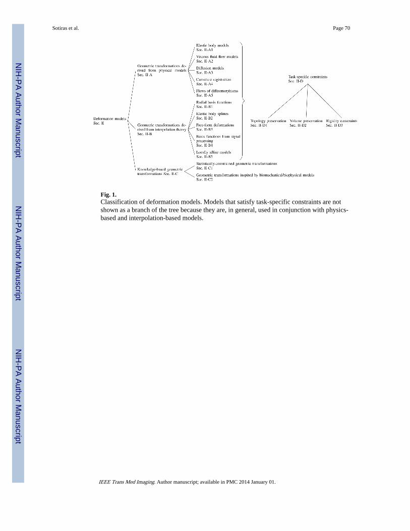
Fig. 1.Classification of deformation models. Models that satisfy task-specific constraints are notshown as a branch of the tree because they are, in general, used in conjunction with physics-based and interpolation-based models.
Sotiras et al. Page 70
IEEE Trans Med Imaging. Author manuscript; available in PMC 2014 January 01.
NIH
-PA Author Manuscript
NIH
-PA Author Manuscript
NIH
-PA Author Manuscript
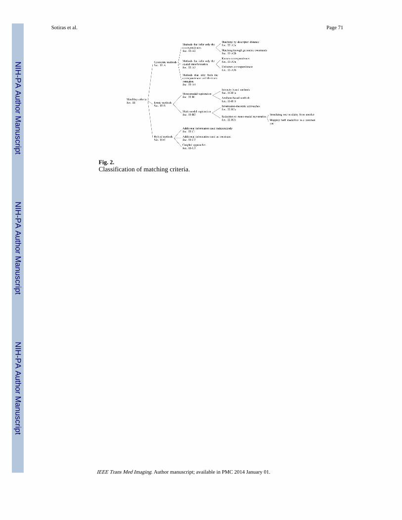
Fig. 2.Classification of matching criteria.
Sotiras et al. Page 71
IEEE Trans Med Imaging. Author manuscript; available in PMC 2014 January 01.
NIH
-PA Author Manuscript
NIH
-PA Author Manuscript
NIH
-PA Author Manuscript
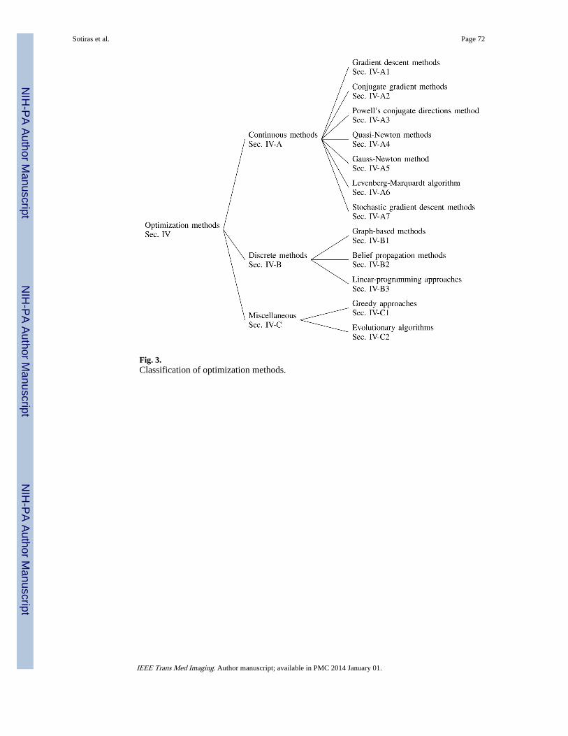
Fig. 3.Classification of optimization methods.
Sotiras et al. Page 72
IEEE Trans Med Imaging. Author manuscript; available in PMC 2014 January 01.
NIH
-PA Author Manuscript
NIH
-PA Author Manuscript
NIH
-PA Author Manuscript