Embed Size (px)
Citation preview
Published online 09 October 2014 Nucleic Acids Research, 2014, Vol. 42, No. 20 12469–12482doi: 10.1093/nar/gku927
Defective histone supply causescondensin-dependent chromatin alterations, SACactivation and chromosome decatenation impairmentMarina Murillo-Pineda1, Marıa J. Cabello-Lobato1, Marta Clemente-Ruiz1,Fernando Monje-Casas2 and Felix Prado1,*
1Departamento de Biologıa Molecular, Centro Andaluz de Biologıa Molecular y Medicina Regenerativa (CABIMER),Consejo Superior de Investigaciones Cientıficas (CSIC), Seville, Spain and 2Universidad de Sevilla (US), Seville,Spain
Received June 09, 2014; Revised September 22, 2014; Accepted September 23, 2014
ABSTRACT
The structural organization of chromosomes is es-sential for their correct function and dynamics duringthe cell cycle. The assembly of DNA into chromatinprovides the substrate for topoisomerases and con-densins, which introduce the different levels of su-perhelical torsion required for DNA metabolism. Inparticular, Top2 and condensin are directly involvedin both the resolution of precatenanes that form dur-ing replication and the formation of the intramolec-ular loop that detects tension at the centromericchromatin during chromosome biorientation. Herewe show that histone depletion activates the spin-dle assembly checkpoint (SAC) and impairs sis-ter chromatid decatenation, leading to chromosomemis-segregation and lethality in the absence of theSAC. We demonstrate that histone depletion impairschromosome biorientation and activates the Aurora-dependent pathway, which detects tension problemsat the kinetochore. Interestingly, SAC activation issuppressed by the absence of Top2 and Smc2, anessential component of condensin. Indeed, smc2-8suppresses catenanes accumulation, mitotic arrestand growth defects induced by histone depletionat semi-permissive temperature. Remarkably, SACactivation by histone depletion is associated withcondensin-mediated alterations of the centromericchromatin. Therefore, our results reveal the impor-tance of a precise interplay between histone supplyand condensin/Top2 for pericentric chromatin struc-ture, precatenanes resolution and centromere biori-entation.
INTRODUCTION
The assembly of DNA into nucleosomes provides the firstlevel of chromosome organization and the substrate fora plethora of enzymatic and structural factors that buildchromosomes. Nucleosomes, topoisomerase II (Top2 inyeast) and condensin are the main determinants of the levelof DNA supercoiling and chromosome compaction. Notsurprisingly, they play essential roles in DNA metabolicprocesses, which involve the continuous accumulation andrelease of torsional stress.
Chromosome packaging starts with the assembly ofnewly replicated DNA into chromatin during S phase. Thenucleosome––the repetitive unit of chromatin––is formedby ∼146 base pairs of DNA wrapped 1.65 times aroundan octamer of histones. This octamer is formed by a core(H3/H4)2 tetramer to which an H2A/H2B dimer binds oneach side (1). In a highly regulated process, histone chap-erones and chromatin assembly factors interact physicallyand genetically with components of the replisome to en-sure a rapid and correct supply of histones at the replica-tion fork (2). Nucleosome assembly introduces an accumu-lation of DNA supercoiling that is further modulated by theactivities of DNA topoisomerases and condensins, whichplay fundamental roles in the structural and functional or-ganization of chromosomes. In particular, Top2 can un-tangle catenated molecules and resolve both positive andnegative supercoils. Condensins are multisubunit complexesformed by two ‘structural maintenance of chromosomes’(SMCs, Smc2 and Smc4 in yeast) ATPases and three non-SMC subunits, which form a V-shaped structure able to trapDNA and introduce positive superhelical tension. Top2 andcondensin are essential for proper chromosome condensa-tion and segregation and play important roles in processessuch as transcription, recombination and DNA repair (3,4).Top2 and condensin are also essential for resolving the pre-catenanes that result from the advance of the replicationforks (5–8). Finally, they provide the tensile properties of the
*To whom correspondence should be addressed. Tel: 34 954468210; Fax: 34 954461664; Email: [email protected]
C© The Author(s) 2014. Published by Oxford University Press on behalf of Nucleic Acids Research.This is an Open Access article distributed under the terms of the Creative Commons Attribution License (http://creativecommons.org/licenses/by-nc/4.0/), whichpermits non-commercial re-use, distribution, and reproduction in any medium, provided the original work is properly cited. For commercial re-use, please [email protected]
at Centro de Inform
ación y D
ocumentaciÃ
³n CientÃ
fica on October 20, 2015
http://nar.oxfordjournals.org/D
ownloaded from
12470 Nucleic Acids Research, 2014, Vol. 42, No. 20
centromeric chromatin, which are essential for the activa-tion of the spindle assembly checkpoint (SAC) in responseto problems in the attachment of the microtubules to thekinetochores (9–14).
Chromatin is directly involved in the response to DNAdamage and chromosome segregation. Specifically, subtlechanges in chromatin structure caused by a deficit or an ex-cess in the pool of available histones have deleterious con-sequences on genome integrity (15–18). In the case of his-tone depletion, yeast mutants display replication fork insta-bility and accumulation of recombinogenic DNA damage,phenotypes that are reminiscent to those displayed by chro-matin assembly mutants lacking either the histone chap-erone Asf1 or the chromatin assembly factors Cac1 andRtt106 (17–20). In addition, cells expressing different mu-tants of histone H4 or lacking Cac1 and the histone chaper-one Hir1 display defects in the centromeric chromatin andthe kinetochores that are associated with increased ratesof chromosome mis-segregation and the activation of theSAC (21–23). Not surprisingly, nucleosome depletion by hi-stone loss causes a G2/M arrest (18,24). Here we show that,unexpectedly, the DNA damage checkpoint does not con-tribute to this arrest; instead, histone depletion activates theSAC. We show that histone depletion affects chromosomebiorientation and activates the Aurora-dependent branchof the SAC, which detects problems of tension at the kine-tochore (25,26). We also show that histone depletion im-pairs sister chromatid decatenation, leading to chromosomemis-segregation and cell lethality in SAC-defective cells. Im-portantly, histone depletion-mediated SAC activation is as-sociated with condensin-mediated alterations of the cen-tromeric chromatin, and accordingly, it is suppressed by theabsence of Smc2 and Top2; likewise, catenanes accumula-tion is suppressed by the absence of Smc2. Altogether, ourresults suggest that histone deposition and condensin/Top2cooperate to assemble a structural and functional chro-matin structure at the centromeric region.
MATERIALS AND METHODS
Yeast strains and plasmids
Yeast strains used in this study are listed in SupplementaryTable S1. Tagged strains and deletion mutants were con-structed by a polymerase chain reaction (PCR)-based strat-egy (27). chr-G::HHF1 strains were constructed by replac-ing in a hhf2Δ strain the HHF1 promoter with a PCR frag-ment containing the GAL1 promoter and the URA3 markerfrom pARSGLB-IN (28). G::HHF2 and M::HHF2 strainswere constructed by replacing plasmid p413TARtetH4 withplasmid pUK421 (G::HHF2) or p416MetH4 (M::HHF2).p413TARtetH4 (18), pUK421 (24) and p416MetH4 areHIS3-, TRP1- and URA3-based centromeric plasmids ex-pressing histone H4 from tet, GAL1 and MET25 pro-moter, respectively. p416MetH4 was constructed in twosteps: first, a BamHI-XhoI PCR fragment containing theORF of HHF2 was inserted at the XbaI (made blunt)-XhoIsite of p426Met25 (29); then, the generated PvuII frag-ment containing the MET25p::HHF2::CYC1t constructwas inserted at PvuII of pRS416. pWJ1344 (R. Roth-stein, Columbia University) and p314R52YFP are LEU2-
and TRP1-based centromeric plasmids expressing RAD52-YFP. p314R52YFP was constructed by inserting a SacI-XhoI fragment from pWJ1213 containing the constructRAD52-YFP at the SacI-XhoI site of pRS314. pRS416 (30)and YCp50 (31) are URA3-based centromeric plasmids;YCpPDED1T2 is a URA3-based centromeric plasmid ex-pressing Top2 from the DED1 promoter (32).
Growth conditions
Yeast cells were grown at 30◦C––unless otherwise stated––insupplemented minimal medium (SMM), except for theanalyses of Mad2-GFP in Figure 2B and Brn1-Pk9 in Fig-ures 4D, 5A and 5B, which required nocodazole (NCD)treatment and were performed in YPD-rich medium. ForG1 synchronization, cells were grown to mid-log-phaseand �-factor was added twice at 60-min intervals at ei-ther 2 (BAR1 strains) or 0.5 �g/ml (bar1Δ strains), ex-cept for t::HHF2/G::HHF2/M::HHF2 strains, which weretreated at 90 (t::HHF2 and G::HHF2) or 120 (M::HHF2)min intervals at either 5 (BAR1) or 1 �g/ml (bar1Δ).Cells were then washed three times and released into freshmedium with 50 �g/ml pronase. To induce histone deple-tion, t::HHF2 cells growing in the presence of 5 �g/mldoxycycline were shifted to 0.25 �g/ml during G1 synchro-nization and release; G::HHF2 cells growing in the pres-ence of 2% galactose were shifted to 2% glucose during thesecond incubation with �-factor and after release, exceptfor Figure 6D where they were shifted to 0.05% galactose;chr-G::HHF1 cells growing in the presence of 2% galactosewere shifted to 2% glucose after �-factor release; M::HHF2cells growing in the absence of methionine were shiftedto medium with 100 �M methionine during G1 synchro-nization and release. To induce Top2 degradation duringG1 synchronization in top2dg strains, mid-log phase cellsgrowing at 26◦C in SMM with 2% raffinose were shiftedto 2% raffinose with �-factor for 2 h at 26◦C, then shiftedto 1.5% raffinose/2% galactose with 50 ng/ml doxycyclineand �-factor for 1 h at 26◦C, and finally shifted to 1.5%raffinose/2% galactose with 50 ng/ml doxycycline and �-factor for 1 h at 37◦C. Cell growth analyses were performedby plating 10-fold serial dilutions from the same number ofmid-log phase cells in SMM plates containing 0.25 �g/mldoxycycline.
Flow cytometry
DNA content analysis was performed by Flow cytometry asreported previously (18). Each cell cycle progression analy-sis by Flow cytometry was repeated two to three times withsimilar results.
Immunofluorescence analysis
Immunolocalization of the spindles was performed byimmunofluorescence using antibodies anti-tubulin (Ab-cam) and anti-rat fluorescein isothiocyanate (Jackson Im-munoResearch Laboratories, Inc.) as previously described(33), except that formaldehyde fixation was overnight.Each cell cycle progression analysis by immunofluores-cence against tubulin was repeated twice with similar re-sults. Rad52-YFP, Mad2-GFP and tetR-GFP signals were
at Centro de Inform
ación y D
ocumentaciÃ
³n CientÃ
fica on October 20, 2015
http://nar.oxfordjournals.org/D
ownloaded from
Nucleic Acids Research, 2014, Vol. 42, No. 20 12471
A270
wt
t::HHF2
mad2∆
t::HHF2mad2∆
Ewt t::HHF2
D
C
% c
ells
wt pds1∆t::HHF2 t::HHF2 pds1∆
time after G1 release (min)0 30 45 60 90 120 150 180 0 30 45 60 90 120 150 180 0 30 45 60 90 120 150 180 0 30 45 60 90 120 150 180
time after G1 release (min)time after G1 release (min) time after G1 release (min)
G1 30 60 90 120 150 180 210 240
time after G1 release (min)
wt
t::HHF2
ndc10-1
t::HHF2ndc10-1
1C 2C 4C
G1 30 60 90 120 150 180 210 240
time after G1 release (min)
% c
ells
mad2∆
0 30 45 60 75 90 105 120
0 30 45 60 75 90 105 120
0 30 45 60 75 90 105 120
0 30 45 60 75 90 105 120
0 30 45 60 75 90 105 120
0 30 45 60 75 90 105 120
time after G1 release (min)
t::HHF2 mad2∆
time after G1 release (min)
Bmetaphaseanaphase
% c
ells
% c
ells
rad53∆ chk1∆
0
20
40
60
80
100
0
20
40
60
80
100
% c
ells
0
20
40
60
80
100
0
20
40
60
80
100
0
20
40
60
80
100
0
20
40
60
80
100
0
20
40
60
80
100
0
20
40
60
80
100
0
20
40
60
80
100
0
20
40
60
80
100
t::HHF2 rad53∆ chk1∆
wt t::HHF2
rad53∆chk1∆
t::HHF2rad53∆ chk1∆
metaphaseanaphase
Pds1
Pgk1
G1 30 60* 90 180150120 G1 30 60 90 180150*120 270240 330300210*
time after G1 release (min)time after G1 release (min)
39
MW
75
51
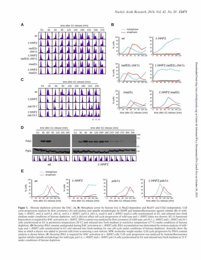
Figure 1. Histone depletion activates the SAC. (A, B) Metaphase arrest by histone loss is Mad2-dependent and Rad53 and Chk1-independent. Cellcycle progression analysis by flow cytometry (A) and nuclear and spindle morphologies by DAPI and immunofluorescence against tubulin (B) of wild-type, t::HHF2, sml1Δ rad53Δ chk1Δ, sml1Δ t::HHF2 rad53Δ chk1Δ, mad2Δ and t::HHF2 mad2Δ cells synchronized in G1 and released into freshmedium under conditions of histone depletion. sml1Δ did not affect cell cycle progression of wild type and t::HHF2 (data not shown). (C) A functionalkinetochore is required for SAC activation in t::HHF2. DNA content was analyzed by flow cytometry of wild-type, ndc10-1, t::HHF2 and t::HHF2 ndc10-1cells synchronized in G1 at permissive temperature (26◦C) and released into fresh medium at restrictive temperature (37◦C) under conditions of histonedepletion. (D) Securine/Pds1 remains undegraded during SAC activation in t::HHF2 cells. Pds1 accumulation was determined by western analysis of wild-type and t::HHF2 cells synchronized in G1 and released into fresh medium for one cell cycle under conditions of histone depletion. Asterisks show thetime at which �-factor was added to prevent cells from re-entering a new mitosis. MW, molecular weight marker. Cell cycle progression by DNA contentanalysis is shown below. (E) Securine/Pds1 is required for SAC activation in t::HHF2 cells. Cell cycle progression was analyzed by immunofluorescenceagainst tubulin (spindle morphology) for wild-type, pds1Δ, t::HHF2 and t::HHF2 pds1Δ cells synchronized in G1 and released into fresh medium at 23◦Cunder conditions of histone depletion.
at Centro de Inform
ación y D
ocumentaciÃ
³n CientÃ
fica on October 20, 2015
http://nar.oxfordjournals.org/D
ownloaded from
12472 Nucleic Acids Research, 2014, Vol. 42, No. 20
B
A
wt
bright field
Mad2-GFP
DAPI
t::HHF2- NCD - NCD + NCD+ NCD
wt
- NCD + NCD
t::HHF2
0
60
20
30
40
50
59
46
2 1.8
70
10
cells
with
Mad
2-G
FP fo
cus
(%)
wtDMSO 15 20
benomyl (μg/ml)
t::HHF2
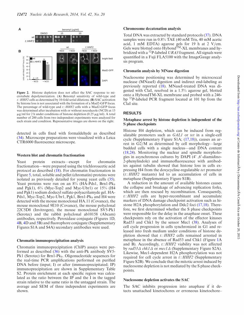
Figure 2. Histone depletion does not affect the SAC response to mi-crotubule depolymerization. (A) Benomyl sensitivity of wild-type andt::HHF2 cells as determined by 10-fold serial dilutions. (B) SAC activationby histone loss is not associated with the formation of a Mad2-GFP focus.The percentage of wild-type and t::HHF2 cells with a Mad2-GFP focuswas determined after incubation with or without nocodazole (NCD) at 15�g/ml for 2 h under conditions of histone depletion (0.25 �g/ml). A totalnumber of 200 cells from two independent experiments were analyzed foreach strain and condition. Representative images are shown on the right.
detected in cells fixed with formaldehyde as described(34). Microscope preparations were visualized with a LeicaCTR6000 fluorescence microscope.
Western blot and chromatin fractionation
Yeast protein extracts––except for chromatinfractionation––were prepared using the trichloroacetic acidprotocol as described (18). For chromatin fractionation inFigure 5, total, soluble and pellet (chromatin) proteins wereisolated as previously detailed for young yeast cells (35).Yeast proteins were run on an 8% (HA-Pds1, Brn1-Pk9and Pgk1), 6% (Myc-Top2 and Myc-Ubr1) or 15% (H4and Pgk1) sodium dodecyl sulfate-polyacrilamyde gel. HA-Pds1, Myc-Top2, Myc-Ubr1, Pgk1, Brn1-Pk9 and H4 weredetected with the mouse monoclonal HA.11 (Covance), themouse monoclonal 9E10 (Covance), the mouse polyclonal22C5D8 (Invitrogen), the mouse monoclonal SV5-Pk1(Serotec) and the rabbit polyclonal ab10158 (Abcam)antibodies, respectively. Peroxidase conjugate (Figures 1D,4B, 4D and 5B) and fluorophore conjugate (SupplementaryFigures S1A and S4A) secondary antibodies were used.
Chromatin immunoprecipitation analysis
Chromatin immunoprecipitation (ChIP) assays were per-formed as described (36) with the anti-Pk antibody SV5-Pk1 (Serotec) for Brn1-Pk9. Oligonucleotide sequences forthe real-time PCR amplifications performed on purifiedDNA before (input; I) or after (immunoprecipitated; IP)immunoprecipitation are shown in Supplementary TableS2. Protein enrichment at each specific region was calcu-lated as the ratio between the IP and the I in the taggedstrain relative to the same ratio in the untagged strain. Theaverage and SEM of three independent experiments areshown.
Chromosome decatenation analysis
Total DNA was extracted by standard protocols (37). DNAsamples were run in 0.8% TAE (40 mM Tris, 40 mM aceticacid, 1 mM EDTA) agarose gels for 19 h at 2 V/cm.Gels were blotted onto HybondTM-XL membranes and hy-bridized with a 32P-labeled URA3 fragment. All signals werequantified in a Fuji FLA5100 with the ImageGauge analy-sis program.
Chromatin analysis by MNase digestion
Nucleosome positioning was determined by micrococcalnuclease (MNaseI) digestion and indirect end-labeling aspreviously reported (18). MNaseI-treated DNA was di-gested with ClaI, resolved in a 1.5% agarose gel, blottedonto a HybondTM-XL membrane and probed with a 246-bp 32P-labeled PCR fragment located at 101 bp from theClaI site.
RESULTS
Metaphase arrest by histone depletion is independent of theS phase checkpoints
Histone H4 depletion, which can be induced from reg-ulatable promoters such as GAL1 or tet in a single-cellcycle (Supplementary Figure S1A; (17,18)), causes an ar-rest in G2/M as determined by cell morphology––largebudded cells with a single nucleus––and DNA content(18,24). Monitoring the nuclear and spindle morpholo-gies in asynchronous cultures by DAPI (4’ ,6’-diamidino-2-phenylindole) and immunofluorescence with antibod-ies against tubulin showed that histone loss in cells ex-pressing H4 from the doxycycline-regulatable tet promoter(t::HHF2 mutants) led to an accumulation of cells inmetaphase (Supplementary Figure S1B).
A reduction in the amount of available histones causesthe collapse and breakage of advancing replication forks,which are then rescued by recombination. Consequently,t::HHF2 cells are hyper-recombinant and accumulatemarkers of DNA damage checkpoint activation such as hi-stone H2A phosphorylation and Ddc2 foci (17,18). There-fore, we first determined whether the S phase checkpointswere responsible for the delay in the anaphase onset. Thesecheckpoints rely on the activation of the effector kinasesRad53 and Chk1 by the sensor Mec1 (38). Analyses ofcell cycle progression in cells synchronized in G1 and re-leased into fresh medium under conditions of histone de-pletion showed that t::HHF2 cells remained arrested inmetaphase in the absence of Rad53 and Chk1 (Figure 1Aand B). Accordingly, t::HHF2 viability was not affectedby rad53Δ chk1Δ or mec1Δ (Supplementary Figure S2A).Likewise, Mec1-dependent H2A phosphorylation was notrequired for cell cycle arrest in t::HHF2 (SupplementaryFigure S2B). We conclude that the mitotic arrest induced bynucleosome depletion is not mediated by the S phase check-points.
Nucleosome depletion activates the SAC
The SAC inhibits progression into anaphase if it de-tects unattached kinetochores or erroneous kinetochore–
at Centro de Inform
ación y D
ocumentaciÃ
³n CientÃ
fica on October 20, 2015
http://nar.oxfordjournals.org/D
ownloaded from
Nucleic Acids Research, 2014, Vol. 42, No. 20 12473
t::HHF2
ipl1-321
t::HHF2ipl1-321
A
B
Ctime after G1 release (min)
wt
G1 30 60 90 120 150 180 210 240 270 300
bright field
foci1 2
cenIV-GFP
cenIV-GFP+ Spc42-Cherry
+ DAPI
DAPIM::CDC20
G::HHF2
M::CDC20
M::CDC20 G::HHF2M::CDC20
G1 60 90 120
0
20
10
30
40
60
50
cells
with
two
cenI
V-G
FP fo
ci (%
)
time after G1 release (min)
type of cells (no cen foci)
*** **
* ***
wt
bright field
amph.amph. synt.
G::HHF2
wt
1 2
type of cells (no cen foci)
1 2
G::HHF2
0
0.7
0.30.40.50.6
0.8
0.20.1
0.91.0
shor
t SP
B-C
en/lo
ng S
PB
-Cen
***
0
10
14
2
4
6
8
13.3
<11.8
12
SP
B-C
en>S
PB
-SP
B (%
)
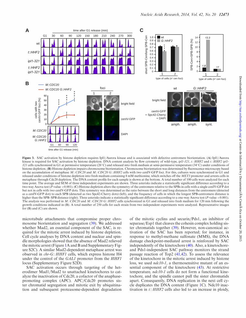
Figure 3. SAC activation by histone depletion requires Ipl1/Aurora kinase and is associated with defective centromere biorientation. (A) Ipl1/Aurorakinase is required for SAC activation by histone depletion. DNA content analysis by flow cytometry of wild-type, ipl1-321, t::HHF2 and t::HHF2 ipl1-321 cells synchronized in G1 at permissive temperature (26◦C) and released into fresh medium at semi-permissive temperature (34◦C) under conditions ofhistone depletion. (B) Histone depletion impairs chromosome biorientation. Chromosome biorientation was determined by fluorescence microscopy basedon the accumulation of metaphase M::CDC20 and M::CDC20 G::HHF2 cells with two cenIV-GFP foci. For this, cultures were synchronized in G1 andreleased under conditions of histone depletion into fresh medium containing 8 mM methionine, which switches off the MET3 promoter and arrests cells inmetaphase through Cdc20 depletion. The DNA content profile for each sample is shown at the bottom. A total number of 100 cells were analyzed for eachtime point. The average and SEM of three independent experiments are shown. Three asterisks indicate a statistically significant difference according to atwo-way Anova test (P-value <0.001). (C) Histone depletion alters the symmetry of the centromere relative to the SPBs in cells with a single cenIV-GFP dotbut not in cells with two cenIV-GFP dots. This symmetry was determined as the ratio between the short and long distances from the centromere (detectedas a cenIV-GFP dot) to each SPB (detected as two Spc42-Cherry dots) (left), and the frequency of cells in which the longest SPB-centromere distance ishigher than the SPB–SPB distance (right). Three asterisks indicate a statistically significant difference according to a one-way Anova test (P-value <0.001).The analysis was performed in M::CDC20 and M::CDC20 G::HHF2 cells synchronized in G1 and released into fresh medium for 120 min following thegrowth conditions indicated in (B). A total number of 250 cells for each strain from two independent experiments were analyzed. Representative imagesfor (B) and (C) are shown.
microtubule attachments that compromise proper chro-mosome biorientation and segregation (39). We addressedwhether Mad2, an essential component of the SAC, is re-quired for the mitotic arrest induced by histone depletion.Cell cycle analyses by DNA content and nuclear and spin-dle morphologies showed that the absence of Mad2 relievedthe mitotic arrest (Figure 1A and B and Supplementary Fig-ure S2C). A similar Mad2-dependent metaphase arrest wasobserved in chr-G::HHF1 cells, which express histone H4under the control of the GAL1 promoter from the HHF1locus (Supplementary Figure S2D).
SAC activation occurs through targeting of the het-erodimer Mad1/Mad2 to unattached kinetochores to cat-alyze the inactivation of Cdc20, a cofactor of the anaphase-promoting complex (APC). APC/Cdc20 promotes sis-ter chromatid segregation and mitotic exit by ubiquitina-tion and subsequent proteasome-dependent degradation
of the mitotic cyclins and securin/Pds1, an inhibitor ofseparase/Esp1 that cleaves the cohesin complex holding sis-ter chromatids together (39). However, non-canonical ac-tivation of the SAC has been reported; for instance, inresponse to methyl-methane sulfonate (MMS), the DNAdamage checkpoint-mediated arrest is reinforced by SACindependently of the kinetochore (40). Also, a kinetochore-and Pds1-independent SAC pathway monitors the strandpassage reaction of Top2 (41,42). To assess the relevanceof the kinetochore in the mitotic arrest induced by histoneloss, we used ndc10-1, a thermosensitive mutant of an es-sential component of the kinetochore (43). At restrictivetemperature, ndc10-1 cells do not form a functional kine-tochore, and the spindle cannot pull the sister chromatidsapart. Consequently, DNA replication in the next cell cy-cle duplicates the DNA content (Figure 1C). Ndc10 inac-tivation in t::HHF2 cells also led to an increase in ploidy,
at Centro de Inform
ación y D
ocumentaciÃ
³n CientÃ
fica on October 20, 2015
http://nar.oxfordjournals.org/D
ownloaded from
12474 Nucleic Acids Research, 2014, Vol. 42, No. 20
A time after G1 release (min)
wt
M::HHF2
top2td
M::HHF2 top2td
E
D
wtYCP50
YCP50
YCpPDED1T2
YCpPDED1T2t::HHF2
G1 30 60 90 120 150 180 210 240 270 300
C
B
F
wt
smc2-8t::HHF2
t::HHF2 smc2-8
time after G1 release (min)
wt
smc2-8
t::HHF2
t::HHF2 smc2-8
37oC
G1 30 60 90 120 150 180 210 240 270 300
G
wt
M::HHF2
top2td
M::HHF2 t
op2td
0
14
12
10
8
2
4
6
cells
with
Rad
52-Y
FP fo
ci (%
)
H
wt
t::HHF2
smc2
-8
t::HHF2 s
mc2-8
0
25
20
15
10
5
budd
ed c
ells
with
Rad
52-Y
FP fo
ci (%
)**
***
***
***
***
+- +-
Top2
Pgk1
Ubr1
top2tdtd:
M::HHF2 top2td
100135MW
Pgk1
Brn1-Pk9
wt- G::HHF2
180
48
245MW
48
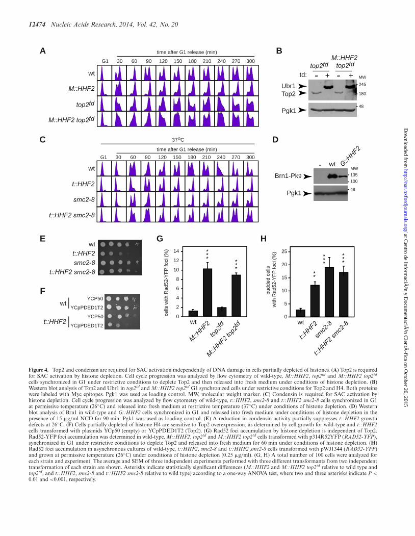
Figure 4. Top2 and condensin are required for SAC activation independently of DNA damage in cells partially depleted of histones. (A) Top2 is requiredfor SAC activation by histone depletion. Cell cycle progression was analyzed by flow cytometry of wild-type, M::HHF2, top2td and M::HHF2 top2td
cells synchronized in G1 under restrictive conditions to deplete Top2 and then released into fresh medium under conditions of histone depletion. (B)Western blot analysis of Top2 and Ubr1 in top2td and M::HHF2 top2td G1 synchronized cells under restrictive conditions for Top2 and H4. Both proteinswere labeled with Myc epitopes. Pgk1 was used as loading control. MW, molecular weight marker. (C) Condensin is required for SAC activation byhistone depletion. Cell cycle progression was analyzed by flow cytometry of wild-type, t::HHF2, smc2-8 and t::HHF2 smc2-8 cells synchronized in G1at permissive temperature (26◦C) and released into fresh medium at restrictive temperature (37◦C) under conditions of histone depletion. (D) Westernblot analysis of Brn1 in wild-type and G::HHF2 cells synchronized in G1 and released into fresh medium under conditions of histone depletion in thepresence of 15 �g/ml NCD for 90 min. Pgk1 was used as loading control. (E) A reduction in condensin activity partially suppresses t::HHF2 growthdefects at 26◦C. (F) Cells partially depleted of histone H4 are sensitive to Top2 overexpression, as determined by cell growth for wild-type and t::HHF2cells transformed with plasmids YCp50 (empty) or YCpPDED1T2 (Top2). (G) Rad52 foci accumulation by histone depletion is independent of Top2.Rad52-YFP foci accumulation was determined in wild-type, M::HHF2, top2td and M::HHF2 top2td cells transformed with p314R52YFP (RAD52-YFP),synchronized in G1 under restrictive conditions to deplete Top2 and released into fresh medium for 60 min under conditions of histone depletion. (H)Rad52 foci accumulation in asynchronous cultures of wild-type, t::HHF2, smc2-8 and t::HHF2 smc2-8 cells transformed with pWJ1344 (RAD52-YFP)and grown at permissive temperature (26◦C) under conditions of histone depletion (0.25 �g/ml). (G, H) A total number of 100 cells were analyzed foreach strain and experiment. The average and SEM of three independent experiments performed with three different transformants from two independenttransformation of each strain are shown. Asterisks indicate statistically significant differences (M::HHF2 and M::HHF2 top2td relative to wild type andtop2td, and t::HHF2, smc2-8 and t::HHF2 smc2-8 relative to wild type) according to a one-way ANOVA test, where two and three asterisks indicate P <
0.01 and <0.001, respectively.
at Centro de Inform
ación y D
ocumentaciÃ
³n CientÃ
fica on October 20, 2015
http://nar.oxfordjournals.org/D
ownloaded from
Nucleic Acids Research, 2014, Vol. 42, No. 20 12475
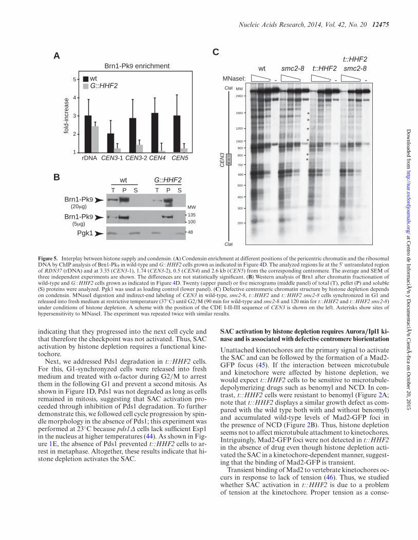
CA
B
wt
rDNA CEN3-1 CEN3-2 CEN4 CEN5
MW
Brn1-Pk9 enrichment
CE
N3
ClaI
ClaI
MNaseI: -
smc2-8
G::HHF2
t::HHF2t::HHF2smc2-8
-
-
-
----
-
-
--
900
800
700
600
500
400
300
200
2000
1500
1200
1000
-
- -
*
*
*
*
*
*
wtG::HHF2
1
3
5
2
4
fold
-incr
ease
IIIIII
100135MW
Pgk1
Brn1-Pk9(20μg)
Brn1-Pk9(5μg)
wtT P S T P S
48
Figure 5. Interplay between histone supply and condensin. (A) Condensin enrichment at different positions of the pericentric chromatin and the ribosomalDNA by ChIP analysis of Brn1-Pk9 in wild-type and G::HHF2 cells grown as indicated in Figure 4D. The analyzed regions lie at the 5′ untranslated regionof RDN37 (rDNA) and at 3.35 (CEN3-1), 1.74 (CEN3-2), 0.5 (CEN4) and 2.6 kb (CEN5) from the corresponding centromere. The average and SEM ofthree independent experiments are shown. The differences are not statistically significant. (B) Western analysis of Brn1 after chromatin fractionation ofwild-type and G::HHF2 cells grown as indicated in Figure 4D. Twenty (upper panel) or five micrograms (middle panel) of total (T), pellet (P) and soluble(S) proteins were analyzed. Pgk1 was used as loading control (lower panel). (C) Defective centromeric chromatin structure by histone depletion dependson condensin. MNaseI digestion and indirect-end labeling of CEN3 in wild-type, smc2-8, t::HHF2 and t::HHF2 smc2-8 cells synchronized in G1 andreleased into fresh medium at restrictive temperature (37◦C) until G2/M (90 min for wild-type and smc2-8 and 120 min for t::HHF2 and t::HHF2 smc2-8)under conditions of histone depletion. A scheme with the position of the CDE I-II-III sequence of CEN3 is shown on the left. Asterisks show sites ofhypersensitivity to MNaseI. The experiment was repeated twice with similar results.
indicating that they progressed into the next cell cycle andthat therefore the checkpoint was not activated. Thus, SACactivation by histone depletion requires a functional kine-tochore.
Next, we addressed Pds1 degradation in t::HHF2 cells.For this, G1-synchronyzed cells were released into freshmedium and treated with �-factor during G2/M to arrestthem in the following G1 and prevent a second mitosis. Asshown in Figure 1D, Pds1 was not degraded as long as cellsremained in mitosis, suggesting that SAC activation pro-ceeded through inhibition of Pds1 degradation. To furtherdemonstrate this, we followed cell cycle progression by spin-dle morphology in the absence of Pds1; this experiment wasperformed at 23◦C because pds1Δ cells lack sufficient Esp1in the nucleus at higher temperatures (44). As shown in Fig-ure 1E, the absence of Pds1 prevented t::HHF2 cells to ar-rest in metaphase. Altogether, these results indicate that hi-stone depletion activates the SAC.
SAC activation by histone depletion requires Aurora/Ipl1 ki-nase and is associated with defective centromere biorientation
Unattached kinetochores are the primary signal to activatethe SAC and can be followed by the formation of a Mad2-GFP focus (45). If the interaction between microtubuleand kinetochore were affected by histone depletion, wewould expect t::HHF2 cells to be sensitive to microtubule-depolymerizing drugs such as benomyl and NCD. In con-trast, t::HHF2 cells were resistant to benomyl (Figure 2A;note that t::HHF2 displays a similar growth defect as com-pared with the wild type both with and without benomyl)and accumulated wild-type levels of Mad2-GFP foci inthe presence of NCD (Figure 2B). Thus, histone depletionseems not to affect microtubule attachment to kinetochores.Intriguingly, Mad2-GFP foci were not detected in t::HHF2in the absence of drug even though histone depletion acti-vated the SAC in a kinetochore-dependent manner, suggest-ing that the binding of Mad2-GFP is transient.
Transient binding of Mad2 to vertebrate kinetochores oc-curs in response to lack of tension (46). Thus, we studiedwhether SAC activation in t::HHF2 is due to a problemof tension at the kinetochore. Proper tension as a conse-
at Centro de Inform
ación y D
ocumentaciÃ
³n CientÃ
fica on October 20, 2015
http://nar.oxfordjournals.org/D
ownloaded from
12476 Nucleic Acids Research, 2014, Vol. 42, No. 20
A
D
wt
wt (proper segregation)
t::HHF2 mad2∆ (missegregation)
mad2∆
G::HHF2
G::HHF2 m
ad2∆
0
10
14
2
4
6
8
11.3
<0.3
12
% c
hrom
osom
e IV
mis
segr
egat
ion
wtmad2∆
t::HHF2t::HHF2 mad2∆
wtmad1∆
t::HHF2t::HHF2 mad1∆
Bwt
t::HHF2ndc10-1
t::HHF2 ndc10-1
34oC26oC 30oC
Cwt
t::HHF2ipl1-321
t::HHF2 ipl1-321
37oC34oC32oC30oC
brightfield
CrIV-GFP+ DAPI
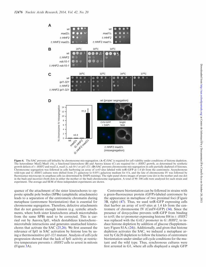
Figure 6. The SAC prevents cell lethality by chromosome mis-segregation. (A–C) SAC is required for cell viability under conditions of histone depletion.The heterodimer Mad2/Mad1 (A), a functional kinetochore (B) and Aurora kinase (C) are required for t::HHF2 growth, as determined by syntheticgrowth defects of t::HHF2 and mad2Δ, mad1Δ, ndc10-1 or ipl1-321. (D) SAC prevents chromosome mis-segregation in cells partially depleted of histones.Chromosome segregation was followed in cells harboring an array of tetO sites labeled with tetR-GFP at 1.4 kb from the centromere. Asynchronouswild-type and G::HHF2 cultures were shifted from 2% galactose to 0.05% galactose medium for 4 h, and the fate of chromosome IV was followed byfluorescence microscopy in anaphase cells (as determined by DAPI staining). The right panel shows images of proper (one dot in the mother and one dotin the bud) and incorrect (both dots in either the mother or the bud) chromosome segregation. A total of 90–100 cells were analyzed for each strain andexperiment. The average and SEM of three independent experiments are shown.
quence of the attachment of the sister kinetochores to op-posite spindle pole bodies (SPBs) (amphitelic attachments)leads to a separation of the centromeric chromatin duringmetaphase (centromere biorientation) that is essential forchromosome segregation. Therefore, defective attachmentsthat do not generate enough tension (e.g. syntelic attach-ments, where both sister kinetochores attach microtubulesfrom the same SPB) need to be corrected. This is car-ried out by Aurora/Ipl1, which destabilizes kinetochore–microtubule interactions and generates unattached kineto-chores that activate the SAC (25,26). We first assessed therelevance of Ipl1 in SAC activation by histone loss by us-ing a thermosensitive ipl1-321 mutant. Analysis of cell cycleprogression showed that the lack of Ipl1 activity at restric-tive temperature prevents t::HHF2 cells to arrest in mitosis(Figure 3A).
Centromere biorientation can be followed in strains witha green-fluorescence protein (GFP)-labeled centromere bythe appearance in metaphase of two proximal foci (Figure3B, right) (47). Thus, we used tetR-GFP expressing cellsthat harbor an array of tetO sites at 1.4 kb from the cen-tromere of chromosome IV (CenIV-GFP) (34). Since thepresence of doxycycline prevents tetR-GFP from bindingto tetO, the tet promoter expressing histone H4 in t::HHF2was replaced with the GAL1 promoter in G::HHF2, to in-duce histone depletion by addition of glucose (Supplemen-tary Figure S1A; (24)). Additionally, and given that histonedepletion activates the SAC, we induced a metaphase ar-rest by Cdc20 depletion to follow the kinetics of centromerebiorientation under similar cell cycle conditions for the mu-tant and the wild type. Thus, synchronous cultures werefirst arrested in G1, where all cells displayed a single GFP
at Centro de Inform
ación y D
ocumentaciÃ
³n CientÃ
fica on October 20, 2015
http://nar.oxfordjournals.org/D
ownloaded from
Nucleic Acids Research, 2014, Vol. 42, No. 20 12477
dot, and then released in the presence of methionine toswitch off the MET3 promoter that drives Cdc20 expression(M::CDC20). As shown in Figure 3B, cells displayed twoclosed GFP dots as they accumulated in metaphase. No-tably, only ∼20% of the metaphase arrested G::HHF2 cellsshowed two dots as compared with the ∼50% detected inthe wild type, indicating that histone depletion affects chro-mosome biorientation.
To further evaluate whether histone depletion-induceddefective chromosome biorientation was associated withsyntelic attachments, the distance between CenIV and eachof the SPBs was measured in M::CDC20 and M::CDC20G::HHF2 metaphase cells expressing both CenIV-GFP andthe SPB protein Spc42 fused to Cherry. In accordance withprevious results (48), we observed that histone depletion in-creased the length of the spindle, in particular in cells with asingle CenIV-GFP dot (Supplementary Figure S3A). As ex-pected if histone depletion led to the formation of syntelicattachments, the ratio between the shorter and the longerCenIV-SPB distance was lower in the mutant than in thewild type for cells with a single CenIV-GFP dot, while it wassimilar for cells with two CenIV-GFP dots (Figure 3C, left,and Supplementary Figure S3B). In addition, the spindlelength was shorter than the distance from the centromereto one of the SPBs in 13.3% of histone-depleted cells with asingle CenIV-GFP dot (Figure 3C, right). Altogether theseresults suggest that histone depletion gives rise to syntelicattachments.
Top2 and condensin are required for SAC activation in re-sponse to histone depletion
The assembly of DNA into chromatin provides a majorsource of topological constraints that demand the activ-ity of topoisomerases during chromosome dynamics. Infact, Top2 binds preferentially to genes with low nucleo-some density in histone-depleted cells (49). Thus, we de-cided to assess the role of Top2 in SAC activation by hi-stone loss. Since TOP2 is essential (50), we used a ‘de-gron’ allele of TOP2 that expresses a Top2 protein thatcan be rapidly degraded in the presence of doxycycline andgalactose at 37◦C (6). Under these conditions Top2 expres-sion from the tet promoter is repressed, and Top2 degra-dation is promoted by the E3 ubiquitin ligase Ubr1 ex-pressed from the GAL1 promoter. This background forcedus to replace the promoter expressing histone H4 with theMET25 promoter, which represses H4 in response to me-thionine (Supplementary Figure S1A; strain M::HHF2). Asshown for t::HHF2 and G::HHF2, M::HHF2 cells arrestedin metaphase when histone expression was repressed with100 �M methionine (Figure 4A). We verified that Top2 wasdegraded in G1-arrested cells prior to their release into freshmedium under restrictive conditions (Figure 4B). As previ-ously demonstrated, the lack of Top2 did not prevent pro-gression through mitosis even though top2td cells enteredinto the following cell cycle with an aberrant DNA con-tent profile due to chromosome mis-segregation by defec-tive sister chromatid decatenation (Figure 4A) (6). Impor-tantly, M::HHF2 top2td progressed through the cell cycleas did top2td, indicating that the absence of Top2 relievedthe SAC-mediated arrest induced by histone depletion. As
shown in Figure 4B, the expression of Top2 was not affectedby histone depletion, indicating that the arrest was not dueto increased levels of Top2 in the mutant.
Proper chromosome segregation depends on the accuracyof two processes that require Top2, namely, sister chromatiddecatenation and chromosome condensation (4). These twoprocesses also require condensin (5,8,51,52). Thus, we ad-dressed the relevance of Smc2, an essential component ofcondensin, in SAC activation by histone loss. Wild-type andt::HHF2 cells expressing either SMC2 or a thermosensi-tive smc2-8 allele were synchronized in G1 and released intofresh medium at restrictive temperature. In accordance withprevious results (14), smc2-8 cells arrested in metaphase at37◦C (Figure 4C). Remarkably, t::HHF2 smc2-8 progressedfaster through mitosis than the single mutants, indicatingthat condensin is required for SAC activation by histone de-pletion. As previously shown for Top2, SAC activation wasnot due to higher levels of condensin; indeed, histone de-pletion reduced the total amount of the condensin subunitBrn1 (Figure 4D). Likewise, suppression of the mitotic ar-rest by the absence of Top2 and condensin was not due toan increase in the levels of histone H4 (Supplementary Fig-ure S4A). Therefore, Top2 and condensin are required forhistone depletion-induced SAC activation.
Notably, a reduction of condensin activity in smc2-8 atpermissive temperature alleviated the mitotic arrest andpartially suppressed the growth defects associated with his-tone loss (Figure 4E and Supplementary Figure S4B). Thissuppression was not observed with other conditions thatcause DNA damage; in fact, smc2-8 was sensitive to hydrox-yurea (HU) and MMS and arrested in G2/M in responseto HU (Supplementary Figure S4C and S4D). These re-sults suggest that condensin generates a chromosomal prob-lem in histone-depleted cells that activates the SAC. Growthanalyses could not be performed with top2td because it islethal at restrictive conditions; however, if Top2 causes achromosomal problem detected by the SAC, t::HHF2 cellsshould be sensitive to high levels of Top2. Consistently,t::HHF2 cells transformed with a plasmid that increasesTop2 by a factor of 10 (32) displayed growth defects (Figure4F).
Although the SAC and not the S phase checkpoints ar-rests t::HHF2 cells, we cannot rule out the possibility thatthe SAC is activated by DNA damage in this mutant. Wethereby tested if DNA damage induced by histone deple-tion depends on Top2 and/or condensin by following theaccumulation of recombinogenic DNA damage (Rad52-YFP foci). Consistent with the fact that DNA damage byTop2 depletion requires cytokinesis (6), top2td cells releasedinto S phase without Top2 did not accumulate Rad52-YFPfoci (Figure 4G). Importantly, Top2 was not responsible forDNA damage in t::HHF2, as evidenced by the accumula-tion of Rad52-YFP foci in t::HHF2 top2td. When we an-alyzed the effect of condensin in Rad52-YFP foci, we ob-served that smc2-8 cells accumulated DNA damage even atpermissive temperature, as previously shown for other con-densin mutants (53), and that this accumulation was main-tained in t::HHF2 smc2-8 cells (Figure 4H). Thus, we can-not establish the relevance of condensin in the accumulationof DNA damage by histone loss. Nevertheless, the fact thatsmc2-8 suppresses the growth defects of t::HHF2 despite
at Centro de Inform
ación y D
ocumentaciÃ
³n CientÃ
fica on October 20, 2015
http://nar.oxfordjournals.org/D
ownloaded from
12478 Nucleic Acids Research, 2014, Vol. 42, No. 20
the cells accumulating DNA damage indicates that DNAdamage does not activate the SAC in histone-depleted cells.
Defective centromeric chromatin structure by histone deple-tion depends on condensin
Histone depletion causes alterations at the centromericchromatin which make DNA more accessible to the mi-crococcal nuclease (MNaseI), which cuts preferentially atnucleosome-free DNA (54). These chromatin modificationsmight alter the occupancy/activity of condensin and Top2thus impairing the tensile properties of the centromericchromatin. An alternative but not mutually exclusive possi-bility is that condensin and Top2 might be required to mod-ify the centromeric chromatin structure in cells with reducedlevels of histones. To explore these possibilities, we first an-alyzed Brn1 binding to previously identified regions (55).The results suggested that histone depletion causes a reduc-tion in the amount of condensin bound at the pericentricchromatin but not at the ribosomal chromatin, even thoughthe differences were not statistically significant (Figure 5A).Likewise, the total amount of Brn1 bound to chromatin wasslightly reduced in the mutant as determined by chromatinfractionation (P lane; Figure 5B).
Next, we analyzed the chromatin structure of CEN3 byMNaseI digestion and indirect-end labeling in mitotic cells(Figure 5C). While the lack of condensin activity in smc2-8cells at restrictive temperature did not affect the chromatinstructure of CEN3, histone depletion increased the acces-sibility of MNaseI to both the centromere DNA elements(CDEs I, II and III)––which are wrapped around an spe-cialized nucleosome––and proximal DNA sequences (Fig-ure 5C; asterisks (54)). Remarkably, these nucleosome al-terations were not detected in t::HHF2 smc2-8. Therefore,histone depletion by itself is not sufficient to alter the cen-tromeric chromatin; condensin activity is required for hi-stone depletion-induced chromatin alterations. This resultsuggests interplay between histone supply and condensin.
The SAC prevents chromosome mis-segregation bycondensin-dependent defective decatenation in responseto histone depletion
As the biological aim of the checkpoints is to provide timeto resolve chromosome-associated problems, the lack of acheckpoint may lead to a loss of viability. Accordingly, theabsence of the heterodimer Mad1/Mad2 led to a loss ofviability in t::HHF2 cells (Figure 6A). Likewise, t::HHF2ndc10-1 and t::HHF2 ipl1-321 were lethal at semipermis-sive temperature (Figure 6B and C). Therefore, the SAC isrequired for t::HHF2 viability.
Cell growth analyses also showed that the lack of eitherthe sensor Mec1 or the effectors Rad53 and Chk1 does notaffect the viability of t::HHF2 mad2Δ cells (SupplementaryFigure S5A and B), indicating that the SAC is not mask-ing a putative effect of the S phase checkpoints on histonedepletion-dependent mitotic arrest.
As a major genetic consequence of defective SAC activa-tion is chromosome mis-segregation, we followed the fateof CrIV in G::HHF2. To prevent a complete metaphaseblock by addition of glucose (24), wild-type and G::HHF2
cells were shifted from 2% galactose to 0.05% galactosefor 4 h. Proper chromosome segregation led to anaphasecells with a dot in the mother and a dot in the bud, whilechromosome mis-segregation led to anaphase cells with thetwo dots either in the mother or in the bud (Figure 6D,right panel). Whereas chromosome IV mis-segregation inG::HHF2, mad2Δ and wild-type cells was barely detectable(<0.3%), it increased to 11.3% in G::HHF2 mad2Δ cells(Figure 6D), indicating that SAC activation ensures properchromosome segregation in response to histone depletion.
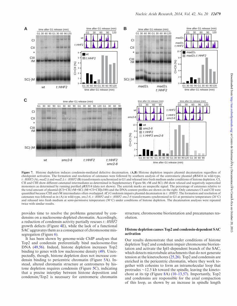
Next, we asked whether chromosome mis-segregation int::HHF2 was due to defective sister chromatid decatena-tion. DNA unwinding during replication generates positivetorsional stress ahead the fork, part of which is diffusedacross the replicated DNA by swiveling of the fork leadingto the formation of intertwined sister chromatids (precate-nanes). Resolution of precatenanes, which occurs right af-ter DNA synthesis and during mitosis as sister chromatidsmove apart, is carried out by Top2 and condensin, and it isessential for proper chromosome segregation (5–8). Forma-tion and resolution of precatenanes during cell cycle can befollowed by studying the accumulation of the different inter-mediates of a centromeric plasmid (8). Wild-type cells tran-siently accumulated different intermediates (Figure 7A; CI,CII and CIII), which represent catenated molecules as in-ferred by their accumulation in cells lacking Top2 (Supple-mentary Figure S6). Most plasmids were monomeric in G1-arrested t::HHF2 cells (rM and SC(-)M intermediates), in-dicating that most precatenanes were properly resolved un-der conditions of histone depletion (note that only a smallfraction of catenated plasmids was detected as comparedto the wild type, which might reflect minor resolution de-fects and/or incomplete synchronization; see fluorescence-activated cell sorting profiles). However, the catenanes ac-cumulated in t::HHF2 relative to the wild type during Sand G2/M, suggesting that histone depletion impaired de-catenation (Figure 7A). While Top2 rapidly resolves mostprecatenanes after their formation, a fraction remains untilmitosis, during which condensins and spindle tension facil-itate Top2 activity; accordingly, checkpoint activation pre-vents their resolution (5,8). Indeed, the entry into mitosisby lack of Mad2 reduced the fraction of catenanes at latetimes, but it did not affect the accumulation of early cate-nanes in t::HHF2 (Figure 7B). Next, we analyzed the roleof condensins in the accumulation of these catenated plas-mids, taking advantage of the fact that the effect of con-densin inactivation is barely detectable in small plasmids(8). As shown in Figure 7C, the accumulation of catenanesin t::HHF2 cells was suppressed by reducing the activity ofcondensins.
DISCUSSION
Here we show that histone depletion causes an unscheduledactivation of the SAC by Top2 and condensins, as well as anaccumulation of catenanes by condensins. Since catenanesaccumulation in the absence of Top2 does not arrest yeastcells (6,42), it is unlikely that checkpoint activation results asa consequence of defective decatenation in histone-depletedcells. Instead, SAC activation is associated with problemsof chromosome biorientation. Nevertheless, SAC activation
at Centro de Inform
ación y D
ocumentaciÃ
³n CientÃ
fica on October 20, 2015
http://nar.oxfordjournals.org/D
ownloaded from
Nucleic Acids Research, 2014, Vol. 42, No. 20 12479
A
G1 30 60 90 G1 30 60 90 G1 30 60 90 G1 30 60 90120
time after G1 release (min)
rM
*
SC(-)M
CII
CI
0
10
2
4
6
8
0
10
2
4
6
8
CI+
CII
(%)
smc2-8
time after G1 release (min)
time after G1 release (min)
wt
G1 30 60 90
t::HHF2smc2-8
time after G1 release (min)
t::HHF2
G1 30 60 90 120
G1 30 60 90 G1 30 60 90G1 30 60 90 G1 30 60 90120
C
wt t::HHF2
wt t::HHF2smc2-8 t::HHF2smc2-8
G1 30 60 90 30 12060 90time after G1 release (min)
G1
rM
*
SC(-)M
CII
CI
wtt::HHF2
0
6
2
3
4
5
1
CI+
CII
(%)
t::HHF2
time after G1 release (min)
time after G1 release (min)
wt
G1 30 60 90 120
G1 30 60 90 G1 30 60 90120
B
mad2∆ mad2∆t::HHF2
mad2∆ t::HHF2
G1 30 60 90 30 12060 90time after G1 release (min)
G1
rM
*
SC(-)M
CII
CI
mad2∆
CI+
CII
(%)
mad2∆t::HHF2
time after G1 release (min)
time after G1 release (min)
mad2∆
G1 30 60 90 120
G1 30 60 90 G1 30 60 90120
wt
t::HHF2smc2-8
t::HHF2 smc2-8
CIII
CIII
CIII
Figure 7. Histone depletion induces condensin-mediated defective decatenation. (A,B) Histone depletion impairs plasmid decatenation regardless ofcheckpoint activation. The formation and resolution of catenanes were followed by southern analysis of the centromeric plasmid pRS416 in wild-type,t::HHF2 (A), mad2Δ and mad2Δ t::HHF2 (B) transformants synchronized in G1 and released into fresh medium under conditions of histone depletion. CI,CII and CIII show different catenated intermediates as determined in Supplementary Figure S6; rM and SC(-)M show relaxed and negatively supercoiledmonomers as determined by running purified pRS314 (data not shown). The asterisk marks an unspecific signal. The percentage of catenanes relative tothe total amount of plasmid ([CI+CII/rM+SC(-)M+CI+CII]x100) and the DNA content profiles are shown on the right. Only catenanes CI and CII werequantified because CIII and rM intermediates often overlapped. (C) Condensin impairs plasmid decatenation in t::HHF2. The formation and resolution ofcatenanes was followed as in (A) in wild-type, smc2-8, t::HHF2 and t::HHF2 smc2-8 transformants synchronized in G1 at permissive temperature (26◦C)and released into fresh medium at semi-permissive temperature (30◦C) under conditions of histone depletion. The decatenation analyses were repeatedtwice with similar results.
provides time to resolve the problems generated by con-densins on a nucleosome-depleted chromatin. Accordingly,a reduction of condensin activity partially rescues t::HHF2growth defects (Figure 4E), while the lack of a functionalSAC aggravates them as a consequence of chromosome mis-segregation (Figure 6).
It has been shown by genome-wide ChIP analyses thatTop2 and condensin preferentially bind nucleosome-freeDNA (49,56). Indeed, histone depletion increases Top2binding to genes with low nucleosome density (49). Unex-pectedly, though, histone depletion does not increase con-densin binding to pericentric chromatin (Figure 5A). In-stead, altered chromatin structure at centromeres by his-tone depletion requires condensin (Figure 5C), indicatingthat a precise interplay between histone deposition andcondensin/Top2 is necessary for centromeric chromatin
structure, chromosome biorientation and precatenanes res-olution.
Histone depletion causes Top2 and condensin-dependent SACactivation
Our results demonstrate that under conditions of histonedepletion Top2 and condensin impair chromosome biorien-tation and activate the Ipl1-dependent branch of the SAC,which detects microtubule attachments that do not generatetension at the kinetochores (25,26). Top2 and condensin areenriched in the pericentric chromatin, where they work to-gether with cohesins to form an intramolecular loop thatprotrudes ∼12.5 kb toward the spindle, leaving the kineto-chore at its tip (Figure 8A) (10–13,57). Importantly, Top2and condensins are responsible for the axial compactionof this loop, as shown by an increase in spindle length
at Centro de Inform
ación y D
ocumentaciÃ
³n CientÃ
fica on October 20, 2015
http://nar.oxfordjournals.org/D
ownloaded from
12480 Nucleic Acids Research, 2014, Vol. 42, No. 20
A B C
Aurora/Ipl1Top2 & Condensin kinetochore
cohesinnucleosome Cse4-nucleosome
DNA
microtubule
D
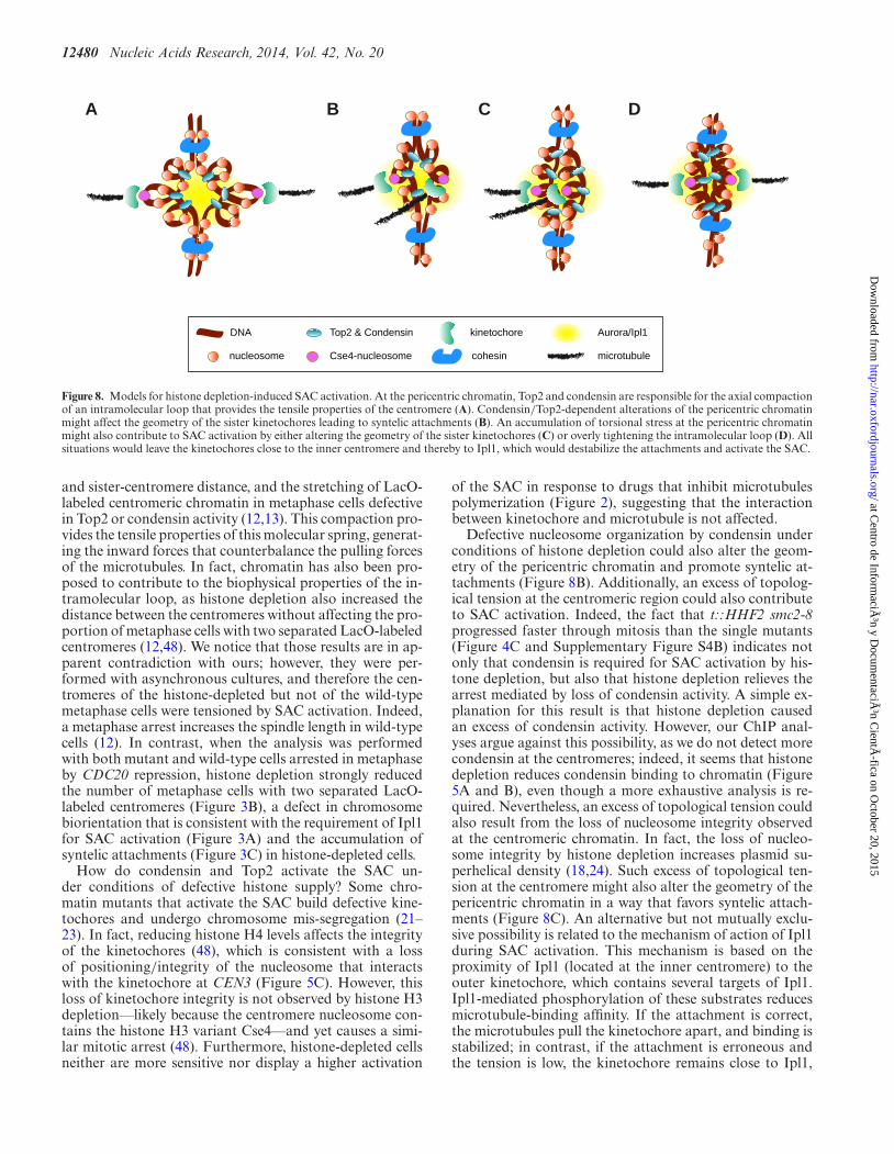
Figure 8. Models for histone depletion-induced SAC activation. At the pericentric chromatin, Top2 and condensin are responsible for the axial compactionof an intramolecular loop that provides the tensile properties of the centromere (A). Condensin/Top2-dependent alterations of the pericentric chromatinmight affect the geometry of the sister kinetochores leading to syntelic attachments (B). An accumulation of torsional stress at the pericentric chromatinmight also contribute to SAC activation by either altering the geometry of the sister kinetochores (C) or overly tightening the intramolecular loop (D). Allsituations would leave the kinetochores close to the inner centromere and thereby to Ipl1, which would destabilize the attachments and activate the SAC.
and sister-centromere distance, and the stretching of LacO-labeled centromeric chromatin in metaphase cells defectivein Top2 or condensin activity (12,13). This compaction pro-vides the tensile properties of this molecular spring, generat-ing the inward forces that counterbalance the pulling forcesof the microtubules. In fact, chromatin has also been pro-posed to contribute to the biophysical properties of the in-tramolecular loop, as histone depletion also increased thedistance between the centromeres without affecting the pro-portion of metaphase cells with two separated LacO-labeledcentromeres (12,48). We notice that those results are in ap-parent contradiction with ours; however, they were per-formed with asynchronous cultures, and therefore the cen-tromeres of the histone-depleted but not of the wild-typemetaphase cells were tensioned by SAC activation. Indeed,a metaphase arrest increases the spindle length in wild-typecells (12). In contrast, when the analysis was performedwith both mutant and wild-type cells arrested in metaphaseby CDC20 repression, histone depletion strongly reducedthe number of metaphase cells with two separated LacO-labeled centromeres (Figure 3B), a defect in chromosomebiorientation that is consistent with the requirement of Ipl1for SAC activation (Figure 3A) and the accumulation ofsyntelic attachments (Figure 3C) in histone-depleted cells.
How do condensin and Top2 activate the SAC un-der conditions of defective histone supply? Some chro-matin mutants that activate the SAC build defective kine-tochores and undergo chromosome mis-segregation (21–23). In fact, reducing histone H4 levels affects the integrityof the kinetochores (48), which is consistent with a lossof positioning/integrity of the nucleosome that interactswith the kinetochore at CEN3 (Figure 5C). However, thisloss of kinetochore integrity is not observed by histone H3depletion––likely because the centromere nucleosome con-tains the histone H3 variant Cse4––and yet causes a simi-lar mitotic arrest (48). Furthermore, histone-depleted cellsneither are more sensitive nor display a higher activation
of the SAC in response to drugs that inhibit microtubulespolymerization (Figure 2), suggesting that the interactionbetween kinetochore and microtubule is not affected.
Defective nucleosome organization by condensin underconditions of histone depletion could also alter the geom-etry of the pericentric chromatin and promote syntelic at-tachments (Figure 8B). Additionally, an excess of topolog-ical tension at the centromeric region could also contributeto SAC activation. Indeed, the fact that t::HHF2 smc2-8progressed faster through mitosis than the single mutants(Figure 4C and Supplementary Figure S4B) indicates notonly that condensin is required for SAC activation by his-tone depletion, but also that histone depletion relieves thearrest mediated by loss of condensin activity. A simple ex-planation for this result is that histone depletion causedan excess of condensin activity. However, our ChIP anal-yses argue against this possibility, as we do not detect morecondensin at the centromeres; indeed, it seems that histonedepletion reduces condensin binding to chromatin (Figure5A and B), even though a more exhaustive analysis is re-quired. Nevertheless, an excess of topological tension couldalso result from the loss of nucleosome integrity observedat the centromeric chromatin. In fact, the loss of nucleo-some integrity by histone depletion increases plasmid su-perhelical density (18,24). Such excess of topological ten-sion at the centromere might also alter the geometry of thepericentric chromatin in a way that favors syntelic attach-ments (Figure 8C). An alternative but not mutually exclu-sive possibility is related to the mechanism of action of Ipl1during SAC activation. This mechanism is based on theproximity of Ipl1 (located at the inner centromere) to theouter kinetochore, which contains several targets of Ipl1.Ipl1-mediated phosphorylation of these substrates reducesmicrotubule-binding affinity. If the attachment is correct,the microtubules pull the kinetochore apart, and binding isstabilized; in contrast, if the attachment is erroneous andthe tension is low, the kinetochore remains close to Ipl1,
at Centro de Inform
ación y D
ocumentaciÃ
³n CientÃ
fica on October 20, 2015
http://nar.oxfordjournals.org/D
ownloaded from
Nucleic Acids Research, 2014, Vol. 42, No. 20 12481
and binding is destabilized (39). Given that the distance be-tween Ipl1 and the kinetochore is essential for SAC activa-tion, an excess of torsional stress might overly tighten theintramolecular loop and leave the kinetochores close to theinner centromere, even though the attachments were correct(Figure 8D).
Histone depletion causes condensin-dependent accumulationof catenanes
The absence of Top2 activity does not prevent nucleosomedeposition in Xenopus egg extracts, indicating that nucle-osomes can be accommodated into the precatenanes gen-erated during DNA replication (58). The authors of thisstudy thus proposed that replication-coupled nucleosomedeposition may facilitate chromatid decatenation by forc-ing the precatenanes nodes to adopt a ‘hooked’ DNA jux-taposition conformation, which is the preferred substratefor the DNA disentangling activity of Top2 (59,60). Ac-cording to this hypothesis, defective nucleosome organiza-tion by condensin under conditions of histone depletioncould alter DNA geometry behind the fork and interferewith Top2-mediated chromatid decatenation. Likewise, theexcess of positive supercoiling that is associated with his-tone depletion (18,24) might interfere with Top2 activity byaffecting DNA geometry and thereby the resolution of cate-nanes. Baxter and Aragon have provided strong evidence fora model in which chromosome decatenation results from anexquisite interplay between the enzymatic activities of Top2and condensin, with the positive supercoiling introduced bycondensins and spindle tension as being necessary for cate-nanes resolution by Top2 (5,51).
In summary, our results reveal the importance of a cor-rect supply of histones for the accuracy of condensin/Top2-mediated DNA processes. Specifically, we show that a pre-cise interplay between histone deposition and condensin isrequired for pericentric chromatin structure, precatenanesresolution and centromere biorientation. An attractive pos-sibility that we are currently evaluating is that this inter-play modulated global chromatin structure and chromo-some condensation. Finally, we anticipate that defects inchromosome dynamics and cell cycle progression may beparticularly severe during physiological processes such asthe replicative aging, which strongly reduce the density ofnucleosomes (35,61).
SUPPLEMENTARY DATA
Supplementary Data are available at NAR Online.
ACKNOWLEDGMENTS
We thank L. Aragon, O. Cohen-Fix, J.F.X. Diffley and J.Nitiss for various strains and reagents, and P. Huertas andF. Cortes-Ledesma for critically reading the manuscript.
FUNDING
Spanish Ministry of Science [BFU2009-09036, BFU2012-38171]; Spanish Government [to M.M.-P., M.J.C.-L.].Funding for open access charge: Spanish Ministry of Sci-ence [BFU2012-38171].
Conflict of interest statement. None declared.
REFERENCES1. Khorasanizadeh,S. (2004) The nucleosome: from genomic
organization to genomic regulation. Cell, 116, 259–272.2. Burgess,R.J. and Zhang,Z. (2013) Histone chaperones in nucleosome
assembly and human disease. Nat. Struct. Mol. Biol., 20, 14–22.3. Hirano,T. (2012) Condensins: universal organizers of chromosomes
with diverse functions. Genes Dev., 26, 1659–1678.4. Vos,S.M., Tretter,E.M., Schmidt,B.H. and Berger,J.M. (2011) All
tangled up: how cells direct, manage and exploit topoisomerasefunction. Nature, 12, 827–841.
5. Baxter,J., Sen,N., Martinez,V.L., De Carandini,M.E.M.,Schvartzman,J.B., Diffley,J.F.X. and Aragon,L. (2011) Positivesupercoiling of mitotic DNA drives decatenation by topoisomerase IIin eukaryotes. Science, 331, 1328–1332.
6. Baxter,J. and Diffley,J.F.X. (2008) Topoisomerase II inactivationprevents the completion of DNA replication in budding yeast. Mol.Cell, 30, 790–802.
7. Bermejo,R., Doksani,Y., Capra,T., Katou,Y.M., Tanaka,H.,Shirahige,K. and Foiani,M. (2007) Top1- and Top2-mediatedtopological transitions at replication forks ensure fork progressionand stability and prevent DNA damage checkpoint activation. GenesDev., 21, 1921–1936.
8. Charbin,A., Bouchoux,C. and Uhlmann,F. (2014) Condensin aidssister chromatid decatenation by topoisomerase II. Nucleic AcidsRes., 42, 340–348.
9. Winey,M. and Bloom,K. (2012) Mitotic spindle form and function.Genetics, 190, 1197–1224.
10. Wang,B.D., Eyre,D., Basrai,M., Lichten,M. and Strunnikov,A. (2005)Condensin binding at distinct and specific chromosomal sites in theSaccharomyces cerevisiae genome. Mol. Cell. Biol., 25, 7216–7225.
11. Takahashi,Y., Yong-Gonzalez,V., Kikuchi,Y. and Strunnikov,A.(2006) SIZ1/SIZ2 control of chromosome transmission fidelity ismediated by the sumoylation of topoisomerase II. Genetics, 172,783–794.
12. Stephens,A.D., Haase,J., Vicci,L., Taylor,R.M. and Bloom,K. (2011)Cohesin, condensin, and the intramolecular centromere loop togethergenerate the mitotic chromatin spring. J. Cell Biol., 193, 1167–1180.
13. Warsi,T.H., Navarro,M.S. and Bachant,J. (2008) DNA topoisomeraseII is a determinant of the tensile properties of yeast centromericchromatin and the tension checkpoint. Mol. Biol. Cell, 19, 4421–4433.
14. Yong-Gonzalez,V., Wang,B.-D., Butylin,P., Ouspenski,I. andStrunnikov,A. (2007) Condensin function at centromere chromatinfacilitates proper kinetochore tension and ensures correct mitoticsegregation of sister chromatids. Genes Cells, 12, 1075–1090.
15. Singh,R.K., Liang,D., Gajjalaiahvari,U.R., Kabbaj,M.-H.M., Paik,J.and Gunjan,A. (2010) Excess histone levels mediate cytotoxicity viamultiple mechanisms. Cell Cycle, 9, 4236–4244.
16. Meeks-Wagner,D. and Hartwell,L.H. (2003) Normal stoichiometryof histone dimer sets is necessary for high fidelity of mitoticchromosome transmission. Cell, 44, 43–52.
17. Clemente-Ruiz,M. and Prado,F. (2009) Chromatin assembly controlsreplication fork stability. EMBO Rep., 10, 790–796.
18. Prado,F. and Aguilera,A. (2005) Partial depletion of histone H4increases homologous recombination-mediated genetic instability.Mol. Cell. Biol., 25, 1526–1536.
19. Clemente-Ruiz,M., Gonzalez-Prieto,R. and Prado,F. (2011) HistoneH3K56 acetylation, CAF1, and Rtt106 coordinate nucleosomeassembly and stability of advancing replication forks. PLoS Genet., 7,e1002376.
20. Prado,F. and Clemente-Ruiz,M. (2012) Nucleosome assembly andgenome integrity: the fork is the link. Bioarchitecture, 2, 6–10.
21. Sharp,J.A., Franco,A.A., Osley,M.A. and Kaufman,P.D. (2002)Chromatin assembly factor I and Hir proteins contribute to buildingfunctional kinetochores in S. cerevisiae. Genes Dev., 16, 85–100.
22. Smith,M.M., Yang,P., Santisteban,M.S., Boone,P.W., Goldstein,A.T.and Megee,P.C. (1996) A novel histone H4 mutant defective innuclear division and mitotic chromosome transmission. Mol. Cell.Biol., 16, 1017–1026.
23. Yu,Y., Srinivasan,M., Nakanishi,S., Leatherwood,J., Shilatifard,A.and Sternglanz,R. (2011) A conserved patch near the C terminus of
at Centro de Inform
ación y D
ocumentaciÃ
³n CientÃ
fica on October 20, 2015
http://nar.oxfordjournals.org/D
ownloaded from
12482 Nucleic Acids Research, 2014, Vol. 42, No. 20
histone H4 is required for genome stability in budding yeast. Mol.Cell. Biol., 31, 2311–2325.
24. Kim,U.J., Han,M., Kayne,P. and Grunstein,M. (1988) Effects ofhistone H4 depletion on the cell cycle and transcription ofSaccharomyces cerevisiae. EMBO J., 7, 2211–2219.
25. Biggins,S. and Murray,A.W. (2001) The budding yeast protein kinaseIpl1/Aurora allows the absence of tension to activate the spindlecheckpoint. Genes Dev., 15, 3118–3129.
26. Pinsky,B.A., Kung,C., Shokat,K.M. and Biggins,S. (2005) TheIpl1-Aurora protein kinase activates the spindle checkpoint bycreating unattached kinetochores. Nat. Cell Biol., 8, 78–83.
27. Longtine,M.S., McKenzie,A., Demarini,D.J., Shah,N.G., Wach,A.,Brachat,A., Philippsen,P. and Pringle,J.R. (1998) Additional modulesfor versatile and economical PCR-based gene deletion andmodification in Saccharomyces cerevisiae. Yeast, 14, 953–961.
28. Prado,F. and Aguilera,A. (2005) Impairment of replication forkprogression mediates RNA polII transcription-associatedrecombination. EMBO J., 24, 1267–1276.
29. Mumberg,D., Muller,R. and Funk,M. (1994) Regulatable promotersof Saccharomyces cerevisiae: comparison of transcriptional activityand their use for heterologous expression. Nucleic Acids Res., 22,5767–5768.
30. Sikorski,R.S. and Hieter,P. (1989) A system of shuttle vectors andyeast host strains designed for efficient manipulation of DNA inSaccharomyces cerevisiae. Genetics, 122, 19–27.
31. Johnston,M. and Davis,R.W. (1984) Sequences that regulate thedivergent GAL1-GAL10 promoter in Saccharomyces cerevisiae. Mol.Cell. Biol. 4, 1440–1448.
32. Nitiss,J.L., Liu,Y.X., Harbury,P., Jannatipour,M., Wasserman,R. andWang,J.C. (1992) Amsacrine and etoposide hypersensitivity of yeastcells overexpressing DNA topoisomerase II. Cancer Res., 52,4467–4472.
33. Valerio-Santiago,M. and Monje-Casas,F. (2011) Tem1 localization tothe spindle pole bodies is essential for mitotic exit and impairs spindlecheckpoint function. J. Cell Biol., 192, 599–614.
34. Monje-Casas,F., Prabhu,V.R., Lee,B.H., Boselli,M. and Amon,A.(2007) Kinetochore orientation during meiosis is controlled byAurora B and the monopolin complex. Cell, 128, 477–490.
35. Feser,J., Truong,D., Das,C., Carson,J.J., Kieft,J., Harkness,T. andTyler,J.K. (2010) Elevated histone expression promotes life spanextension. Mol. Cell, 39, 724–735.
36. Hecht,A. and Grunstein,M. (1999) Mapping DNA interaction sitesof chromosomal proteins using immunoprecipitation and polymerasechain reaction. Methods Enzymol., 304, 399–414.
37. Amberg,D.C., Burke,D.J. and Strathern,N.J. (2005) Methods in YeastGenetics. Cold Spring harbor Laboratory Press, Cold Spring Harbor,New York.
38. Branzei,D. and Foiani,M. (2009) The checkpoint response toreplication stress. DNA Repair, 8, 1038–1046.
39. Foley,E.A. and Kapoor,T.M. (2013) Microtubule attachment andspindle assembly checkpoint signalling at the kinetochore. Nature, 14,25–37.
40. Kim,E.M. and Burke,D.J. (2008) DNA damage activates the SAC inan ATM/ATR-dependent manner, independently of the kinetochore.PLoS Genet., 4, e1000015.
41. Andrews,C.A., Vas,A.C., Meier,B., Gimenez-Abian,J.F.,Dıaz-Martınez,L.A., Green,J., Erickson,S.L., Vanderwaal,K.E.,Hsu,W.-S. and Clarke,D.J. (2006) A mitotic topoisomerase IIcheckpoint in budding yeast is required for genome stability but actsindependently of Pds1/securin. Genes Dev., 20, 1162–1174.
42. Furniss,K.L., Tsai,H.-J., Byl,J.A.W., Lane,A.B., Vas,A.C., Hsu,W.-S.,Osheroff,N. and Clarke,D.J. (2013) Direct monitoring of the strand
passage reaction of DNA topoisomerase II triggers checkpointactivation. PLoS Genet., 9, e1003832.
43. Phuay-YeeGoh, and Kilmartin,J.V. (1993) NDC10: a gene involvedin chromosome segregation in Saccharomyces cerevisiae. J. Cell Biol.,121, 503–512.
44. Jensen,S., Segal,M., Clarke,D.J. and Reed,S.I. (2001) A novel role ofthe budding yeast separin Esp1 in anaphase spindle elongation:evidence that proper spindle association of Esp1 is regulated by Pds1.J. Cell Biol., 152, 27–40.
45. Iouk,T., Kerscher,O., Scott,R.J., Basrai,M.A. and Wozniak,R.W.(2002) The yeast nuclear pore complex functionally interacts withcomponents of the spindle assembly checkpoint. J. Cell Biol., 159,807–819.
46. Waters,J.C., Chen,R.H., Murray,A.W. and Salmon,E.D. (1998)Localization of Mad2 to kinetochores depends on microtubuleattachment, not tension. J. Cell Biol., 141, 1181–1191.
47. Tanaka,T., Fuchs,J., Loidl,J. and Nasmyth,K. (2000) Cohesin ensuresbipolar attachment of microtubules to sister centromeres and resiststheir precocious separation. Nat. Cell Biol., 2, 492–499.
48. Bouck,D.C. and Bloom,K. (2007) Pericentric chromatin is an elasticcomponent of the mitotic spindle. Curr. Biol., 17, 741–748.
49. Sperling,A.S., Jeong,K.S., Kitada,T. and Grunstein,M. (2011)Topoisomerase II binds nucleosome-free DNA and acts redundantlywith topoisomerase I to enhance recruitment of RNA Pol II inbudding yeast. Proc. Natl Acad. Sci. U.S.A., 108, 12693–12698.
50. Holm,C., Goto,T., Wang,J.C. and Botstein,D. (1985) DNAtopoisomerase II is required at the time of mitosis in yeast. Cell, 41,553–563.
51. Baxter,J. and Aragon,L. (2012) A model for chromosomecondensation based on the interplay between condensin andtopoisomerase II. Trends Genet., 28, 110–117.
52. Vas,A.C.J., Andrews,C.A., Kirkland Matesky,K. and Clarke,D.J.(2007) In vivo analysis of chromosome condensation inSaccharomyces cerevisiae. Mol. Biol. Cell, 18, 557–568.
53. Huang,D. (2003) Chromosome integrity in Saccharomyces cerevisiae:the interplay of DNA replication initiation factors, elongationfactors, and origins. Genes Dev., 17, 1741–1754.
54. Saunders,M.J., Yeh,E., Grunstein,M. and Bloom,K. (1990)Nucleosome depletion alters the chromatin structure ofSaccharomyces cerevisiae centromeres. Mol. Cell. Biol., 10, 5721–5727.
55. Cuylen,S., Metz,J. and Haering,C.H. (2011) Condensin structureschromosomal DNA through topological links. Nat. Struct. Mol.Biol., 18, 894–901.
56. Piazza,I., Rutkowska,A., Ori,A., Walczak,M., Metz,J., Pelechano,V.,Beck,M. and Haering,C.H. (2014) Association of condensin withchromosomes depends on DNA binding by its HEAT-repeatsubunits. Nat. Struct. Mol. Biol., 21, 560–568.
57. Yeh,E., Haase,J., Paliulis,L.V., Joglekar,A., Bond,L., Bouck,D.,Salmon,E.D. and Bloom,K.S. (2008) Pericentric chromatin isorganized into an intramolecular loop in mitosis. Curr. Biol., 18,81–90.
58. Germe,T. and Hyrien,O. (2005) Topoisomerase II–DNA complexestrapped by ICRF-193 perturb chromatin structure. EMBO Rep., 6,729–735.
59. Buck,G.R. and Lynn Zechiedrich,E. (2004) DNA disentangling bytype-2 topoisomerases. J. Mol. Biol., 340, 933–939.
60. Liu,Z., Zechiedrich,L. and Chan,H.S. (2010) Action at hooked ortwisted–hooked DNA juxtapositions rationalizes unlinkingpreference of type-2 topoisomerases. J. Mol. Biol., 400, 963–982.
61. O’Sullivan,R.J., Kubicek,S., Schreiber,S.L. and Karlseder,J. (2010)Reduced histone biosynthesis and chromatin changes arising from adamage signal at telomeres. Nat. Struct. Mol. Biol., 17, 1218–1225.
at Centro de Inform
ación y D
ocumentaciÃ
³n CientÃ
fica on October 20, 2015
http://nar.oxfordjournals.org/D
ownloaded from