Embed Size (px)
Citation preview
1
GE Healthcare
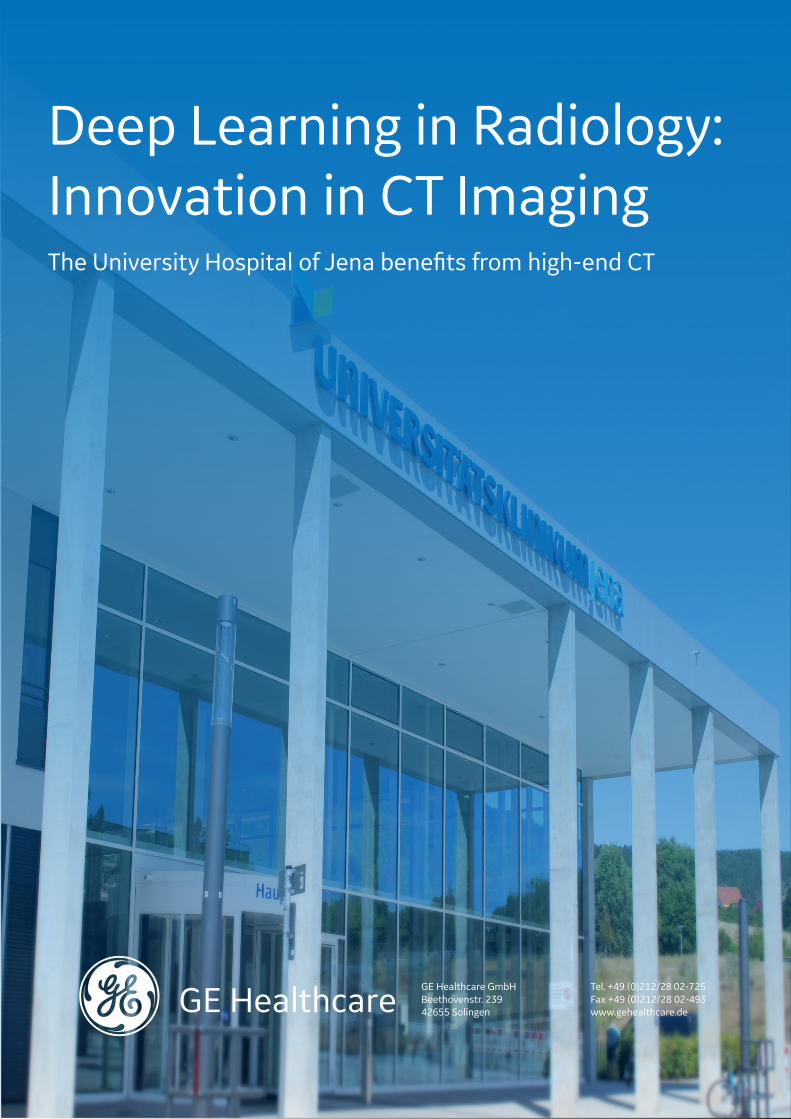
Deep Learning in Radiology: Innovation in CT ImagingThe University Hospital of Jena benefits from high-end CT
GE Healthcare GmbHBeethovenstr. 23942655 Solingen
Tel. +49 (0)212/28 02-725Fax +49 (0)212/28 02-493www.gehealthcare.de

2
The spectrum of possible conditions ranges from harmless to life-threatening: When a
new patient arrives at the emergency room (ER), the medical staff has to categorize and diagnose a possible disease as quickly and accurately as pos-sible. Worldwide, one of the top reasons for an ER visit is chest pain. But not every patient com-plaining of tightness, pain, pressure in the chest or shortness in breath is suffering from a heart condition such as a heart attack. "It‘s exactly the-se at-risk patients that we can screen and treat as soon as they arrive at the ER," says Professor Wilhelm Behringer, Director of the Center for Emergency Medicine at the University Hospital in Jena, Germany (UKJ).
For the past five years, this critical care provider in the east German state of Thuringia has been using GE Healthcare‘s Revolution CT at the Cen-tral Emergency Department – making the facili-ty one of the pioneers in deep learning radiology. In April 2019, the hospital was one of six users worldwide to pilot artificial intelligence in the re-construction of CT images. Thanks to artificial in-telligence, this high-end CT routinely delivers very sharp images with low noise, including accurate
imaging of the heart and surrounding blood ves-sels.
Faster and better diagnostics in radiology with deep learning Although commissioning this CT initially requi-red new workflows, it then sped up processes for the benefit of patients. "The CT images are much clearer than before, enabling more rapid diagnoses and an immediate start of necessary therapies," explains Dr. Ioannis Diamantis, senior physician at the hospital‘s Institute of Diagno-stic and Interventional Radiology. "We no longer discuss artifacts or blurred images, for example, while analyzing the aorta to measure the lumen of a thrombus."
Together with GE Health-care, we have developed
various measurement protocols, so that now one click is all it takes to store a large volume of data correctly and make complex evaluations reusable for every-one.Professor Ulf Teichgraeber,Director at the Institute of Diagnostic and Interventional Radiology
The University Hospital of Jena benefits from high-end CT:
Deep Learning in RadiologyArtificial intelligence has paved the way for the next steps of innovation in CT imaging. The use of a computer tomography (CT) scanner with Deep Learning Image Reconstruction (DLIR) in the central emergency room at the University Hospital of Jena, Germany, shows how both doctors and patients can benefit from this new technology. In addition, this high-end device increases cost efficiency in hospital workflow.

3
The main innovation is image reconstruction with the help of artificial intelligence: Today, "True Fi-delity," a Deep Learning Image Reconstruction (DLIR), makes it possible to reconstruct data sets in an very sharp, low-noise and high-contrast manner. In the past, radiologists were used to images reconstructed with filtered back projec-tion. However, recording these images required a much higher radiation dose in order to achieve the same quality and thus diagnostic significance. The new image reconstruction technology is integrated into a unique imaging chain with the highest technological level – from the detector element to data transmission and deep learning image reconstruction (DLIR).
DLIR is a complex form of machine learning based on a deep neural network that is particular-ly similar to the human brain. The network is fed with both sample data from phantom images and high-resolution patient images. DLIR then pro-cesses the information independently on several levels – and learns. During the training process algorithms are derived, adapted and further opti-mized. In this way, the potential of large amounts of data (big data) can be fully exploited. The fully trained algorithm is then validated and put into clinical use. In addition, the new DLIR technology keeps radiation exposure for patients and medi-cal staff as low as possible. "Deep Learning-based reconstruction significantly reduces image noise, compared to the established model-based itera-tive reconstruction. In many cases we manage to reduce the standard deviation by 50%," says Felix Guettler, Financial and Technical Director at the Institute for Diagnostic and interventional Ra-diology at UKJ. "In the past, we had to increase radiation dose for enhance image quality. With DLIR we can significantly reduce the radiation dose, and at the same time we can achieve a bet-ter image quality and a shorter reconstruction time. We now reduce radiation dosages by 30%
on average, compared to examinations without DLIR. But we are still working on optimizing the protocols. Patients with a high BMI especially benefit from this," adds Diamantis.
Deep learning saves radiology costsThe decision to launch the Revolution CT was clearly also driven by economic reasons. "Many follow-up examinations are no longer necessary now, which saves enormous costs," says Guettler. Highly complex evaluations such as a three-di-mensional vascular reconstruction are now stan-dard procedures in Jena – day and night.
This level of success was possible because the manufacturer regularly assisted the central ER employees on site for six months. "This exten-sive support after commissioning has allowed a faster exploitation of the full technical poten-tial. Through optimized processes, we have also achieved economic advantages," says GuettlerThe University Hospital in Jena now operates two Revolution CT as well as one Revolution EVO.
Central data storage enables follow-up evaluations Through its equipment, GE Healthcare also has an eye on IT infrastructure. All measured data is stored securely in a digital thin-film archive as well as on the AW (Advantage Workstation) server. "This web-based solution can now be accessed and used by many members of the hospital staff,
Thanks to the new CT, only relevant – that is, re-
ally sick – patients come to us for cardiac surgery. Patients with diffuse chest pain without cardiovascular findings have already been examined in the emergency room.Professor Torsten Doenst, Director at the Clinic for Cardiac and Thoracic Surgery
About 6.3 million CT scans were performed on patients admitted to German hospitals in 2018. This reflects an increase of around 70 per-cent within 10 years. With the increasing number of CT examinations, dose reduction for individual applications is increasingly important.
gbe-bund.de

4
including our residents," says Diamantis. Previously, there were only one or two work-stations. This resulted in limited access and, often, additional costs for new image reconstructions such as 3D visualization. Today, GE Healthcare technicians also have remote access to the AW server and can respond quickly in case of technical issues. In addition, all measured data is archived for follow-up evaluations and future AI applications.
The associated technology platform can be expanded and scaled as required: GE Health-care’s "Edison" platform integrates all AI and analytics applications such as intelligent apps and smart systems into existing devices. Deve-lopers can find on this platform services for big data management as well as for data security for patients and physicians.
Radiology in Jena participates in deep learning development"The potential of Deep Learning in the radiology field is enormous. Thanks to DLIR, Deep Learning has now for the first time a broad influence on the clinical care at UKJ. We are in close contact with GE Healthcare. We can quickly participate in the technical development process of the CT scanners in our facility," explains Guettler, who is a computer scientist himself. The UKJ made a conscious decision to purchase two high-end CTs with an innovative 16-centimeter detector and Deep Learning Image Reconstruction (DLIR). Now, a complete heart examination takes only 0.14 seconds. The heart, aorta and lungs can be fully scanned in one second – regardless of the
heart rate and almost without respiratory pau-ses. Each year this device performs 26,000 ex-aminations in Jena, providing significant imaging of patients with a high pulse rate, irregular heart-beat or extreme restlessness. "We want to offer our patients and physicians real innovation. We regard this as our responsibility in medical care as well as in specialist training for physicians," adds Guettler. Medical training has a strong reputation in Jena. The introduction of the new CT scanners and of DLIR caused particular interest among the employees in the radiology department.
A good reputation among experts and the general publicInterdisciplinary cooperation among doctors at the UKJ has also greatly benefitted from this innovation. The exchange between the emer-gency department and the cardiology depart-ment has grown enormously, and other depart-ments such as oncology and vascular surgery also benefit from the new, exact imaging. "Now, we’re observing a much closer communication among doctors from different departments. Mutual trust has grown, which, of course, also improves the work environment," says Teichgraeber. And word has spread far beyond Jena about how well the new diagnostics system is working – and not just within specialist circles. "We also receive inqui-ries from patients about the possibilities of the new, non-invasive cardiac diagnostics," explains Teichgraeber. As a result, the device is also used for outpatients in special departments. This example underlines how artificial intelligence allows doctors to diagnose medical conditions more precisely and efficiently, in the shortest time possible and on a single device.
50 petabytes of data
is generated by a 500-bed hospital each year.
DeepLearning
MachineLearning
Intelligence
We have many dedicated young doctors here, who
are interested in technology. We can train them quickly and in a future oriented manner with up-to-date devices and techno-logies.MUDr. Ioannis Diamantis,Senior Physician at the Institute of Diagnostic and Interventional Radiology
gehealthcare.com

5
Why did you choose GE Healthcare‘s Revolution CT for the radiology in the central emergency room?
In our work at the University Hospital, we have to meet the highest clinical and scientific require-ments while fulfilling the necessary financial tar-gets. These requirements also apply to the devices and the technologies we use, especially in the ER. Therefore, only the most innovative systems of the manufacturers were in competition.
What has improved for your doctors?
We see huge potential in AI not only for image reconstruction but also for supporting clinical diagnostics. Reliable and fast technologies are important for both working results and em- ployees’ satisfaction. We must offer to the future specialists high-level training with state-of-the-art technology in order to allow them to shape the future and to have an easy access to science.
How can patients benefit from these innovations?
Our patients benefit from a better imaging as they get an accurate diagnosis from the begin-ning of their hospital stay. Moreover, our pro-cesses are more efficient. Now, more people can leave the emergency room and go directly back home instead of staying at the hospital. The pa-tients are diagnosed quicker and can eventually receive their therapy sooner than in the past.
"3 Questions for..." A short interview with Felix V. Guettler, Financial and Technical Director at the Institute for Diagnostic and Interventional Radiology, University Hospital in Jena, Germany.
Felix V. Guettler,M.Sc. in Informatics
A CT heart examination reduces the need for a cardiac catheterization procedure from 100% to 14% in a group of patients with suspected coronary heart disease (CHD). A CT examination was five times more likely to diagnose a CHD in the group of patients that re-ceived a CT examination before a cardiac catheterization procedure than in the group of patients that received a cardiac catheterization procedure directly.
October 2019 – subject to changes. © GE Healthcare GmbH. All rights reserved. A company of General Electric Company, known as GE Healthcare. JB71181DE
www.bmj.com