Embed Size (px)
Citation preview

OR I G I N A L S T UD I E S
Dedicated plug based closure for large boreaccess –The MARVEL prospective registry
Herbert G. Kroon MD1 | Pim A.L. Tonino MD PhD2 | Mikko Savontaus MD PhD3 |
Giovanni Amoroso MD PhD4 | Mika Laine MD PhD5 |
Evald H. Christiansen MD PhD6 | Stefan Toggweiler MD PhD7 |
Jur ten Berg MD PhD8 | Janarthanan Sathananthan MBChB MPH9 |
Joost Daemen MD, PhD1 | Peter P. de Jaegere MD, PhD1 |
Guus B.R.G. Brueren MD PhD2 | Markus Malmberg MD, PhD3 |
Ton Slagboom MD PhD4 | Noriaki Moriyama MD5 |
Christian J. Terkelsen MD PhD6 | Federico Moccetti MD7 | Livia Gheorghe MD8 |
John Webb MD9 | David Wood MD9 | Nicolas M. Van Mieghem MD, PhD1
1Department of Cardiology, Erasmus University Medical Center, Rotterdam, The Netherlands
2Department of Cardiology, Catharina Hospital, Eindhoven, The Netherlands
3Department of Cardiology, Turku University Hospital, Turku, Finland
4Department of Cardiology, Onze Lieve Vrouwe Hospital, Amsterdam, The Netherlands
5Department of Cardiology, Helsinki University Hospital, Helsinki, Finland
6Department of Cardiology, Aarhus University Hospital, Aarhus, Denmark
7Department of Cardiology, Luzern Kantonsspital, Luzern, Switzerland
8Department of Cardiology, St. Antonius Hospital, Nieuwegein, The Netherlands
9Centre for Cardiovascular and Heart Valve Innovation, St. Paul's and Vancouver General Hospital, Vancouver, Canada
Correspondence
Nicolas M. Van Mieghem MD, PhD,
Department of Interventional Cardiology,
Thoraxcenter, ErasmusMC, Room Nt-645, Dr
Molewaterplein 40, 3015 GD Rotterdam, The
Netherlands.
Email: [email protected]
Abstract
Objectives: To study safety and performance of the MANTA Vascular closure device
(VCD) under real world conditions in 10 centers.
Background: The MANTA is a novel plug-based device for large bore arteriotomy closure.
Methods: We included all eligible patients who underwent transfemoral large bore
percutaneous procedures. Exclusion criteria were per operator's discretion and
included severe calcification or marked tortuosity of the access vessel, presence of
marked obesity/cachexia or a systolic blood pressure above 180 mmHg. The primary
performance endpoint was time to hemostasis. Primary and secondary safety end-
points were major and minor access site related vascular complications up to 30 days,
respectively. Vascular complications were adjudicated by an independent clinical
event committee according to VARC-2 criteria. We performed multivariable logistic
regression to estimate the effect of baseline and procedural characteristics on any
and major vascular complications.
Received: 7 August 2020 Revised: 24 October 2020 Accepted: 29 November 2020
DOI: 10.1002/ccd.29439
This is an open access article under the terms of the Creative Commons Attribution-NonCommercial-NoDerivs License, which permits use and distribution in any
medium, provided the original work is properly cited, the use is non-commercial and no modifications or adaptations are made.
© 2020 The Authors. Catheterization and Cardiovascular Interventions published by Wiley Periodicals LLC.
Catheter Cardiovasc Interv. 2020;1–9. wileyonlinelibrary.com/journal/ccd 1

Results: Between February 2018 and July 2019 500 patients were enrolled undergo-
ing Transcatheter aortic valve replacement (TAVR, N = 496), Balloon aortic
valvuloplasty (BAV, N = 2), Mechanical circulatory support (MCS, N = 1) or Endo-
vascular aneurysm repair (EVAR, N = 1). Mean age was 80.8 ± 6.6 years with a
median STS-score of 2.7 [IQR 2.0–4.3] %. MANTA access site complications were
major in 20 (4%) and minor in 28 patients (5.6%). Median time to hemostasis was
50 [IQR 20–120] sec. Severe femoral artery calcification, scar presence in groin, lon-
ger procedure duration, female gender and history of hypertension were independent
predictors for vascular complications.
Conclusion: In this study, MANTA appeared to be a safe and effective device for
large bore access closure under real-world conditions.
K E YWORD S
transcatheter valve implantation, vascular closure, vascular complications
1 | INTRODUCTION
Catheter based techniques have emerged to treat aorta and left-sided
heart valve disease which may include mechanical circulatory support
(MCS). These procedures typically require large bore arterial access. A
completely percutaneous approach implies the use of closure devices
to secure the arteriotomy after catheter removal, in which most expe-
rience has been accrued with suture-based closure techniques. Trans-
catheter aortic valve replacement (TAVR) is arguably the most
common large bore arterial intervention. Recent TAVR trials in lower
risk patients reported major vascular complication rates ranging
between 2.2% and 7.9%.1,2 The majority of TAVR related access site
complications seem related to closure device failure.3 A comparison of
various early generation suture-based techniques resulted in con-
flicting results but suggested a 20% vascular complication rate.4,5 The
MANTA vascular closure device (VCD, Teleflex Inc., PA) is a novel col-
lagen plug-based device dedicated for large bore arteriotomy closure.
The CE Mark study demonstrated favorable outcomes in a small sam-
ple of 50 patients undergoing large bore interventions with MANTA
closure.6 The US based SAFE MANTA IDE trial reported high techni-
cal success with MANTA closure and a 4.2% major vascular complica-
tion rate in a selected cohort of 263 patients undergoing large bore
arterial interventions.7 The international, multi-center, prospective
observational MARVEL (MAnta Registry for Vascular Large-borE CLo-
sure) study aimed to evaluate safety and performance of MANTA clo-
sure for large bore arteriotomies in contemporary clinical practice.
2 | METHODS
2.1 | Patient selection and study protocol
Patients undergoing completely percutaneous transfemoral large bore
arterial interventions were eligible for the study. A total of
500 patients were enrolled by 10 hospitals in Canada, Denmark,
Netherlands, Finland and Switzerland. All patients were discussed in a
multidisciplinary (heart) team meeting including (interventional) cardi-
ologists and cardiothoracic or vascular surgeons. Preprocedural plan-
ning with multi-detector computed tomography was mandatory for all
TAVR procedures and recommended for other interventions such as
MCS, balloon aortic valvuloplasty (BAV) or endovascular aneurysm
repair (EVAR). Femoral artery calcification at access site level was clas-
sified according to the semi-quantitative MANTA Femoral Artery Cal-
cification Score (MFACS, Supplementary Figure 1) as follows:
Stage 0: No calcification.
Stage 1: Small calcium spots dispersed over the vessel surface.
Stage 2: Calcium plaques dispersed over the vessel surface.
Stage 3: Large calcium plaque at the posterior wall.
Stage 4: Large calcium plaque at the anterior wall.
Stage 5: Excessive and circumferential calcium.
Relative exclusion criteria for MANTA use were per operator's
discretion and included (1) excessive calcification of the access vessel;
(2) severe peripheral artery disease preluding safe introduction of a
large arterial sheath; (3) marked tortuosity of the femoral or iliac
artery; (4) body mass index > 40 kg/m2 (5) body mass index < 20 kg/
m2 and (6) uncontrolled hypertension at baseline (systolic blood
pressure > 180 mmHg). All participating operators had performed at
least 10 MANTA closures prior to study entry. All interventions were
performed with unfractionated heparin and target activated clotting
time at the time of MANTA closure needed to be < 250 s. Protamine
use was per operator's decision. A femoral angiogram post-MANTA
deployment was recommended for all patients. Clinical examination of
the MANTA access site for complications was mandatory directly and
24 hours after the index procedure and also before hospital discharge.
Clinical follow up was planned at 30 days. Every patient signed
informed consent for the index procedure and trial enrollment. MAR-
VEL was conducted in compliance with the declaration of Helsinki,
Good Clinical Practice principles and current International Standard
2 KROON ET AL.

for Clinical Investigations of medical devices for human subjects (EN-
ISO 14155:2011). The Institutional Review Board of each institution
approved the design of present study. MARVEL was registered under
the Identifier NCT03330002.
2.2 | The MANTA device
The MANTA VCD consists of a delivery system and an implantable
closing unit which comprises a bioresorbable (poly-lactic-co-glycolic
acid) toggle inside the vessel and a hemostatic bovine collagen plug
outside the vessel (Figure 1). The latter two are connected with a
non-resorbable polyester suture and a stainless-steel lock. Prior to
large sheath insertion, arteriotomy depth is determined with an 8F
depth locator with centimeter markers (Supplementary Figure 2). At
the end of the procedure the large sheath is exchanged for the dedi-
cated 14F or 18F MANTA sheath with centimeter markers that
accommodate the delivery unit. The combined assembly is then with-
drawn over the wire to the designated arteriotomy depth. The toggle
is released by turning the lever on the handle. Subsequently, the
assembly is then further withdrawn until a green color code is seen on
the handle, that denotes sufficient pulling force. The collagen plug is
delivered towards the vessel by sliding down the tamper tube. The
wire is removed, and the MANTA suture is cut at skin level. All
MANTA components are resorbed within 6 months apart from the
stainless-steel lock. Arterial sheaths sizes 10 – 14F (maximum outer
diameter of 18F) and 14 – 20F (maximum outer diameter of 25F)
require a 14F and 18F MANTA device, respectively.
2.3 | Clinical endpoints
The primary and secondary safety endpoints were the occurrence of
major and minor MANTA access site related vascular complications up to
30 days, respectively. All clinically relevant endpoints related to the
MANTA access site were adjudicated according to the Valve Academic
Research Consortium (VARC) 2 criteria by an independent Clinical Event
Committee.8 The adjudication committee consisted of physicians outside
the participating centers who received a fee by the investigators from the
unrestricted grant to execute the trial. The primary performance endpoint
was time to hemostasis, which was defined as the elapsed time between
MANTA deployment (withdrawal of dedicated MANTA sheath from
artery) and observed arterial hemostasis (no or minimal subcutaneous
oozing and the absence of expanding or developing hematoma).
2.4 | Data collection and monitoring
Baseline and procedural characteristics, follow-up data as well as
adverse events were entered into an electronic data capture system.
Accuracy of data and compliance with regulatory agreements related
to study conduction was monitored by an independent clinical
research organization, Factory-CRO (Bilthoven, The Netherlands).
Additionally, an independent auditor (Phase More, Nijmegen, The
Netherlands) visited the highest enrolling center to ensure compliance
with Good Clinical Practice principles and ISO 14155:2011 and to
perform source verification as well.
2.5 | Statistical analysis
Continuous variables were presented as mean (±SD) or median (inter-
quartile range) and categorical variables as n (%). The distribution of
continuous variables was examined for normality through histograms
and Q-Q plots. For the comparison of categorical variables between the
different transcatheter heart valves we used the Pearson χ2 or Fisher
exact test as appropriate. For ensuing pairwise comparisons, we applied
Bonferroni corrections to account for multiple testing. Additionally, we
performed multivariable logistic regression to estimate the effect of rel-
evant baseline and procedural characteristics on occurrence of any
(major) vascular complications after MANTA closure. We entered those
variables that displayed a p-value of less than 0.10 in univariate analy-
sis. When limited number of events were present, we chose those vari-
ables that had a p-value less than 0.10 and are known risk factors for
F IGURE 1 The 18F MANTA device. 1. Delivery system andclosing unit. 2. Puncture location dilator 3. Dedicated MANTA sheath
4. Sheath introducer. The 18F system has a blue color, the 14Fsystem has an orange color. The lower part illustrates the toggle onthe inside of the vessel and the collagen plug on the outside. Alsonotice the green color on the delivery unit when sufficient pullingforce is applied when withdrawing the MANTA assembly (dedicatedsheath and delivery unit) from the artery [Color figure can be viewedat wileyonlinelibrary.com]
KROON ET AL. 3

the occurrence of peripheral artery disease or vascular complications or
could theoretically increase the risk of vascular complications (severe
femoral artery calcification defined as MFACS ≥ 3, groin scarring from
previous procedures, arteriotomy depth). All statistical analyses were
performed with SPSS version 25.0 (IBM Corp., N Y).
3 | RESULTS
Between February 2018 and July 2019, a total of 500 patients were
included. A flowchart describing patient inclusion is displayed in
Figure 2. Table 1 summarizes baseline demographics. Almost half of
patients were female (N = 226, 45%), mean age was 80.8 ± 6.6 years
and the Society of Thoracic Surgery (STS) predicted risk of mortality
was 2.7 [IQR 2.0–4.3] %. Most patients (N = 496, 99%) underwent a
TAVR procedure with either the Sapien 3 (Ultra) (N = 203–41%,
Edwards Lifesciences, CA), Evolut R/PRO (N = 202–40%, Medtronic
Inc., MN) or ACURATE neo valve (N = 72–14%, Boston Scientific,
MA), while two patients underwent BAV. One patient underwent an
EVAR procedure and another patient underwent a high risk percuta-
neous coronary intervention (PCI) requiring MCS. Procedural data are
tabulated in Table 2 and used sheaths in Supplementary Table 1.
3.1 | Primary endpoint
The primary performance endpoint of time to hemostasis was a
median of 50 [IQR 20–120] sec as displayed in Table 3 and Supple-
mentary Figure 3. Overall, time to hemostasis ranged from 0 s up to
75 min. More than half of patients (N = 269, 54%) reached hemostasis
within 1 min and cumulatively 421 (84%) patients had hemostasis
within 5 min. When manual compression was required, the median
time to hemostasis was 7 [IQR 2 to 12] min (range 55 s - 16 min). The
primary endpoint of major vascular complications related to the
MANTA access site occurred in 20 patients (4.0%): immediate
MANTA closure device failure in 7 (sometimes combined), large hema-
toma requiring red blood cell (RBC) transfusion in 4 and flow limiting
dissection or stenosis in 4. The remaining patients suffered from rup-
ture or perforation (N = 3), distal embolization (N = 2), pseudo-
aneurysm (N = 2) or ipsilateral lower extremity ischemia (N = 1). These
patients were treated with surgery (N = 12), ballooning (N = 2) or
deployment of a covered stent (N = 3). One patient required ambula-
tory surgery due to a pseudoaneurysm 2 weeks after a major vascular
complication and subsequent bleeding. Five patients were in need of
surgery due to uncontrolled bleeding. One of these suffered a retro-
peritoneal bleeding and needed 8 RBC transfusions. Four patients
F IGURE 2 Flowchart describing study inclusion [Color figure can be viewed at wileyonlinelibrary.com]
4 KROON ET AL.

needed vascular repair surgery for a large hematoma or flow limiting
dissection/stenosis. Two patients required surgery due to MANTA
embolization distally. The MANTA needed to be explanted in five and
thrombendarteriectomy was performed in four subjects. Three
patients required no treatment or just prolonged manual compression.
A total of 12 patients required RBC transfusions. All major vascular
complications occurred on the procedure day. No significant differ-
ences in major vascular complication rates were observed between
valve types (Supplementary Figure 4).
3.2 | Secondary endpoint and overall 30 dayclinical outcomes
Twenty-eight patients (5.6%) suffered a minor vascular complication
after MANTA deployment and included (sometimes combined) nine dis-
sections, seven pseudoaneurysms, six hematomas, five immediate
MANTA closure device failures and three stenoses. These patients
were treated with minor surgery (N = 2), ballooning (N = 5) or a covered
stent (N = 4). One patient underwent surgery due to uncontrolled
bleeding and one required vascular repair due to a flow limiting steno-
sis. Five patients required lidocaine/epinephrine combination or throm-
bin injection, but the majority did not require treatment (N = 6) or just
prolonged manual compression (N = 6). Three patients required a RBC
transfusion. All minor vascular complication occurred on the procedure
day, except for three patients 1 day later. Seven patients (1%) died dur-
ing the follow-up period of 40 ± 13 days. One patient died due to
shock because of external iliac artery rupture after BAV. That patient
had severe femoral artery calcification and a too high femoral puncture
was performed. There was no hemostasis after MANTA closure. Hemo-
stasis was achieved with an occlusion balloon in the abdominal aorta.
After leaving the catheterization laboratory, the patient developed
gradual hypotension and underwent urgent vascular surgery. However,
the patient developed multi-organ failure and died the same day in the
ICU. Twenty-five (5.0%) patients suffered a bleeding event, 10 major
and 6 life-threatening. In total, 16 patients needed red blood cell trans-
fusions. In one patient the bleeding was not access related. Four out of
six patients with a disabling bleeding needed red blood cell transfusions
(2–8 transfusions). Seventy percent (N = 7 out of 10) of the patients
with a major bleeding required blood transfusions and eventually ste-
nting or surgery. Thirteen percent of patients needed a new permanent
pacemaker implantation (61/457, Supplementary Table 2). Acute kid-
ney injury (stage I or II) occurred in three patients and major stroke
struck 2% of patients (N = 9). Overall 22 patients (4.4%) suffered a
vascular complication not related to the MANTA access site
(i.e., pericardial tamponade or related to contralateral access closure), of
which six were major. Some patients who suffered a vascular complica-
tion on the MANTA side also developed non-MANTA related vascular
complications elsewhere.
3.3 | Predictors of vascular complications withMANTA closure
In univariate analysis age, STS-score, history of hypertension, chronic
kidney disease, groin scar from previous surgery, a MFACS score ≥ 3,
female gender and longer procedure duration were associated with
the occurrence of any vascular complication after MANTA closure
(Supplementary Table 3). In multivariable analysis, groin scarring from
a previous procedure (OR 16.55, 95% CI 2.72–100.59, p = .002),
MFACS score ≥ 3 (OR 2.72, 95% CI 1.06–7.03, p = .038), duration of
procedure (OR 1.04, 95% CI 1.02–1.05, p = .0005), hypertension (OR
2.82, 95% CI 1.14–6.97, p = .025) and female gender (OR 2.13, 95%
CI 1.05–4.34, p = .037) remained independent predictors for any
vascular complication (Table 4). For major vascular complications the
predicting variables were similar, however gender and history of
hypertension seemed less important (Supplementary Tables 4 and 5).
4 | DISCUSSION
The main findings of this prospective, multicenter, post-market study
can be summarized as follows: (a) The MANTA VCD appeared to be
TABLE 1 Baseline characteristics of the enrolled patients
Number of patients
(N = 500)
Baseline characteristics
Female gender 226 (45%)
Age (years) 80.8 ± 6.6
STS score (%) 2.7 [IQR 2.0–4.3]a
Body mass index (kg/m2) 26.9 ± 4.3
History of PCI 144 (29%)
History of CABG 53 (11%)
Hypertension 323 (65%)
History of stroke 85 (17%)
Peripheral artery disease 40 (8%)
Pacemaker at baseline 43 (9%)
Glomerular filtration rate < 60 mL/min 226 (45%)
Left ventricular ejection fraction <20% 2 (< 1%)
Antithrombotic therapy at baseline
Acetylsalicylic acid 286 (57%)
Clopidogrel or other P2Y12 inhibitor 200 (40%)
Oral anticoagulation 66 (13%)
New oral anticoagulation 61 (12%)
Heparin or low molecular weight heparin 14 (3%)
Note: Categorical variables are shown as N (%). Continuous variables are
displayed as mean ± SD or median [IQR].
Abbreviations: CABG, coronary artery bypass grafting; PCI, percutaneous
coronary intervention; STS, society of thoracic surgeons.aAarhus University Hospital and Vancouver General Health did not
provide STS-scores.
KROON ET AL. 5
safe for usage in completely percutaneous large bore access proce-
dures. (b) Time to hemostasis is short with a median time of 50 [IQR
20–120] sec. (c) MANTA use was associated with a 4% major and
5.6% minor vascular complication rate. (d) Corrective vascular surgery
was required in 2.8% of cases. (e) Scarring around the puncture site
from previous surgery, procedure duration, excessive femoral calcifi-
cations, hypertension and female gender were associated with
MANTA access site vascular complications.
The MARVEL trial mirrors the SAFE MANTA IDE trial that
reported a 4.2% major MANTA vascular complication rate in
263 selected patients aged 79.4 ± 8.4 years old with a mean STS-
score of 4.45 ± 3.04%. Notably, the SAFE MANTA trial applied more
stringent inclusion/exclusion criteria and MARVEL doubled the sam-
ple size. MARVEL therefore appears to confirm MANTA safety in a
broader population and its feasibility in real world clinical practice.
Multiple retrospective studies on MANTA reported minor and major
vascular complication rates between 0.0%–6.0% and 1.1%–9.3%,
respectively.6,9-12 Notably, the majority of patients in MARVEL under-
went TAVR. Major vascular complications rates in recent TAVR trials
including patients at low or intermediate operative risk vary between
2.0% and 7.9%.1,2 With a mean age of 80.8 ± 6.6 years old, an STS-
score of 2.7 [IQR 2.0–4.3] % and peripheral artery disease prevalence
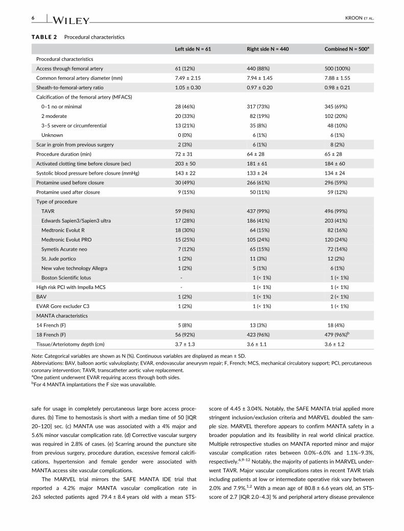
TABLE 2 Procedural characteristics
Left side N = 61 Right side N = 440 Combined N = 500a
Procedural characteristics
Access through femoral artery 61 (12%) 440 (88%) 500 (100%)
Common femoral artery diameter (mm) 7.49 ± 2.15 7.94 ± 1.45 7.88 ± 1.55
Sheath-to-femoral-artery ratio 1.05 ± 0.30 0.97 ± 0.20 0.98 ± 0.21
Calcification of the femoral artery (MFACS)
0–1 no or minimal 28 (46%) 317 (73%) 345 (69%)
2 moderate 20 (33%) 82 (19%) 102 (20%)
3–5 severe or circumferential 13 (21%) 35 (8%) 48 (10%)
Unknown 0 (0%) 6 (1%) 6 (1%)
Scar in groin from previous surgery 2 (3%) 6 (1%) 8 (2%)
Procedure duration (min) 72 ± 31 64 ± 28 65 ± 28
Activated clotting time before closure (sec) 203 ± 50 181 ± 61 184 ± 60
Systolic blood pressure before closure (mmHg) 143 ± 22 133 ± 24 134 ± 24
Protamine used before closure 30 (49%) 266 (61%) 296 (59%)
Protamine used after closure 9 (15%) 50 (11%) 59 (12%)
Type of procedure
TAVR 59 (96%) 437 (99%) 496 (99%)
Edwards Sapien3/Sapien3 ultra 17 (28%) 186 (41%) 203 (41%)
Medtronic Evolut R 18 (30%) 64 (15%) 82 (16%)
Medtronic Evolut PRO 15 (25%) 105 (24%) 120 (24%)
Symetis Acurate neo 7 (12%) 65 (15%) 72 (14%)
St. Jude portico 1 (2%) 11 (3%) 12 (2%)
New valve technology Allegra 1 (2%) 5 (1%) 6 (1%)
Boston Scientific lotus - 1 (< 1%) 1 (< 1%)
High risk PCI with Impella MCS - 1 (< 1%) 1 (< 1%)
BAV 1 (2%) 1 (< 1%) 2 (< 1%)
EVAR Gore excluder C3 1 (2%) 1 (< 1%) 1 (< 1%)
MANTA characteristics
14 French (F) 5 (8%) 13 (3%) 18 (4%)
18 French (F) 56 (92%) 423 (96%) 479 (96%)b
Tissue/Arteriotomy depth (cm) 3.7 ± 1.3 3.6 ± 1.1 3.6 ± 1.2
Note: Categorical variables are shown as N (%). Continuous variables are displayed as mean ± SD.
Abbreviations: BAV, balloon aortic valvuloplasty; EVAR, endovascular aneurysm repair; F, French; MCS, mechanical circulatory support; PCI, percutaneous
coronary intervention; TAVR, transcatheter aortic valve replacement.aOne patient underwent EVAR requiring access through both sides.bFor 4 MANTA implantations the F size was unavailable.
6 KROON ET AL.

of 8.0%, MARVEL arguably included patients at intermediate risk and
complication rates might be lower in younger and truly low risk
patients. In MARVEL all access complications clustered within the first
24 hours after closure. This is relevant in view of contemporary trends
to implement early discharge (i.e., < 48 hours) protocols with TAVR.
The Multidisciplinary, Multimodality, But Minimalist Transcatheter
Aortic Valve Replacement (3 M TAVR) trial prospectively confirmed
safety and efficacy of early discharge after the index procedure with a
minimalistic or simplified TAVR approach.13 Inherent to early dis-
charge is safe, reliable and durable arteriotomy closure. Because we
did not observe any late MANTA failures, plug-based closure may
complement early discharge. Conversely, patients suffering vascular
complications would demand close clinical follow-up.
Most clinical experience in terms of large bore arteriotomy clo-
sure accumulated with suture-based Proglide and Prostar (both
Abbott Vascular, CA) devices. The CONTROL multi-center study was
a propensity matched comparison of Proglide vs. Prostar XL in
944 patients and reported an overall 20% vascular complication rate
with more major vascular complications with Prostar XL (7.4%
vs. 1.9%, p < .001).5 Single-center data from Catania presented con-
flicting data with more vascular complications with Proglide
vs. Prostar (24% vs. 11.4%, p = .007).4 Both studies were performed
with early generation suture based closure devices and enrolled
patients had more often peripheral artery disease. Thus, these results
may not be a true reflection of current practice. Recently, an observa-
tional study comparing MANTA (N = 168) with Prostar XL (N = 198)
found much lower major vascular and bleeding complication rates
with the suture based device than reported earlier and comparable
between both closure techniques (0.6% vs. 1.0%, p = .661 and 0.6%
vs. 1.5%, p = .102, respectively).14 Minor vascular and bleeding com-
plications seemed somewhat more frequently observed with Prostar
XL (10.7% vs 18.8%, p = .003 and 13.7% vs. 19.7%, p = .08, respec-
tively).14 In a retrospective TAVR study, Hoffmann et al compared
75 Manta with 76 Proglide closures and reported fewer vascular com-
plications and need for bail out surgery/interventions with suture-
based closure.9 Three other comparative retrospective studies
reported no difference in vascular complications between MANTA
and Proglide closure strategies albeit MANTA was consistently associ-
ated with less bleeding events, fewer RBC transfusions and shorter
in-hospital stay.10-12 However, the non-randomized fashion of these
studies precludes any definite conclusions. Importantly, our corrective
surgery rate of 2.8% is comparable with the 2.0% and 0.8% found in
the highly selected patients in the CE MARK and SAFE MANTA tri-
als.6,7 Surgery rates varied between 1.3% to 5.6% for the ProStar and
1.1% to 4.0% for ProGlide closures in the literature.4,5 Of note, the
retrospective studies comparing MANTA with ProGlide show compa-
rable rates of bail-out corrective surgery.10,12 Besides corrective sur-
gery, the risk for infection due to the collagen pad may be relevant.
This pad may function as a culture medium with a direct pathway to
the skin. Also, stenosis or occlusion may occur at the site of MANTA
deployment. In our study, we did not observe plug infections. Femoral
artery stenosis/occlusion occurred in 7 patients (1.4%), which can be
treated with ballooning from the contralateral side or eventually sur-
gery. The SAFE MANTA trial followed up patients at 30 and 60 days
with femoral ultrasound and ankle brachial index.7 The ankle brachial
index remained comparable throughout the study period and no major
complications occurred after discharge date. However, CoreLab ultra-
sound analysis found 18 access site related events, all requiring no
treatment. This included 4 cases of oozing (1.5%), 5 cases of hema-
toma/ecchymosis (1.9%), femoral artery stenosis (N = 1, 0.4%), intimal
defect (N = 1, 0.4%) and arterial thrombosis (N = 1, 0.4%). No
TABLE 3 Overview of 30-day clinical outcomes
Number of patients
(N = 500)
30-day outcomes
Mean follow-up (days) 40 ± 13
Manta related major vascular complication 20 (4.0%)
Manta related minor vascular complication 28 (5.6%)
Bleeding
Minor bleeding 9 (1.8%)
Major bleeding 10 (2.0%)
Disabling bleeding 6 (1.2%)
Red blood cell transfusions
1 packed cell 4 (0.8%)
2 packed cells 8 (1.6%)
3 packed cells 2 (0.4%)
4 packed cells 1 (0.2%)
8 packed cells 1 (0.2%)
Percutaneous closure device failure 12 (2.4%)
Median time to hemostasis (s) 50 [20–120]
Note: Categorical variables are shown as N (%). Continuous variables are
displayed as mean ± SD or median [IQR].
TABLE 4 Multivariable analysis on the occurrence of any vascularcomplication
Any vascular complication
OR (95% CI)
P
value
Baseline characteristics
Scar in groin from previous
procedure
16.55 (2.72–100.59) .002
Glomerular filtration
rate < 60 mL/min
1.74 (0.87–3.51) .12
Severe calcification (MFACS
score 3 or greater)
2.72 (1.06–7.03) .038
Duration of procedure (per
minute increase)
1.04 (1.02–1.05) .0005
Female gender 2.13 (1.05–4.34) .037
Hypertension 2.82 (1.14–6.97) .025
Note: Variables are shown as odds ratio OR (95% confidence interval).
Abbreviations: MFACS, MANTA femoral artery calcification score.
KROON ET AL. 7

infection of the collagen plug was reported. In our study, four patients
underwent thrombendarteriectomy during vascular surgery. No cases
of late thrombosis were reported. Arguably, suture-based closure may
come with a longer learning curve including the need for multiple
devices and experience with a preclosure technique. Conversely and
as opposed to MANTA closure, suture-based closure may herald more
catheter-based bail out options in case of failed arteriotomy closure
with the application of multiple suture- or plug based closure devices.
To be able to react quickly in case of failed MANTA closure, contralat-
eral access may provide ways to stop the bleeding (i.e., contralateral
ballooning). Multivariable logistic regression analysis found several risk
factors for developing vascular complications after MANTA closure.
Scarring in the groin due to previous procedures with femoral access
may result in adhesions and more difficulty to access the femoral
artery. Therefore, presence of subcutaneous scar tissue may impede
complete apposition of the collagen plug. This mechanism has been
shown to lead to bleeding complications and pseudoaneurysm forma-
tion in a paper analyzing MANTA-associated vascular complications.15
Also, severe femoral artery calcification is an established risk factor
for TAVR related vascular complications.16 We strongly recommend a
femoral angiogram after large bore arteriotomy closure, particularly in
patients with moderate to severe femoral calcification and/or history
of percutaneous femoral procedures. Also, in case of severe (anterior)
femoral artery calcification, ultrasound guidance may help to select
the most preferable segments to access. Importantly, as suggested by
others, we would advise against too high femoral punctures close to
the inguinal ligament.15 Presence of the (more dense) inguinal liga-
ment may impede or result in more difficulty achieving complete colla-
gen plug apposition. Furthermore, advanced chronic kidney disease is
associated with accelerated atherosclerosis and thus related to worse
outcomes after TAVR on the short- and long-term.17 Longer proce-
dure time may imply added complexity, including challenging access,
and therefore might indicate a higher risk for vascular complications.
Lastly, female gender consistently was associated with more TAVR
related vascular complications. Women have on average smaller artery
sizes and, with the relatively large sheath diameters needed for TAVR,
this may increase the risk for vascular complications. Further research
is required to optimize procedural planning, risk stratification for vas-
cular complications and potentially patient specific closure device
selection. In that regard, the MASH (MAnta versus Suture-based clo-
sure after transcatHeter aortic valve implantation) trial
(NCT03811119) is a prospective 2-center randomized comparison
between plug and suture based arteriotomy closure that recently
completed its enrollment of 210 TAVR patients.
4.1 | Study limitations
MARVEL was a multicenter, prospective study without randomization
and could be prone to selection bias. Also, femoral angiography after
large bore closure was recommended but not mandated and also site
reported. Femoral angiography assessment by an independent core lab-
oratory could have enhanced the overall phenotyping of the access site
and further scrutinized closure success. Still, it is the largest prospective
study on this topic to date. Its multi-center design and only relative
exclusion criteria make it a realistic reflection of contemporary clinical
practice. Access technique (i.e., ultrasound guidance) may affect clinical
outcome but was not captured in this trial to reflect each center's every
day clinical practice. MARVEL was a post-market study and aimed to
enroll a relatively unselected every-day practice population. However,
patient enrollment needs to be seen in perspective of local practices
and dynamics and thus selection bias cannot be excluded.
5 | CONCLUSION
In this present study, MANTA VCD appeared to be a safe and effec-
tive device for large bore access closure under real-world conditions.
ACKNOWLEDGEMENTS
We like to thank Maarten van Wiechen, Joris F Ooms and Thijmen
Hokken (all from Erasmus University Medical Center) for their valu-
able help in data acquisition and management. We also like to thank
Darra Bigelow, Todd Sorzano, Dr Chris Buller, Edward Ramirez, Jill
Hutton-Pugh, Karis Oasan and Dara Bigelow (Teleflex Inc.) for assis-
tance with trial logistics.
CONFLICTS OF INTEREST
Herbert G. Kroon MD: No conflicts of interest to declare.
Pim A.L. Tonino MD PhD: No conflicts of interest to declare.
Mikko Savontaus MD PhD: Is a proctor/consultant for Boston Scien-
tific and Medtronic.
Giovanni Amoroso MD PhD: No conflicts of interest to declare.
Mika Laine MD PhD: Received a research grant from Teleflex.
Evald H. Christiansen MD PhD: No conflicts of interest to declare.
Stefan Toggweiler MD PhD: Is a proctor/consultant for Boston Scien-
tific, New Valve Technology and Abbott Vascular, has received
unrestricted research grants from Boston Scientific and Fumedica AG,
and is holding equity in Hi-D Imaging AG.
Jur ten Berg MD PhD: No conflicts of interest to declare.
Janarthanan Sathananthan MD: No conflicts of interest to declare.
Joost Daemen MD PhD: No conflicts of interest to declare.
Peter de Jaegere MD PhD: Is proctor for Boston Scientific.
Guus B.R.G. Brueren MD PhD: No conflicts of interest to declare.
Markus Malmberg MD: No conflicts of interest to declare.
Ton Slagboom MD PhD: No conflicts of interest to declare.
Noriaki Moriyama MD: No conflicts of interest to declare.
Christian J. Terkelsen MD PhD: No conflicts of interest to declare.
Federico Moccetti MD: No conflicts of interest to declare
Livia Gheorghe MD: No conflicts of interest to declare
John Webb MD: No conflicts of interest to declare
David Wood MD: No conflicts of interest to declare
Nicolas M. Van Mieghem MD PhD: Dr Van Mieghem has received
research grants from Medtronic, Boston Scientific, Edwards
Lifesciences, Abbott, PulseCath, Essential Medical and Claret. He is
advisor to PulseCath, Claret and Essential Medical.
8 KROON ET AL.

DATA AVAILABILITY STATEMENT
Data is available upon reasonable request from the corresponding
author.
ORCID
Herbert G. Kroon https://orcid.org/0000-0003-1866-5855
Janarthanan Sathananthan https://orcid.org/0000-0001-9513-
9440
Joost Daemen https://orcid.org/0000-0001-8628-1410
Federico Moccetti https://orcid.org/0000-0002-5794-5676
REFERENCES
1. Leon MB, Smith CR, Mack MJ, et al. Transcatheter or surgical aortic-
valve replacement in intermediate-risk patients. N Engl J Med. 2016;
374:1609-1620.
2. Mack MJ, Leon MB, Thourani VH, et al. Transcatheter aortic-valve
replacement with a balloon-expandable valve in low-risk patients. N
Engl J Med. 2019;380:1695-1705.
3. Van Mieghem NM, Tchetche D, Chieffo A, et al. Incidence, predictors,
and implications of access site complications with transfemoral trans-
catheter aortic valve implantation. Am J Cardiol. 2012;110:1361-
1367.
4. Barbanti M, Capranzano P, Ohno Y, et al. Comparison of suture-based
vascular closure devices in transfemoral transcatheter aortic valve
implantation. EuroIntervention. 2015;11:690-697.
5. Barbash IM, Barbanti M, Webb J, et al. Comparison of vascular clo-
sure devices for access site closure after transfemoral aortic valve
implantation. Eur Heart J. 2015;36:3370-3379.
6. Van Mieghem NM, Latib A, van der Heyden J, et al. Percutaneous
plug-based Arteriotomy closure device for large-bore access. A
Multicenter Prospective Study JACC Cardiovasc Interv. 2017;10:
613-619.
7. Wood DA, Krajcer Z, Sathananthan J, et al. Pivotal clinical study to
evaluate the safety and effectiveness of the MANTA percutaneous
vascular closure device. Circ Cardiovasc Interv. 2019;12:e007258.
8. Kappetein AP, Head SJ, Genereux P, et al. Updated standardized end-
point definitions for transcatheter aortic valve implantation: the valve
academic research Consortium-2 consensus document. Eur Heart J.
2012;33:2403-2418.
9. Hoffmann P, Al-Ani A, von Lueder T, et al. Access site complications
after transfemoral aortic valve implantation - a comparison of Manta
and ProGlide. CVIR Endovasc. 2018;1:20.
10. Biancari F, Romppanen H, Savontaus M, et al. MANTA versus
ProGlide vascular closure devices in transfemoral transcatheter aortic
valve implantation. Int J Cardiol. 2018;263:29-31.
11. De Palma R, Settergren M, Ruck A, Linder R, Saleh N. Impact of per-
cutaneous femoral arteriotomy closure using the MANTA(TM) device
on vascular and bleeding complications after transcatheter aortic
valve replacement. Catheter Cardiovasc Interv. 2018;92:954-961.
12. Moriyama N, Lindstrom L, Laine M. Propensity-matched comparison
of vascular closure devices after transcatheter aortic valve replace-
ment using MANTA versus ProGlide. EuroIntervention. 2019;14:
e1558-e1565.
13. Wood DA, Lauck SB, Cairns JA, et al. The Vancouver 3M (multi-
disciplinary, multimodality, but minimalist) clinical pathway facilitates
safe next-day discharge home at low-, medium-, and high-volume
Transfemoral Transcatheter aortic valve replacement centers: the 3M
TAVR study. JACC Cardiovasc Interv. 2019;12:459-469.
14. Gheorghe L, Brouwer J, Mathijssen H, et al. Early outcomes after per-
cutaneous closure of access site in transfemoral transcatheter valve
implantation using the novel vascular closure device collagen plug-
based MANTA. Am J Cardiol. 2019;124:1265-1271.
15. Moccetti F, Brinkert M, Seelos R, et al. Insights from a multi-
disciplinary introduction of the MANTA vascular closure device. JACC
Cardiovasc Interv. 2019;12:1730-1736.
16. Sinning JM, Horack M, Grube E, et al. The impact of peripheral arterial
disease on early outcome after transcatheter aortic valve implanta-
tion: results from the German Transcatheter aortic valve interven-
tions registry. Am Heart J. 2012;164:102-10 e1.
17. Dumonteil N, van der Boon RM, Tchetche D, et al. Impact of preoper-
ative chronic kidney disease on short- and long-term outcomes after
transcatheter aortic valve implantation: a pooled-RotterdAm-Milano-
Toulouse in collaboration plus (PRAGMATIC-plus) initiative substudy.
Am Heart J. 2013;165:752-760.
SUPPORTING INFORMATION
Additional supporting information may be found online in the
Supporting Information section at the end of this article.
How to cite this article: Kroon HG, Tonino PAL, Savontaus M,
et al. Dedicated plug based closure for large bore access –The
MARVEL prospective registry. Catheter Cardiovasc Interv.
2020;1–9. https://doi.org/10.1002/ccd.29439
KROON ET AL. 9