Embed Size (px)
Citation preview

M
D
MD
a
ARAA
KRPCDMRMDAMC
C
(tp
1d
DNA Repair 11 (2012) 12– 21
Contents lists available at SciVerse ScienceDirect
DNA Repair
j ourna l ho me pag e: www.elsev ier .com/ locate /dnarepai r
ini review
eath by protein damage in irradiated cells
ichael J. Daly ∗
epartment of Pathology, Uniformed Services University of the Health Sciences (USUHS), School of Medicine, 4301 Jones Bridge Road, Bethesda, MD 20814, USA
r t i c l e i n f o
rticle history:eceived 17 October 2011ccepted 19 October 2011vailable online 23 November 2011
eywords:adiationrotein oxidationarbonylationNA double strand break (DSB) repair
a b s t r a c t
A founding concept of radiobiology that deals with X-rays, �-rays and ultraviolet light is that radiationindiscriminately damages cellular macromolecules. Mounting experimental evidence does not fit into thistheoretical framework. Whereas DNA lesion-yields in cells exposed to a given dose and type of radiationappear to be fixed, protein lesion-yields are highly variable. Extremely radiation resistant bacteria suchas Deinococcus radiodurans have evolved extraordinarily efficient antioxidant chemical defenses whichspecifically protect proteins and the functions they catalyze. In diverse prokaryotes, the lethal effects ofradiation appear to be governed by oxidative protein damage, which inactivates enzymes including thoseneeded to repair and replicate DNA. These findings offer fresh insight into the molecular mechanisms ofradiation resistance and present themselves as new opportunities to study and control oxidative stress
anganese (II) antioxidant complexeseactive oxygen species (ROS)etabolite accumulationeinococcusrchaeaammalian cells
ancer cells
in eukaryotes, including mammalian cells and their cancer cell counterparts.Published by Elsevier B.V.
ontents
1. Introduction . . . . . . . . . . . . . . . . . . . . . . . . . . . . . . . . . . . . . . . . . . . . . . . . . . . . . . . . . . . . . . . . . . . . . . . . . . . . . . . . . . . . . . . . . . . . . . . . . . . . . . . . . . . . . . . . . . . . . . . . . . . . . . . . . . . . . . . . . . 122. D. radiodurans . . . . . . . . . . . . . . . . . . . . . . . . . . . . . . . . . . . . . . . . . . . . . . . . . . . . . . . . . . . . . . . . . . . . . . . . . . . . . . . . . . . . . . . . . . . . . . . . . . . . . . . . . . . . . . . . . . . . . . . . . . . . . . . . . . . . . . . . 133. The role of DNA damage and repair in radiation-induced toxicity . . . . . . . . . . . . . . . . . . . . . . . . . . . . . . . . . . . . . . . . . . . . . . . . . . . . . . . . . . . . . . . . . . . . . . . . . . . . . . . . . . 134. Protein oxidation as the basis for the efficiency of DSB repair . . . . . . . . . . . . . . . . . . . . . . . . . . . . . . . . . . . . . . . . . . . . . . . . . . . . . . . . . . . . . . . . . . . . . . . . . . . . . . . . . . . . . . 145. Protein damage: cause or effect of radiation toxicity? . . . . . . . . . . . . . . . . . . . . . . . . . . . . . . . . . . . . . . . . . . . . . . . . . . . . . . . . . . . . . . . . . . . . . . . . . . . . . . . . . . . . . . . . . . . . . . 156. The role of Mn antioxidants and metabolites . . . . . . . . . . . . . . . . . . . . . . . . . . . . . . . . . . . . . . . . . . . . . . . . . . . . . . . . . . . . . . . . . . . . . . . . . . . . . . . . . . . . . . . . . . . . . . . . . . . . . . . 167. Evolution of radiation resistance . . . . . . . . . . . . . . . . . . . . . . . . . . . . . . . . . . . . . . . . . . . . . . . . . . . . . . . . . . . . . . . . . . . . . . . . . . . . . . . . . . . . . . . . . . . . . . . . . . . . . . . . . . . . . . . . . . . . 178. Fresh insights into the induction of DNA repair and mutagenesis . . . . . . . . . . . . . . . . . . . . . . . . . . . . . . . . . . . . . . . . . . . . . . . . . . . . . . . . . . . . . . . . . . . . . . . . . . . . . . . . . . 18
9. Conclusion and future perspectives . . . . . . . . . . . . . . . . . . . . . . . . . . . . . . . . . . . . . . . . . . . . . . . . . . . . . . . . . . . . . . . . . . . . . . . . . . . . . . . . . . . . . . . . . . . . . . . . . . . . . . . . . . . . . . . . . 18Conflict of interest statement. . . . . . . . . . . . . . . . . . . . . . . . . . . . . . . . . . . . . . . . . . . . . . . . . . . . . . . . . . . . . . . . . . . . . . . . . . . . . . . . . . . . . . . . . . . . . . . . . . . . . . . . . . . . . . . . . . . . . . . . 19. . . . . . . . . . . . . . . . . . . . . . . . . . . . . . . . . . . . . . . . . . . . . . . . . . . . . . . . . . . . . . . . . . . . . . . . . . . . . . . 19
. . . . .
ultraviolet light) forms of radiation have been used as a primaryool to study oxidative stress responses in organisms spanningrokaryotes to higher eukaryotes. Molecular insights into how
∗ Corresponding author. Tel.: +1 301 295 3750; fax: +1 301 295 1640.E-mail addresses: [email protected], [email protected]
568-7864/$ – see front matter. Published by Elsevier B.V.oi:10.1016/j.dnarep.2011.10.024
. . . . . . . . . . . . . . . . . . . . . . . . . . . . . . . . . . . . . . . . . . . . . . . . . . . . . . . . . . . . . . . . . . . . . . . . . 19
reactive oxygen species (ROS) generated by radiation elicit theirtoxic effects have served as the foundation for investigating thenature of mutagenesis, disease, aging, and a myriad of biologicalprocesses ending in cell death. Recently, experimental evidencefrom several independent groups has converged on the conclu-sion that proteins in mammalian cells [1] and prokaryotes [2–5] aremore probable initial targets of cellular radiation damage than DNA.
Acknowledgements . . . . . . . . . . . . . . . . . . . . . . . . . . . . . . . . . . . . . . . . . . . . . . . . . . .
References . . . . . . . . . . . . . . . . . . . . . . . . . . . . . . . . . . . . . . . . . . . . . . . . . . . . . . . . . . . . .
1. Introduction
For seventy years, ionizing (X-rays, �-rays) and non-ionizing
While classical radiation toxicity models identify DNA damage asthe universal critical lesion in cells [6,7], studies now support thatthe survival of many organisms is governed by the level of oxidative
epair
pfr
dtietwatttrepbatc
2
oElvdisErdintdr
FatfiMw(hyi[l
M.J. Daly / DNA R
rotein damage caused during irradiation [2–5], which limits theunctionality and efficiency of enzymes, including those needed toepair and replicate DNA.
In this review, experimental studies are presented whichemonstrate that the critical “target” molecules in irradiated bac-eria are proteins. Whereas the levels of DNA damage inflictedn naturally radiation-sensitive and radiation-resistant bacteriaxposed to a given dose of radiation are very similar, resistant bac-eria have evolved highly efficient antioxidant chemical defenseshich specifically protect proteins and the functions they cat-
lyze. The thesis that radiation toxicity is determined mainly byhe level of protein oxidation caused during irradiation rather thanhe amount of DNA damage, developed from comparisons betweenhe extremely radiation resistant bacterium Deinococcus radiodu-ans [8] and other bacteria representing the full range of resistancesncountered in the natural world [2,9]. The trends which now sup-ort a critical role of protein oxidation in the survival of irradiatedacteria and archaea, the predominant forms of life on Earth, par-llel the trends developing for irradiated mammalian cells – andhe inferences carry with them the prospects of new strategies toombat oxidative stress in humans.
. D. radiodurans
The bacterium D. radiodurans is capable of surviving huge dosesf X-rays or �-rays (12,000 Gy), 20 times greater than the bacteriumscherichia coli, and 3000 times greater than most human cells iniquid culture (Fig. 1) [8]. Survival curves for D. radiodurans displayery large shoulders (Fig. 1), but the mutation frequency of the cellsoes not increase significantly until very high doses [8]. Reason-
ng that DNA in D. radiodurans might be unusually protected, earlytudies compared the amount of DNA damage in D. radiodurans and. coli exposed to ionizing radiation or ultraviolet C (UVC) (254 nm)adiation [8], and later with other organisms [9–11]. For a givenose of X-rays or �-rays, or UVC, the relatively small differences
n DNA breaks and DNA base damages between the bacteria were
ot nearly sufficient to explain the great differences in their resis-ance [12]. D. radiodurans exposed to �-rays can survive ∼160 DNAouble strand breaks (DSBs) per haploid genome whereas highlyadiosensitive bacteria can survive only a few �-ray-induced DSBsig. 1. Survival curves of representative organisms exposed to �-radiation. Therrows indicate the approximate number of DSBs inflicted per haploid genome athe dose which kills 90% of the organisms. Yields of DSBs were determined by pulsedeld gel electrophoresis (Table 1). Bacteria: S. oneidensis (ATCC 700550), E. coli (K12,G1655), D. radiodurans (ATCC BAA-816); human lung fibroblasts in liquid culture:ild-type (European Collection of Cell Culture, MRC-5); bdelloid rotifer: A. vaga
Table 1). Many explanations of the cause of the shoulders on cell survival curvesave been proposed. The most favored hypotheses begin with the premise that theield of DSBs is linear with dose and that the non-linearity of the cell survival curvess caused by dose-dependent changes in the efficiency/accuracy of enzymatic repair30]. The vast majority of organisms on Earth are radiation sensitive, killed by dosesess than 500 Gy [8,12].
11 (2012) 12– 21 13
(Table 1). The paradoxical survival of irradiated D. radiodurans andother extremely radiation resistant organisms has been rational-ized under the hypothesis of enhanced DNA repair [8,12].
D. radiodurans also is very resistant to desiccation, showing85% viability after two years in the presence of less than 5%humidity [9,13]. It has long been recognized that extreme radia-tion resistance and desiccation tolerance are closely aligned [8].Desiccation-tolerant bacteria are very resistant to protein oxida-tion caused during drying, and studies support that the mutualnature of radiation and desiccation resistance resides in cytoso-lic Mn-dependent antioxidant processes which selectively protectproteins from ROS [14]. Since life on earth most likely did notcommonly encounter extremes of ionizing radiation over geologictimes, the extreme radiation resistance phenotypes frequentlyobserved in desert soil-inhabiting organisms likely evolved inresponse to oxidative stress caused during cycles of drying andrehydration [15]. Based on functional and comparative genomics,it has been proposed that the extreme resistance phenotypes ofthe family Deinococcaceae stem from a subtle regulatory interplaybetween diverse processes including Mn homeostasis, metaboliteregulation, respiratory control, macromolecular degradation, andother oxidative stress response pathways [8,12,16]. In Deinococcusbacteria, these functions manifest themselves as protein protec-tion, which preserves the high efficiency of its DNA repair enzymesduring irradiation or desiccation [2,4,12]. In contrast, irradiated ordesiccated bacteria lacking these antioxidant processes are readilyoverwhelmed by protein oxidation, which renders even minor DNAdamage irreparable [2,8,12,14]. This model was offered on the basisthat a system which protected and preserved the activity of diverserepair enzymes would more likely be evolved to provide multipleresistances than would a series of separate repair mechanisms beevolved for each extremophilic character noted for D. radiodurans[2,12].
3. The role of DNA damage and repair in radiation-inducedtoxicity
The biological effects of ionizing radiation on DNA are usuallyascribed to the sum of two indiscriminately destructive processes.‘Direct action’ refers to the unavoidable damaging effects of energydeposited by photons, damage which predominates in deeplyfrozen (−80 ◦C) or dry preparations [17,18]. In contrast, the over-whelming majority of lesions in cells irradiated in the aqueous stateare caused by the ‘indirect action’ of ROS – principally short-livedhydroxyl radicals (HO•) formed from water that react with DNAat near diffusion-limited rates [12]. The yields of DSBs and single-strand breaks (SSBs) in DNA by �-rays in aqueous solution aretypically 2–3 orders of magnitude greater than those for dry DNA[18] (Fig. 2). Yet, at high concentrations, potent HO•-scavengingagents such as dimethyl sulfoxide (DMSO) prevent less than 80% ofdamage to purified DNA in aqueous preparations exposed to ion-izing radiation [19] (Fig. 2). The non-scavengable indirect effectson DNA by ionizing radiation are presumed to be caused by prox-imal HO• formed from water molecules which are bound tightlyto DNA [17,18], and also by ultrashort-lived prehydrated electrons[20,21], which are not easily scavenged due to their extremely shortlifetimes. In cells and viruses, DNA is bound and condensed by pro-teins, and is highly protected from ROS [17,22,23] (Fig. 2). As theyields of ionizing radiation-induced DSBs are very similar acrossphylogenetically diverse cell-types with greatly differing antioxi-dant statuses (Table 1), it is clear that DSBs in irradiated cells and
viruses are caused mainly by non-scavengable indirect effects.The nature of the “target” molecules in cells – the alterationof which by radiation leads to cell death – was first studiedin radiation-sensitive bacteria, and the conclusions were broadly
14 M.J. Daly / DNA Repair 11 (2012) 12– 21
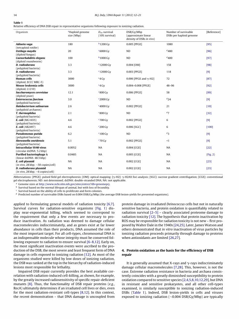
Table 1Relative efficiency of DNA DSB repair in representative organisms following exposure to ionizing radiation.
Organism aHaploid genomesize (Mbp)
D10 survival(10% survival)
DSB/Gy/Mbp(approximate lineardensity of DSBs in vivo)
Number of survivableDSBs per haploid genome
[Reference]
Adineta vaga(tetraploid rotifer)
180 b1200 Gy 0.005 [PFGE] 1080 [95]
Ustilago maydis(diploid fungus)
20 c6000 Gy ND d480 [96]
Caenorhabditis elegans(diploid roundworm)
100 b1000 Gy ND d400 [97]
D. radiodurans(polyploid bacteria)
3.3 c12000 Gy 0.004 [OM] 158 [98]
D. radiodurans(polyploid bacteria)
3.3 c12000 Gy 0.003 [PFGE] 118 [9]
Human cells(diploid, ECCC MRC-5)
3000 c4 Gy 0.006 [PFGE and �-H2] 72 [87]
Mouse leukemia cells(diploid, L1210)
3000 c4 Gy 0.004–0.008 [PFGE] 48–96 [92]
Saccharomyces cerevisiae(diploid yeast)
12.1 c800 Gy 0.006 [PFGE] 58 [99]
Enterococcus faecium(polyploid bacteria)
3.0 c2000 Gy ND d24 [9]
Halobacterium salinarum(polyploid archaeon)
2.6 c4000 Gy 0.002 [PFGE] 21 [10]
T. thermophilus(polyploid bacteria)
2.1 c800 Gy ND d7 [32]
E. coli (MG1655)(polyploid bacteria)
4.6 c700 Gy 0.002 [PFGE] 6 [9]
E. coli (AB2497)(polyploid bacteria)
4.6 c200 Gy 0.006 [SGC] 6 [100]
Pseudomonas putida(polyploid bacteria)
6.2 c100 Gy ND d3 [9]
Shewanella oneidensis(polyploid bacteria)
5.1 c70 Gy 0.002 [PFGE] <1 [9]
Intracellular SV40 virus(circular dsDNA: 5.2 kbp)
0.0052 NA 0.004 [CGE] NA [22]
Purified bacteriophage �(linear dsDNA: 48.5 kbp)
0.0485 NA 0.005 [CGE] NA (Fig. 2)
E. coli plasmid(in vivo, 28 kbp; ∼50 copies/cell)
NA NA 0.002 [CGE] NA [23]
D. radiodurans plasmid(in vivo, 28 kbp; ∼6 copies/cell)
NA NA 0.002 [CGE] NA [23]
Abbreviations: [PFGE]: pulsed field gel electrophoresis; [OM]: optical mapping; [�-H2]: �-H2AX foci analysis; [SGC]: sucrose gradient centrifugation; [CGE]: conventionalgel electrophoresis; ND: not determined; dsDNA: double-stranded DNA; NA: not applicable.
a Genome sizes at http://www.ncbi.nlm.nih.gov/sites/entrez?db=genomeprj.
DSB l
aSptdmataltddotl
rbmRft
b Survival based on the normal lifespan of animal, but with loss of fecundity.c Survival based on the ability of cells to proliferate and form colonies.d Predicted number of survivable DSBs based on 0.004 DSB/Gy/Mbp (the average
pplied to formulating general models of radiation toxicity [6,7].urvival curves for radiation-sensitive organisms (Fig. 1) dis-lay near-exponential killing, which seemed to correspond tohe requirement that only a few events are necessary to pro-uce inactivation. As radiation was deemed to damage cellularacromolecules indiscriminately, and as genes exist at far lower
bundance in cells than their products, DNA assumed the role ofhe most important target. For all cell-types, chromosomal DNA isn indispensable molecule whose integrity must be conserved fol-owing exposure to radiation to ensure survival [6–8,12]. Early on,he most significant inactivation events were ascribed to the pro-uction of the DSB, the most severe and least frequent form of DNAamage in cells exposed to ionizing radiation [12]. As most of therganisms studied were killed by low doses of ionizing radiation,he DSB was ranked at the top in the hierarchy of radiation-inducedesions most responsible for lethality.
Impaired DSB repair currently provides the best available cor-elation with radiation-induced cell-killing, as shown, for example,y the greatly increased radiosensitivity of specific repair-deficient
utants [8]. Thus, the functionality of DSB repair proteins (e.g.,ecA) ultimately determines if an irradiated cell lives or dies, evenor the most radiation-resistant cell-types [8,12]. In this context,he recent demonstration – that DNA damage is uncoupled from
esion-yields for presented organisms).
protein damage in irradiated Deinococcus cells but not in naturallysensitive bacteria, and protein oxidation is quantifiably related toradiation survival [2–5] – clearly associated proteome damage toradiation toxicity [12]. The hypothesis that protein inactivation byROS may be responsible for radiation toxicity is not new – first pro-posed by Walter Dale in the 1940s [24,25]. Later, James Watson andothers demonstrated that in vitro inactivation of virus particles byionizing radiation proceeds primarily through damage to proteinswhen antioxidants are limited [26,27].
4. Protein oxidation as the basis for the efficiency of DSBrepair
It is generally assumed that X-rays and �-rays indiscriminatelydamage cellular macromolecules [7,28]. This, however, is not thecase. Extreme radiation resistance in bacteria and archaea consis-tently coincides with a greatly diminished susceptibility to proteinoxidation compared to sensitive species [2,4,5,8,10,12,29], but DNA
in resistant and sensitive prokaryotes, and all other cell-typesexamined, is similarly susceptible to ionizing radiation-inducedDSBs (Table 1). Indeed, DSB lesion-yields in cells and virusesexposed to ionizing radiation (∼0.004 DSB/Gy/Mbp) are typically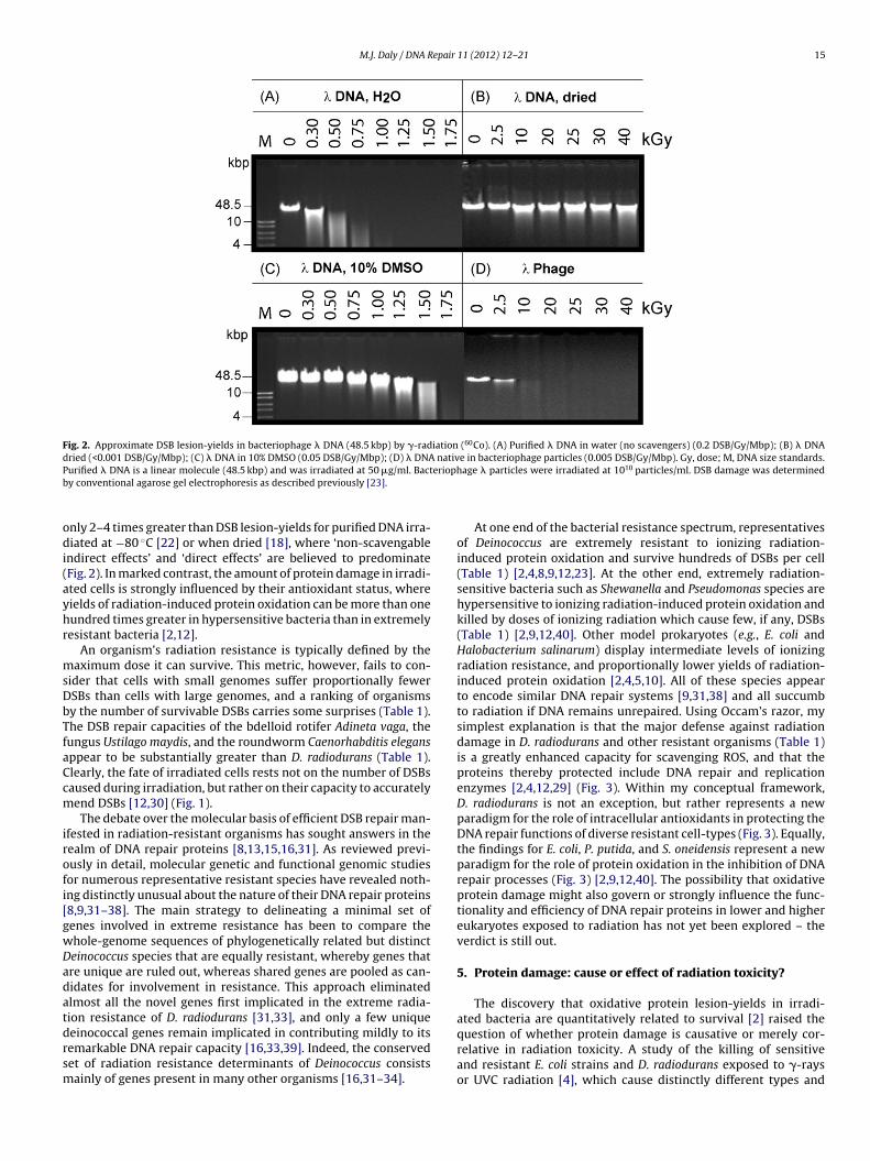
M.J. Daly / DNA Repair 11 (2012) 12– 21 15
Fig. 2. Approximate DSB lesion-yields in bacteriophage � DNA (48.5 kbp) by �-radiation (60Co). (A) Purified � DNA in water (no scavengers) (0.2 DSB/Gy/Mbp); (B) � DNAdried (<0.001 DSB/Gy/Mbp); (C) � DNA in 10% DMSO (0.05 DSB/Gy/Mbp); (D) � DNA native in bacteriophage particles (0.005 DSB/Gy/Mbp). Gy, dose; M, DNA size standards.P eriophb
odi(ayhr
msDbTfaCcm
irofi[gwDadatdrsm
urified � DNA is a linear molecule (48.5 kbp) and was irradiated at 50 �g/ml. Bacty conventional agarose gel electrophoresis as described previously [23].
nly 2–4 times greater than DSB lesion-yields for purified DNA irra-iated at −80 ◦C [22] or when dried [18], where ‘non-scavengable
ndirect effects’ and ‘direct effects’ are believed to predominateFig. 2). In marked contrast, the amount of protein damage in irradi-ted cells is strongly influenced by their antioxidant status, whereields of radiation-induced protein oxidation can be more than oneundred times greater in hypersensitive bacteria than in extremelyesistant bacteria [2,12].
An organism’s radiation resistance is typically defined by theaximum dose it can survive. This metric, however, fails to con-
ider that cells with small genomes suffer proportionally fewerSBs than cells with large genomes, and a ranking of organismsy the number of survivable DSBs carries some surprises (Table 1).he DSB repair capacities of the bdelloid rotifer Adineta vaga, theungus Ustilago maydis, and the roundworm Caenorhabditis elegansppear to be substantially greater than D. radiodurans (Table 1).learly, the fate of irradiated cells rests not on the number of DSBsaused during irradiation, but rather on their capacity to accuratelyend DSBs [12,30] (Fig. 1).The debate over the molecular basis of efficient DSB repair man-
fested in radiation-resistant organisms has sought answers in theealm of DNA repair proteins [8,13,15,16,31]. As reviewed previ-usly in detail, molecular genetic and functional genomic studiesor numerous representative resistant species have revealed noth-ng distinctly unusual about the nature of their DNA repair proteins8,9,31–38]. The main strategy to delineating a minimal set ofenes involved in extreme resistance has been to compare thehole-genome sequences of phylogenetically related but distincteinococcus species that are equally resistant, whereby genes thatre unique are ruled out, whereas shared genes are pooled as can-idates for involvement in resistance. This approach eliminatedlmost all the novel genes first implicated in the extreme radia-ion resistance of D. radiodurans [31,33], and only a few unique
einococcal genes remain implicated in contributing mildly to itsemarkable DNA repair capacity [16,33,39]. Indeed, the conservedet of radiation resistance determinants of Deinococcus consistsainly of genes present in many other organisms [16,31–34].age � particles were irradiated at 1010 particles/ml. DSB damage was determined
At one end of the bacterial resistance spectrum, representativesof Deinococcus are extremely resistant to ionizing radiation-induced protein oxidation and survive hundreds of DSBs per cell(Table 1) [2,4,8,9,12,23]. At the other end, extremely radiation-sensitive bacteria such as Shewanella and Pseudomonas species arehypersensitive to ionizing radiation-induced protein oxidation andkilled by doses of ionizing radiation which cause few, if any, DSBs(Table 1) [2,9,12,40]. Other model prokaryotes (e.g., E. coli andHalobacterium salinarum) display intermediate levels of ionizingradiation resistance, and proportionally lower yields of radiation-induced protein oxidation [2,4,5,10]. All of these species appearto encode similar DNA repair systems [9,31,38] and all succumbto radiation if DNA remains unrepaired. Using Occam’s razor, mysimplest explanation is that the major defense against radiationdamage in D. radiodurans and other resistant organisms (Table 1)is a greatly enhanced capacity for scavenging ROS, and that theproteins thereby protected include DNA repair and replicationenzymes [2,4,12,29] (Fig. 3). Within my conceptual framework,D. radiodurans is not an exception, but rather represents a newparadigm for the role of intracellular antioxidants in protecting theDNA repair functions of diverse resistant cell-types (Fig. 3). Equally,the findings for E. coli, P. putida, and S. oneidensis represent a newparadigm for the role of protein oxidation in the inhibition of DNArepair processes (Fig. 3) [2,9,12,40]. The possibility that oxidativeprotein damage might also govern or strongly influence the func-tionality and efficiency of DNA repair proteins in lower and highereukaryotes exposed to radiation has not yet been explored – theverdict is still out.
5. Protein damage: cause or effect of radiation toxicity?
The discovery that oxidative protein lesion-yields in irradi-ated bacteria are quantitatively related to survival [2] raised the
question of whether protein damage is causative or merely cor-relative in radiation toxicity. A study of the killing of sensitiveand resistant E. coli strains and D. radiodurans exposed to �-raysor UVC radiation [4], which cause distinctly different types and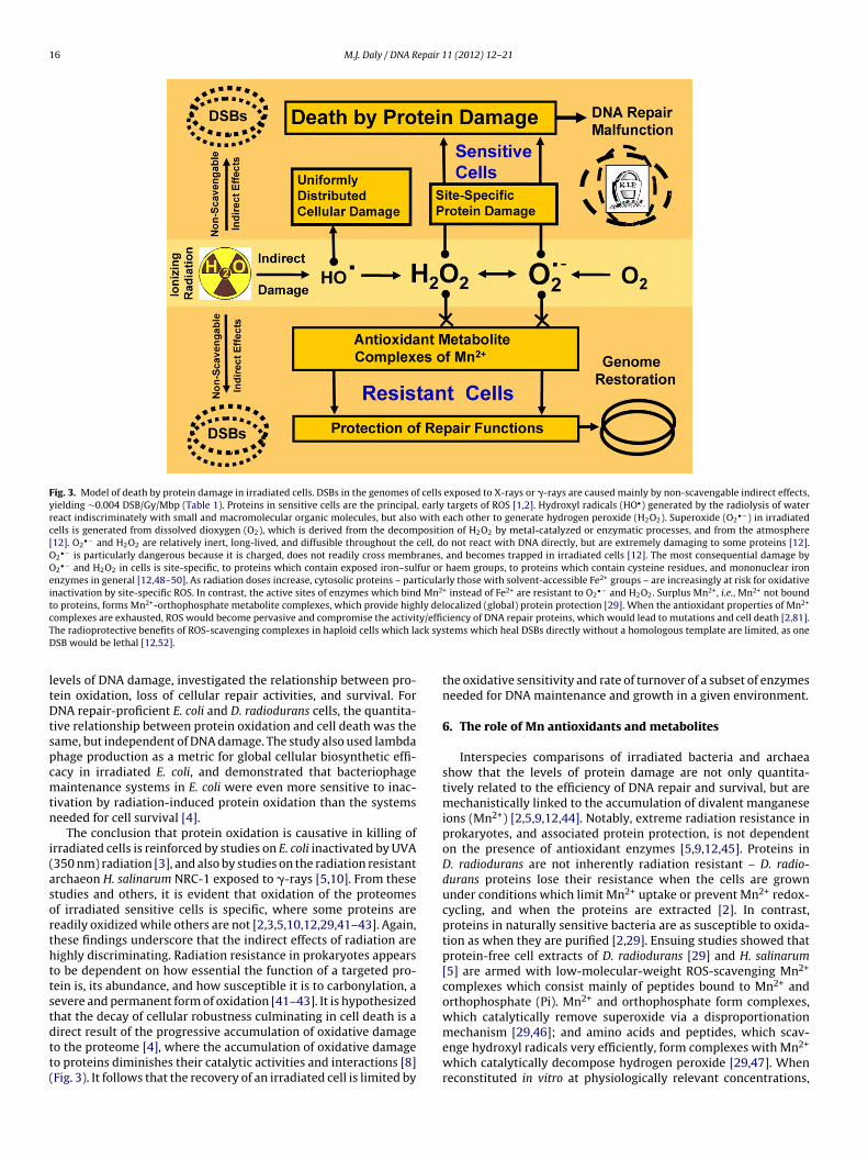
16 M.J. Daly / DNA Repair 11 (2012) 12– 21
Fig. 3. Model of death by protein damage in irradiated cells. DSBs in the genomes of cells exposed to X-rays or �-rays are caused mainly by non-scavengable indirect effects,yielding ∼0.004 DSB/Gy/Mbp (Table 1). Proteins in sensitive cells are the principal, early targets of ROS [1,2]. Hydroxyl radicals (HO•) generated by the radiolysis of waterreact indiscriminately with small and macromolecular organic molecules, but also with each other to generate hydrogen peroxide (H2O2). Superoxide (O2
•−) in irradiatedcells is generated from dissolved dioxygen (O2), which is derived from the decomposition of H2O2 by metal-catalyzed or enzymatic processes, and from the atmosphere[12]. O2
•− and H2O2 are relatively inert, long-lived, and diffusible throughout the cell, do not react with DNA directly, but are extremely damaging to some proteins [12].O2
•− is particularly dangerous because it is charged, does not readily cross membranes, and becomes trapped in irradiated cells [12]. The most consequential damage byO2
•− and H2O2 in cells is site-specific, to proteins which contain exposed iron–sulfur or haem groups, to proteins which contain cysteine residues, and mononuclear ironenzymes in general [12,48–50]. As radiation doses increase, cytosolic proteins – particularly those with solvent-accessible Fe2+ groups – are increasingly at risk for oxidativeinactivation by site-specific ROS. In contrast, the active sites of enzymes which bind Mn2+ instead of Fe2+ are resistant to O2
•− and H2O2. Surplus Mn2+, i.e., Mn2+ not boundto proteins, forms Mn2+-orthophosphate metabolite complexes, which provide highly delocalized (global) protein protection [29]. When the antioxidant properties of Mn2+
c y/effiT ck sysD
ltDtspcmtn
i(asorthttstdtt(
omplexes are exhausted, ROS would become pervasive and compromise the activithe radioprotective benefits of ROS-scavenging complexes in haploid cells which laSB would be lethal [12,52].
evels of DNA damage, investigated the relationship between pro-ein oxidation, loss of cellular repair activities, and survival. ForNA repair-proficient E. coli and D. radiodurans cells, the quantita-
ive relationship between protein oxidation and cell death was theame, but independent of DNA damage. The study also used lambdahage production as a metric for global cellular biosynthetic effi-acy in irradiated E. coli, and demonstrated that bacteriophageaintenance systems in E. coli were even more sensitive to inac-
ivation by radiation-induced protein oxidation than the systemseeded for cell survival [4].
The conclusion that protein oxidation is causative in killing ofrradiated cells is reinforced by studies on E. coli inactivated by UVA350 nm) radiation [3], and also by studies on the radiation resistantrchaeon H. salinarum NRC-1 exposed to �-rays [5,10]. From thesetudies and others, it is evident that oxidation of the proteomesf irradiated sensitive cells is specific, where some proteins areeadily oxidized while others are not [2,3,5,10,12,29,41–43]. Again,hese findings underscore that the indirect effects of radiation areighly discriminating. Radiation resistance in prokaryotes appearso be dependent on how essential the function of a targeted pro-ein is, its abundance, and how susceptible it is to carbonylation, aevere and permanent form of oxidation [41–43]. It is hypothesizedhat the decay of cellular robustness culminating in cell death is a
irect result of the progressive accumulation of oxidative damageo the proteome [4], where the accumulation of oxidative damageo proteins diminishes their catalytic activities and interactions [8]Fig. 3). It follows that the recovery of an irradiated cell is limited byciency of DNA repair proteins, which would lead to mutations and cell death [2,81].tems which heal DSBs directly without a homologous template are limited, as one
the oxidative sensitivity and rate of turnover of a subset of enzymesneeded for DNA maintenance and growth in a given environment.
6. The role of Mn antioxidants and metabolites
Interspecies comparisons of irradiated bacteria and archaeashow that the levels of protein damage are not only quantita-tively related to the efficiency of DNA repair and survival, but aremechanistically linked to the accumulation of divalent manganeseions (Mn2+) [2,5,9,12,44]. Notably, extreme radiation resistance inprokaryotes, and associated protein protection, is not dependenton the presence of antioxidant enzymes [5,9,12,45]. Proteins inD. radiodurans are not inherently radiation resistant – D. radio-durans proteins lose their resistance when the cells are grownunder conditions which limit Mn2+ uptake or prevent Mn2+ redox-cycling, and when the proteins are extracted [2]. In contrast,proteins in naturally sensitive bacteria are as susceptible to oxida-tion as when they are purified [2,29]. Ensuing studies showed thatprotein-free cell extracts of D. radiodurans [29] and H. salinarum[5] are armed with low-molecular-weight ROS-scavenging Mn2+
complexes which consist mainly of peptides bound to Mn2+ andorthophosphate (Pi). Mn2+ and orthophosphate form complexes,which catalytically remove superoxide via a disproportionation
mechanism [29,46]; and amino acids and peptides, which scav-enge hydroxyl radicals very efficiently, form complexes with Mn2+which catalytically decompose hydrogen peroxide [29,47]. Whenreconstituted in vitro at physiologically relevant concentrations,

epair
ti5tnsniisRdcdMcgoiroapp
7
gdto
Fpmi
M.J. Daly / DNA R
hese constituents interacted synergistically in preventing thenactivation of enzymes during high-dose irradiation [29]. At0,000 Gy, Mn2+–peptide–Pi complexes preserved 50% activity ofhe dodecameric enzyme glutamine synthetase (466 kDa), which isormally inactivated by 150 Gy; however, Mn–peptide–Pi did notignificantly protect DNA from DSBs [29]. Evidently, the quater-ary structures of proteins and their functions can be preserved
n aqueous solution by Mn2+–metabolite complexes at doses ofonizing radiation which destroy similarly treated DNA [29]. Inummary, the action of Mn2+ in protecting cytosolic proteins fromOS appears to occur at two levels: (i) by replacing Fe2+ and otherivalent cations (e.g., Mg2+ and Cu2+) with Mn2+ as mononuclearofactors in enzymes, active sites are protected from oxidativeamage [48–50]; and (ii) surplus Mn2+ (i.e., the portion of a cell’sn2+ budget which is not bound to proteins) forms ROS-scavenging
omplexes with various metabolites [29,51–53], which providelobal protein protection and preserve the quaternary structuresf irradiated enzymes [29] (Fig. 3). It is important to note, based on
n vitro enzyme studies, that high (not extreme) levels of radiationesistance are predicted to occur in cells which accumulate sec-ndary metabolites without Mn2+ [29]; Mn2+ accumulation is not
singular determinant of radiation resistance. Rather, Mn2+ boostsrotein protection in cells by interacting synergistically with theool of small-molecule metabolites built up in cells.
. Evolution of radiation resistance
The distribution of extreme radiation resistance in the phylo-
enetic tree of life is not domain specific. Surprisingly, there areramatic differences in radiation resistance among organisms fromhe same order and even between species which share a large coref genes and which evolved from a proximal common ancestorig. 4. Phylogenetic distribution of radiation resistant organisms. Among bacteria, D. raroximal common ancestor, forming a clade in the gene-content tree [32]. Yet, T. thermopembers. The existence of so many unrelated radioresistant species suggests that the mol
ndependently in these organisms [13].
11 (2012) 12– 21 17
[13,32]. Notably, D. radiodurans forms a clade in the gene-contenttree with Thermus thermophilus (Fig. 4), which is as radiation sen-sitive as E. coli (Fig. 1). The genetic basis for the great differencesin radiation resistance within the Deinococcus–Thermus group isunknown, but appears not to be the result of acquisition or loss ofDNA repair genes [16,32,33], nor by elevated expression levels ofthe corresponding repair proteins in Deinococcus [54,55].
Over the last fifty years, members of the family Deinococcaceaehave been isolated worldwide, from very diverse nutrient-poorenvironments [8,12]. It was, therefore, surprising to find thatextremely radiation-resistant deinococcal isolates display severemetabolic defects [56]. Could radiation resistance develop throughthe loss of metabolic functions? D. radiodurans is predictedto accumulate a large pool of small molecules based on itsmetabolic configuration, which includes defects in biosynthesis,and expanded gene families encoding phosphatases and proteases[16,31,33,56]. D. radiodurans also displays a remarkable shift in theregulation of metabolic flux through the tricarboxylic acid (TCA)cycle following irradiation [57]. Potent antioxidant complexes con-sisting of Mn2+, Pi, and small organic molecules specifically protectproteins from oxidation in D. radiodurans [29]. This raises the pos-sibility that a major route to extreme radiation resistance in cellswhich express Mn2+ uptake systems is via metabolite accumula-tion, which may represent a widespread strategy for efficientlycombating oxidative stress [29,51,52]. Numerous organisms whichaccumulate “compatible solutes” fit this model, including represen-tative archaea, cyanobacteria, lichens, black yeast and fungi, andtardigrades [58–65], which are well-known for their radiation and
desiccation resistance.Intermediary organic metabolites are ordinarily present atextremely low intracellular concentration. However, mutationswhich block biosynthetic reactions present themselves as routes
diodurans and T. thermophilus share a large core of genes, which evolved from ahilus is as sensitive to radiation as E. coli (Fig. 1). Trefoils indicate radiation resistantecular mechanisms that protect against ionizing radiation-induced damage evolved

1 epair 1
tonrttrcTtadrattbct–whatst
rerlfsoahccamTt
8m
riuroww[tttisoAtr[
8 M.J. Daly / DNA R
o radiation resistance by promoting the constitutive accumulationf precursors which precede the defective reactions. This idea linksot only naturally radiation resistant prokaryotes, but also highlyadiation-resistant mutants evolved from radiation-sensitive bac-eria in the laboratory. For example, for Bacillus pumilus, Salmonellayphimurium and E. coli, directed evolution of highly radiation-esistant mutants has been achieved by the successive passage ofells through fractionated sublethal exposures to �-rays [66–68].he most radiation resistant mutants of B. pumilus displayed mul-iple amino acid auxotrophies and a requirement for nicotinamidedenine dinucleotide (NAD) [66] – similar to the natural metaboliceficiencies of many Deinococcus species [56]. In contrast, the mostesistant S. typhimurium mutants displayed no requirements formino acids or NAD, and grew in minimal medium with glucose ashe sole carbon-energy source [67]. However, the most resistant S.yphimurium mutants lost their ability to grow on non-glucose car-ohydrates in minimal medium. Recent whole-genome sequenceomparisons between radioresistant E. coli mutants and their sensi-ive founder strain (K-12) also lend early support to this hypothesis(i) many mutations in metabolic genes of the resistant mutantsere reported, but they have not yet been characterized compre-ensively for their effects on substrate utilization or metaboliteccumulation [68]; and (ii) consistent with a role of protein oxida-ion in the efficiency of DSB repair, the resistant E. coli mutants wereomewhat less susceptible to radiation-induced protein oxidationhan their sensitive parent [4].
As for bacteria, there are now several lines of evidence thatadioresistance in cancer cells can develop during fractionatedxposures to �-rays [69]. Failure to control tumor growth andecurrence remains a major obstacle to recovery in many cases fol-owing radiation therapy. One can reasonably extend the inferencesrom metabolic defects in naturally and evolved resistant bacterialtrains to the development of resistant cancer cells. The resistancef tumors to both radiotherapy and chemotherapy can often bettributed to its aberrant metabolism [70]. Precursor accumulationas been reported in cancer cells including grossly elevated con-entrations of succinate, lactate and citrate [71,72], which can formatalytic ROS-scavenging Mn-complexes [53]; ceramide precursorsre accumulated in multidrug-resistant cancer cell lines [73]; and inany adenocarcinomas, precursor accumulation also occurs [74].
he extent to which metabolism plays a role in radioresistance andumorigenesis should not be underestimated [56,70].
. Fresh insights into the induction of DNA repair andutagenesis
Within the Death by Protein Damage model (Fig. 3), DNAemains the sovereign molecule which needs to be rebuilt follow-ng irradiation to ensure survival, but the level of protein oxidationltimately determines the functionality and efficiency of DNAepair proteins. Generally, any process which inhibits the activityf DNA repair – by mutation of repair genes [8], by compoundshich specifically inhibit repair enzymes [17], by mechanismshich sequester repair proteins away from chromosomal DNA
75,76], by epigenetic inactivation of repair genes [77], or by oxida-ive damage to repair enzymes [2,4] – will limit a cell’s abilityo recover from DNA damage caused by radiation. The findinghat proteins are more probable initial targets of cellular ion-zing radiation damage than DNA in mammalian cells [1] andensitive prokaryotes [2] also raises the possibility that proteinxidation might trigger cellular responses involved in DNA repair.
lthough there is no direct evidence in the literature that damageo proteins is directly involved in the induction of DNA damageesponses, results of existing studies do not rule out this possibility78–80].
1 (2012) 12– 21
A distinctive feature of radiation sensitive bacteria is error-prone DNA repair. For example, E. coli is readily mutated by X-raysand UVC over the entire range of survival, but D. radioduransexposed to far harsher radiation treatments displays approximatelythe same low level of mutagenesis that occurs during one normalround of replication [8,81]. Unlike D. radiodurans, upon exposure toradiation, E. coli induces low-fidelity DNA polymerases as part of theSOS response [78]. This is mediated by the cleavage of the LexA pro-tein and its release from DNA–protein repressor complexes locatedupstream of several DNA repair and replication genes. While DSBsare strongly implicated among the primary lesions which triggerthe SOS response, SOS functions in bacteria have been inducedby doses of ionizing radiation as low as 1 Gy, which do not gen-erate DNA breaks in bacteria [40,82,83]. As DNA repair proficientradiation-sensitive bacteria are highly susceptible to protein oxi-dation, it is conceivable that protein damage primes the inductionof the SOS response – perhaps by promoting the destabilization ofLexA binding to its DNA consensus sequence. DNA–protein com-plexes are readily disrupted by ionizing radiation, mainly due tooxidative protein modifications [1,84–86]. This could help explainhow bacteria manage to mount a strong SOS response under con-ditions which are only minimally genotoxic [40,79]. By contrast, inhuman and other mammalian cells, which have genomes on theorder of 3 billion bp, DSBs do occur at doses less than 0.5 Gy (0.004DSB/Gy/Mbp), but those DSBs are not repaired efficiently [87,88].Based on ideas presented here, it is conceivable that the levels ofprotein oxidation induced in mammalian cells exposed to doses(∼10 mGy) which cause few DSBs are too low to trigger DNA repairresponses mediated by protein damage.
9. Conclusion and future perspectives
Two definitive insights into the reparability of broken genomeswere gained recently by comparisons of DNA and protein dam-age in irradiated bacteria. First, the yields of DSBs per dose amongnaturally sensitive and extremely resistant bacteria, and for othercell types with very different antioxidant statuses are relativelyconstant (Table 1) – this supports that DSBs in irradiated cellsare caused mainly by ‘non-scavengable indirect effects’ (Fig. 2).Second, the yields of protein oxidation in sensitive and resistantbacteria exposed to radiation are highly variable and quantita-tively related to survival [2,4,12] – this supports that variationsin radiosensitivity and efficiency of DNA repair in wild-type bac-teria may be determined mainly by protein oxidation, which isgoverned by the antioxidant status of a cell (Fig. 3) [12]. Indeed,for many oxidative stress conditions, DNA is no longer consid-ered the principal target of ROS that accounts for their toxicity[2–4,44,89–91]. These trends parallel some emerging for irradiatedmammalian cells. For example, the relationship between DSBs and�-ray dose in human cells is about the same as in other cell-types[87,92] (Table 1). In cultured mouse cells exposed to �-rays, pro-tein oxidation precedes DNA damage, and is implicated as a criticaland very early event in radiotoxicity [1]. Moreover, mouse cellswhich maintain low levels of ROS, either naturally or by treatmentwith antioxidants, are consistently more resistant to ionizing radi-ation than cells with high ROS levels, but with no overt effects onDSB yields [93]; and, highly radiation resistant cancer cells (e.g.,osteosarcoma cells) display high ROS-scavenging capacities andhighly efficient DNA repair compared to normal mammalian cells[94]. However, the degree to which protein oxidation is expectedto influence recovery of irradiated mammalian cells at low doses
is greater than for bacteria because of the impact of genome size(Table 1). For example, 16 Gy does not cause DSBs in an E. coligenome (4.6 Mbp), and any oxidative damage to DSB repair pro-teins in E. coli exposed to 16 Gy would be inconsequential. Not so
epair
folhcela(
wtfeehpp
C
A
1tma
R
M.J. Daly / DNA R
or most human cells in liquid culture, where a typically lethal dosef 16 Gy is expected to cause approximately 190 DSBs per hap-oid genome [87,92]. In this context, it has been demonstrated thatuman Jurkat T cells exposed to 16 Gy in liquid culture can be res-ued by Deinococcus protein-free cell extracts, which are highlynriched in Mn2+–peptide–phosphate complexes [29]. Thus, theevel of protein protection may also set the efficiency of DSB repairnd radiation resistance in human cells exposed to higher doses0.5–20 Gy).
Since the 1960s the overriding goal of the field radiobiologyas to develop medical countermeasures against ionizing radia-
ion – for medical purposes and national defense. Unfortunately,ew advances have been made in radioprotection in the last sev-ral decades and large gaps persist in the treatment of radiationxposure. Today, radiation resistant prokaryotes stand poised toelp expand radiation countermeasures in diverse settings, fromre-exposure prophylactic interventions to post-exposure thera-eutics.
onflict of interest statement
Michael J. Daly declares that there are no conflicts of interest.
cknowledgements
Work in M.J. Daly’s laboratory is supported by grant FA9550-07--0218 from the Air Force Office of Scientific Research. The authorhanks Yuri I. Wolf of the National Center for Biotechnology Infor-
ation, NIH, Bethesda, MD, USA for support in assembling Fig. 3,nd Elena K. Gaidamakova of USUHS for preparing Fig. 2.
eferences
[1] J. Du, J.M. Gebicki, Proteins are major initial cell targets of hydroxyl freeradicals, Int. J. Biochem. Cell Biol. 36 (2004) 2334–2343.
[2] M.J. Daly, E.K. Gaidamakova, V.Y. Matrosova, A. Vasilenko, M. Zhai, R.D. Leap-man, B. Lai, B. Ravel, S.M. Li, K.M. Kemner, J.K. Fredrickson, Protein oxidationimplicated as the primary determinant of bacterial radioresistance, PLoS Biol.5 (2007) e92.
[3] F. Bosshard, K. Riedel, T. Schneider, C. Geiser, M. Bucheli, T. Egli, Protein oxi-dation and aggregation in UVA-irradiated Escherichia coli cells as signs ofaccelerated cellular senescence, Environ. Microbiol. 12 (2010) 2931–2945.
[4] A. Krisko, M. Radman, Protein damage and death by radiation inEscherichia coli and Deinococcus radiodurans, Proc. Natl. Acad. Sci. U.S.A. 107(2010) 14373–14377.
[5] C.K. Robinson, K. Webb, A. Kaur, P. Jaruga, M. Dizdaroglu, N.S. Baliga, A.Place, J. Diruggiero, A major role for non-enzymatic antioxidant processesin the radioresistance of Halobacterium salinarum, J. Bacteriol. 193 (2011)1653–1662.
[6] F. Hutchinson, The molecular basis for radiation effects on cells, Cancer Res.26 (1966) 2045–2052.
[7] J. Blok, H. Loman, The effects of gamma-radiation in DNA, Curr. Top. Radiat.Res. Q. 9 (1973) 165–245.
[8] D. Slade, M. Radman, Oxidative stress resistance in Deinococcus radiodurans,Microbiol. Mol. Biol. Rev. 75 (2011) 133–191.
[9] M.J. Daly, E.K. Gaidamakova, V.Y. Matrosova, A. Vasilenko, M. Zhai, A.Venkateswaran, M. Hess, M.V. Omelchenko, H.M. Kostandarithes, K.S.Makarova, L.P. Wackett, J.K. Fredrickson, D. Ghosal, Accumulation of Mn(II) inDeinococcus radiodurans facilitates gamma-radiation resistance, Science 306(2004) 1025–1028.
[10] A. Kish, G. Kirkali, C. Robinson, R. Rosenblatt, P. Jaruga, M. Dizdaroglu, J.DiRuggiero, Salt shield: intracellular salts provide cellular protection againstionizing radiation in the halophilic archaeon Halobacterium salinarum NRC-1,Environ. Microbiol. 11 (2009) 1066–1078.
[11] R. Moeller, T. Douki, P. Rettberg, G. Reitz, J. Cadet, W.L. Nicholson, G. Hor-neck, Genomic bipyrimidine nucleotide frequency and microbial reactions togermicidal UV radiation, Arch. Microbiol. 192 (2010) 521–529.
[12] M.J. Daly, A new perspective on radiation resistance based on Deinococcusradiodurans, Nat. Rev. Microbiol. 7 (2009) 237–245.
[13] F. Confalonieri, S. Sommer, Bacterial and archaeal resistance to ionizing
radiation, J. Phys.: Conf. Ser. 261 (2011) 012005, doi:10.1088/1742-6596/261/1/012005.[14] J.K. Fredrickson, S.M. Li, E.K. Gaidamakova, V.Y. Matrosova, M. Zhai, H.M. Sul-loway, J.C. Scholten, M.G. Brown, D.L. Balkwill, M.J. Daly, Protein oxidation:key to bacterial desiccation resistance? ISME J. 2 (2008) 393–403.
11 (2012) 12– 21 19
[15] M.M. Cox, J.R. Battista, Deinococcus radiodurans—the consummate survivor,Nat. Rev. Microbiol. 3 (2005) 882–892.
[16] K.S. Makarova, M.J. Daly, Comparative genomics of stress response systems inDeinococcus bacteria, in: G. Storz, R. Hennge (Eds.), Bacterial Stress Responses,ASM Press, Washington, DC, 2010, pp. 445–457.
[17] J.F. Ward, DNA damage produced by ionizing radiation in mammalian cells:identities, mechanisms of formation, and reparability, Prog. Nucleic Acid Res.Mol. Biol. 35 (1988) 95–125.
[18] T. Ito, S.C. Baker, C.D. Stickley, J.D. Peak, M.J. Peak, Dependence of the yieldof strand breaks induced by gamma-rays in DNA on the physical conditionsof exposure: water content and temperature, Int. J. Radiat. Biol. 63 (1993)289–296.
[19] J.E. Repine, O.W. Pfenninger, D.W. Talmage, E.M. Berger, D.E. Pettijohn,Dimethyl sulfoxide prevents DNA nicking mediated by ionizing radiationor iron/hydrogen peroxide-generated hydroxyl radical, Proc. Natl. Acad. Sci.U.S.A. 78 (1981) 1001–1003.
[20] L. Sanche, Biological chemistry: beyond radical thinking, Nature 461 (2009)358–359.
[21] Q.B. Lu, Effects and applications of ultrashort-lived prehydrated electronsin radiation biology and radiotherapy of cancer, Mutat. Res. 704 (2010)190–199.
[22] R.E. Krisch, M.B. Flick, C.N. Trumbore, Radiation chemical mechanisms ofsingle- and double-strand break formation in irradiated SV40 DNA, Radiat.Res. 126 (1991) 251–259.
[23] M.J. Daly, L. Ouyang, P. Fuchs, K.W. Minton, In vivo damage and recA-dependent repair of plasmid and chromosomal DNA in the radioresistantbacterium Deinococcus radiodurans, J. Bacteriol. 176 (1994) 3508–3517.
[24] W.M. Dale, The effect of X-rays on enzymes, Biochem. J. 34 (1940) 1367–1373.
[25] W.M. Dale, The effect of X-rays on the conjugated protein d-amino-acid oxi-dase, Biochem. J. 36 (1942) 80–85.
[26] J.D. Watson, The properties of X-ray-inactivated bacteriophage II: inactivationby indirect effects, J. Bacteriol. 63 (1952) 473–485.
[27] L.H. Luthjens, J. Blok, The indirect action of gamma-rays on the coat proteinof bacteriophage particles, Int. J. Radiat. Biol. Relat. Stud. Phys. Chem. Med. 16(1969) 101–111.
[28] C. von Sonntag, The Chemical Basis of Radiation Biology, Taylor & Francis,London, 1987.
[29] M.J. Daly, E.K. Gaidamakova, V.Y. Matrosova, J.G. Kiang, R. Fukumoto, D.Y.Lee, N.B. Wehr, G.A. Viteri, B.S. Berlett, R.L. Levine, Small-molecule antiox-idant proteome-shields in Deinococcus radiodurans, PLoS One 5 (2010)e12570.
[30] J.F. Ward, The yield of DNA double-strand breaks produced intracel-lularly by ionizing radiation: a review, Int. J. Radiat. Biol. 57 (1990)1141–1150.
[31] K.S. Makarova, L. Aravind, Y.I. Wolf, R.L. Tatusov, K.W. Minton, E.V. Koonin,M.J. Daly, Genome of the extremely radiation resistant bacterium Deinococcusradiodurans viewed from the perspective of comparative genomics, Microbiol.Mol. Biol. Rev. 65 (2001) 44–79.
[32] M.V. Omelchenko, Y.I. Wolf, E.K. Gaidamakova, V.Y. Matrosova, A. Vasilenko,M. Zhai, M.J. Daly, E.V. Koonin, K.S. Makarova, Comparative genomics ofThermus thermophilus and Deinococcus radiodurans: divergent routes of adap-tation to thermophily and radiation resistance, BMC Evol. Biol. 5 (2005)57.
[33] K.S. Makarova, M.V. Omelchenko, E.K. Gaidamakova, V.Y. Matrosova, A.Vasilenko, M. Zhai, A. Lapidus, A. Copeland, E. Kim, M. Land, K. Mavrom-matis, S. Pitluck, P.M. Richardson, C. Detter, T. Brettin, E. Saunders, B. Lai,B. Ravel, K.M. Kemner, Y.I. Wolf, A. Sorokin, A.V. Gerasimova, M.S. Gelfand,J.K. Fredrickson, E.V. Koonin, M.J. Daly, Deinococcus geothermalis: the pool ofextreme radiation resistance genes shrinks, PLoS One 2 (2007) e955.
[34] A. de Groot, R. Dulermo, P. Ortet, L. Blanchard, P. Guérin, B. Fernandez, B.Vacherie, C. Dossat, E. Jolivet, P. Siguier, M. Chandler, M. Barakat, A. Dedieu, V.Barbe, T. Heulin, S. Sommer, W. Achouak, J. Armengaud, Alliance of proteomicsand genomics to unravel the specificities of Sahara bacterium Deinococcusdeserti, Plos Genet. 5 (2009) e10000434.
[35] S. Jung, M. Joe, S. Im, D. Kim, S. Lim, Comparison of the genomes of deinococcalspecies using oligonucleotide microarrays, J. Microbiol. Biotechnol. 20 (2010)1637–1646.
[36] W.K. Holloman, J. Schirawski, R. Holliday, Towards understanding theextreme radiation resistance of Ustilago maydis, Trends Microbiol. 15 (2007)525–529.
[37] C.R. Busch, J. DiRuggiero, MutS and MutL are dispensable for maintenanceof the genomic mutation rate in the halophilic archaeon Halobacterium sali-narum NRC-1, PLoS One 5 (2010) e9045.
[38] A. Kish, J. DiRuggiero, Rad50 is not essential for the Mre11-dependent repairof DNA double-strand breaks in Halobacterium sp. strain NRC-1, J. Bacteriol.190 (2008) 5210–5216.
[39] C. Bouthier de la Tour, S. Boisnard, C. Norais, M. Toueille, E. Bentchikou, F.Vannier, M.M. Cox, S. Sommer, P. Servant, The deinococcal DdrB protein isinvolved in an early step of DNA double strand break repair and in plas-mid transformation through its single-strand annealing activity, DNA Repair
(Amst.) (2011), doi:10.1016/j.dnarep.2011.09.010 (Epub ahead of print).[40] X. Qiu, M.J. Daly, A. Vasilenko, M.V. Omelchenko, E.K. Gaidamakova, L. Wu,J. Zhou, G.W. Sundin, J.M. Tiedje, Transcriptome analysis applied to survivalof Shewanella oneidensis MR-1 exposed to ionizing radiation, J. Bacteriol. 188(2006) 1199–1204.
2 epair 1
0 M.J. Daly / DNA R[41] F. Bosshard, M. Bucheli, Y. Meur, T. Egli, The respiratory chain is the cell’sAchilles’ heel during UVA inactivation in Escherichia coli, Microbiology 156(2010) 2006–2015.
[42] L.J. Yan, M.J. Forster, Chemical probes for analysis of carbonylated pro-teins: a review, J. Chromatogr. B: Anal. Technol. Biomed. Life Sci. 879 (2010)1308–1315.
[43] E. Maisonneuve, A. Ducret, P. Khoueiry, S. Lignon, S. Longhi, E. Talla, S.Dukan, Rules governing selective protein carbonylation, PLoS One 4 (2009)e7269.
[44] H. Sun, G. Xu, H. Zhan, H. Chen, Z. Sun, B. Tian, Y. Hua, Identification and eval-uation of the role of the manganese efflux protein in Deinococcus radiodurans,BMC Microbiol. 10 (2010) 319.
[45] R. Shashidhar, S.A. Kumar, H.S. Misra, J.R. Bandekar, Evaluation of the role ofenzymatic and nonenzymatic antioxidant systems in the radiation resistanceof Deinococcus, Can. J. Microbiol. 56 (2010) 195–201.
[46] K. Barnese, E.B. Gralla, D.E. Cabelli, J.S. Valentine, Manganous phosphate actsas a superoxide dismutase, J. Am. Chem. Soc. 130 (2008) 4604–4606.
[47] B.S. Berlett, P.B. Chock, M.B. Yim, E.R. Stadtman, Manganese(II) catalyzes thebicarbonate-dependent oxidation of amino acids by hydrogen peroxide andthe amino acid-facilitated dismutation of hydrogen peroxide, Proc. Natl. Acad.Sci. U.S.A. 87 (1990) 389–393.
[48] A. Anjem, S. Varghese, J.A. Imlay, Manganese import is a key element of theOxyR response to hydrogen peroxide in Escherichia coli, Mol. Microbiol. 72(2009) 844–858.
[49] J. Sobota, J. Imaly, The iron enzyme ribulose-5-phosphate 3-epimerase inE. coli is rapidly damaged by hydrogen peroxide but can be protected bymanganese, Proc. Natl. Acad. Sci. U.S.A. 108 (2011) 5402–5407.
[50] J.A. Imlay, Cellular defenses against superoxide and hydrogen peroxide, Annu.Rev. Biochem. 77 (2008) 755–776.
[51] A.R. Reddi, V.C. Culotta, Regulation of manganese antioxidants by nutri-ent sensing pathways in Saccharomyces cerevisiae, Genetics (2010),doi:10.1534/genetics.111.134007 (Epub ahead of print).
[52] A.C. Granger, E.K. Gaidamakova, V.Y. Matrosova, M.J. Daly, P. Setlow, Effects ofMn and Fe levels on Bacillus subtilis spore resistance and effects of Mn2+, otherdivalent cations, orthophosphate, and dipicolinic acid on protein resistanceto ionizing radiation, Appl. Environ. Micriobiol. 77 (2011) 32–40.
[53] F.S. Archibald, I. Fridovich, The scavenging of superoxide radical bymanganous complexes: in vitro, Arch. Biochem. Biophys. 214 (1982)452–463.
[54] M. Blasius, S. Sommer, U. Hübscher, Deinococcus radiodurans: what belongsto the survival kit? Crit. Rev. Biochem. Mol. Biol. 43 (2008) 221–238.
[55] E. Jolivet, F. Lecointe, G. Coste, K. Satoh, I. Narumi, A. Bailone, S. Sommer,Limited concentration of RecA delays DNA double-strand break repair inDeinococcus radiodurans R1, Mol. Microbiol. 59 (2006) 338–349.
[56] D. Ghosal, M.V. Omelchenko, E.K. Gaidamakova, V.Y. Matrosova, A. Vasilenko,A. Venkateswaran, M. Zhai, H.M. Kostandarithes, H. Brim, K.S. Makarova,L.P. Wackett, J.K. Fredrickson, M.J. Daly, How radiation kills cells: survivalof Deinococcus radiodurans and Shewanella oneidensis under oxidative stress,FEMS Microbiol. Rev. 29 (2005) 361–375.
[57] Y. Liu, J. Zhou, M.V. Omelchenko, A.S. Beliaev, A. Venkateswaran, J. Stair,L. Wu, D.K. Thompson, D. Xu, I.B. Rogozin, E.K. Gaidamakova, M. Zhai, K.S.Makarova,.E.V. Koonin, M.J. Daly, Transcriptome dynamics of Deinococcusradiodurans recovering from ionizing radiation, Proc. Natl. Acad. Sci. U.S.A.100 (2003) 4191–4196.
[58] S. Klähn, M. Hagemann, Compatible solute biosynthesis in cyanobacteria,Environ. Microbiol. 13 (2011) 551–562.
[59] D. Billi, E.I. Friedmann, K.G. Hofer, M.G. Caiola, R. Ocampo-Friedmann,Ionizing-radiation resistance in the desiccation-tolerant cyanobacteriumChroococcidiopsis, Appl. Environ. Microbiol. 66 (2000) 1489–1492.
[60] N. Empadinhas, M.S. da Costa, Diversity, biological roles and biosyntheticpathways for sugar-glycerate containing compatible solutes in bacteria andarchaea, Environ. Microbiol. 13 (2010) 2056–2077.
[61] S. Aubert, C. Juge, A.M. Boisson, E. Gout, R. Bligny, Metabolic processes sus-taining the reviviscence of lichen Xanthoria elegans (Link) in high mountainenvironments, Planta 226 (2007) 1287–1297.
[62] L. Rebecchi, M. Cesari, T. Altiero, A. Frigieri, R. Guidetti, Survival and DNAdegradation in anhydrobiotic tardigrades, J. Exp. Biol. 212 (2009) 4033–4039.
[63] A. Oren, N. Gunde-Cimerman, Mycosporines and mycosporine-like aminoacids: UV protectants or multipurpose secondary metabolites, FEMS Micro-biol. Lett. 269 (2007) 1–10.
[64] S. Hohmann, M. Krantz, B. Nordlander, Yeast osmoregulation, Methods Enzy-mol. 428 (2007) 29–45.
[65] A. Khajo, R.A. Bryan, M. Friedman, R.M. Burger, Y. Levitsky, A. Casadevall, R.S.Magliozzo, E. Dadachova, Protection of Melanized Cryptococcus neoformansfrom lethal dose gamma irradiation involves changes in melanin’s chemicalstructure and paramagnetism, PLoS One 6 (2011) e25092.
[66] A. Parisi, A.D. Antoine, Increased radiation resistance of vegetative Bacilluspumilus, Appl. Microbiol. 28 (1974) 41–46.
[67] R. Davies, A.J. Sinskey, Radiation-resistant mutants of Salmonellatyphimurium LT2: development and characterization, J. Bacteriol. 113 (1973)
133–144.[68] D.R. Harris, S.V. Pollock, E.A. Wood, R.J. Goiffon, A.J. Klingele, E.L. Cabot,W. Schackwitz, J. Martin, J. Eggington, T.J. Durfee, C.M. Middle, J.E. Norton,M.C. Popelars, H. Li, S.A. Klugman, L.L. Hamilton, L.B. Bane, L.A. Pennac-chio, T.J. Albert, N.T. Perna, M.M. Cox, J.R. Battista, Directed evolution of
1 (2012) 12– 21
ionizing radiation resistance in Escherichia coli, J. Bacteriol. 191 (2009)5240–5252.
[69] A.G. Pearce, T.M. Segura, A.C. Rintala, N.D. Rintala-Maki, H. Lee, The gener-ation and characterization of a radiation-resistant model system to studyradioresistance in human breast cancer cells, Radiat. Res. 156 (2001) 739–750.
[70] D.A. Tennant, R.V. Durán, E. Gottlieb, Targeting metabolic transformation forcancer therapy, Nat. Rev. Cancer 10 (2010) 267–277.
[71] P.J. Pollard, J.J. Brière, N.A. Alam, J. Barwell, E. Barclay, N.C. Wortham, T. Hunt,M. Mitchell, S. Olpin, S.J. Moat, I.P. Hargreaves, S.J. Heales, Y.L. Chung, J.R.Griffiths, A. Dalgleish, J.A. McGrath, M.J. Gleeson, S.V. Hodgson, R. Poulsom, P.Rustin, I.P. Tomlinson, Accumulation of Krebs cycle intermediates and over-expression of HIF1alpha in tumours which result from germline FH and SDHmutations, Hum. Mol. Genet. 14 (2005) 2231–2239.
[72] R. Moreno-Sánchez, S. Rodríguez-Enríquez, A. Marín-Hernández, E. Saavedra,Energy metabolism in tumor cells, FEBS J. 274 (2007) 1393–1418.
[73] Y. Lavie, H. Cao, S.L. Bursten, A.E. Giuliano, M.C. Cabot, Accumulation of glu-cosylceramides in multidrug-resistant cancer cells, J. Biol. Chem. 271 (1996)19530–19536.
[74] A. Singhal, S. Hakomori, Molecular changes in carbohydrate antigens associ-ated with cancer, BioEssays 12 (1990) 223–230.
[75] M. Quanz, N. Berthault, C. Roulin, M. Roy, A. Herbette, C. Agrario, C. Alberti, V.Josserand, J.L. Coll, X. Sastre-Garau, J.M. Cosset, L. Larue, J.S. Sun, M. Dutreix,Small-molecule drugs mimicking DNA damage: a new strategy for sensitizingtumors to radiotherapy, Clin. Cancer Res. 15 (2009) 1308–1316.
[76] M. Quanz, D. Chassoux, N. Berthault, C. Agrario, J.S. Sun, M. Dutreix, Hyperacti-vation of DNA-PK by double-strand break mimicking molecules disorganizesDNA damage response, PLoS One 4 (2009) e6298.
[77] C. Lahtz, G.P. Pfeifer, Epigenetic changes of DNA repair genes in cancer, J. Mol.Cell Biol. 3 (2011) 51–58.
[78] B. Michel, After 30 years of study, the bacterial SOS response still surprisesus, PLoS Biol. 3 (2005) e255.
[79] R.S. Galhardo, P.J. Hastings, S.M. Rosenberg, Mutation as a stress responseand the regulation of evolvability, Crit. Rev. Biochem. Mol. Biol. 42 (2007)399–435.
[80] M. Patel, A new model for SOS-induced mutagenesis: how RecA proteinactivates DNA polymerase V, Crit. Rev. Biochem. Mol. Biol. 45 (2010) 171–184.
[81] J. Repar, S. Cvjetan, D. Slade, M. Radman, D. Zahradka, K. Zahradka, RecAprotein assures fidelity of DNA repair and genome stability in Deinococcusradiodurans, DNA Repair (Amst.) 9 (2010) 1151–1161.
[82] D. Ewing, Can an X-ray dose threshold be measured for the induction of SOSrepair activity in Escherichia coli? Biochem. Biophys. Res. Commun. 206 (1995)781–785.
[83] M. Brena-Valle, J. Serment-Guerrero, SOS induction by �-radiation inEscherichia coli strains defective in repair and/or recombination mechanisms,Mutagenesis 13 (1998) 637–641.
[84] S. Aci-Sèche, N. Garnier, S. Goffinont, D. Genest, M. Spotheim-Maurizot, M.Genest, Comparing native and irradiated E. coli lactose repressor-operatorcomplex by molecular dynamics simulation, Eur. Biophys. J. 39 (2010)1375–1384.
[85] S. Goffinont, M. Davidkova, M. Spotheim-Maurizot, Radiation-inducedtetramer-to-dimer transition of Escherichia coli lactose repressor, Biochem.Biophys. Res. Commun. 386 (2009) 300–304.
[86] V. Stísová, S. Goffinont, M. Spotheim-Maurizot, M. Davídková, Radiation dam-age to DNA–protein specific complexes: estrogen response element-estrogenreceptor complex, Radiat. Prot. Dosimetry 122 (2007) 106–109.
[87] K. Rothkamm, M. Löbrich, Evidence for a lack of DNA double-strand breakrepair in human cells exposed to very low X-ray doses, Proc. Natl. Acad. Sci.U.S.A. 100 (2003) 5057–5062.
[88] S. Grudzenski, A. Raths, S. Conrad, C.E. Rübe, M. Löbrich, Inducible responserequired for repair of low-dose radiation damage in human fibroblasts, Proc.Natl. Acad. Sci. U.S.A. 107 (2010) 14205–14210.
[89] S.V. Avery, Molecular targets of oxidative stress, Biochem. J. 434 (2011)201–210.
[90] C. Espírito Santo, E.W. Lam, C.G. Elowsky, D. Quaranta, D.W. Domaille, C.J.Chang, G. Grass, Bacterial killing by dry metallic copper surfaces, Appl. Envi-ron. Microbiol. 77 (2011) 794–802.
[91] L.I. Leichert, F. Gehrke, H.V. Gudiseva, T. Blackwell, M. Ilbert, A.K. Walker,J.R. Strahler, P.C. Andrews, U. Jakob, Quantifying changes in the thiol redoxproteome upon oxidative stress in vivo, Proc. Natl. Acad. Sci. U.S.A. 105 (2008)8197–8202.
[92] K. Erixon, B. Cedervall, Linear induction of DNA double-strand breakage withX-ray dose, as determined from DNA fragment distribution, Radiat. Res. 142(1995) 153–162.
[93] R.D. Pearlstein, Y. Higuchi, M. Moldovan, K. Johnson, S. Fukuda, D.S. Gridley,J.D. Crapo, D.S. Warner, J.M. Slater, Metalloporphyrin antioxidants amelioratenormal tissue radiation damage in rat brain, Int. J. Radiat. Biol. 86 (2010)145–163.
[94] Y. Ogawa, T. Takahashi, T. Kobayashi, S. Kariya, A. Nishioka, S. Hamasato,T. Moriki, H. Seguchi, S. Yoshida, H. Sonobe, Immunocytochemical char-
acteristics of human osteosarcoma cell line HS-Os-1: possible implicationin apoptotic resistance against irradiation, Int. J. Mol. Med. 14 (2004)397–403.[95] E. Gladyshev, M. Meselson, Extreme resistance of bdelloid rotifers to ionizingradiation, Proc. Natl. Acad. Sci. U.S.A. 105 (2008) 5139–5144.

epair
the genome, Proc. Natl. Acad. Sci. U.S.A. 105 (2008) 11845–11850.
M.J. Daly / DNA R
[96] R. Holliday, Early studies on recombination and DNA repair in Ustilago maydis,DNA Repair (Amst.) 3 (2004) 671–682.
[97] T.E. Johnson, P.S. Hartman, Radiation effects on life span in Caenorhabditis
elegans, J. Gerontol. 43 (1988) B137–B141.[98] J. Lin, R. Qi, C. Aston, J. Jing, T.S. Anantharaman, B. Mishra, O. White, M.J. Daly,K.W. Minton, J.C. Venter, D.C. Schwartz, Whole-genome shotgun optical map-ping of Deinococcus radiodurans using genomic DNA molecules, Science 285(1999) 1558–1562.
11 (2012) 12– 21 21
[99] J.L. Argueso, J. Westmoreland, P.A. Mieczkowski, M. Gawel, T.D. Petes, M.A.Resnick, Double-strand breaks associated with repetitive DNA can reshape
[100] K.M. Ulmer, R.F. Gomez, A.J. Sinskey, Ionizing radiation damage to the foldedchromosome of Escherichia coli K-12: sedimentation properties of irradiatednucleoids and chromosomal deoxyribonucleic acid, J. Bacteriol. 138 (1979)475–485.


![Hepatic irradiation persistently eliminates liver resident ... · irradiated during radiation therapy for tumors [2]. Subsequent damage to tissues ultimately cul-minates in fibrosis](https://img.pdfslide.us/doc/110x75/6057a46f043ce5736843420e/hepatic-irradiation-persistently-eliminates-liver-resident-irradiated-during.jpg)