Embed Size (px)
Citation preview

10/7/2013
1
CCRN Review:The Pulmonary System
The pulmonary system exists for the purpose of gas exchange
Pulmonary: 18%
• Acute Lung Injury (ARDS)
• Acute Pulmonary Embolus
• Acute Respiratory Failure
• Acute Respiratory Infections
• Air‐leak Syndromes (Pneumo)
• COPD, Status Asthmaticus, Chronic Bronchitis, Emphysema
• Pulmonary HTN
• Thoracic Surgery and Trauma
CNS Control of Respirations
• Respiratory generator– Located in the medulla
• Medulla responds to changes in CO2 and pH
• Respond, not directly to PCO2, but the pH of the ECF surrounding chemoreceptor
• Input from other CNS regions– Pons
• Normal, coordinated breathing
– Cerebral Cortex• Exerts a conscious or voluntary control over ventilation
CNS Control of Respirations
• Peripheral chemoreceptors located in carotid body and aortic body
• Sensitive to changes in the PO2, with hypoxemia stimulating chemoreceptor discharge
• Minor role in sensing PCO2
Gas Exchange Process: 4 Steps
• Step 1 – Ventilation
• Step 2 – Diffusion
• Step 3 – Perfusion
• Step 4 – Diffusion
Step 1 – Ventilation
• Moving air between atmosphere & alveoli
• Measures
– Minute ventilation
• RR x TV
– Indirect pCO2
10/7/2013
2
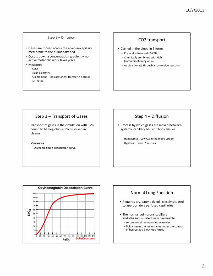
Step 2 – Diffusion
• Gases are moved across the alveolar‐capillary membrane to the pulmonary bed
• Occurs down a concentration gradient – no active metabolic work takes place
• Measures– ABGs
– Pulse oximetry
– A‐a gradient – indicates if gas transfer is normal
– P/F Ratio ‐
Step 3 – Transport of Gases
• Transport of gases in the circulation with 97% bound to hemoglobin & 3% dissolved in plasma
• Measures
– Oxyhemoglobin dissociation curve
CO2 transport
• Carried in the blood in 3 forms
– Physically dissolved (PaCO2)
– Chemically combined with Hgb (carbaminohemoglobin)
– As bicarbonate through a conversion reaction
Step 4 – Diffusion
• Process by which gases are moved between systemic capillary bed and body tissues
– Hypoxemia – Low O2 in the blood stream
– Hypoxia – Low O2 in tissue
Normal Lung Function
• Requires dry, patent alveoli, closely situated to appropriately perfused capillaries
• The normal pulmonary capillary endothelium is selectively permeable– serum protein remains intravascular
– fluid crosses the membranes under the control of hydrostatic & osmotic forces
10/7/2013
3
Balance of Hydrostatic & Osmotic Forces
• Allows small quantities of fluid into the interstitium
• Mechanisms to prevent alveolar edema– Retained intravascular protein
• maintains osmotic gradient favoring reabsorption
– Interstitial lymphatics • return large quantities of fluid to the circulation
– Tight junctions between alveolar epithelial cells• prevent leakage into the air spaces
www.uptodate.com
Other General Principles
• Transport of oxygen to body tissues mostly influenced by CO, Hgb concentration and O2/Hgb binding and releasing factors.
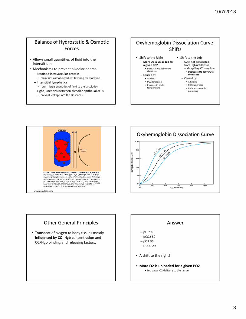
Oxyhemoglobin Dissociation Curve: Shifts
• Shift to the Right– More O2 is unloaded for a given PO2
• Increases O2 delivery to the tissue
– Caused by• Acidosis
• PCO2 increase
• Increase in body temperature
• Shift to the Left– O2 is not dissociated from Hgb until tissue and capillary O2 very low
• Decreases O2 delivery to the tissues
– Caused by• Alkalosis
• PCO2 decrease
• Carbon monoxide poisoning
Oxyhemoglobin Dissociation Curve
Answer
– pH 7.18
– pCO2 80
– pO2 35
– HCO3 29
• A shift to the right!
• More O2 is unloaded for a given PO2• Increases O2 delivery to the tissue

10/7/2013
4
• Questions thus far?????
Acute Respiratory Failure
• Impairment of oxygenation and / or ventilation
– Pao2 < 55 mmHg or Sao2 < 88%
– Paco2 > 50‐55 mmHg with accompanying acidemia, or pH < 7.30
Signs & Symptoms
• Dyspnea• Neuro:
– Hypoxemia• Anxiety, irritability, restlessness, confusion
– Hypercarbia• H/A, lethargy, confused, obtunded, coma
• Pulmonary– Flared nostrils, increased respiratory rate, use of accessory muscles, dyspnea, SOB
• Cardiovascular– Tachycardia, bounding pulses, dysrhythmias
Diagnostic Studies
• ABG
– Decreased PaO2 and / or
– Hypercapnia
• Radiologic
– Findings depend on primary disease
Management
• Noninvasive VS invasive ventilation
• Positioning– HOB elevated 30°
• Maximize ventilation
• Prevent aspiration
• Skin care
• Pain management / sedation
• Nutrition
• ATBs as appropriate
Indications for Mechanical Ventilation
• Pneumonia
• COPD
• ARDS
• Pulmonary edema
• Lung trauma
• Asthma
• Near Drowning
• Multiple sclerosis
• Muscular dystrophy
• Myasthenia gravis
• Spinal cord injury
• General anesthesia
• Overdose
• Obesity

10/7/2013
5
Goals of Mechanical Ventilation
• Provide adequate ventilation
– direct measure is minute ventilation (6‐8 liters per minute)
– indirect measure is CO2 of arterial blood ( 35‐45)
• Provide adequate oxygenation
– measured by pO2 of arterial blood gas
– if correlation has been established, can be inferred by peripheral oxygen saturation
Provide Adequate Ventilation
• Hypoventilation
– High PaCO2
– Respiratory acidosis
– Inadequate minute ventilation
• How do we correct???
– Assure adequate minute ventilation
– TV x RR = minute ventilation
• Hyperventilation
– Low PaCO2
– Respiratory alkalosis
– Excessive minute ventilation
• How do we correct???– Assure appropriate minute
ventilation
– TV x RR = Minute Ventilation
Provide Adequate Oxygenation
• Maximal alveolar ventilation increase oxygen exchange
• Deliver high level of oxygen
• Add PEEP to better inflate alveoli thus improving oxygen exchange
Complications from O2 Therapy
• Oxygen toxicity
– An oxygen concentration in excess of 50% for > 24 hours increases the potential for development of oxygen toxicity & lung damage
– Can impair Alveolar type II cells
• These cells produce surfactant!
Other Considerations
• Nitrogen
– Most plentiful gas (normally)
– Promotes alveolar expansion
• When completely displaced by 100% oxygen
– Can result in atelectasis
PEEP
• Improves oxygenation by maintaining alveolar airflow during expiration
– Airways have a tendency to collapse during expiration as a result of increasing pressure outside the airway
• Optimal levels are achieved by the lowest level of PEEP needed to raise the PaO2 without resulting in cardiovascular compromise (5‐15 common)

10/7/2013
6
ARDS
• A syndrome of acute respiratory failure characterized by non‐cardiac pulmonary edema and manifested by refractory hypoxemia caused by intrapulmonary shunt
• Nearly always occurs suddenly
• Overall mortality of ARDS ranges from 25% to 58%
ARDS Definition
• A syndrome of acute respiratory failure characterized by non‐cardiac pulmonary edema and manifested by refractory hypoxemia caused by intrapulmonary shunt
• ARDS refers to the severe end of the spectrum of ‘Acute Lung Injury’ (ALI).
What is Acute Lung Injury?
• Defined as syndrome of acute and persistent lung inflammation with increased vascular permeability.
• Characterized by 3 clinical features
– Bilateral radiographic infiltrates
– PaO2/FiO2 ratio between 201‐300 mmHg.
– No clinical evidence of an elevated left atrial pressure. If measured, PAOP in 18 mmHg.
PaO2/FiO2 Ratio
• A ratio of the partial pressure of arterial oxygen to the fraction of inspired oxygen.
• Normal is 300‐500 mmHg
• FiO2 is expressed as a decimal.
Examples
• WNL ‐ a patient has PaO2 of 90 (normal 80‐100) on room air (21%)
– P/F ratio = 90 / 0.21 = 428 mmHg
• ALI ‐ a patient has a PaO2 of 90 on FIO2 of 40%
– P/F ratio = 90/0.40 = 225 mmHg
ARDS = ALI + Worse Hypoxia
• ARDS– Hypoxia is WORSE
– PaO2/FiO2 200 mmHg
• Distinction between ALI and ARDS is somewhat arbitrary, since the degree of gas exchange disturbance does not correlate reliably with the extent of the underlying pathology.

10/7/2013
7
• Example #1 – a patient has a PaO2 of 60 on FiO2 of 80%
– P/F ratio = 60 / .80 = 75 mmHg
• Example #2 – a patient has a PaO2 of 50 on FiO2 of 100%
– P/F ratio = 50 / 1.0 = 50mmHg
www.uptodate.com
www.uptodate.com
Epidemiology
• Within ICUs, approximately 10‐15% of admitted patients meet criteria for ARDS
• Up to 20% of mechanically ventilated patients meet criteria of ARDS!
Causes
• More than 60 causes of ARDS have been identified
• Additional causes continue to emerge as adverse pulmonary reactions to new therapies are discovered.
Common Causes
• Sepsis – most common cause
• Aspiration
• Infectious pneumonia
• Severe trauma
• Surface burns
• Multiple blood transfusions (>15)
• Leukoagglutin reactions
• Pancreatitis
• Drug overdose
• Near drowning
• Smoke inhalation
• Cardiopulmonary bypass
• Pulmonary contusion
• Multiple fractures
• Following upper airway obstruction
• Drug reaction
• Venous air embolism
• Neurogenic pulmonary edema
• Acute esoinophilic pneumonia
• Bronchiotitis obliterans organizing pneumonia (BOOP)
• Miliary TB

10/7/2013
8
ARDS: Pathophysiology
• Acute phase ‐ damage to integrity of the blood‐gas barrier
– Damage to type 1 alveolar epithelial cells
– Increased endothelial permeability
– Interstitial edema is found
– Leakage of protein‐containing fluid into the alveoli
– Impaired production and fx of surfactant
www.uptodate.com
www.uptodate.com
www.uptodate.com
ARDS: Pathophysiology
• Resultant physiologic abnormalities
– Shunting of blood through atelectatic or fluid‐filled lung units
– Increased physiologic dead space
• Frequently exceeding 60% of each breath
– Compliance is reduced
– Increased resistance to blood flow
Results In:
• Impaired Gas Exchange
• Decrease lung compliance
• Pulmonary hypertension

10/7/2013
9
Decreased Lung Compliance
• Hallmark of ARDS
• Low compliance due to the stiffness of poorly or non‐aerated lung.
• Any questions????
Pulmonary Hypertension
• Occurs in about 25% of pts with ARDS subjected to mechanical ventilation.
• Contributing factors
– hypoxic vasoconstriction
– vascular compression (by + pressure vent)
Signs & Symptoms
• Severe dyspnea
• Increased work of breathing
• Refractory hypoxemia
• Diminished LOC if hypoxemia is severe
• Radiographic diffuse bilateral infiltrates – interstitial and alveolar
– without cardiomegaly
• PAOP – normal or low
Treatment
• The mainstay of therapy for ARDS is:
– Management of the underlying disorder causing it.
• ID treatable sources
– Main treatment is supportive.
Pharmocological Treatment
• Antibiotics
• Steroids (stress dose, watch glucose for osmotic changes)
• Diuretics
• Avoid excessive fluid administration
10/7/2013
10
Mechanical Ventilation• Tidal Volume
– initial 6 ml/kg– Permissive hypercapnea
• FiO2 100% (maintain Sa02 92‐94%)
• PEEP
– 5‐10 cm H2O is effective in reducing intrapulmonary shunting and improving oxygenation
– Frequently see PEEP > 12
Mechanical Ventilation
• Sedation
–May require Neuromuscular Blockade
• May use Pressure Controlled Ventilation
– sets pressure limits, allowing TV to fluctuate and prevents alveolar over distension
Pneumonia
• Inflammation of lung parenchyma often characterized by consolidation
• Exudate, inflammatory cells, fibrosis
• Usually caused by infectious agents or microbes,
– Can be caused by aspiration of gastric contents
Etiology
• Organisms
– Streptococcus pneumoniae• Most common cause
– Mycoplasma pneumoniae
– Haemophilus influenzae
• Viruses
– Relatively uncommon, accounting for 25‐50% of nonbacterial pneumonias
– Influenza A – most common
Signs & Symptoms
• Symptoms– Dyspnea– Chest pain– Wheezing cough– Fever chills rigors weight loss
– Night sweats– Fatigue– Weakness– Decreased energy – anorexia
• Signs– Consolidation– Pleural effusion– Airway involvement (wheezing)
– Crackles, rhonchi– Tachypnea– Tarchycardia– Fever– Purulent sputum– Dry cough– H/A, fatigue, sore throat– N/V, diarrhea
Diagnosis
• Sputum gram stain
– determine organisms and coverage
• Sputum cultures / sensitivities
• CBC with Diff
• Blood cultures
• Chest x‐ray
– Generally shows localized infiltrates
• Bronch / invasive diagnostics

10/7/2013
11
Treatment
• Antibiotics
• Fluids
• Oxygen
• Mechanical Ventilation
Asthma
• Chronic disease
• Characterized by airway hyper‐reactivity
• Produces airway narrowing of a reversiblenature
Pathophysiology
• Increased responsiveness to stimuli• Widespread narrowing of airway• Cellular infiltration and mucosal edema• Airway hyperreactivity
– Smooth muscle contraction – excessive mucus production – Diminished secretion clearance
• V/Q abnormalities• Increased work of breathing & airway resistance• Hyperinflation of the lung, increased residual volume• Host defect of altered immunologic state
Status Asthmaticus
• Characteristics
– Unrelenting acute asthma
– Broncho‐constriction despite treatment
– Predominantly high deadspace region
– Increased work of breathing
• Bronchospasm, inflammation, mucous production / plugs
– Increased minute ventilation
Pathophysiology
• Airway narrowing from
– Bronchial smooth muscle spasm
– Inflammation of bronchial walls, leading to increased permeability and thickening
– Mucous plugging from increased production and decreased clearance of secretions
Precipitating Events
• Infection, sinusitis
• Smoking
• Recent exposure (pollens, dust mites, animals, beta blockers)
• Emotional factors
• Gastroesophageal reflux
• Exercise

10/7/2013
12
Impending Status Asthmaticus
• Recurrent episodes over a short period (2‐7 days)
• Change in pattern of symptoms– Wheezing more severe or frequent
• Worsening dyspnea– Exercise limitation, at rest
• Cough with tenacious sputum
• Irritability
• Extremely dyspneic
• Inspiratory and expiratory wheezing usually audible
• Prolonged expiratory phase– Pt tries to exhale trapped air through narrow airways
• Tachypnea
• Tachycardia
• Use of accessory muscles
• Flaring nares, pallor, cyanosis, increased work of breathing, and fatigue
• Disappearance of wheezing may be ominous sign
– Airways completely obstructed
Treatment
• Bronchodilators
• Corticosteroids
• Oxygen
• Hydration
• Antibiotics if infection suspected
• Mechanical ventilation – Low tidal volumes
– May need sedatives
Acute Pulmonary Embolus
• Characteristics– Obstruction of the pulmonary arteries by emboli
– Effects lung tissue, the pulmonary circulation and the fx of the right and left side of the heart
– Most thromboembolic (>95%) originate in the ileofemoral veins
– Complication of DVT
Other predisposing factors
• Age > 40
• Immobility
• Previous DVT
• Anesthesia / surgery
• Pregnancy / post‐partum
• Trauma

10/7/2013
13
Hemodynamic Consequences
• Obstruction stimulates neurohumoral stimuli
– Increases PA pressures & PVR
– Results in increased RV work
• Pulmonary HTN (mean PAP > 20 mmHg)
• RV will fail if mean PA pressure > 40 mmHg
Disruption in blood flow
• Alveoli become nonfunctioning units
– Don’t participate in gas exchange
– Increases deadspace
• To maintain gas exchange
– Ventilation is shifted to the noninvolved areas of the lung
– Results in constriction of distal airways
– Leads to alveolar collapse and atelectasis
Signs & Symptoms
• Depends on severity– Sudden onset of chest pain
– Cough
– Hemoptysis
• Massive PE– >50% vascular occlusion
• Mental clouding
• Anxiety
• Feeling of impending doom
• Apprehension
• Dyspnea, tachypnea, increased work of breathing, tachycardia, reduced blood pressure, restlessness, syncope, asymmetric chest expansion
PE Diagnostics
• ABG– May indicate respiratory alkalosis
• CXR– Nonspecific, frequently normal
• V/Q scan – not definitive, but suggestive
• Search for DVT – anticoagulation
• Pulmonary angiogram – most definitive test
• CT‐angio may also be done
• ECG – usually normal, except in massive PE (new RBBB)
Goals of Care
• Restore pulmonary artery blood flow
• Maintain / Restore hemodynamic stability
• Relieve chest pain
Pneumothorax
• Characteristics– Air in the pleural space
– Leading causes• + pressure ventilation
• Diagnostic procedure
– Tension pneumo present when intrapleural pressure exceeds atmospheric pressure throughout expiration
– Severity depends on size, underlying lung dz, whether a tension pneumo is present

10/7/2013
14
Pneumothorax
• Symptoms– Dyspnea
– Chest pain
• Ususally pleuritic
• Typically acute onset
• Signs– Tachypnea
– Tachycardia
– Hypotension
– Decreased respiratory excursion
– Wider intercostal spaces
– Absent or redcued brath sounds
– Hyperresonant to percussion
– Tracheal shift to contralateral side
– Hypoxemia +/‐ hypercapnia on ABG
Therapy
• Re‐expansion of the collapsed lung
– Chest tube insertion
• Adequate oxygenation
• Maintain cardiac output
• Reduce / control pain
• Observation
Chest Trauma
• Pathophysiology depends on type and extent of injury
• Trauma to the chest or lungs may interfere with any of the components involved with inspiration, gas exchange and expiration
Chest Trauma
• Blunt injuries
• Etiology & Risk factors– Auto accidents
– Falls
– Assaults
– Explosions
• Penetrating injuries
• Etiology & Risk factors– All those causing blunt
injuries &
– Bullets
– Knives
– Shell fragments
– Free‐flying objects
– Industrial accidents
Signs & Symptoms
• Varies with specific injury
– Tachypnea, dyspnea, pain, respiratory distress may occur with any injury
Blunt injuries
• Visceral injuries without chest wall damage
– Pneumothorax, hemothorax
– Lung contusion
– Diaphragmatic injury
– Myocardial contusion, aortic rupture
– Rupture of the trachea or bronchus

10/7/2013
15
Blunt injuries
• Soft tissue injuries
– Possibly a sign of severe underlying damage
• Cutaneous abrasion
• Ecchymosis
• Laceration of superficial layers
• Burns
• Hematoma
Blunt injuries
• Others– Fracture of the sternum
• Occurs either as a result of direct impact or as the indirect result of overflexion of the trunk
– Rib fractures• As a result of overflexion or straightening.• Can be unifocal or multifocal • Multiple fractures result in flail chest
– Often complicated by injuries to soft tissues and pleura
– S/SX• Pain accentuated by chest wall movement, deep inspiration or touch
• Flair chest – dyspnea and localized pain
Penetrating injuries
• Pleural cavity & chest wall entered
• Damage to deeper structures – more serious
• Extent of injury and organs injured predicted by course of wound and nature of penetrating instrument
• High velocity projectiles do more damage
Penetrating injuries
• Open sucking chest wounds
– If opening is < diameter of trachea, minimal symptoms
– If opening is > more air enters the pleural space, collapses the lung, impairs ventilation and gas exchange, results in dyspnea
Penetrating injuries
• Hemothorax, hemopneumothorax
• Combined thoraco‐abdominal injuries
– Bowel sounds may be heard in chest
• Trachea / large airway damage
– Sub‐Q emphysema
• Wounds of heart / great vessels
– Dyspnea and backache, intense pain in chest or back unaffected by respiration
Diagnostic Studies
• CXR
• MRI or CT if stable enough
• Aortography – confirms dx of rupture of aorta or other great vessels
• Bronchoscopy – dx rupture of trachea or bronchus
• ECG

10/7/2013
16
Goals of Care
• Patent airway
• ABG levels and pulmonary parameters restored and maintained
• Chest wall integrity and stability restored
• Establish integrity of pleural space
• Minimize chest pain and dyspnea
• Questions…………………….