Embed Size (px)
Citation preview
IntroductionDaxx is a ubiquitously expressed protein (Hollenbach et al.,1999; Kiriakidou et al., 1997) that was originally isolated froma yeast two-hybrid screen aimed at identifying proteins thatinteract with the cytoplasmic tail of the Fas receptor (FasR),the region of the receptor known as the ‘death domain’. It wasoriginally proposed that hDaxx promoted apoptosis, asoverexpression of hDaxx was observed to enhance Fas-dependent apoptosis (Yang et al., 1997). However, in contrastto its proposed role in promoting apoptosis, Daxx-null micewere embryonic lethal by day 9.5 of gestation and exhibitedextensive apoptosis (Michaelson et al., 1999). This observationis contradictory to a role for hDaxx in Fas-dependent apoptosisand argues against a role for Daxx in promoting Fas-dependentcell death. The Daxx knockout embryos also exhibited a highlydisorganized physiology (Michaelson et al., 1999), suggestingthat Daxx is essential for mouse development.
In addition to the originally proposed role for hDaxx inpromoting apoptosis, it was also suggested that it acted as alink between FasR and ASK1 (Chang et al., 1998), adownstream signaling kinase involved in Fas-dependentapoptosis. This observation predicted a cytoplasmic cellularlocalization for hDaxx. However, it has since beendemonstrated that hDaxx is a strictly nuclear protein(Hollenbach et al., 1999; Kiriakidou et al., 1997; Pluta et al.,1998), with at least two identified functions in the nucleus.First, hDaxx has been identified as a component of nuclear
promyelocytic leukemia protein (PML) oncogenic domains(PODs) (Everett et al., 1999; Zhong et al., 2000). It wasdemonstrated that the observed sensitivity of cellsoverexpressing hDaxx to apoptosis is mediated through thenuclear interaction of hDaxx with PODs and not through adirect interaction with the Fas receptor in the cytoplasm (Toriiet al., 1999; Villunger et al., 2000; Zhong et al., 2000). Daxxassociates with PODs through a direct interaction with PML,a critical component of PODs (Everett et al., 1999; Ishov et al.,1999; Li et al., 2000a). The interaction is a dynamic, cell cycleregulated event and is dependent on the post-translationalmodification of PML by the small ubiquitin-related modifierSUMO-1 (Ishov et al., 1999; Lehembre et al., 2001; Li et al.,2000a).
In addition to its presence in PODs, we showed that hDaxxcould act as a transcriptional co-repressor. Consistent with thisrole hDaxx contains four structural domains commonly foundin transcriptional regulatory proteins: two predicted pairedamphipathic helices, an acid-rich domain and a Ser/Pro/Thr(SPT)-rich domain (Hollenbach et al., 1999). The tethering ofhDaxx to DNA by fusing it to the GAL4 DNA-binding domain(GAL4-DBD) resulted in an 85% repression of transcriptionalactivity from the constitutively active thymidine kinasepromoter that contained a GAL4-binding site (Hollenbach etal., 1999; Li et al., 2000; Torii et al., 1999). Furthermore,hDaxx exists as three distinctly migrating forms with apparentmolecular weights of 70 kDa, 97 kDa and 120 kDa. The 120
3319
Human Daxx is a protein that functions, in part, as atranscriptional co-repressor through its interaction with agrowing number of nuclear, DNA-associated proteins. Todetermine the mechanism by which hDaxx repressestranscription, we used conventional chromatography toisolate endogenous hDaxx. We determined that hDaxx hasan apparent molecular weight of 360 kDa, which isconsistent with the fact that multiple domains of hDaxx arerequired for transcriptional repression and suggests thathDaxx associates with multiple proteins. Using co-fractionation and co-immunoprecipitation we demonstratethat hDaxx associates with proteins that are critical fortranscriptional repression, such as histone deacetylase II,constituents of chromatin such as core histones H2A, H2B,
H3 and H4, and Dek, a chromatin-associated proteinreported to change the topology of DNA in chromatin invitro. We also demonstrate a requirement for the SPTdomain and the first paired amphipathic helix of hDaxx forits association with histone deacetylase II and acetylatedhistone H4, respectively. Finally, we provide evidencesuggesting that the association of hDaxx with chromatin-related proteins is dependent on the post-translationalphosphorylation status of hDaxx. A working model for therepressive action of hDaxx through its association withchromatin related proteins is presented.
Key words: Daxx, HDAC II, Histone, Dek
Summary
Daxx and histone deacetylase II associate withchromatin through an interaction with core histonesand the chromatin-associated protein DekAndrew D. Hollenbach, Craig J. McPherson*, Edwin J. Mientjes, Rekha Iyengar and Gerard Grosveld ‡
1Department of Genetics, St. Jude Children’s Research Hospital, 332 North Lauderdale, Memphis, TN 38105, USA*Present address: Department of Infectious Diseases, Parke Davis Pharmaceuticals, 2800 Plymouth Road, Ann Arbor, MI 28105, USA‡Author for correspondence (e-mail: [email protected])
Accepted 20 May 2002Journal of Cell Science 115, 3319-3330 (2002) © The Company of Biologists Ltd
Research Article
3320
kDa form was shown to result, in part, from a post-translationalphosphorylation (Hollenbach et al., 1999). However, only thenon-phosphorylated 70 kDa form of hDaxx interacts with theDNA-bound transcription factor Pax3, which represses thetranscriptional activity of Pax3 by nearly 80% (Hollenbach etal., 1999). In a later report, hDaxx was also shown to repressthe transcriptional activity of ETS-1, a member of the etsfamily of transcription factors (Li et al., 2000b), thussupporting the role of hDaxx as a transcriptional co-repressor.
To date, the mechanism by which hDaxx exerts itsrepression activity is poorly understood. Histone deacetylation,which plays a critical role in transcriptional silencing ofactively transcribed chromatin by inducing chromatincondensation onto naked DNA (Pazin and Kadonaga, 1997;Wolffe and Hayes, 1999), has been implicated in hDaxx-mediated transcriptional repression (Li et al., 2000a). However,an exact mechanism describing the repression activity ofhDaxx has yet to be determined. Therefore, to betterunderstand the mechanism of hDaxx repression, we havecreated a series of hDaxx deletion mutants fused to the GAL4DNA-binding domain (DBD). In the present work, wedemonstrate that multiple domains of hDaxx exerttranscriptional repression, which suggests that hDaxx mayassociate with multiple proteins. Using standardchromatography and co-immunoprecipitation analyses, wedemonstrate that hDaxx associates with the core histones H2A,H2B, H3 and H4; histone deacetylase II (HDAC II), which iscritical for transcriptional repression; and Dek, a protein thatalters the superhelical density of DNA in vitro (Alexiadis et al.,2000) and associates with chromatin in vivo (Kappes et al.,2001). Finally, consistent with the requirement of multipledomains for the repression activity of hDaxx, we demonstratethat both the SPT domain and the first paired amphipathic helixof hDaxx are necessary for its association with HDAC II andacetylated histone H4, respectively.
Materials and MethodsConstruction of hDaxx deletion mutants and protein expressionAll deletion mutant constructs were cloned in frame with the GAL4DBD (amino acids 1-144) in the vector pM2. The N-terminal and C-terminal deletions are identified by the specific amino acids that wereremoved. The hDaxx∆574-740 deletion was constructed by removalof the C-terminal HindIII fragment from GAL4-hDaxx. ThehDaxx∆1-573 deletion was constructed by cloning the C-terminalHindIII fragment of hDaxx into pM2. The remaining C- and N-terminal deletions were generated by PCR amplification and standardcloning techniques. The domain-specific and nuclear-localization-specific deletion mutants were constructed by overlap extension PCR(Ho et al., 1989). Primers were complementary to the regions ofhDaxx to be deleted and were engineered to utilize unique restrictionsites, allowing the efficient cloning of the amplified products as‘cassettes’. Specifically, the hDaxx∆PAH1 PCR product removed thefirst predicted paired amphipathic helix (nucleotides 189-324), and theresulting PCR cassette was cloned into the AccI site present in thepM2 polylinker and at position 375 in hDaxx. The hDaxx∆PAH2 PCRproduct removed the second predicted paired amphipathic helix(nucleotides 574-726), and the resulting PCR cassette was cloneddirectionally into the hDaxx NgoMI site at position 354 and the hDaxxEagI site at position 748. The hDaxx∆AD PCR product removed theacid-rich domain (nucleotides 1299-1479), and the resulting PCRcassette was cloned into the hDaxx KasI site at position 1077 and thehDaxx BstXI site at position 2056. The hDaxx∆SPT PCR product
removed the final 495 bp of sequence (nucleotides 1725-stop), and theresulting PCR cassette was cloned into the KasI site at position 1077of hDaxx and the XbaI site in the pM2 polylinker. Finally, the ∆NLS1PCR product removed the first predicted nuclear localization sequence(nucleotides 1162-1185), and the ∆NLS2 PCR product removed thesecond predicted nuclear localization sequence (nucleotides 1888-1902). Both PCR products were cloned into the hDaxx KasI site atposition 1077 and the hDaxx BstXI site at position 2056. The ‘deletioncassettes’ were then used to construct the multiple domain deletionmutants. For example, the hDaxx∆PAH1-2 deletion construct wascreated by cloning the NgoMI-EagI ∆PAH2 cassette into the NgoMI-EagI site of hDaxx∆PAH1. All constructs were sequenced to confirmthat they are in frame with the GAL4 DBD and that no point mutationswere introduced during PCR amplification.
To determine protein expression, NIH3T3 cells (5×10⁵) were platedon 100 mm dishes and transfected the following day with 10 µg ofthe pM2-hDaxx deletion constructs using the Fugene6 method(Boehringer Mannheim, Germany) according to the manufacturer’sspecifications. After 48 hours, the cells were harvested, resuspendedin PBS containing both protease and phosphatase inhibitors (0.1 mMPMSF, 10 µg/ml leupeptin, 2 µg/ml Aprotinin, 1 mM β-glycerophosphate, 1 mM NaF and 0.1 mM NaVO4) and lysed by fourrounds of freezing and thawing. Cell lysates (30 µg) were resolved byeither 15% or 8% SDS-PAGE, blotted to Immobilon-P membrane(Millipore, Bedford, MA), and the protein was detected by westernanalysis using the mouse anti-GAL4 DBD monoclonal antibody(Santa Cruz Biotechnology, CA).
ImmunofluorescenceTo determine the cellular localization of the individual deletionmutants, NIH3T3 cells (5×10⁴) were plated on one-chamberpolystyrene vessel tissue-culture-treated glass slides (BectonDickinson, NJ) and transfected the following day with 2 µg of each ofthe pM2-hDaxx deletion constructs. After 48 hours, the cells werefixed and permeablized as previously described (Lam et al., 1999) andincubated for 1.5 hours with a 1:2000 dilution of the anti-GAL4 DBDantibody. After extensively washing with PBS, the cells were incubatedwith FITC-conjugated goat anti-mouse antibody (1:250 dilution) for45 minutes, washed with PBS and mounted with Vectashield
mounting medium (Vector Laboratories, Burlingame, CA) thatcontained 3 µM 4′,6-diamindino-2phenylindole (DAPI, Sigma, MO).Slides were examined using an Olympus BX50 fluorescent scope.
Transcriptional assays To determine the repression activity of the hDaxx deletion mutants,NIH3T3 cells (3×10⁵) were plated on 60 mm dishes and transfectedthe following day using the Fugene6 method. The Fugene6-DNAcomplex consisted of 10 fmol of the indicated pM2-hDaxx deletionconstructs, 500 ng of the secreted alkaline phosphatase (SEAP)control plasmid under control of the MAP1 promoter and either 1 µgof the 1X-GAL4-TK-CAT reporter plasmid containing one GAL4-DNA-binding site or 0X-GAL4-TK-CAT reporter plasmid containingno GAL4-DNA-binding sites. This reporter contained thechloramphenicol acetyl transferase (CAT) gene under control of theconstitutively active thymidine kinase promoter containing either oneor no GAL4-DNA-binding sites. After 48 hours, the medium wasassayed for SEAP activity as previously described (Bram et al., 1993),and the cells were harvested and assayed for CAT activity aspreviously described (Ausubel et al., 1996). The percentage of[14C]chloramphenicol acetylation was quantified from thin-layerchromatography plates using a Molecular Dynamics Phosphorimager(Amersham Pharmacia Biotech, Piscataway, NJ). The transfectionefficiency was normalized relative to the SEAP activity, and valuesrepresent the average and standard deviation from four independentdeterminations.
Journal of Cell Science 115 (16)
3321Daxx, HDAC II, histones and Dek association
Co-fractionation of hDaxx, HDAC II, core histones and DekTo determine whether hDaxx, HDACII, histones and Dek co-fractionated, U937T cells (1.2×10⁸) (Boer et al., 1998) stablyexpressing FLAG-epitope tagged Dek were harvested andresuspended in 10 ml of a buffer containing 20 mM HEPES (pH 7.9),150 mM KCl and all protease and phosphatase inhibitors as describedabove. The cells were sonicated two times for 20 seconds on powerlevel three with a 550 Sonic Dismembrator (Fisher, Pittsburgh, PA),separated into 10×1 ml aliquots, and each aliquot was sonicated foran additional three rounds. The debris was removed by centrifugationin a Eppendorf Centrifuge 5415D microfuge at 16,100 g for 10minutes. The supernatant was removed, and glycerol and Tween 20were added to final concentrations of 20% and 0.1%, respectively(Buffer PLB). The total cell lysate (70 mg) was loaded onto a HiPrep
Sephacryl S-300 size exclusion column (26×600 mm, Pharmacia,Belgium) pre-equilibrated with buffer PLB. Proteins were eluted witha flow rate of 1 ml/minute into 5 ml fractions. The presence of hDaxxwas determined by Deoxycholate/Trichloroacetic acid (DOC/TCA)precipitating 20 microliters of each individual fraction followed byacetone precipitation and separating the precipitated proteins on a 4-20% SDS-PAGE gradient gel. The gel was blotted to Immobilon-Pmembrane (Millipore, Bedford, MA), and hDaxx was detected bywestern analysis using a rabbit polyclonal antibody describedpreviously (Hollenbach et al., 1999). Fractions were also analyzed forthe presence of HDACII using a mouse monoclonal anti-HDACIIantibody (Santa Cruz sc-9959, CA), acetylated histone H4 using arabbit polyclonal anti-acetylated histone H4 antibody (UpstateBiotechnology #06-866, Lake Placid, NY) and Dek-FLAG using amouse monoclonal anti-FLAG antibody (Sigma, St. Louis, MO). Theapparent native molecular weight of endogenous hDaxx wasdetermined from a standard curve of proteins of known molecularweights.
The fractions containing hDaxx were combined, and the protein (20mg, 3.5-fold purification over crude lysate) was loaded onto aResource-Q anion exchange column (Pharmacia, Belgium) 6 ml bedvolume) previously equilibrated with buffer PLB. The column waswashed with 10 column volumes of buffer PLB (60 ml) at a flow rateof 1 ml/minute into 5 ml fractions, and proteins were eluted with alinear gradient of 0.15-1 M KCl in buffer PLB at the same flow rate.The presence of hDaxx, HDACII, acetylated histone H4 and Dek-FLAG in each fraction was determined as described above. Thefractions containing hDaxx were combined, concentrated to 2 ml, andthe protein (2 mg, 35-fold purification over crude lysate) was loadedonto a Superdex HR-200 gel filtration column (20×320 mm,Pharmacia, Belgium) previously equilibrated with buffer PLB.Proteins were eluted with a flow rate of 1 ml/minute into 2.5 mlfractions, and the presence of all proteins was determined as describedabove. The apparent native molecular weight of hDaxx wasdetermined from a standard curve of proteins of known molecularweights. Approximately 120 µg of hDaxx and its associated proteinswere obtained from 140 mg of total cellular extract, resulting in anapproximately 1150-fold purification.
Mass spectral analysis of histones We determined the presence of histones by mass spectral analysisbecause commercially available antibodies did not recognize non-acetylated histones by western analysis in our hands. 20 µg of proteinfrom the final fraction of the purification were separated by 4-20%SDS-PAGE, and the proteins were visualized by Coomassie staining.Following electrophoresis and visualization, the gel was dried and theproteins corresponding to the molecular weights of histones H2A,H2B, H3 and H4 were excised from the gel. The protein in the excisedgel piece was reduced, alkylated with iodoacetamide and digestedwith trypsin. Tryptic peptides were extracted and subjected tocombined capillary liquid chromatography/tandem mass spectrometry.Mass spectrometry was performed using a ThermoQuest LCQ-DECA
ion-trap mass spectrometer with an electrospray ion source. Fragmention (MS2) spectra were subjected to search of the NCBI non-redundant protein database using the SEQUEST program of Eng andYates marketed by ThermoQuest.
Co-immunoprecipitation of hDaxx, HDAC II, Dek andacetylated histone H4 To demonstrate a physical association of hDaxx, HDAC II, Dek andacetylated histone H4 and to map the domains of hDaxx responsiblefor its association with HDACII and acetylated histone H4, co-immunoprecipitation experiments were performed. Because U937Tcells did not transfect well by the Fugene6 method, 293T cells wereused. 293T cells (1×10⁶) were plated in 100 mm dishes andtransfected the following day with 5 µg of the mammalian expressionvectors encoding either GAL4-hDaxx, GAL4-hDaxx∆1-132, GAL4-hDaxx∆1-352 or GAL4-hDaxx-SPT using the Fugene6 methodaccording to the manufacturer’s specifications. After 48 hours, thecells were lysed on ice in NP40 lysis buffer (0.4 M NaCl, 0.2 mMEGTA, 10% glycerol, 1% NP40 and all protease and phosphataseinhibitors as described above), the cell debris was removed bycentrifugation at maximum speed in a Sorvall RMC 1Y microfuge for15 minutes at 4°C, and the resulting cell extracts were precleared byincubating them with Gamma Bind plus Sepharose resin (Pharmacia,Belgium) for 2 hours at 4°C. Total cell lysate (150 µg) was incubatedwith a mouse monoclonal anti-GAL4 DBD antibody (clone RK5C1,Santa Cruz, CA) overnight at 4°C on a rotary shaker; immunecomplexes were collected with Gamma Bind Plus Sepharose beads;and the beads were washed four times with ice-cold NP40 lysis buffer.The pellets were resuspended in 30 µl of SDS-PAGE buffer and boiledfor 5 minutes. Supernatants were resolved by 4-20% gradient SDS-PAGE, blotted to Immobilon-P membrane and either HDAC II oracetylated histone H4 were detected by western analysis using amouse monoclonal anti-HDAC II or rabbit polyclonal anti-acetylatedhistone H4 antibody, respectively. Unfortunately, we were unable toanalyze the presence of endogenous Dek owing to the non-specificinteraction of Dek with the gamma-bind plus resin. To confirmoverexpression of all deletion constructs, 20 µg of total cell lysate wasresolved by 4-20% SDS-PAGE, blotted to Immobilon-P membrane,and protein was detected by western analysis using the mousemonoclonal anti-GAL4 DBD antibody as described above.
Alternatively, associated proteins were also analyzed byimmunoprecipitation of Dek-FLAG. Fractions from the Sephacryl S-300 column that contained hDaxx, Dek, acetylated histone H4 andHDAC II (fractions 9-13) were combined and passed over an anti-FLAG antibody affinity column (Sigma, St. Louis, MO). The columnwas washed extensively with buffer PLB, and proteins that wereretained by the column were competitively eluted with PLB buffercontaining 100 µg/ml FLAG peptide. The eluted proteins wereDOC/TCA/acetone precipitated, separated by 4-20% SDS-PAGE,transferred to Immobilon-P membrane, and hDaxx, HDAC II andacetylated histone H4 were detected by western analysis as describedabove, or the core-histones H2A, H2B, and H3 were visualized bysilver stain.
Histone deacetylase activity assaysTo determine the ability of GAL4-hDaxx to immunoprecipitatehistone deacetylase activity, 293T cells (1×10⁶) were plated in 100mm dishes and transfected the following day with 5 µg of themammalian expression vectors encoding either GAL4-hDaxx orGAL4 by the Fugene6 method according to the manufacturer’sspecifications. After 48 hours, the cells were lysed with RIPA buffercontaining all protease and phosphatase inhibitors and precleared, asdescribed above. Total cell lysate (100 µg) was immunoprecipitatedwith the anti-GAL4 antibody, and after extensive washing with lysisbuffer, the beads containing the immune complexes were resuspended
3322
in 200 µl of HDAC assay buffer (10 mM Tris-HCl, pH 8.0, 150 mMNaCl, 10% glycerol and all protease). [3H]acetyl histone H4 (730,000cpm/µl), previously prepared according to the manufacturer’sspecifications (Upstate Biotechnology, Lake Placid, NY), was addedto the immune complexes, and the reactions were incubated for 36hours at room temperature. The reactions were stopped by the additionof 50 µl of quenching solution (1.0 M HCl, 160 mM acetic acid), andthe released [3H]acetate was extracted and quantified according to themanufacturer’s specifications (Upstate Biotechnology, Lake Placid,NY). As a positive control for the histone deacetylase reaction, 10 µgof HeLa nuclear extract was incubated with the HDAC assay bufferand [3H]acetyl histone H4, and released [3H]acetate was analyzed asdescribed above. The specificity of the histone deactylase reaction wasdetermined by parallel reactions that were incubated with 50 mMsodium butyrate, a specific histone deacetylase inhibitor.
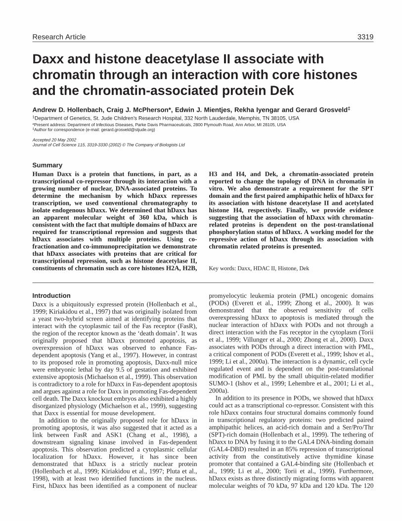
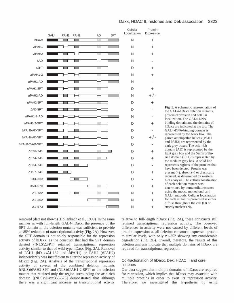
ResultsGAL4-hDaxx deletion constructsTo understand the mechanism by which hDaxx exerts itstranscriptional repression activity, we created a series ofdeletion mutants that target the four identified structuraldomains of hDaxx. All of the deletion constructs were fusedin frame to the GAL4-DNA-binding domain (DBD) (Fig. 1).These mutants included successive C-terminal (hDaxx∆574-740, hDaxx∆334-740, and hDaxx∆157-740) and N-terminal(hDaxx∆1-132, hDaxx∆1-352, and hDaxx∆1-573) deletions,which removed regions surrounding the identified structuraldomains of hDaxx. We also included the C-terminal deletionhDaxx∆626-740 that was previously described to be aconstitutively active mutant in its putative role in Fas-dependent apoptosis (Yang et al., 1997). In addition, weconstructed two mutants that retain only the regionssurrounding the paired amphipathic helix number two(hDaxx133-333) and the acidic domain (hDaxx353-573).Finally, we generated a series of small deletion mutants thatinclude the removal of each individual domain (∆PAH1,∆PAH2, ∆AD, and ∆SPT), the removal of two domains(∆PAH1-2, ∆PAH1-AD, ∆PAH1-SPT, ∆PAH2-AD, ∆PAH2-SPT, and ∆AD-SPT), the removal of three domains (∆PAH1-2-AD, ∆PAH1-2-SPT, ∆PAH1-AD-SPT, and ∆PAH2-AD-SPT) and the removal of all four domains of hDaxx (∆PAH1-2-AD-SPT).
Transcriptional repression activity of GAL4-hDaxxdeletion mutants Before we could use the deletion constructs in transcriptionalrepression assays, several controls needed to be performed.First, to demonstrate that all deletion mutants were expressedat similar levels, each of the constructs was individuallytransfected into NIH3T3 fibroblasts, and the presence ofmutant protein was detected by western analysis using a mouseanti-GAL4 DBD monoclonal antibody. The majority of thedeletion constructs expressed similar levels of protein (Fig. 1)except for ∆1-352, which in addition to expressing protein ofthe correct molecular weight also demonstrated a considerableamount of protein degradation (Fig. 2B). Interestingly, wenoted barely detectable levels of protein with the majority ofconstructs in which the acid-rich domain had been removed(∆AD, ∆PAH1-AD, ∆AD-SPT, ∆PAH1-2-AD, ∆PAH1-AD-SPT, ∆PAH1-2-AD-SPT, hDaxx∆334-740, and hDaxx133-
333) (Fig. 1). In addition, the deletion constructs that lackedthe acid-rich domain but still retained the N-terminal pairedamphipathic helix (∆PAH2-AD, ∆PAH2-AD-SPT) expressedprotein of the correct molecular weight at a lower level thanfull-length hDaxx (Fig. 1). A pulse-chase analysis of selectdeletion mutants confirmed that the proteins were beingexpressed and that the observed reduction in the steady-statelevel of protein expression was caused by a decrease in thestability of deletion mutants lacking the acid-rich domain. Thespecific removal of the acid-rich domain decreased the normalhalf-life of GAL4-hDaxx by greater than four-fold (data notshown), suggesting that the acid-rich domain is essential forthe stable expression of hDaxx.
Second, hDaxx contains two predicted nuclear localizationsignals (NLS): one present in the SPT domain (NLS2,K630KSRK634) and one immediately N-terminal to the acid-rich domain (NLS1, R391KKRR395) (Kiriakidou et al., 1997;Li et al., 2000a). Therefore, to confirm that the hDaxx deletionmutants maintained a nuclear localization, we transientlytransfected NIH3T3 cells with each individual construct,and the localization of the protein was visualized byimmunofluorescence using the anti-GAL4 DBD monoclonalantibody. Consistent with previous reports (Everett et al., 1999;Hollenbach et al., 1999; Ishov et al., 1999; Kiriakidou et al.,1997; Pluta et al., 1998), GAL4-hDaxx demonstrated a strictnuclear staining (Fig. 1). In addition, nuclear staining was alsoobserved for each of the deletion mutants containing an intactSPT domain (Fig. 1). By contrast, despite the presence of apredicted NLS in hDaxx immediately N-terminal to the acid-rich domain, hDaxx deletion mutants missing all or part of theSPT domain demonstrated a diffuse staining throughout thecell (Fig. 1). The loss of strict nuclear staining upon removalof the SPT domain indicated that NLS2 is critical for thenuclear localization of hDaxx. Consistent with thisobservation, we found that the specific deletion of NLS2 wassufficient to remove the strict nuclear localization of GAL4-hDaxx. However, the removal of NLS1 had no effect on thenuclear localization of GAL4-hDaxx (data not shown). Thisobservation provided direct evidence that the NLS2 isnecessary and sufficient for the localization of hDaxx to thenucleus.
Finally, the deletion constructs that localized strictly to thenucleus and produced stable protein (hDaxx, hDaxx∆1-573,hDaxx∆1-132, ∆PAH1-2, ∆PAH2-AD, hDaxx∆1-352, ∆PAH2,and ∆PAH1) were tested for their ability to repress transcription.Deletion mutants that expressed stable protein but displayeda diffuse cellular localization were cloned in frame with theSV40 NLS between the GAL4 DBD and the hDaxx mutantconstruct [(NLS)∆PAH1-SPT, (NLS)∆SPT, (NLS)∆PAH2-SPT,(NLS)∆PAH1-2-SPT, and (NLS)hDaxx353-573]. The presenceof the SV40 NLS did not affect protein stability (Fig. 2B) nordid it affect the transcriptional repression activity of GAL4-hDaxx (data not shown). It was, however, sufficient to restorethe strict nuclear localization of the deletion mutants (data notshown). Consistent with previous reports (Hollenbach et al.,1999; Li et al., 2000a), expression of full-length GAL4-hDaxxresulted in an 85% reduction of transcriptional activity (Fig.2A). This reduction was dependent on GAL4-hDaxx binding toDNA and was not caused by titration of other essential factorsby hDaxx as minimal repression (≤15%) was observed wheneither the GAL4 DBD or the GAL4-DNA-binding sites were
Journal of Cell Science 115 (16)
3323Daxx, HDAC II, histones and Dek association
removed (data not shown) (Hollenbach et al., 1999). In the samemanner as with full-length GAL4-hDaxx, the presence of theSPT domain in the deletion mutants was sufficient to providean 85% reduction of transcriptional activity (Fig. 2A). However,the SPT domain is not solely responsible for the repressionactivity of hDaxx, as the construct that had the SPT domaindeleted ((NLS)∆SPT) retained transcriptional repressionactivity similar to that of wild-type hDaxx (Fig. 2A). Removalof PAH1 (hDaxx∆1-132 and ∆PAH1) or PAH2 (∆PAH2)independently was insufficient to alter the repression activity ofhDaxx (Fig. 2A). Analysis of the transcriptional repressionactivity of several of the combined deletion mutants[(NLS)∆PAH2-SPT and (NLS)∆PAH1-2-SPT] or the deletionmutant that retained only the region surrounding the acid-richdomain [(NLS)hDaxx353-573] demonstrated that althoughthere was a significant increase in transcriptional activity
relative to full-length hDaxx (Fig. 2A), these constructs stillretained transcriptional repression activity. The observeddifferences in activity were not caused by different levels ofprotein expression as all deletion constructs expressed proteinto similar levels, with only ∆1-352 showing any considerabledegradation (Fig. 2B). Overall, therefore, the results of thisdeletion analysis indicate that multiple domains of hDaxx arerequired for transcriptional repression.
Co-fractionation of hDaxx, Dek, HDAC II and corehistonesOur data suggest that multiple domains of hDaxx are requiredfor repression, which implies that hDaxx may associate withmultiple proteins in order to exert its repression activity.Therefore, we investigated this hypothesis by using
CellularLocalization
ProteinExpression
hDaxx N +∆PAH1 N +∆PAH2 N +
∆AD N -∆SPT D +
∆PAH1-2 N +∆PAH1-AD N -
∆PAH1-SPT D +∆PAH2-AD N + / -
∆PAH2-SPT D +∆AD-SPT D -
∆PAH1-2-AD N -∆PAH1-2-SPT D +
∆PAH1-AD-SPT D -∆PAH2-AD-SPT D + / -
∆PAH1-2-AD-SPT D -∆626-740 D +∆574-740 D +∆334-740 D -∆157-740 D +
133-333 D -353-573 D +
∆1-132 N +∆1-352 N +∆1-573 N +
GAL4 PAH1 PAH2 AD SPT
Fig. 1.A schematic representation ofthe GAL4-hDaxx deletion mutants,protein expression and cellularlocalization. The GAL4-DNA-binding domain and the domains ofhDaxx are indicated at the top. TheGAL4-DNA-binding domain isrepresented by the black box. Thepaired amphipathic helices (PAH1and PAH2) are represented by thedark gray boxes. The acid-richdomain (AD) is represented by thelight gray box and the Ser/Pro/Thr-rich domain (SPT) is represented bythe medium gray box. A solid linerepresents regions of the proteins thathave been deleted. Protein waspresent (+), absent (–) or drasticallyreduced, as determined by westernblot analysis. The cellular localizationof each deletion mutant wasdetermined by immunofluorescenceusing the mouse monoclonal anti-GAL4 antibody. Cellular localizationfor each mutant is presented as eitherdiffuse throughout the cell (D) orstrictly nuclear (N).
3324
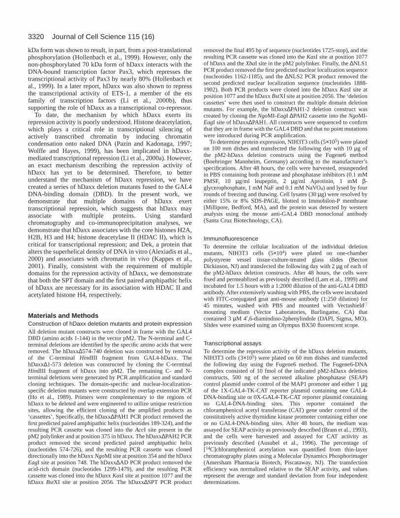
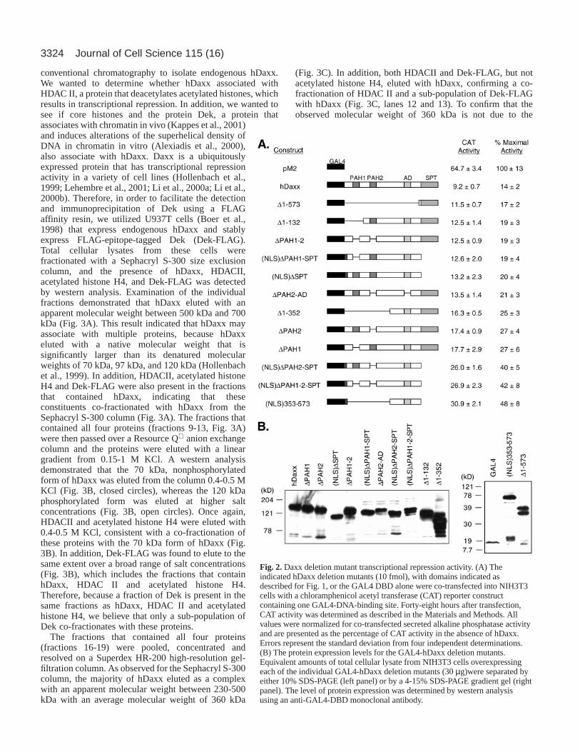
conventional chromatography to isolate endogenous hDaxx.We wanted to determine whether hDaxx associated withHDAC II, a protein that deacetylates acetylated histones, whichresults in transcriptional repression. In addition, we wanted tosee if core histones and the protein Dek, a protein thatassociates with chromatin in vivo (Kappes et al., 2001)and induces alterations of the superhelical density ofDNA in chromatin in vitro (Alexiadis et al., 2000),also associate with hDaxx. Daxx is a ubiquitouslyexpressed protein that has transcriptional repressionactivity in a variety of cell lines (Hollenbach et al.,1999; Lehembre et al., 2001; Li et al., 2000a; Li et al.,2000b). Therefore, in order to facilitate the detectionand immunoprecipitation of Dek using a FLAGaffinity resin, we utilized U937T cells (Boer et al.,1998) that express endogenous hDaxx and stablyexpress FLAG-epitope-tagged Dek (Dek-FLAG).Total cellular lysates from these cells werefractionated with a Sephacryl S-300 size exclusioncolumn, and the presence of hDaxx, HDACII,acetylated histone H4, and Dek-FLAG was detectedby western analysis. Examination of the individualfractions demonstrated that hDaxx eluted with anapparent molecular weight between 500 kDa and 700kDa (Fig. 3A). This result indicated that hDaxx mayassociate with multiple proteins, because hDaxxeluted with a native molecular weight that issignificantly larger than its denatured molecularweights of 70 kDa, 97 kDa, and 120 kDa (Hollenbachet al., 1999). In addition, HDACII, acetylated histoneH4 and Dek-FLAG were also present in the fractionsthat contained hDaxx, indicating that theseconstituents co-fractionated with hDaxx from theSephacryl S-300 column (Fig. 3A). The fractions thatcontained all four proteins (fractions 9-13, Fig. 3A)were then passed over a Resource Q anion exchangecolumn and the proteins were eluted with a lineargradient from 0.15-1 M KCl. A western analysisdemonstrated that the 70 kDa, nonphosphorylatedform of hDaxx was eluted from the column 0.4-0.5 MKCl (Fig. 3B, closed circles), whereas the 120 kDaphosphorylated form was eluted at higher saltconcentrations (Fig. 3B, open circles). Once again,HDACII and acetylated histone H4 were eluted with0.4-0.5 M KCl, consistent with a co-fractionation ofthese proteins with the 70 kDa form of hDaxx (Fig.3B). In addition, Dek-FLAG was found to elute to thesame extent over a broad range of salt concentrations(Fig. 3B), which includes the fractions that containhDaxx, HDAC II and acetylated histone H4.Therefore, because a fraction of Dek is present in thesame fractions as hDaxx, HDAC II and acetylatedhistone H4, we believe that only a sub-population ofDek co-fractionates with these proteins.
The fractions that contained all four proteins(fractions 16-19) were pooled, concentrated andresolved on a Superdex HR-200 high-resolution gel-filtration column. As observed for the Sephacryl S-300column, the majority of hDaxx eluted as a complexwith an apparent molecular weight between 230-500kDa with an average molecular weight of 360 kDa
(Fig. 3C). In addition, both HDACII and Dek-FLAG, but notacetylated histone H4, eluted with hDaxx, confirming a co-fractionation of HDAC II and a sub-population of Dek-FLAGwith hDaxx (Fig. 3C, lanes 12 and 13). To confirm that theobserved molecular weight of 360 kDa is not due to the
Journal of Cell Science 115 (16)
Fig. 2.Daxx deletion mutant transcriptional repression activity. (A) Theindicated hDaxx deletion mutants (10 fmol), with domains indicated asdescribed for Fig. 1, or the GAL4 DBD alone were co-transfected into NIH3T3cells with a chloramphenicol acetyl transferase (CAT) reporter constructcontaining one GAL4-DNA-binding site. Forty-eight hours after transfection,CAT activity was determined as described in the Materials and Methods. Allvalues were normalized for co-transfected secreted alkaline phosphatase activityand are presented as the percentage of CAT activity in the absence of hDaxx.Errors represent the standard deviation from four independent determinations.(B) The protein expression levels for the GAL4-hDaxx deletion mutants.Equivalent amounts of total cellular lysate from NIH3T3 cells overexpressingeach of the individual GAL4-hDaxx deletion mutants (30 µg)were separated byeither 10% SDS-PAGE (left panel) or by a 4-15% SDS-PAGE gradient gel (rightpanel). The level of protein expression was determined by western analysisusing an anti-GAL4-DBD monoclonal antibody.
3325Daxx, HDAC II, histones and Dek association
Fig. 3.Co-fractionation of hDaxx, HDAC II, Dek and core histones. (A) Sephacryl S-300 size exclusion chromatography. Total cellular extractsof U937 cells (Boer et al., 1998) stably expressing FLAG-epitope tagged Dek were sonicated and separated by Sephacryl S-300 size exclusionchromatography. Each fraction was analyzed for the presence of hDaxx, Dek-FLAG, HDACII and acetylated histone H4 by western blotanalysis. The elution volumes for molecular weight standards are noted below the gel. The amount of total protein (absorbance 280 nm, dottedline), hDaxx (circle, solid line), HDAC II (triangle, solid line) and acetylated histone H4 (square, solid line) in each fraction was determined bydensitometry and is presented graphically as arbitrary densitometric units for each fraction. (B) Resource Q anion exchange chromatography.Fractions 9-13 of the Sephacryl S-300 column were fractionated over a Resource Q anion exchange column, and proteins were eluted with a0.15-1 M KCl linear gradient. Equivalent volumes of protein from each fraction were DOC/TCA/acetone precipitated, separated on a 4-20%SDS-PAGE gel and analyzed for the presence of all four proteins. In addition to HDAC II and acetylated histone H4, both the 70 kDa form ofhDaxx (closed circle, solid line) and the 120 kDa form of hDaxx (open circle, solid line) are presented. (C) Superdex HR 200 gel filtrationchromatography. Fractions 16-19 of the Resource Q column were concentrated and fractionated over a Superdex HR 200 gel filtrationcolumn. Each fraction was analyzed for the presence of all four proteins as described above. In panels A-C the faster-migrating band observedfor Dek-FLAG most probably consists of a degradation product as described by others (Alexiadis et al., 2000; McGarvey et al., 2000). (D) ACoomassie-blue-stained SDS-PAGE of the purified hDaxx complex. Lane 1 contains 1 µg of each of the purified histones H1, H2A, H2B, H3and H4. Lane 2 contains 20 µg of DOC/TCA-precipitated protein from fraction 13 of the Superdex HR200 column.
3326
presence of oligo-nucleosomes, an aliquot of the peak fractioncontaining hDaxx (Fig. 3C, fraction 13) was deproteinized,and the presence of DNA was determined by 2% agarosegel electrophoresis. This analysis demonstrated that a smallamount of DNA was present in this fraction and consisted ofa fragment of approximately 180 bp, a size consistent with theDNA present in a mono-nucleosome (data not shown). Inaddition to the elution of hDaxx with an apparent molecularweight of 360 kDa, a small amount of hDaxx eluted with anapparent molecular weight of 670 kDa and, in addition toHDACII and Dek-FLAG, co-fractionated with acetylatedhistone H4 (Fig. 3C, lanes 10 and 11). The co-fractionation ofacetylated histone H4 with only a small subset of hDaxxsuggests that if hDaxx associates with acetylated histone H4,the association is of a transient nature, potentially becauseof the presence of HDAC II in these fractions. The co-fractionation of HDAC II with hDaxx and acetylated histoneH4 would potentially bring HDAC II and acetylated histone H4into close proximity, allowing the deacetylation of histone H4.Alternatively, co-fractionation may be fortuitous, withacetylated histone H4 and hDaxx being part of different largecomplexes.
The potential deacetylation of histones by HDAC II presentin the complex suggests that non-acetylated histones may alsoco-fractionate with hDaxx, Dek and HDAC II in the 360 kDafraction. Because commercially available antibodies did notrecognize non-acetylated histones by western analysis in ourhands, we determined the presence of non-acetylated histonesin the 360 kDa hDaxx fractions by mass spectral analysis. TheCoomassie-stained gel of proteins present in the peak hDaxxfraction from the Superdex HR-200 column demonstrated thatin addition to several unidentified proteins of higher molecularweight, there was also the presence of bands consistent withthe molecular weights of all histones (Fig. 3D). To confirm theidentity of these proteins as the core histones H2A, H2B, H3and H4, the bands corresponding to these proteins were excisedfrom the gel, digested with trypsin, and the resulting peptidefragments were sequenced and identified by liquidchromatography and tandem mass spectral analysis. Thisanalysis identified fragments corresponding to non-acetylatedhistones H2A, H2B, H3 and H4. A similar analysis of the bandcorresponding to histone H1 did not identify any peptidefragments corresponding to histone H1. Therefore, this resultdemonstrated that the core histones H2A, H2B, H3 and H4 co-fractionate with hDaxx, Dek and HDAC II in the 360 kDahDaxx fraction.
Daxx, Dek, HDAC II and core histones physicallyassociateOur results demonstrate that hDaxx, acetylated and non-acetylated core histones, HDAC II and a subpopulation of Dek-FLAG co-fractionate through a series of chromatographicseparations. However, it is possible that their co-fractionationis merely fortuitous and that these proteins do not physicallyassociate in vivo. Therefore we performed a series ofco-immunoprecipitation experiments, which unlike co-fractionation depend on physical associations, using twoindependent components to confirm the association of hDaxx,Dek, HDAC II and core histones. First, we immunoprecipitatedDek-FLAG from the fractions of the Sephacryl S-300 column
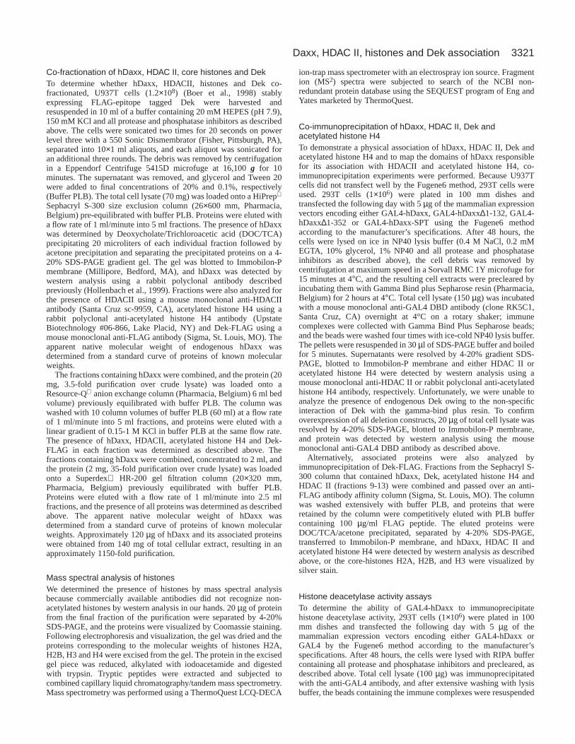
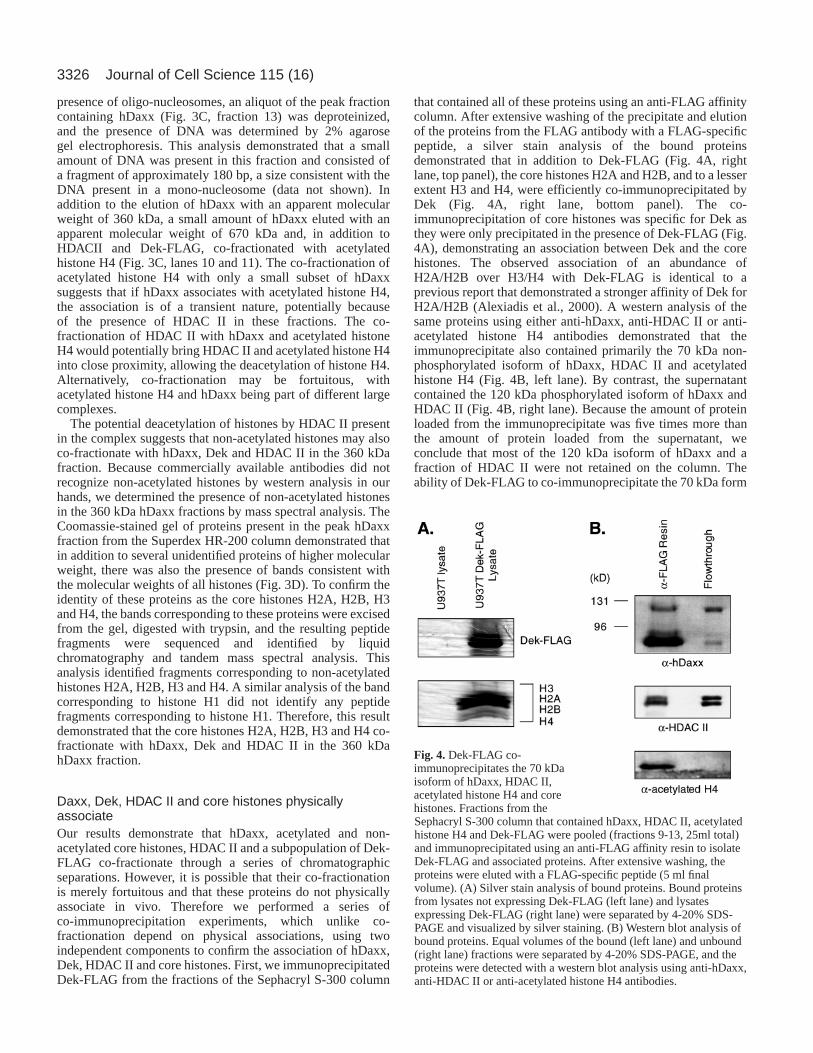
that contained all of these proteins using an anti-FLAG affinitycolumn. After extensive washing of the precipitate and elutionof the proteins from the FLAG antibody with a FLAG-specificpeptide, a silver stain analysis of the bound proteinsdemonstrated that in addition to Dek-FLAG (Fig. 4A, rightlane, top panel), the core histones H2A and H2B, and to a lesserextent H3 and H4, were efficiently co-immunoprecipitated byDek (Fig. 4A, right lane, bottom panel). The co-immunoprecipitation of core histones was specific for Dek asthey were only precipitated in the presence of Dek-FLAG (Fig.4A), demonstrating an association between Dek and the corehistones. The observed association of an abundance ofH2A/H2B over H3/H4 with Dek-FLAG is identical to aprevious report that demonstrated a stronger affinity of Dek forH2A/H2B (Alexiadis et al., 2000). A western analysis of thesame proteins using either anti-hDaxx, anti-HDAC II or anti-acetylated histone H4 antibodies demonstrated that theimmunoprecipitate also contained primarily the 70 kDa non-phosphorylated isoform of hDaxx, HDAC II and acetylatedhistone H4 (Fig. 4B, left lane). By contrast, the supernatantcontained the 120 kDa phosphorylated isoform of hDaxx andHDAC II (Fig. 4B, right lane). Because the amount of proteinloaded from the immunoprecipitate was five times more thanthe amount of protein loaded from the supernatant, weconclude that most of the 120 kDa isoform of hDaxx and afraction of HDAC II were not retained on the column. Theability of Dek-FLAG to co-immunoprecipitate the 70 kDa form
Journal of Cell Science 115 (16)
Fig. 4. Dek-FLAG co-immunoprecipitates the 70 kDaisoform of hDaxx, HDAC II,acetylated histone H4 and corehistones. Fractions from theSephacryl S-300 column that contained hDaxx, HDAC II, acetylatedhistone H4 and Dek-FLAG were pooled (fractions 9-13, 25ml total)and immunoprecipitated using an anti-FLAG affinity resin to isolateDek-FLAG and associated proteins. After extensive washing, theproteins were eluted with a FLAG-specific peptide (5 ml finalvolume). (A) Silver stain analysis of bound proteins. Bound proteinsfrom lysates not expressing Dek-FLAG (left lane) and lysatesexpressing Dek-FLAG (right lane) were separated by 4-20% SDS-PAGE and visualized by silver staining. (B) Western blot analysis ofbound proteins. Equal volumes of the bound (left lane) and unbound(right lane) fractions were separated by 4-20% SDS-PAGE, and theproteins were detected with a western blot analysis using anti-hDaxx,anti-HDAC II or anti-acetylated histone H4 antibodies.
3327Daxx, HDAC II, histones and Dek association
of hDaxx, HDAC II and core histones therefore confirmsthat there is a direct association between Dek and theseconstituents.
Next we performed independent co-immunoprecipitationexperiments using GAL4-hDaxx, as our hDaxx antibodieswere raised against the SPT domain of hDaxx, the regiondemonstrated to interact with a variety of proteins (Chang etal., 1998; Hollenbach et al., 1999; Ishov et al., 1999;Kiriakidou et al., 1997; Li et al., 2000a; Pluta et al., 1998; Toriiet al., 1999; Yang et al., 1999). 293T cells were transfectedwith full-length GAL4-hDaxx or GAL4 DBD alone andequivalent amounts of total cell lysate immunoprecipitatedwith the anti-GAL4 antibody. Consistent with the co-fractionation of hDaxx with both acetylated histone H4 andHDACII (Fig. 3), both proteins were co-immunoprecipitatedwith full-length GAL4-hDaxx (Fig. 5C,D, lane 2). Thisassociation was specific for hDaxx since neither acetylatedhistone H4 nor HDACII were detected when the identicalco-immunoprecipitations were carried out with cellsoverexpressing GAL4 DBD alone (Fig. 5C and 5D, lane 1).This association was also independent of the presence ofDNA since the same trace amount of low molecular weightDNA (~180 bp) was present regardless of whether theimmunoprecipitation was performed with the anti-hDaxx orcontrol antiserum (data not shown).
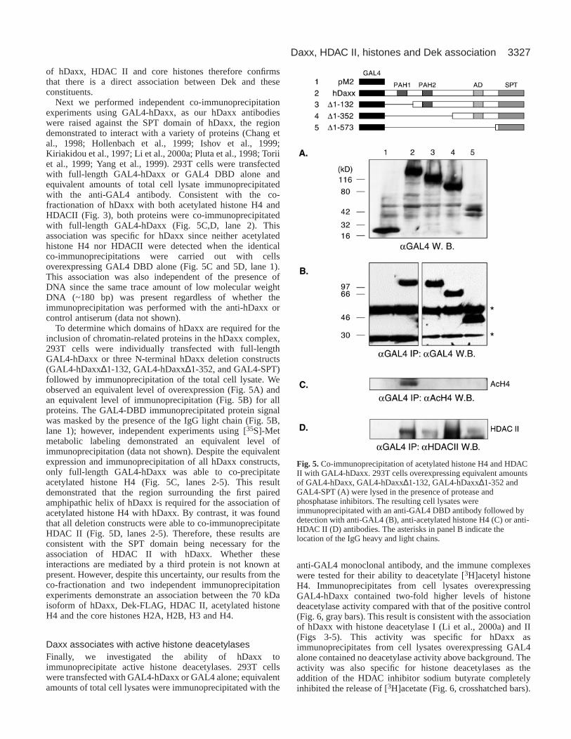
To determine which domains of hDaxx are required for theinclusion of chromatin-related proteins in the hDaxx complex,293T cells were individually transfected with full-lengthGAL4-hDaxx or three N-terminal hDaxx deletion constructs(GAL4-hDaxx∆1-132, GAL4-hDaxx∆1-352, and GAL4-SPT)followed by immunoprecipitation of the total cell lysate. Weobserved an equivalent level of overexpression (Fig. 5A) andan equivalent level of immunoprecipitation (Fig. 5B) for allproteins. The GAL4-DBD immunoprecipitated protein signalwas masked by the presence of the IgG light chain (Fig. 5B,lane 1); however, independent experiments using [35S]-Metmetabolic labeling demonstrated an equivalent level ofimmunoprecipitation (data not shown). Despite the equivalentexpression and immunoprecipitation of all hDaxx constructs,only full-length GAL4-hDaxx was able to co-precipitateacetylated histone H4 (Fig. 5C, lanes 2-5). This resultdemonstrated that the region surrounding the first pairedamphipathic helix of hDaxx is required for the association ofacetylated histone H4 with hDaxx. By contrast, it was foundthat all deletion constructs were able to co-immunoprecipitateHDAC II (Fig. 5D, lanes 2-5). Therefore, these results areconsistent with the SPT domain being necessary for theassociation of HDAC II with hDaxx. Whether theseinteractions are mediated by a third protein is not known atpresent. However, despite this uncertainty, our results from theco-fractionation and two independent immunoprecipitationexperiments demonstrate an association between the 70 kDaisoform of hDaxx, Dek-FLAG, HDAC II, acetylated histoneH4 and the core histones H2A, H2B, H3 and H4.
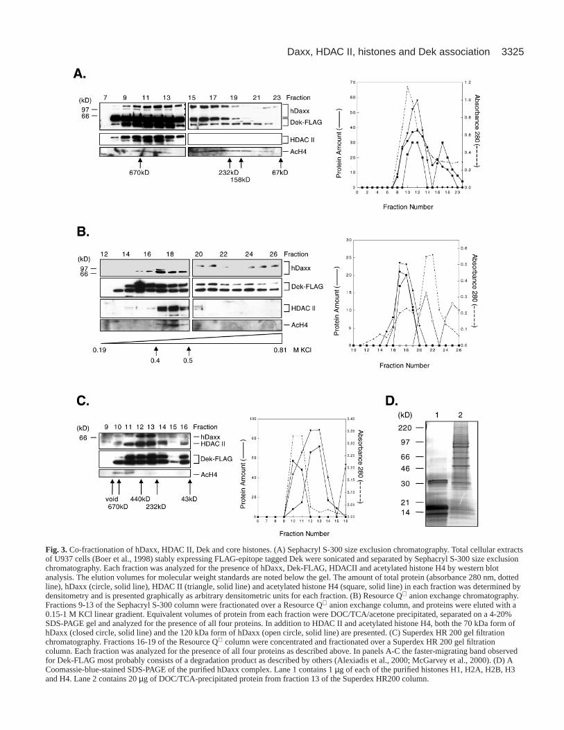
Daxx associates with active histone deacetylasesFinally, we investigated the ability of hDaxx toimmunoprecipitate active histone deacetylases. 293T cellswere transfected with GAL4-hDaxx or GAL4 alone; equivalentamounts of total cell lysates were immunoprecipitated with the
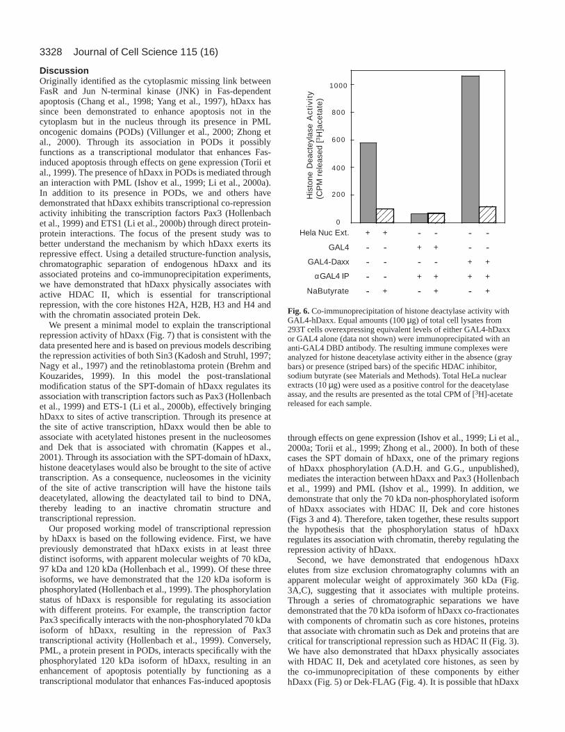
anti-GAL4 monoclonal antibody, and the immune complexeswere tested for their ability to deacetylate [3H]acetyl histoneH4. Immunoprecipitates from cell lysates overexpressingGAL4-hDaxx contained two-fold higher levels of histonedeacetylase activity compared with that of the positive control(Fig. 6, gray bars). This result is consistent with the associationof hDaxx with histone deacetylase I (Li et al., 2000a) and II(Figs 3-5). This activity was specific for hDaxx asimmunoprecipitates from cell lysates overexpressing GAL4alone contained no deacetylase activity above background. Theactivity was also specific for histone deacetylases as theaddition of the HDAC inhibitor sodium butyrate completelyinhibited the release of [3H]acetate (Fig. 6, crosshatched bars).
Fig. 5. Co-immunoprecipitation of acetylated histone H4 and HDACII with GAL4-hDaxx. 293T cells overexpressing equivalent amountsof GAL4-hDaxx, GAL4-hDaxx∆1-132, GAL4-hDaxx∆1-352 andGAL4-SPT (A) were lysed in the presence of protease andphosphatase inhibitors. The resulting cell lysates wereimmunoprecipitated with an anti-GAL4 DBD antibody followed bydetection with anti-GAL4 (B), anti-acetylated histone H4 (C) or anti-HDAC II (D) antibodies. The asterisks in panel B indicate thelocation of the IgG heavy and light chains.
3328
DiscussionOriginally identified as the cytoplasmic missing link betweenFasR and Jun N-terminal kinase (JNK) in Fas-dependentapoptosis (Chang et al., 1998; Yang et al., 1997), hDaxx hassince been demonstrated to enhance apoptosis not in thecytoplasm but in the nucleus through its presence in PMLoncogenic domains (PODs) (Villunger et al., 2000; Zhong etal., 2000). Through its association in PODs it possiblyfunctions as a transcriptional modulator that enhances Fas-induced apoptosis through effects on gene expression (Torii etal., 1999). The presence of hDaxx in PODs is mediated throughan interaction with PML (Ishov et al., 1999; Li et al., 2000a).In addition to its presence in PODs, we and others havedemonstrated that hDaxx exhibits transcriptional co-repressionactivity inhibiting the transcription factors Pax3 (Hollenbachet al., 1999) and ETS1 (Li et al., 2000b) through direct protein-protein interactions. The focus of the present study was tobetter understand the mechanism by which hDaxx exerts itsrepressive effect. Using a detailed structure-function analysis,chromatographic separation of endogenous hDaxx and itsassociated proteins and co-immunoprecipitation experiments,we have demonstrated that hDaxx physically associates withactive HDAC II, which is essential for transcriptionalrepression, with the core histones H2A, H2B, H3 and H4 andwith the chromatin associated protein Dek.
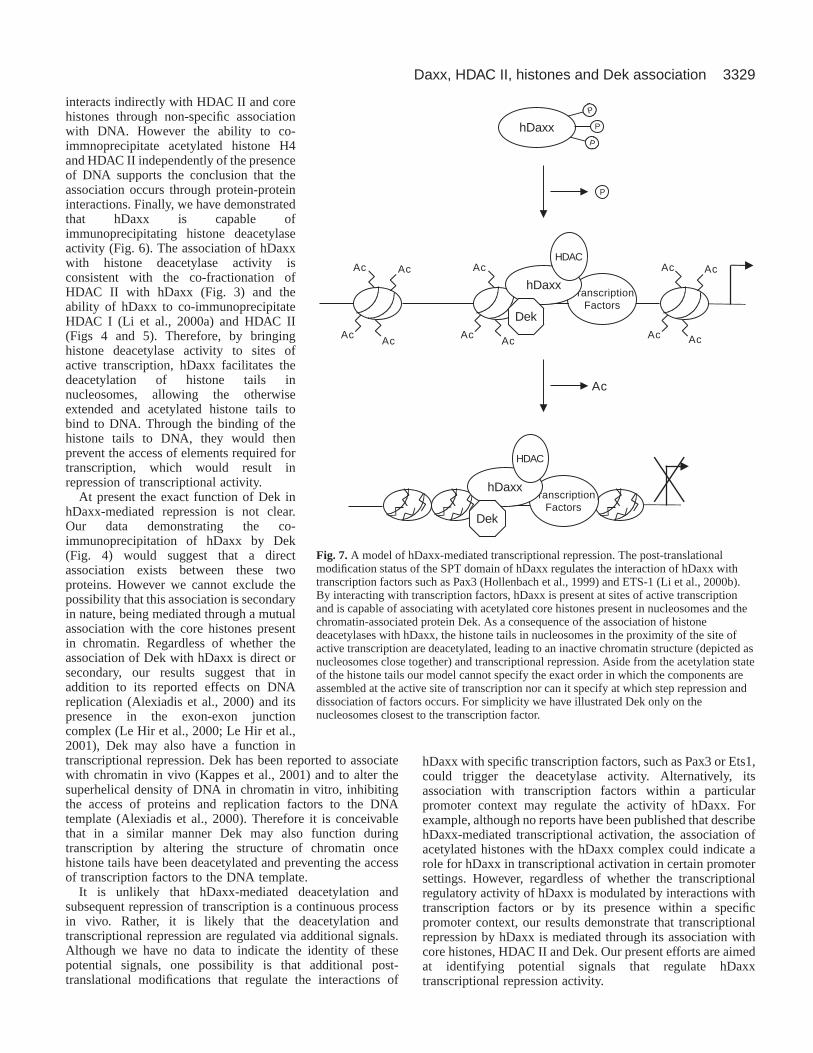
We present a minimal model to explain the transcriptionalrepression activity of hDaxx (Fig. 7) that is consistent with thedata presented here and is based on previous models describingthe repression activities of both Sin3 (Kadosh and Struhl, 1997;Nagy et al., 1997) and the retinoblastoma protein (Brehm andKouzarides, 1999). In this model the post-translationalmodification status of the SPT-domain of hDaxx regulates itsassociation with transcription factors such as Pax3 (Hollenbachet al., 1999) and ETS-1 (Li et al., 2000b), effectively bringinghDaxx to sites of active transcription. Through its presence atthe site of active transcription, hDaxx would then be able toassociate with acetylated histones present in the nucleosomesand Dek that is associated with chromatin (Kappes et al.,2001). Through its association with the SPT-domain of hDaxx,histone deacetylases would also be brought to the site of activetranscription. As a consequence, nucleosomes in the vicinityof the site of active transcription will have the histone tailsdeacetylated, allowing the deactylated tail to bind to DNA,thereby leading to an inactive chromatin structure andtranscriptional repression.
Our proposed working model of transcriptional repressionby hDaxx is based on the following evidence. First, we havepreviously demonstrated that hDaxx exists in at least threedistinct isoforms, with apparent molecular weights of 70 kDa,97 kDa and 120 kDa (Hollenbach et al., 1999). Of these threeisoforms, we have demonstrated that the 120 kDa isoform isphosphorylated (Hollenbach et al., 1999). The phosphorylationstatus of hDaxx is responsible for regulating its associationwith different proteins. For example, the transcription factorPax3 specifically interacts with the non-phosphorylated 70 kDaisoform of hDaxx, resulting in the repression of Pax3transcriptional activity (Hollenbach et al., 1999). Conversely,PML, a protein present in PODs, interacts specifically with thephosphorylated 120 kDa isoform of hDaxx, resulting in anenhancement of apoptosis potentially by functioning as atranscriptional modulator that enhances Fas-induced apoptosis
through effects on gene expression (Ishov et al., 1999; Li et al.,2000a; Torii et al., 1999; Zhong et al., 2000). In both of thesecases the SPT domain of hDaxx, one of the primary regionsof hDaxx phosphorylation (A.D.H. and G.G., unpublished),mediates the interaction between hDaxx and Pax3 (Hollenbachet al., 1999) and PML (Ishov et al., 1999). In addition, wedemonstrate that only the 70 kDa non-phosphorylated isoformof hDaxx associates with HDAC II, Dek and core histones(Figs 3 and 4). Therefore, taken together, these results supportthe hypothesis that the phosphorylation status of hDaxxregulates its association with chromatin, thereby regulating therepression activity of hDaxx.
Second, we have demonstrated that endogenous hDaxxelutes from size exclusion chromatography columns with anapparent molecular weight of approximately 360 kDa (Fig.3A,C), suggesting that it associates with multiple proteins.Through a series of chromatographic separations we havedemonstrated that the 70 kDa isoform of hDaxx co-fractionateswith components of chromatin such as core histones, proteinsthat associate with chromatin such as Dek and proteins that arecritical for transcriptional repression such as HDAC II (Fig. 3).We have also demonstrated that hDaxx physically associateswith HDAC II, Dek and acetylated core histones, as seen bythe co-immunoprecipitation of these components by eitherhDaxx (Fig. 5) or Dek-FLAG (Fig. 4). It is possible that hDaxx
Journal of Cell Science 115 (16)
1000
800
600
400
200His
tone
Dea
ctey
lase
Act
ivit
y(C
PM
rel
ease
d [3
H]a
ceta
te)
0
- --- + +GAL4
+ +- - - -GAL4-Daxx
- - + + + +αGAL4 IP
- - -+ + +NaButyrate
+ + - - - -Hela Nuc Ext.
Fig. 6.Co-immunoprecipitation of histone deactylase activity withGAL4-hDaxx. Equal amounts (100 µg) of total cell lysates from293T cells overexpressing equivalent levels of either GAL4-hDaxxor GAL4 alone (data not shown) were immunoprecipitated with ananti-GAL4 DBD antibody. The resulting immune complexes wereanalyzed for histone deacetylase activity either in the absence (graybars) or presence (striped bars) of the specific HDAC inhibitor,sodium butyrate (see Materials and Methods). Total HeLa nuclearextracts (10 µg) were used as a positive control for the deacetylaseassay, and the results are presented as the total CPM of [3H]-acetatereleased for each sample.
3329Daxx, HDAC II, histones and Dek association
interacts indirectly with HDAC II and corehistones through non-specific associationwith DNA. However the ability to co-immnoprecipitate acetylated histone H4and HDAC II independently of the presenceof DNA supports the conclusion that theassociation occurs through protein-proteininteractions. Finally, we have demonstratedthat hDaxx is capable ofimmunoprecipitating histone deacetylaseactivity (Fig. 6). The association of hDaxxwith histone deacetylase activity isconsistent with the co-fractionation ofHDAC II with hDaxx (Fig. 3) and theability of hDaxx to co-immunoprecipitateHDAC I (Li et al., 2000a) and HDAC II(Figs 4 and 5). Therefore, by bringinghistone deacetylase activity to sites ofactive transcription, hDaxx facilitates thedeacetylation of histone tails innucleosomes, allowing the otherwiseextended and acetylated histone tails tobind to DNA. Through the binding of thehistone tails to DNA, they would thenprevent the access of elements required fortranscription, which would result inrepression of transcriptional activity.
At present the exact function of Dek inhDaxx-mediated repression is not clear.Our data demonstrating the co-immunoprecipitation of hDaxx by Dek(Fig. 4) would suggest that a directassociation exists between these twoproteins. However we cannot exclude thepossibility that this association is secondaryin nature, being mediated through a mutualassociation with the core histones presentin chromatin. Regardless of whether theassociation of Dek with hDaxx is direct orsecondary, our results suggest that inaddition to its reported effects on DNAreplication (Alexiadis et al., 2000) and itspresence in the exon-exon junctioncomplex (Le Hir et al., 2000; Le Hir et al.,2001), Dek may also have a function intranscriptional repression. Dek has been reported to associatewith chromatin in vivo (Kappes et al., 2001) and to alter thesuperhelical density of DNA in chromatin in vitro, inhibitingthe access of proteins and replication factors to the DNAtemplate (Alexiadis et al., 2000). Therefore it is conceivablethat in a similar manner Dek may also function duringtranscription by altering the structure of chromatin oncehistone tails have been deacetylated and preventing the accessof transcription factors to the DNA template.
It is unlikely that hDaxx-mediated deacetylation andsubsequent repression of transcription is a continuous processin vivo. Rather, it is likely that the deacetylation andtranscriptional repression are regulated via additional signals.Although we have no data to indicate the identity of thesepotential signals, one possibility is that additional post-translational modifications that regulate the interactions of
hDaxx with specific transcription factors, such as Pax3 or Ets1,could trigger the deacetylase activity. Alternatively, itsassociation with transcription factors within a particularpromoter context may regulate the activity of hDaxx. Forexample, although no reports have been published that describehDaxx-mediated transcriptional activation, the association ofacetylated histones with the hDaxx complex could indicate arole for hDaxx in transcriptional activation in certain promotersettings. However, regardless of whether the transcriptionalregulatory activity of hDaxx is modulated by interactions withtranscription factors or by its presence within a specificpromoter context, our results demonstrate that transcriptionalrepression by hDaxx is mediated through its association withcore histones, HDAC II and Dek. Our present efforts are aimedat identifying potential signals that regulate hDaxxtranscriptional repression activity.
hDaxx P
P
P
P
Ac Ac
AcAc
Ac
AcAc
Ac Ac
AcAc
TranscriptionFactors
hDaxx
Dek
HDAC
Ac
TranscriptionFactors
hDaxx
Dek
HDAC
Fig. 7.A model of hDaxx-mediated transcriptional repression. The post-translationalmodification status of the SPT domain of hDaxx regulates the interaction of hDaxx withtranscription factors such as Pax3 (Hollenbach et al., 1999) and ETS-1 (Li et al., 2000b).By interacting with transcription factors, hDaxx is present at sites of active transcriptionand is capable of associating with acetylated core histones present in nucleosomes and thechromatin-associated protein Dek. As a consequence of the association of histonedeacetylases with hDaxx, the histone tails in nucleosomes in the proximity of the site ofactive transcription are deacetylated, leading to an inactive chromatin structure (depicted asnucleosomes close together) and transcriptional repression. Aside from the acetylation stateof the histone tails our model cannot specify the exact order in which the components areassembled at the active site of transcription nor can it specify at which step repression anddissociation of factors occurs. For simplicity we have illustrated Dek only on thenucleosomes closest to the transcription factor.
3330
All mass spectral analyses were performed in the Hartwell Centerfor Bioinformatics and Biotechnology at St. Jude Children’s ResearchHospital. We thank Erik Bonten for technical assistance with theisolation of endogenous hDaxx, and Joe Vaccaro for his criticalreading of the manuscript. This work was supported, in part, by NIHgrants CA71907-05 and CA76480-03, the Cancer Center (CORE)support grant CA-21765 and the American Lebanese SyrianAssociated Charities (ALSAC) of St. Jude Children’s ResearchHospital.
ReferencesAlexiadis, V., Waldmann, T., Andersen, J., Mann, M., Knippers, R. and
Gruss, C.(2000). The protein encoded by the proto-oncogene DEK changesthe topology of chromatin and reduces the efficiency of DNA replication ina chromatin-specific manner. Genes Dev. 14, 1308-1312.
Ausubel, F. M., Brent, R., Kingston, R. E., Moore, D. D., Seidman, J. G.,Smith, J. A. and Struhl, K. (1996). Current Protocols in MolecularBiology. Boston, MA: John Wiley and Sons.
Boer, J., Bonten-Surtel, J. and Grosveld, G.(1998). Overexpression of thenucleoporin CAN/NUP214 induces growth arrest, nucleocytoplasmictransport defects, and apoptosis. Mol. Cell. Biol. 18, 1236-1247.
Bram, R. J., Hung, D. T., Martin, P. K., Schreiber, S. L. and Crabtree, G.R. (1993). Identification of the immunophilins capable of mediatinginhibition of signal transduction by cyclosporin A and FK506: roles ofcalcineurin binding and cellular location. Mol. Cell. Biol. 13, 4760-4769.
Brehm, A. and Kouzarides, T. (1999). Retinoblastoma protein meetschromatin. Trends Biochem. Sci. 24, 142-145.
Chang, H. Y., Nishitoh, H., Yang, X., Ichijo, H. and Baltimore, D.(1998).Activation of apoptosis signal-regulating kinase 1 (ASK1) by the adapterprotein Daxx. Science281, 1860-1863.
Everett, R. D., Earnshaw, W. C., Pluta, A. F., Sternsdorf, T., Ainsztein, A.M., Carmena, M., Ruchaud, S., Hsu, W.-L. and Orr, A. (1999). Adynamic connection between centromeres and ND10 proteins. J. Cell Sci.112, 3443-3454.
Ho, S. N., Hunt, H. D., Horton, R. M., Pullen, J. K. and Pease, L. R.(1989).Site-directed mutagenesis by overlap extension using the polymerase chainreaction. Gene77, 51-59.
Hollenbach, A. D., Sublett, J. E., McPherson, C. J. and Grosveld, G.(1999). The Pax3-FKHR oncoprotein is unresponsive to the Pax3-Associated repressor hDaxx. EMBO J. 18, 3702-3711.
Ishov, A. M., Sotnikov, A. G., Negorev, D., Vladimirova, O. V., Neff, N.,Kamitani, T., Yeh, E. T. H., Strauss III, J. F. and Maul, G. G.(1999).PML is critical for ND10 formation and recruits the PML-interacting proteinDaxx to this nuclear structure when modified by SUMO-1. J. Cell Biol. 147,221-233.
Kadosh, D. and Struhl, K. (1997). Repression by Ume6 involves recruitmentof a complex containing Sin3 corepressor and Rpd3 histone deacetylase totarget promoters. Cell 89, 365-371.
Kappes, F., Burger, K., Baack, M., Fackelmayer, F. O. and Gruss, C.(2001). Subcellular localization of the human proto-oncogene protein DEK.J. Biol. Chem. 276, 26317-26323.
Kiriakidou, M., Driscoll, D. A., Lopez-Guisa, J. M. and Strauss III, J. F.(1997). Cloning and expression of primate Daxx cDNAs and mapping of
the human gene to chromosome 6p21.3 in the MHC region. DNA Cell Biol.16, 1289-1298.
Lam, P. Y. P., Sublett, J. E., Hollenbach, A. D. and Roussel, M. F.(1999).The oncogenic potential of the Pax3-FKHR fusion protein requires the Pax3homeodomain recognition helix but not the Pax3 paired-box DNA bindingdomain. Mol. Cell. Biol. 19, 594-601.
Lehembre, F., Muller, S., Pandolfi, P. P. and Dejean, A. (2001). Regulationof Pax3 transcriptional activity by SUMO-1 modified PML. Oncogene20,1-9.
Le Hir, H., Izaurralde, E., Maquat, L. E. and Moore, M. J. (2000). Thespliceosome deposits multiple proteins 20-24 nucleotides upstream ofmRNA exon-exon junctions. EMBO J. 19, 6860-6869.
Le Hir, H., Gatfield, D., Izaurralde, E. and Moore, M. J. (2001). The exon-exon junction complex provides a binding platform for factors involved inmRNA export and nonsense-mediated mRNA decay. EMBO J. 20, 4987-4997.
Li, H., Leo, C., Zhu, J., Wu, X. Y., O’Neil, J., Park, E.-J. and Chen, J. D.(2000a). Sequestration and inhibition of Daxx-mediated transcriptionalrepression by PML. Mol. Cell. Biol. 20, 1784-1796.
Li, R., Pei, H., Watson, D. K. and Papas, T. S.(2000b). EAP1/Daxx interactswith ETS1 and represses transcriptional activation of ETS1 target genes.Oncogene19, 745-753.
McGarvey, T., Rosonina, E., McCracken, S., Li, Q., Arnaout, R., Mientjes,E., Nickerson, J. A., Awrey, D., Greenblatt, J., Grosveld, G. andBlencowe, B. J. (2000). The acute myeloid leukemia-associated protein,DEK, forms a splicing-dependent interactions with exon-productcomplexes. J. Cell Biol. 150, 309-320.
Michaelson, J. S., Bader, D., Kuo, F., Kozak, C. and Leder, P.(1999). Lossof Daxx, a promiscuously interacting protein, results in extensive apoptosisin early mouse development. Genes Dev. 13, 1918-1923.
Nagy, L., Kao, H.-Y., Chakravarti, D., Lin, R. J., Hassig, C. A., Ayer, D.E., Schreiber, S. L. and Evans, R. M.(1997). Nuclear receptor repressionmediated by a complex containing SMRT, mSin3A, and histone deacetylase.Cell 89, 373-380.
Pazin, M. J. and Kadonaga, J. T.(1997). What’s up and down with histonedeacetylation and transcription? Cell 89, 325-328.
Pluta, A. F., Earnshaw, W. C. and Goldberg, I. G.(1998). Interphase-specific association of intrinsic centromere protein CENP-C with hDaxx, adeath domain-binding protein implicated in Fas-mediated cell death. J. CellSci. 111, 2029-2041.
Torii, S., Egan, D. A., Evans, R. A. and Reed, J. C.(1999). Human Daxxregulates Fas-induced apoptosis from nuclear PML oncogenic domains(PODs). EMBO J. 18, 101-113.
Villunger, A., Huang, D. C. S., Holler, N., Tschopp, J. and Strasser, A.(2000). Fas ligand-induced c-Jun kinase activation in lymphoid cellsrequires extensive receptor aggregation but is independent of Daxx, and Fas-mediated cell death does not involve Daxx, RIP, or RAIDD. J. Immunol.165, 1337-1343.
Wolffe, A. P. and Hayes, J. J.(1999). Chromatin disruption and modification.Nucleic Acids Res. 27, 711-720.
Yang, X., Khosravi-Far, R., Chang, H. Y. and Baltimore, D.(1997). Daxx,a novel Fas-binding protein that activates JNK and apoptosis. Cell 89, 1067-1076.
Zhong, S., Salomoni, P., Ronchetti, S., Guo, A., Ruggero, D. and Pandolfi,P. P.(2000). Promyelocytic Leukemia Protein (PML) and Daxx participatein a novel nuclear pathway for apoptosis. J. Exp. Med. 191, 631-639.
Journal of Cell Science 115 (16)