Embed Size (px)
Citation preview
Evolving paradigms for repair of tissues by adult stem/progenitor cells (MSCs)
Darwin J. Prockop *, Daniel J. Kota, Nikolay Bazhanov, Roxanne L. Reger
Texas A & M Health Science Center College of Medicine Institute for Regenerative Medicine at Scott & White, Temple, TX, USA
Received: June 25, 2010; Accepted: July 2, 2010
Abstract
In this review, we focus on the adult stem/progenitor cells that were initially isolated from bone marrow and first referred to as colonyforming units-fibroblastic, then as marrow stromal cells and subsequently as either mesenchymal stem cells or multipotent mesenchy-mal stromal cells (MSCs). The current interest in MSCs and similar cells from other tissues is reflected in over 10,000 citations inPubMed at the time of this writing with 5 to 10 new publications per day. It is also reflected in over 100 registered clinical trials withMSCs or related cells (http//www.clinicaltrials.gov). As a guide to the vast literature, this review will attempt to summarize many of thepublications in terms of three paradigms that have directed much of the work: an initial paradigm that the primary role of the cells wasto form niches for haematopoietic stem cells (paradigm I); a second paradigm that the cells repaired tissues by engraftment and differ-entiation to replace injured cells (paradigm II); and the more recent paradigm that MSCs engage in cross-talk with injured tissues andthereby generate microenvironments or ‘quasi-niches’ that enhance the repair tissues (paradigm III).
Keywords: mesenchymal stem cells • multipotent mesenchymal stromal cells • bone marrow • anti-inflammatory •anti-apoptotic • TSG-6 • STC-1
J. Cell. Mol. Med. Vol 14, No 9, 2010 pp. 2190-2199
© 2010 The AuthorsJournal compilation © 2010 Foundation for Cellular and Molecular Medicine/Blackwell Publishing Ltd
doi:10.1111/j.1582-4934.2010.01151.x
Guest Editor: N.I. Moldovan
Paradigm I: the haematopoietic niche
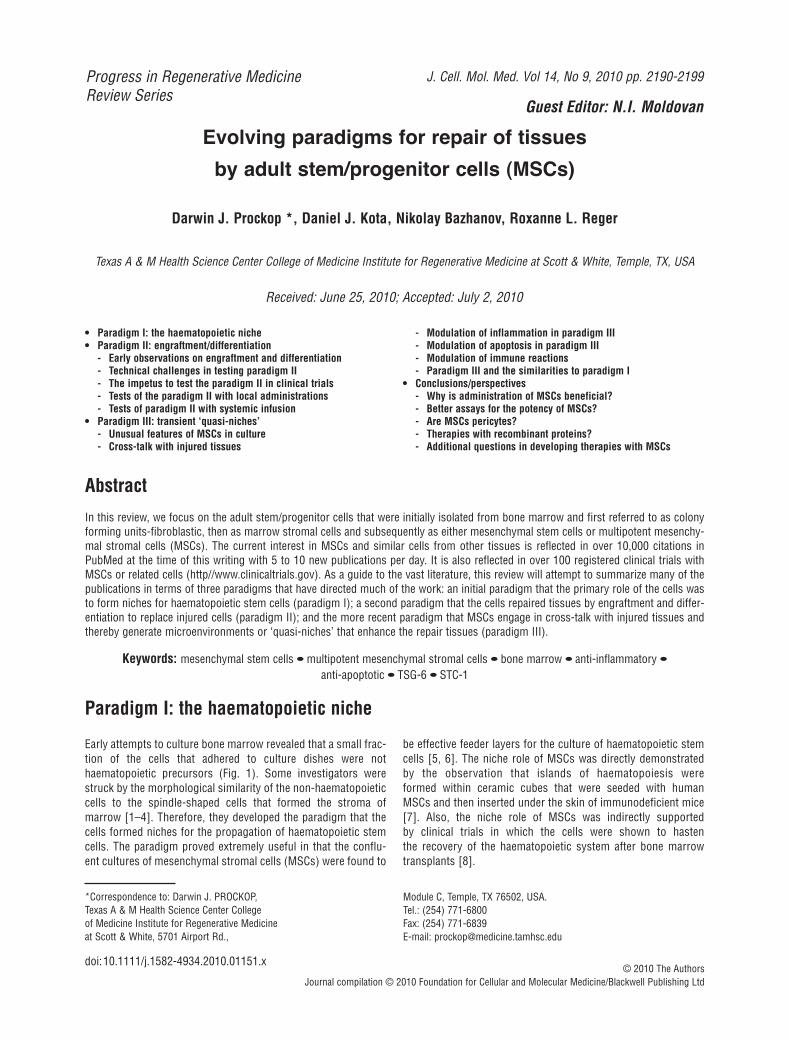
Early attempts to culture bone marrow revealed that a small frac-tion of the cells that adhered to culture dishes were nothaematopoietic precursors (Fig. 1). Some investigators werestruck by the morphological similarity of the non-haematopoieticcells to the spindle-shaped cells that formed the stroma of marrow [1–4]. Therefore, they developed the paradigm that thecells formed niches for the propagation of haematopoietic stemcells. The paradigm proved extremely useful in that the conflu-ent cultures of mesenchymal stromal cells (MSCs) were found to
be effective feeder layers for the culture of haematopoietic stemcells [5, 6]. The niche role of MSCs was directly demonstratedby the observation that islands of haematopoiesis were formed within ceramic cubes that were seeded with humanMSCs and then inserted under the skin of immunodeficient mice[7]. Also, the niche role of MSCs was indirectly supported by clinical trials in which the cells were shown to hasten the recovery of the haematopoietic system after bone marrowtransplants [8].
*Correspondence to: Darwin J. PROCKOP, Texas A & M Health Science Center College of Medicine Institute for Regenerative Medicine at Scott & White, 5701 Airport Rd.,
Module C, Temple, TX 76502, USA. Tel.: (254) 771-6800Fax: (254) 771-6839E-mail: [email protected]
Progress in Regenerative MedicineReview Series
• Paradigm I: the haematopoietic niche• Paradigm II: engraftment/differentiation
- Early observations on engraftment and differentiation- Technical challenges in testing paradigm II- The impetus to test the paradigm II in clinical trials- Tests of the paradigm II with local administrations- Tests of paradigm II with systemic infusion
• Paradigm III: transient ‘quasi-niches’- Unusual features of MSCs in culture- Cross-talk with injured tissues
- Modulation of inflammation in paradigm III- Modulation of apoptosis in paradigm III- Modulation of immune reactions- Paradigm III and the similarities to paradigm I
• Conclusions/perspectives- Why is administration of MSCs beneficial?- Better assays for the potency of MSCs?- Are MSCs pericytes?- Therapies with recombinant proteins?- Additional questions in developing therapies with MSCs
J. Cell. Mol. Med. Vol 14, No 9, 2010
2191© 2010 The AuthorsJournal compilation © 2010 Foundation for Cellular and Molecular Medicine/Blackwell Publishing Ltd
Paradigm II: engraftment/differentiation
Early investigators studying cultures of bone marrow wereimpressed with the facility with which the adherent, spindle-shaped cells differentiated into distinct cellular phenotypes. In particular, Friedenstein and others [1] demonstrated that the cellsreadily became mineralizing cells or chondrocytes both in cultureand after implantation in diffusion chambers in vivo. These obser-vations suggested the paradigm that MSCs might repair injuredtissues by engraftment and differentiation (Fig. 1). The paradigmhad broad implications for medical therapies in part because of theease with which the cells could be isolated from a small sample ofhuman bone marrow and then rapidly expanded in culture through30 or more population doublings [9–11].
Early observations on engraftment and differentiation
Repair by paradigm II was supported by early observations thatlocal administrations of MSCs improved bone repair [12]. Thepotential therapeutic implications of the paradigm were expandedby the observation that after systemic infusions of MSCs contain-ing a mutated human gene into irradiated young mice, the mutatedgene was detected in multiple tissues of the mice [13]. Also, fur-ther support for the therapeutic potentials was provided by theobservation that infusions of MSCs from wild-type mice producedsmall but significant improvements in the bones of a transgenicmouse model for osteogenesis imperfecta [14]. The potentialtherapeutic implications were expanded still further by the obser-vation that, after BrdU-labelled MSCs were injected into the cere-
bral ventricles of newborn mice, the cells migrated throughout thebrain, and a few of the cells became astrocytes [15].
These early observations prompted a clinical trial in which children with severe osteogenesis imperfecta first received bonemarrow transplants from a haplotype-matched normal donor andthen were treated, several years later, with intravenous infusionsof a large number of MSCs from the same donors [16]. The therapyproduced a transient but significant improvement in the clinicalcourse of the children. Most importantly, there was only oneadverse event: one of the children developed a mild allergic reac-tion to foetal calf serum in which the MSCs were expanded. Theresults were followed by a clinical trial in which administration ofMSCs produced encouraging results in children with severe lyso-somal storage diseases [17]. These initial observations raised thepossibility that paradigm II might provide new therapies for abroad spectrum of human diseases.
Technical challenges in testing paradigm II
The early efforts to test the paradigm encountered a series of tech-nical challenges: (1) No endogenous markers for MSCs wereavailable that could be used to track the cells in vivo [18].Exogenous markers such as dyes or transduced genes wereemployed instead, but most produced unexpected artefacts[19–21]. (2) Only a small number of antibodies and other markerswere available to follow differentiation of the cells in vivo. Also, themicroscopes and algorithms to overcome some of the artefacts ofimmunohistochemistry were not commonly available. (3) Speciesdifferences in MSCs created a significant experimental barrier.Cultures of human MSCs were relatively easy to purify fromhaematopoietic precursors by simply re-plating the cells. Cultures
Fig. 1 Schematic summarizing three evolving paradigms for the repair of tissues by MSCs. Themorphology of a small number of adherent cellsfrom bone marrow suggested the paradigm thatthe cells served as a niche for haematopoieticcells (paradigm I). The ready differentiation ofthe cells in culture suggested that the cells couldrepair tissues by engrafting and differentiating(paradigm II). Clinical trials using the cells toimprove bone marrow transplants unexpectedlydemonstrated that they improved graft-versus-host diseases in a few patients and thereby drewattention to their immune modulatory properties.Functional improvement without significantengraftment in animal models and a few patientssuggested that MSCs enhanced repair by forming microenvironments or ‘quasi-niches’(paradigm III).
2192 © 2010 The AuthorsJournal compilation © 2010 Foundation for Cellular and Molecular Medicine/Blackwell Publishing Ltd
of mouse MSCs remained contaminated by haematopoietic pre-cursors through several passages. Also, as was observed muchearlier with mouse fibroblasts [22], cultures of mouse MSCsexpanded slowly until they underwent ‘crisis’ during which a fewcells were transformed and then expanded rapidly [23]. Rat MSCsinitially resembled human MSCs but at a later stage also under-went crisis and transformation [24, 25]. (4) MSCs were not read-ily transplanted into marrow ablated mice and therefore presenteda further limitation in the use of transgenic mice. (5) Most impor-tantly, tissue repair is a highly complex biological process thatvaries with the type of injury and the tissue injured [26]. Also,there are marked species differences in inflammatory and immuneresponses [27] and as a result many experimental animals, espe-cially rodents, repair tissues much more efficiently than humanbeings. In effect, there were several serious barriers to definitiveexperiments to test paradigm II.
The impetus to test the paradigm II in clinical trials
Despite these technical challenges, there continues to be greatinterest in testing the medical implications inherent in paradigm II.The paradigm has been pursued against the history that discover-ies of new therapies in medicine have rarely been linear processes.Initial tests of a potential therapy in vitro are rarely as convincingas one would like, because of the limitations of experiments withpurified molecular components and the artefacts inherent in cultur-ing cells. The data from animal experiments are usually even morelimited because of the difficulty of mimicking human diseases. Thehistory of medicine is replete with examples of therapies that failedin the patients despite the extensive basic and preclinical research.However, the history of medicine also includes examples of thera-pies that were not fully developed or whose beneficial effects werenot understood until after they were first tested in patients [28].The examples include discovery of the anti-thrombotic effects ofaspirin [29, 30], the need of HLA typing in bone marrow trans-plants [31], the revised rationale and design of bisphosphonatesfor therapy of bone diseases [32] and the failure of sildenafil(Viagra) as a therapy for angina despite the Nobel prize researchthat led to its development [33, 34] (see Supporting Information).
Tests of the paradigm II with local administrations
Engraftment and differentiation of MSCs, as predicted by paradigm II, were seen in several settings. In models for bone andcartilage defects, a series of reports demonstrated that directimplantation of MSCs themselves or MSCs embedded in scaffoldsenhanced repair [35–38]. There is a consensus that some of theadministered cells differentiated into osteoblasts or chondrocytes.However, most reports indicated the MSCs disappeared in severalweeks [36, 39], and most of the differentiated cells seen in long-term grafts are host cells, at least in part because of the normalturnover of the tissues.
In models of cardiac defects, several reports indicated that
locally implanted MSCs engrafted and differentiated into cardiomy-ocytes [40, 41]. However, it has not been conclusively establishedthat locally administered MSCs provide a sufficient number of fullyintegrated cardiomyocytes to account for the improvements in ventricular function observed in many experiments [42].
In the central nervous system, some experiments indicatedthat MSCs injected into the ventricles of embryos or of newbornpups migrated throughout the brain and differentiated as theorgan developed [15, 43, 44]. In one series of experiments, quan-titative PCR assays indicated that the number of MSCs or MSC-derived cells increased as much as 30-fold in a few days after maleMSCs were injected into the ventricles of newborn female mice[43]. The possibility of neural differentiation was supported by theobservation that some preparations of MSCs differentiated in cul-ture into dopaminergic-like neurons with the appropriate electro-physiological properties [45]. However, it was difficult to establishdifferentiation of MSCs into functional neural cells in vivo [46, 47].
In contrast to transplants into embryonic brains, very fewMSCs injected into the brains of adult rodents survived more than1 or 2 weeks [21, 48, 49]. Surprisingly, the rate of disappearancewas about the same with human MSCs injected into the hip-pocampi of both immunodeficient and wild-type mice [49].
In models for spinal cord injury, local administration of MSCsproduced improved motor function but few, if any, of the cellsengrafted for prolonged periods or differentiated into neural cells[50, 51]. One initial impression was that the cells formed a scaf-fold for regeneration of nerve tracts in the cord [51]. A recentstudy suggested that the therapeutic benefits were explained byanti-inflammatory effects of the cells [52].
Tests of paradigm II with systemic infusion
Tests of paradigm II with systemic infusions of the cells provedproblematic. Numerous reports described functional improve-ments after systemic infusions of MSCs in models for human dis-eases that included osteogenensis imperfecta [53]; stroke [54];myocardial infarction [55]; acute kidney injury [56] and diabetes[57, 58]. The initial interpretations of the data were based on par-adigm II and assumed that the cells had homed to injured tissues,engrafted and differentiated to replace injured cells. However, itwas difficult to demonstrate extensive engraftment of the cells.Also, the interpretations were not intuitively consistent with sev-eral reports about the fate of systemically infused MSCs:Observations with whole body imaging techniques indicated thatmost MSCs were trapped in the lungs after intravenous infusionsinto rodents, the route used in most of the experiments [59–61].Therefore, the functional improvement of distal organs after intra-venous infusions of the cells was paradoxical.
To explore the paradox, we recently employed quantitative PCRassays for human MSCs infused into mice [62], a strategy intro-duced earlier by Phinney and associates for tracking MSCs infusedinto the brain [43]. (Previous data developed from gel-based PCRassays probably overestimated engraftment of MSCs after sys-temic infusion [13].) An improved protocol for quantitative PCR
J. Cell. Mol. Med. Vol 14, No 9, 2010
2193© 2010 The AuthorsJournal compilation © 2010 Foundation for Cellular and Molecular Medicine/Blackwell Publishing Ltd
assay of human Alu sequences demonstrated that after i.v. infu-sion of the human MSCs, essentially all of the cells were clearedfrom the circulation within 5 min. [62]. Most of the human cellswere recovered in the lungs. The cells in the lungs disappearedwith a half-life of about 24 hrs but only trace amounts were recov-ered in the six other tissues that were assayed. Therefore, theresults questioned whether paradigm II could account for thefunctional improvement observed after intravenous infusions ofMSCs in animal models for diseases of distal organs. In addition,paradigm II could not account for reports that conditionedmedium from cultures of MSCs was as effective in some diseasemodels as the cells themselves [63–65].
Paradigm III: transient ‘quasi-niches’
The accumulating evidence that MSCs could repair injured tissueswithout significant engraftment and differentiation called for a newparadigm that required re-examination of some of the early obser-vations on cultures of the cells and more detailed examination oftheir effects in vivo (Fig. 1).
Unusual features of MSCs in culture
The early observations that confluent and non-propagating MSCsprovided effective feeder layers for cultures of haematopoieticcells were explained in part by the cells secreting paracrine factors[5, 6, 66, 67]. However, the effectiveness of MSCs as feeder layers was not entirely explained by secretion of soluble factors;cell to cell contact was also required for reasons that were notapparent [5, 6].
Unusual features of MSCs in culture were also apparent fromobserving the cells after they were plated at clonal densities. Thecells expanded as single-cell derived colonies but the properties ofthe cells changed as the colonies expanded. In the many of thecolonies that formed, distinct inner and outer regions were appar-ent. The outer regions consisted of rapidly self-renewing cells andthe inner regions consisted of slowly replicating cells that werepartially differentiated [68]. Moreover, the cells displayed aremarkable plasticity in that the cells from both the inner and outerregions generated single-cell derived colonies with the same char-acteristics if they were lifted and re-plated at low density.Therefore, the MSCs expanded at clonal densities appeared toreversibly create their own microenvironments or ‘quasi-niches’ inculture in a manner that paralleled their ability to provide nichesfor haematopoietic stem cells.
Cross-talk with injured tissues
Although MSCs in culture secreted many cytokines [66, 67], it wasnot initially apparent that MSCs responded to injured tissues by
being activated to express high levels of additional therapeuticproteins. In effect, there was cross-talk in which signals frominjured cells activated MSCs to alter expression of large families of genes. At the same time signals from the activated MSCs bothup-regulated and down-regulated large families of genes in theinjured cells.
One of the first examples of cross-talk was observed betweenMSCs and multiple myeloma cells [69]. Co-culture experimentsdemonstrated that signals from the myeloma cells stimulated theMSCs to increase secretion of interleukin (IL)-6 and this IL-6 inturn, increased the proliferation of the myeloma cells. At the sametime, the myeloma cells secreted high levels of Dkk-1, an inhibitorof Wnt signalling, that kept the MSCs in cell cycle and inhibitedthem from differentiating into osteoblasts. The cross-talk providedan explanation for why patients with multiple myeloma developosteolytic lesions in which the cancer cells proliferate butosteoblasts are not recruited to fill the lesions [69].
A second example of cross-talk was encountered in experi-ments in which human MSCs were injected into the hippocampi ofmice following transient cerebral ischemia. The human MSCsreduced neuronal death and improved the neurological deficits[49]. Assays of RNA from the hippocampus with human-specificmRNA/cDNA microarrays demonstrated that in the ischemiainjured brain, the human MSCs increased expression of genes thatmodulated immune and inflammatory responses. Assays of thesame RNA on mouse-specific microarrays demonstrated that thepresence of the human MSCs modulated expression of mouse genesinvolved in immune responses to the ischemic environment.
A similar example of cross-talk was obtained by using species-specific mRNA/cDNA microarrays to survey the lungs of mice afew hours after intravenous infusions of human MSCs [62]. Byproducing microemboli, the human cells altered expression ofhundreds of mouse genes in the lung. At the same time, signalsfrom the mouse cells altered expression of hundreds of genes inthe human MSCs. In parallel with these observations, reports fromseveral laboratories demonstrated that the expression of poten-tially therapeutic cytokines was markedly increased by exposingMSCs to cytokines typically released by injured tissues [70, 71].
Modulation of inflammation in paradigm III
The experiments in which human MSCs were infused intra-venously into mice with myocardial infarcts provided a clue to howthey enhanced tissue repair. One of the most interesting genes up-regulated in human MSCs that were trapped in the lung afterintravenous infusion [62] was tumour necrosis factor (TNF)-�stimulated gene/protein-6 (TSG-6) [72, 73]. Extensive previousresearch demonstrated that TSG-6 had remarkable anti-inflammatoryproperties in a number of experimental settings, including in bothwild-type and transgenic mice [72, 73]. Experiments with recombinant TSG-6 and siRNAs demonstrated that the secretionof TSG-6 by MSCs trapped in the lung largely accounted for previous reports that intravenously administered MSCs improvedmice with myocardial infarcts [55, 74–76]. TSG-6 decreased
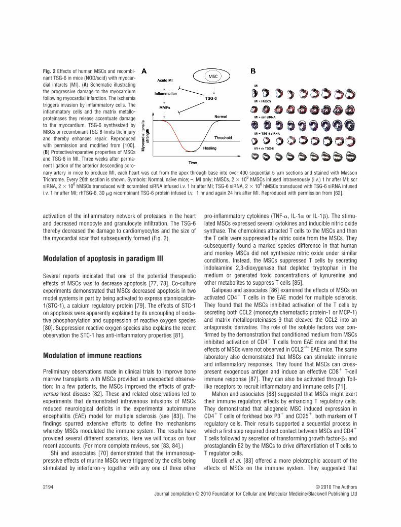
2194 © 2010 The AuthorsJournal compilation © 2010 Foundation for Cellular and Molecular Medicine/Blackwell Publishing Ltd
activation of the inflammatory network of proteases in the heartand decreased monocyte and granulocyte infiltration. The TSG-6thereby decreased the damage to cardiomyocytes and the size ofthe myocardial scar that subsequently formed (Fig. 2).
Modulation of apoptosis in paradigm III
Several reports indicated that one of the potential therapeuticeffects of MSCs was to decrease apoptosis [77, 78]. Co-cultureexperiments demonstrated that MSCs decreased apoptosis in twomodel systems in part by being activated to express stanniocalcin-1(STC-1), a calcium regulatory protein [79]. The effects of STC-1on apoptosis were apparently explained by its uncoupling of oxida-tive phosphorylation and suppression of reactive oxygen species[80]. Suppression reactive oxygen species also explains the recentobservation the STC-1 has anti-inflammatory properties [81].
Modulation of immune reactions
Preliminary observations made in clinical trials to improve bonemarrow transplants with MSCs provided an unexpected observa-tion: In a few patients, the MSCs improved the effects of graft-versus-host disease [82]. These and related observations led toexperiments that demonstrated intravenous infusions of MSCsreduced neurological deficits in the experimental autoimmuneencephalitis (EAE) model for multiple sclerosis (see [83]). Thefindings spurred extensive efforts to define the mechanismswhereby MSCs modulated the immune system. The results haveprovided several different scenarios. Here we will focus on fourrecent accounts. (For more complete reviews, see [83, 84].)
Shi and associates [70] demonstrated that the immunosup-pressive effects of murine MSCs were triggered by the cells beingstimulated by interferon-� together with any one of three other
pro-inflammatory cytokines (TNF-�, IL-1� or IL-1�). The stimu-lated MSCs expressed several cytokines and inducible nitric oxidesynthase. The chemokines attracted T cells to the MSCs and thenthe T cells were suppressed by nitric oxide from the MSCs. Theysubsequently found a marked species difference in that humanand monkey MSCs did not synthesize nitric oxide under similarconditions. Instead, the MSCs suppressed T cells by secretingindoleamine 2,3-dioxygenase that depleted tryptophan in themedium or generated toxic concentrations of kynurenine andother metabolites to suppress T cells [85].
Galipeau and associates [86] examined the effects of MSCs onactivated CD4� T cells in the EAE model for multiple sclerosis.They found that the MSCs inhibited activation of the T cells bysecreting both CCL2 (monocyte chemotactic protein-1 or MCP-1)and matrix metalloproteinases-9 that cleaved the CCL2 into anantagonistic derivative. The role of the soluble factors was con-firmed by the demonstration that conditioned medium from MSCsinhibited activation of CD4� T cells from EAE mice and that theeffects of MSCs were not observed in CCL2–/– EAE mice. The samelaboratory also demonstrated that MSCs can stimulate immuneand inflammatory responses. They found that MSCs can cross-present exogenous antigen and induce an effective CD8� T-cellimmune response [87]. They can also be activated through Toll-like receptors to recruit inflammatory and immune cells [71].
Mahon and associates [88] suggested that MSCs might exerttheir immune regulatory effects by enhancing T regulatory cells.They demonstrated that allogeneic MSC induced expression inCD4� T cells of forkhead box P3� and CD25�, both markers of Tregulatory cells. Their results supported a sequential process inwhich a first step required direct contact between MSCs and CD4�
T cells followed by secretion of transforming growth factor-�1 andprostaglandin E2 by the MSCs to drive differentiation of T cells toT regulator cells.
Uccelli et al. [83] offered a more pleiotrophic account of theeffects of MSCs on the immune system. They suggested that
Fig. 2 Effects of human MSCs and recombi-nant TSG-6 in mice (NOD/scid) with myocar-dial infarcts (MI). (A) Schematic illustratingthe progressive damage to the myocardiumfollowing myocardial infarction. The ischemiatriggers invasion by inflammatory cells. Theinflammatory cells and the matrix metallo-proteinases they release accentuate damageto the myocardium. TSG-6 synthesized byMSCs or recombinant TSG-6 limits the injuryand thereby enhances repair. Reproducedwith permission and modified from [100].(B) Protective/reparative properties of MSCsand TSG-6 in MI. Three weeks after perma-nent ligation of the anterior descending coro-nary artery in mice to produce MI, each heart was cut from the apex through base into over 400 sequential 5 �m sections and stained with MassonTrichrome. Every 20th section is shown. Symbols: Normal, naïve mice; –, MI only; hMSCs, 2 � 106 hMSCs infused intravenously (i.v.) 1 hr after MI; scrsiRNA, 2 � 106 hMSCs transduced with scrambled siRNA infused i.v. 1 hr after MI; TSG-6 siRNA, 2 � 106 hMSCs transduced with TSG-6 siRNA infusedi.v. 1 hr after MI; rhTSG-6, 30 �g recombinant TSG-6 protein infused i.v. 1 hr and again 24 hrs after MI. Reproduced with permission from [62].
J. Cell. Mol. Med. Vol 14, No 9, 2010
2195© 2010 The AuthorsJournal compilation © 2010 Foundation for Cellular and Molecular Medicine/Blackwell Publishing Ltd
MSCs produced a variety of effects such as (1) decreased prolifer-ation, cytotoxicity and cytokine production by NK cells; (2)impaired maturation and antigen presentation by dendritic cells; (3)decreased proliferation of T cells and impaired T helper cells and(4) decreased proliferation and antibody production by B cells.
At the moment, it is not clear which of the proposals bestaccounts for the immune modulatory effects of MSCs in vivo.
Paradigm III and the similarities to paradigm I
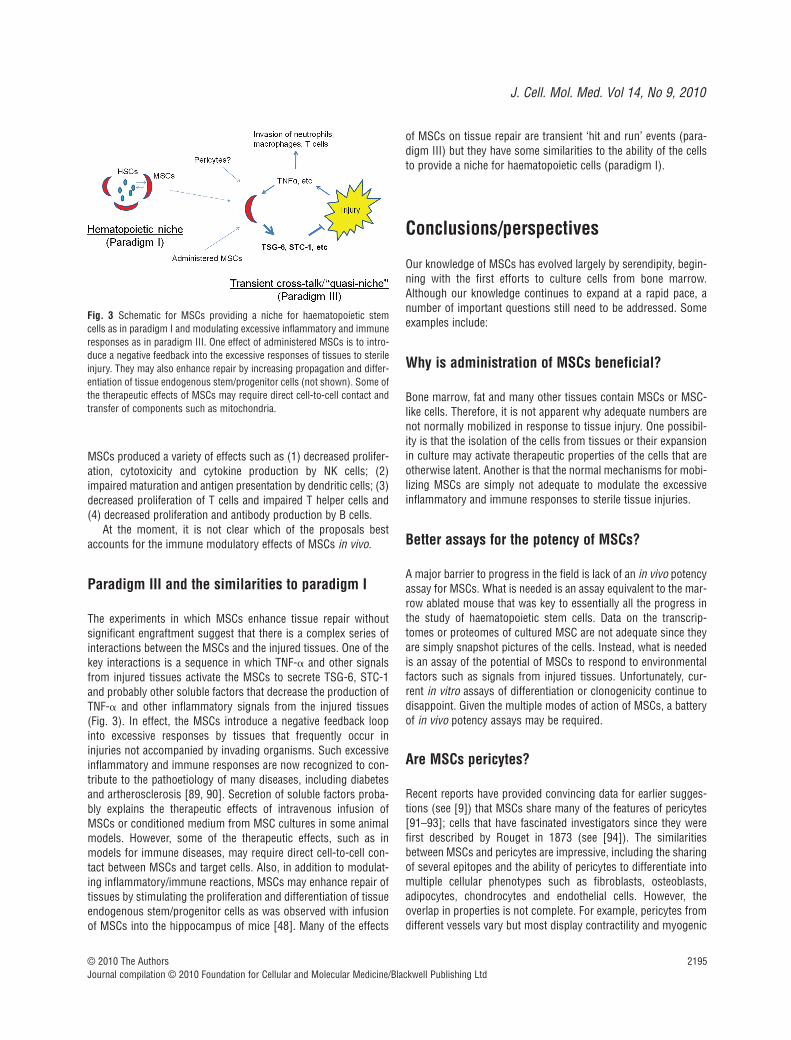
The experiments in which MSCs enhance tissue repair withoutsignificant engraftment suggest that there is a complex series ofinteractions between the MSCs and the injured tissues. One of thekey interactions is a sequence in which TNF-� and other signalsfrom injured tissues activate the MSCs to secrete TSG-6, STC-1and probably other soluble factors that decrease the production ofTNF-� and other inflammatory signals from the injured tissues(Fig. 3). In effect, the MSCs introduce a negative feedback loopinto excessive responses by tissues that frequently occur ininjuries not accompanied by invading organisms. Such excessiveinflammatory and immune responses are now recognized to con-tribute to the pathoetiology of many diseases, including diabetesand artherosclerosis [89, 90]. Secretion of soluble factors proba-bly explains the therapeutic effects of intravenous infusion ofMSCs or conditioned medium from MSC cultures in some animalmodels. However, some of the therapeutic effects, such as inmodels for immune diseases, may require direct cell-to-cell con-tact between MSCs and target cells. Also, in addition to modulat-ing inflammatory/immune reactions, MSCs may enhance repair oftissues by stimulating the proliferation and differentiation of tissueendogenous stem/progenitor cells as was observed with infusionof MSCs into the hippocampus of mice [48]. Many of the effects
of MSCs on tissue repair are transient ‘hit and run’ events (para-digm III) but they have some similarities to the ability of the cellsto provide a niche for haematopoietic cells (paradigm I).
Conclusions/perspectives
Our knowledge of MSCs has evolved largely by serendipity, begin-ning with the first efforts to culture cells from bone marrow.Although our knowledge continues to expand at a rapid pace, anumber of important questions still need to be addressed. Someexamples include:
Why is administration of MSCs beneficial?
Bone marrow, fat and many other tissues contain MSCs or MSC-like cells. Therefore, it is not apparent why adequate numbers arenot normally mobilized in response to tissue injury. One possibil-ity is that the isolation of the cells from tissues or their expansionin culture may activate therapeutic properties of the cells that areotherwise latent. Another is that the normal mechanisms for mobi-lizing MSCs are simply not adequate to modulate the excessiveinflammatory and immune responses to sterile tissue injuries.
Better assays for the potency of MSCs?
A major barrier to progress in the field is lack of an in vivo potencyassay for MSCs. What is needed is an assay equivalent to the mar-row ablated mouse that was key to essentially all the progress inthe study of haematopoietic stem cells. Data on the transcrip-tomes or proteomes of cultured MSC are not adequate since theyare simply snapshot pictures of the cells. Instead, what is neededis an assay of the potential of MSCs to respond to environmentalfactors such as signals from injured tissues. Unfortunately, cur-rent in vitro assays of differentiation or clonogenicity continue todisappoint. Given the multiple modes of action of MSCs, a batteryof in vivo potency assays may be required.
Are MSCs pericytes?
Recent reports have provided convincing data for earlier sugges-tions (see [9]) that MSCs share many of the features of pericytes[91–93]; cells that have fascinated investigators since they werefirst described by Rouget in 1873 (see [94]). The similaritiesbetween MSCs and pericytes are impressive, including the sharingof several epitopes and the ability of pericytes to differentiate intomultiple cellular phenotypes such as fibroblasts, osteoblasts,adipocytes, chondrocytes and endothelial cells. However, theoverlap in properties is not complete. For example, pericytes fromdifferent vessels vary but most display contractility and myogenic
Fig. 3 Schematic for MSCs providing a niche for haematopoietic stemcells as in paradigm I and modulating excessive inflammatory and immuneresponses as in paradigm III. One effect of administered MSCs is to intro-duce a negative feedback into the excessive responses of tissues to sterileinjury. They may also enhance repair by increasing propagation and differ-entiation of tissue endogenous stem/progenitor cells (not shown). Some ofthe therapeutic effects of MSCs may require direct cell-to-cell contact andtransfer of components such as mitochondria.
2196 © 2010 The AuthorsJournal compilation © 2010 Foundation for Cellular and Molecular Medicine/Blackwell Publishing Ltd
properties not observed with isolated MSCs. Also, pericytes prop-agate much more slowly than MSCs, i.e. initial population dou-bling rates as slow as 162 hrs [92] versus 12 to 20 hrs for MSCs.Therefore, pericytes and MSCs clearly have similar but perhapsnot identical properties.
Therapies with recombinant proteins?
Recent observations suggest that therapies with some of the pro-teins produced by MSCs could replace therapies with the cellsthemselves. Use of the proteins has many attractions, but MSCsmay provide major advantages in many situations by their respon-siveness to the particular injury and their ability to deliver factorsin high local concentrations. Also, as suggested by paradigm III,some of the therapeutic benefits of MSCs may require cell-to-cellcontact for transfer of vesicles or other components such as mito-chondria [95] that have not yet been defined.
Additional questions in developing therapies with MSCs
A number of additional questions need to be resolved to developtherapies with MSCs. Although no significant adverse events havebeen reported from clinical trials to date, all interventional thera-pies have some inherent risks and questions about the potentialrisks of therapies with MSCs must be carefully weighed againstthe potential benefits to patients. One question about the potentialrisks is whether MSCs, like embryonic stem cells or inducedpluripotent stem cells, can cause tumours and malignancies [96].The risk cannot be ignored, particularly since MSCs wereobserved to enhance the growth of some tumours [97]. However,MSCs in culture differ from embryonic stem cells and inducedpluripotent cells in that they are not immortal cells and undergosenescence when expanded in culture. (A recent report indicated
that a previous observation of malignant transformation of humanMSCs during expansion in culture was explained by contaminationof the cultures by small numbers of malignant cells [98].) Anotherquestion still under debate is whether autologous MSCs should beused or whether therapies with heterologous MSCs from ‘univer-sal donors’ can be employed, a strategy currently embraced byseveral biotech companies. We all await the data from carefullyconducted clinical trials and from additional basic research toresolve these and other remaining questions about MSCs.
Acknowledgements
The experimental data included in this Review was supported in partby NIH grants P40RR17447, P01HL075161, and R01HL073755.
Conflict of interest
The authors confirm that there are no conflicts of interest.
Supporting Information
Additional Supporting Information may be found in the online ver-sion of this article:
Examples of Medical Therapies Developed after First Trials inPatients.
Please note: Wiley-Blackwell are not responsible for the content orfunctionality of any supporting materials supplied by the authors.Any queries (other than missing material) should be directed tothe corresponding author for the article.
References
1. Owen M, Friedenstein AJ. Stromal stemcells: marrow-derived osteogenic precur-sors. Ciba Found Symp. 1988; 136: 42–60.
2. Allen TD, Dexter TM. Long term bonemarrow cultures: an ultrastructural review.Scan Electron Microsc. 1983; 4: 1851–66.
3. Caplan AI. Mesenchymal stem cells. JOrthop Res. 1991; 9: 641–50.
4. Dominici M, Le Blanc K, Mueller I, et al.Minimal criteria for defining multipotentmesenchymal stromal cells. TheInternational Society for Cellular Therapyposition statement. Cytotherapy. 2006; 8:315–7.
5. Eaves CJ, Cashman JD, Sutherland HJ, et al. Molecular analysis of primitivehematopoietic cell proliferation controlmechanisms. Ann NY Acad Sci. 1991; 628:298–306.
6. Whetton AD, Dexter TM. Influence ofgrowth factors and substrates on differen-tiation of haemopoietic stem cells. CurrOpin Cell Biol. 1993; 5: 1044–9.
7. Sacchetti B, Funari A, Michienzi S, et al.Self-renewing osteoprogenitors in bonemarrow sinusoids can organize ahematopoietic microenvironment. Cell.2008; 131: 324–36.
8. Koç ON, Gerson SL, Cooper BW, et al.Rapid hematopoietic recovery after coinfu-sion of autologous-blood stem cells andculture-expanded marrow mesenchymalstem cells in advanced breast cancerpatients receiving high-dose chemother-apy. J Clin Oncol. 2000; 18: 307–16.
9. Prockop DJ. Marrow stromal cells as stemcells for nonhematopoietic tissue. Science.1997; 276: 71–4.
10. Pittenger MF, Mackay AM, Beck SC, et al. Multilineage potential of adulthuman mesenchymal stem cells. Science.1999; 284: 143–7.
J. Cell. Mol. Med. Vol 14, No 9, 2010
2197© 2010 The AuthorsJournal compilation © 2010 Foundation for Cellular and Molecular Medicine/Blackwell Publishing Ltd
11. Colter DC, Class R, DiGirolamo CM, et al. Rapid expansion of recycling stemcells in cultures of plastic-adherent cellsfrom human bone marrow. Proc Natl AcadSci USA. 2000; 97: 3213–8.
12. Bruder SP, Fink DJ, Caplan AI.Mesenchymal stem cells in bone development, bone repair, and skeletalregeneration therapy. J Cell Biochem. 1994;56: 283–94.
13. Pereira RF, Halford KW, O’Hara MD, et al. Cultured adherent cells from marrowcan serve as long-lasting precursor cellsfor bone, cartilage, and lung in irradiatedmice. Proc Natl Acad Sci USA. 1995; 92:4857–61.
14. Pereira RF, O’Hara MD, Laptev AV, et al.Marrow stromal cells as a source of pro-genitor cells for nonhematopoietic tissuesin transgenic mice with a phenotype ofosteogenesis imperfecta. Proc Natl AcadSci USA. 1998; 95: 1142–7.
15. Kopen GC, Prockop DJ, Phinney DG.Marrow stromal cells migrate throughoutforebrain and cerebellum, and they differ-entiate into astrocytes after injection intoneonatal mouse brains. Proc Natl Acad SciUSA. 1999; 96: 10711–6.
16. Horwitz EM, Prockop DJ, Fitzpatrick LA,et al. Transplantability and therapeuticeffects of bone marrow-derived mes-enchymal cells in children with osteogene-sis imperfecta. Nat Med. 1999; 5: 309–13.
17. Koç ON, Peters C, Aubourg P, et al. Bonemarrow-derived mesenchymal stem cellsremain host-derived despite successfulhematopoietic engraftment after allogeneictransplantation in patients with lysosomaland peroxisomal storage diseases. ExpHematol. 2000; 27: 1675–81.
18. Lee RH, Hsu SC, Munoz J, et al. A subsetof human rapidly self-renewing marrowstromal cells preferentially engraft in mice.Blood. 2006; 107: 2153–61.
19. Krause DS. Bone marrow-derived lungepithelial cells. Proc Am Thorac Soc. 2008;5: 699–702.
20. Kuhn HG, Cooper-Kuhn CM. Bromode -oxyuridine and the detection of neurogenesis.Curr Pharm Biotechnol. 2007; 8: 127–31.
21. Coyne TM, Marcus AJ, Woodbury D, et al. Marrow stromal cells transplanted tothe adult brain are rejected by an inflam-matory response and transfer donor labelsto host neurons and glia. Stem Cells. 2006;24: 2483–92.
22. Rubin H. Multistage carcinogenesis in cellculture. Dev Biol. 2001; 106: 61–6.
23. Tolar J, Nauta AJ, Osborn MJ, et al.Sarcoma derived from cultured mesenchy-
mal stem cells. Stem Cells. 2007; 25:371–9.
24. Foudah D, Redaelli S, Donzelli E, et al.Monitoring the genomic stability of in vitrocultured rat bone-marrow-derived mes-enchymal stem cells. Chromosome Res.2009; 17: 1025–39.
25. Furlani D, Li W, Pittermann E, et al. Atransformed cell population derived fromcultured mesenchymal stem cells has nofunctional effect after transplantation intothe injured heart. Cell Transplant. 2008; 18:319–31.
26. Serhan CN, Chiang N, Van Dyke TE.Resolving inflammation: dual anti-inflam-matory and pro-resolution lipid mediators.Nat Rev Immunol. 2008; 8: 349–61.
27. Mestas J, Hughes CC. Of mice and notmen: differences between mouse andhuman immunology. J Immunol. 2008;172: 2731–8.
28. Ban TA. The role of serendipity in drug dis-covery. Dialogues Clin Neurosci. 2006; 8:335–44.
29. Morris CDW. Acetylsalicyclic acid andplatelet sickness. Lancet. 1967; 289:279–80.
30. Patrono C, Rocca B. Aspirin, 110 yearslater. J Thromb Haemost. 2009; 7: 258–61.
31. Thomas ED, Blume KG. Historical mark-ers in the development of allogeneichematopoietic cell transplantation. BiolBlood Marrow Transplant. 1999; 5: 341–6.
32. Fleisch H. Development of bisphospho-nates. Breast Cancer Res. 2002; 4: 30–4.
33. Bryan NS, Bian K, Murad F. Discovery ofthe nitric oxide signaling pathway and tar-gets for drug development. Front Biosci.2009; 14: 1–18.
34. Ghofrani HA, Osterloh IH, Grimminger F.Sildenafil: from angina to erectile dysfunc-tion to pulmonary hypertension andbeyond. Nat Rev Drug Discov. 2006; 5:689–702.
35. Mankani MH, Kuznetsov SA, Wolfe RM,et al. In vivo bone formation by humanbone marrow stromal cells: reconstructionof the mouse calvarium and mandible.Stem Cells. 2006; 24: 2140–9.
36. Horie M, Sekiya I, Muneta T, et al. Intra-articular injected synovial stem cells differ-entiate into meniscal cells directly and pro-mote meniscal regeneration without mobi-lization to distant organs in rat massivemeniscal defect. Stem Cells. 2009; 27:878–87.
37. Deschaseaux F, Pontikoglou C, SensébéL. Bone regeneration: the stem/progenitorcells point of view. J Cell Mol Med. 2010;14: 103–15.
38. Krause U, Harris S, Green A, et al.Pharmaceutical modulation of canonicalWnt signaling in multipotent stromal cells for improved osteoinductive therapy.Proc Natl Acad Sci USA. 2010; 107:4147–52.
39. Oshima Y, Harwood FL, Coutts RD, et al.Variation of mesenchymal cells in polylac-tic acid scaffold in an osteochondral repairmodel. Tissue Eng C Methods. 2009; 15:595–604.
40. Fukuda K. Regeneration of cardiomy-ocytes from bone marrow: use of mes-enchymal stem cell for cardiovascular tis-sue engineering. Cytotechnology. 2003;41: 165–75.
41. Quevedo HC, Hatzistergos KE, OskoueiBN, et al. Allogeneic mesenchymal stemcells restore cardiac function in chronic ischemic cardiomyopathy viatrilineage differentiating capacity. ProcNatl Acad Sci USA. 2009; 106: 14022–7.
42. Stamm C, Nasseri B, Choi YH, et al. Celltherapy for heart disease: great expecta-tions, as yet unmet. Heart Lung Circ. 2009;18: 245–56.
43. McBride C, Gaupp D, Phinney DG.Quantifying levels of transplanted murineand human mesenchymal stem cells invivo by real-time PCR. Cytotherapy. 2003;5: 7–18.
44. Muñoz-Elias G, Marcus AJ, Coyne TM, et al. Adult bone marrow stromal cells inthe embryonic brain: engraftment, migra-tion, differentiation, and long-term sur-vival. J Neurosci. 2004; 24: 4585–95.
45. Tatard VM, D’Ippolito G, Diabira S, et al.Neurotrophin-directed differentiation ofhuman adult marrow stromal cells todopaminergic-like neurons. Bone. 2007;40: 360–73.
46. Phinney DG, Prockop DJ. Concise review:mesenchymal stem/multipotent stromalcells: the state of transdifferentiation andmodes of tissue repair – current views.Stem Cells. 2007; 25: 2896–902.
47. Parr AM, Tator CH, Keating A. Bone mar-row-derived mesenchymal stromal cellsfor the repair of central nervous systeminjury. Bone Marrow Transplant. 2007; 40:609–19.
48. Munoz JR, Stoutenger BR, Robinson AP,et al. Human stem/progenitor cells frombone marrow promote neurogenesis ofendogenous neural stem cells in the hip-pocampus of mice. Proc Natl Acad SciUSA. 2005; 102: 18171–6.
49. Ohtaki H, Ylostalo JH, Foraker JE, et al.Stem/progenitor cells from bone marrowdecrease neuronal death in global
2198 © 2010 The AuthorsJournal compilation © 2010 Foundation for Cellular and Molecular Medicine/Blackwell Publishing Ltd
ischemia by modulation of inflammatory/immune responses. Proc Natl Acad SciUSA. 2008; 105: 14638–43.
50. Chopp M, Zhang XH, Li Y, et al. Spinalcord injury in rat: treatment with bonemarrow stromal cell transplantation.Neuroreport. 2000; 11: 3001–5.
51. Hofstetter CP, Schwarz EJ, Hess D, et al.Marrow stromal cells form guiding strandsin the injured spinal cord and promoterecovery. Proc Natl Acad Sci USA. 2002;99: 2199–204.
52. Abrams MB, Dominguez C, Pernold K, et al. Multipotent mesenchymal stromalcells attenuate chronic inflammation andinjury-induced sensitivity to mechanicalstimuli in experimental spinal cord injury.Restor Neurol Neurosci. 2007; 27: 307–21.
53. Pereira RF, O’Hara MD, Laptev AV, et al.Marrow stromal cells as a source of pro-genitor cells for nonhematopoietic tissuesin transgenic mice with a phenotype ofosteogenesis imperfecta. Proc Natl AcadSci USA. 1998; 95: 1142–7.
54. Chopp M, Li Y, Zhang ZG. Mechanismsunderlying improved recovery of neurolog-ical function after stroke in the rodent aftertreatment with neurorestorative cell-basedtherapies. Stroke. 2009; 40: S143–5.
55. Krause U, Harter C, Seckinger A, et al.Intravenous delivery of autologous mes-enchymal stem cells limits infarct size andimproves left ventricular function in theinfarcted porcine heart. Stem Cells Dev.2007; 16: 31–7.
56. Tögel F, Westenfelder C. Stem cells inacute kidney injury repair. Minerva UrolNefrol. 2009; 61: 205–13.
57. Lee RH, Seo MJ, Reger RL, et al.Multipotent stromal cells from humanmarrow home to and promote repair ofpancreatic islets and renal glomeruli in dia-betic NOD/scid mice. Proc Natl Acad SciUSA. 2006; 103: 17438–43.
58. Ezquer F, Ezquer M, Simon V, et al.Endovenous administration of bone-mar-row-derived multipotent mesenchymalstromal cells prevents renal failure in dia-betic mice. Biol Blood Marrow Transplant.2009; 15: 1354–65.
59. Gao J, Dennis JE, Muzic RF, et al. Thedynamic in vivo distribution of bone mar-row-derived mesenchymal stem cells afterinfusion. Cells Tissues Organs. 2001; 169:12–20.
60. Schrepfer S, Deuse T, Reichenspurner H,et al. Stem cell transplantation: the lungbarrier. Transplant Proc. 2007; 39: 573–6.
61. Kidd S, Spaeth E, Dembinski JL, et al.Direct evidence of mesenchymal stem cell
tropism for tumor and wounding microen-vironments using in vivo bioluminescentimaging. Stem Cells. 2009; 27: 2614–23.
62. Lee RH, Pulin AA, Seo MJ, et al.Intravenous hMSCs improve myocardialinfarction in mice because cells embolizedin lung are activated to secrete the anti-inflammatory protein TSG-6. Cell StemCell. 2009; 5: 54–63.
63. Lee JW, Fang X, Gupta N, et al.Allogeneic human mesenchymal stemcells for treatment of E. coli endotoxin-induced acute lung injury in the ex vivoperfused human lung. Proc Natl Acad SciUSA. 2009; 106: 16357–62.
64. Oh JY, Kim MK, Shin MS, et al. The anti-inflammatory and anti-angiogenic role ofmesenchymal stem cells in corneal woundhealing following chemical injury. StemCells. 2008; 26: 1047–55.
65. Timmers L, Lim SK, Arslan F, et al.Reduction of myocardial infarct size byhuman mesenchymal stem cell condi-tioned medium. Stem Cell Res. 2007; 1:129–37.
66. Haynesworth SE, Baber MA, Caplan AI.Cytokine expression by human marrow-derived mesenchymal progenitor cells invitro: effects of dexamethasone and IL-1alpha. J Cell Physiol. 1996; 166: 585–92.
67. Caplan AI, Dennis JE. Mesenchymal stemcells as trophic mediators. J Cell Biochem.2006; 98: 1076–84.
68. Ylöstalo J, Bazhanov N, Prockop DJ.Reversible commitment to differentiationby human multipotent stromal cells in sin-gle-cell-derived colonies. Exp Hematol.2008; 36: 1390–402.
69. Gunn WG, Conley A, Deininger L, et al. Acrosstalk between myeloma cells and mar-row stromal cells stimulates production ofDKK1 and interleukin-6: a potential role inthe development of lytic bone disease andtumor progression in multiple myeloma.Stem Cells. 2006; 24: 986–91.
70. Ren G, Zhang L, Zhao X, et al.Mesenchymal stem cell-mediatedimmunosuppression occurs via concertedaction of chemokines and nitric oxide. CellStem Cell. 2008; 2: 141–50.
71. Romieu-Mourez R, François M, BoivinMN, et al. Cytokine modulation of TLRexpression and activation in mesenchymalstromal cells leads to a proinflammatoryphenotype. J Immunol. 2009; 18212: 963–73.
72. Wisniewski HG, Vilcek J. Cytokine-induced gene expression at the crossroadsof innate immunity, inflammation and fer-tility: TSG-6 and PTX3/TSG-14. CytokineGrowth Factor Rev. 2004; 15: 129–46.
73. Mahoney DJ, Mikecz K, Ali T, et al. TSG-6 regulates bone remodeling through inhi-bition of osteoblastogenesis and osteo-clast activation. J Biol Chem. 2008; 283:25952–62.
74. Halkos ME, Zhao ZQ, Kerendi F, et al.Intravenous infusion of mesenchymalstem cells enhances regional perfusionand improves ventricular function in aporcine model of myocardial infarction.Basic Res Cardiol. 2008; 103: 525–36.
75. Iso Y, Spees JL, Serrano C, et al.Multipotent human stromal cells improvecardiac function after myocardial infarctionin mice without long-term engraftment.Biochem Biophys Res Commun. 2007;354: 700–6.
76. Wolf D, Reinhard A, Seckinger A, et al.Regenerative capacity of intravenousautologous, allogeneic and human mes-enchymal stem cells in the infarcted pigmyocardium-complicated by myocardialtumor formation. Scand Cardiovasc J.2009; 43: 39–45.
77. Mirotsou M, Zhang Z, Deb A, et al.Secreted frizzled related protein 2 (Sfrp2)is the key Akt-mesenchymal stem cell-released paracrine factor mediatingmyocardial survival and repair. Proc NatlAcad Sci USA. 2007; 104: 1643–8.
78. Hung SC, Pochampally RR, Chen SC, et al. Angiogenic effects of human multi-potent stromal cell conditioned mediumactivate the PI3K-Akt pathway in hypoxicendothelial cells to inhibit apoptosis,increase survival, and stimulate angiogen-esis. Stem Cells. 2007; 25: 2363–70.
79. Block GJ, Ohkouchi S, Fung F, et al.Multipotent stromal cells (MSCs) are acti-vated to reduce apoptosis in part by upreg-ulation and secretion of stanniocalcin-1(STC-1). Stem Cells. 2009; 27: 670–81.
80. Wang Y, Huang L, Abdelrahim M, et al.Stanniocalcin-1 suppresses superoxidegeneration in macrophages through induc-tion of mitochondrial UCP2. J Leukoc Biol.2009; 86: 981–8.
81. Huang L, Garcia G, Lou Y, et al. Anti-inflammatory and renal protective actionsof stanniocalcin-1 in a model of anti-glomerular basement membrane glomeru-lonephritis. Am J Pathol. 2009; 174:1368–78.
82. Le Blanc K, Rasmusson I, Sundberg B, et al. Treatment of severe acute graft-versus-host disease with third party haploidentical mesenchymal stem cells.Lancet. 2004; 363: 1439–41.
83. Uccelli A, Moretta L, Pistoia V.Mesenchymal stem cells in health and
J. Cell. Mol. Med. Vol 14, No 9, 2010
2199© 2010 The AuthorsJournal compilation © 2010 Foundation for Cellular and Molecular Medicine/Blackwell Publishing Ltd
disease. Nat Rev Immunol. 2008; 8:726–36.
84. Bernardo ME, Locatelli F, Fibbe WE.Mesenchymal stromal cells. Ann NY AcadSci. 2009; 1176: 101–17.
85. Ren G, Helwani FM, Verma S, et al.Species variation in the mechanisms of mesenchymal stem cell-mediatedimmunosuppression. Stem Cells. 2009;27: 1954–62.
86. Rafei M, Helwani FM, Verma S, et al.Mesenchymal stromal cells ameliorateexperimental autoimmune encephalomyelitisby inhibiting CD4 Th17 T cells in a CCchemokine ligand 2-dependent manner. J Immunol. 2009; 182: 5994–6002.
87. François M, Romieu-Mourez R, Stock-Martineau S, et al. Mesenchymal stromalcells cross-present soluble exogenousantigens as part of their antigen-presenting cell properties. Blood. 2009;114: 2632–8.
88. English K, Ryan JM, Tobin L, et al.Cell contact, prostaglandin E(2) and trans-forming growth factor beta 1 play non-redundant roles in human mesenchymalstem cell induction of CD4�CD25(High)
forkhead box P3� regulatory T cells. ClinExp Immunol. 2009; 156: 149–60.
89. Shoelson SE, Herrero L, Naaz A.Obesity, inflammation, and insulin resistance. Gastroenterology. 2007; 132:2169–80.
90. Theuma P, Fonseca VA. Inflammation,insulin resistance, and atherosclerosis.Metab Syndr Relat Disord. 2004; 2:105–13.
91. Shi S, Gronthos S. Perivascular niche of postnatal mesenchymal stem cells in human bone marrow and dental pulp. J Bone Miner Res. 2003; 18:696–704.
92. Crisan M, Yap S, Casteilla L, et al. Aperivascular origin for mesenchymal stemcells in multiple human organs. Cell StemCell. 2008; 3: 301–13.
93. Díaz-Flores L, Gutiérrez R, Madrid JF, et al. Pericytes. Morphofunction, interac-tions and pathology in a quiescent andactivated mesenchymal cell niche. HistolHistopathol. 2009; 24: 909–69.
94. Krueger M, Bechmann I. CNS pericytes:concepts, misconceptions, and a way out.Glia. 2010; 58: 1–10.
95. Spees JL, Olson SD, Whitney MJ, et al.Mitochondrial transfer between cells canrescue aerobic respiration. Proc Natl AcadSci USA. 2006; 103: 1283–8.
96. Park IH, Daley GQ. Human iPS cell deriva-tion/reprogramming. Curr Protoc StemCell Biol. 2009; 4: 1–8.
97. Kidd S, Spaeth E, Klopp A, et al. The (in)auspicious role of mesenchymal stromalcells in cancer: be it friend or foe.Cytotherapy. 2008; 10: 657–67.
98. Garcia S, Martín MC, de la Fuente R, et al. Pitfalls in spontaneous in vitrotransformation of human mesenchymalstem cells. Exp Cell Res. 2010; 316:1648–50.
99. Fang L, Gao XM, Moore XL, et al.Differences in inflammation, MMP activation and collagen damage accountfor gender difference in murine cardiac rupture following myocardialinfarction. J Mol Cell Cardiol. 2007; 43:535–44.
100. Prockop DJ. Repair of tissues by adultstem/progenitor cells (MSCs): controver-sies, myths, and changing paradigms. MolTher. 2009; 17: 939–46.