Embed Size (px)
Citation preview
George A. Taylor1.
2
Roger C. Sanders 1
Received October 21 , 198 2; accepted after revision April 4 , 1983.
1 Department of Radiology and Radiolog ical Science, Johns Hopkins Hospital, Baltimore, MD 21205. Address reprint requests to R. C. Sanders.
2 Present address: Department of Radiology, Children 's Hospital, Boston, MA 0 2 11 5.
AJNR 4 :1203-1206, Nov/ Dec 1983 0195-6 108/83/ 0 406-1203 $ 00.00 © American Roentgen Ray Soc iety
120 3
Dandy-Walker Syndrome: Recognition by Sonography
The sonographic appearance of the Dandy-Walker malformation is not well known. Experience with sonography in the recognition of the Dandy-Walker syndrome in four neonates is presented. Three cases were discovered serendipitously: one during obstetric evaluation for uncertain gestational age and two in asymptomatic neonates. The typical sonographic features are a triangular posterior fossa cyst, a dilated
aqueduct of Sylvius in communication with the cyst, elevation and hypoplasia of the cerebellum, and variable dilatation of the third and lateral ventricles. Sonography is often the first diagnostic procedure performed on these patients and can be very useful in recognizing the anomaly.
The Dandy-Walker malformation is an unusual cystic deformity in the posterior fossa ; it is thought to be caused by a developmental abnormality of the fourth ventricle and cerebellum . Although the ability of sonography to detect thi s lesion has been reported in si x patients [1-3], its sonographic appearance is not well known. Since sonography is often the first method in the evaluation of intracranial pathology, both in utero and in the neonatal period , we report our experience with its use in the recognit ion of Dandy-Walker cysts .
Materials and Methods
We reviewed the medical and sonographic records of four neonates with the DandyWalker malfo rmation seen at our institution between March 1981 and August 1982. All neonatal examinations were performed with an ATL real-time unit using a 5 MHz transducer and standard coronal, sag ittal, and axial views. Obstetric examinations were undertaken with a variety of commerc ially available real-time and B-mode static scanners. All sonographic diagnoses were confirmed by computed tomography (CT), c linica l examination, or autopsy.
Case Reports
Case 1
A 30-day-old full - term female infant was transferred to the Johns Hopkins Hospital for evaluation of cyanotic congenital heart di sease and multiple congenital anomalies. Her head c ircumference was within normal limits. An intrac ranial sonog raphic examination was performed to exclude intraventricular hemorrhage as the cause of her temperature instability and hypotonia. Sonography (figs. 1 A and 1 B) showed a small cystic lesion in the posterio r fossa with superior displacement and hypopl asia of the cerebell ar vermis. There was very minimal dilatation of the lateral ventric les. She died 6 days later because of worsening cyanosis and heart failure. A Dandy-Walker cyst was confirmed at autopsy (fig . 1 C) .
1204 TAYLOR AND SANDERS AJNR:4, Nov./ Dec. 1983
A B c Fig . 1.-Case 1. A , Midline sagitla l sonogram. Small cystic lesion (c) in posterior fossa communicates with third ventricle through dilated aqueduct of sylvius
(arrows ). B , Coronal sonogram with posterior angulat ion shows fourth ventricle cysl (c) oullined anterolaterally by small mounds o f hypoplastic ce rebellum (arrows). Minimallemporal horn di latation. C, Midline sag itlal sect ion of gross autopsy specimen confirms large fourth ventricle cyst with dilatation of aqueduct and hypoplastic vermis.
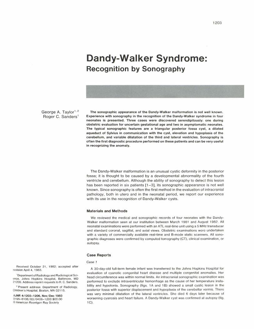
A B c Fig. 2.-Case 2. A, Midline sag itlal sonog ram shows large fourth ventricle cyst (c ). Vermis is displaced superiorly (arrows ). B, Coronal sonogram. Large
Iriangular cysl ic structure occupies mosl of posterior fossa (c). Dilated atria of lateral ventricle (arrows ). C, CT scan shows cystic di latation of fourth ventricle , wilh hypoplasia of cerebellar midline structures.
Case 2
A 31-week-old premature female infant with respiratory distress was examined by sonography because she was at high ri sk for intracranial hemorrhage from her prematurity . There were no neurologic symptoms. The study showed a triangular cyst of the posterior fossa with characteristic superior displacement and hypoplasia of the vermis and minimal hydrocephalus (figs. 2A and 28). CT (fig . 2C) confirmed the diagnosis of cystic d ilatation of the fourth ventricle. Serial follow-up examinations showed no progression of the hydrocephalus, and the patient remained asymptomatic without ventricular shunting.
Case 3
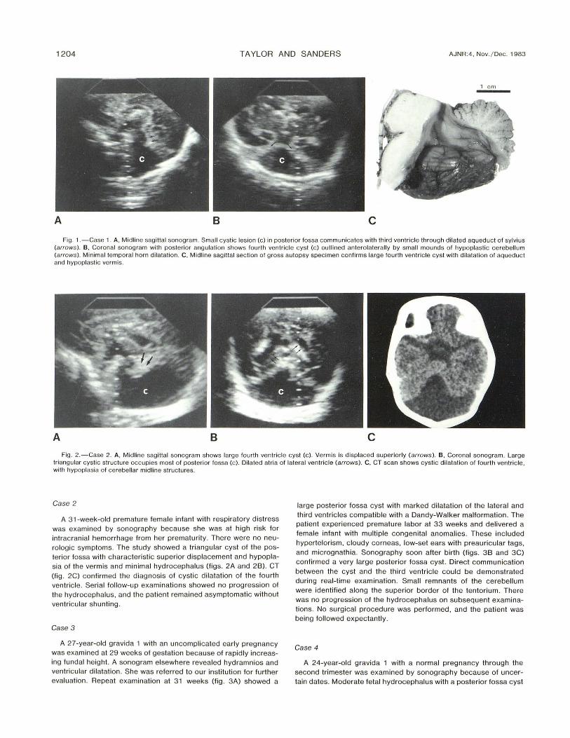
A 27-year-old gravida 1 with an uncomplicated early pregnancy was examined at 29 weeks of gestation because of rapidly increasing fundal height. A sonogram elsewhere revealed hydramnios and ventricular dilatation. She was referred to our institution for further evaluation . Repeat examination at 31 weeks (fig . 3A) showed a
large posterior fossa cyst with marked dilatation of the lateral and third ventricles compatible with a Dandy-Walker malformation. The patient experienced premature labor at 33 weeks and delivered a female infant with multiple congenita l anomalies. These included hypertelorism, cloudy corneas , low-set ears with preauricular tags, and micrognathia. Sonography soon after birth (figs . 38 and 3C) confirmed a very large posterior fossa cyst. Direct communication between the cyst and the third ventric le cou ld be demonstrated during real-time examination. Small remnants of the cerebellum were identified along the superior border of the tentorium . There was no progression of the hydrocephalus on subsequent examinations. No surgical procedure was performed , and the patient was being followed expectant ly.
Case 4
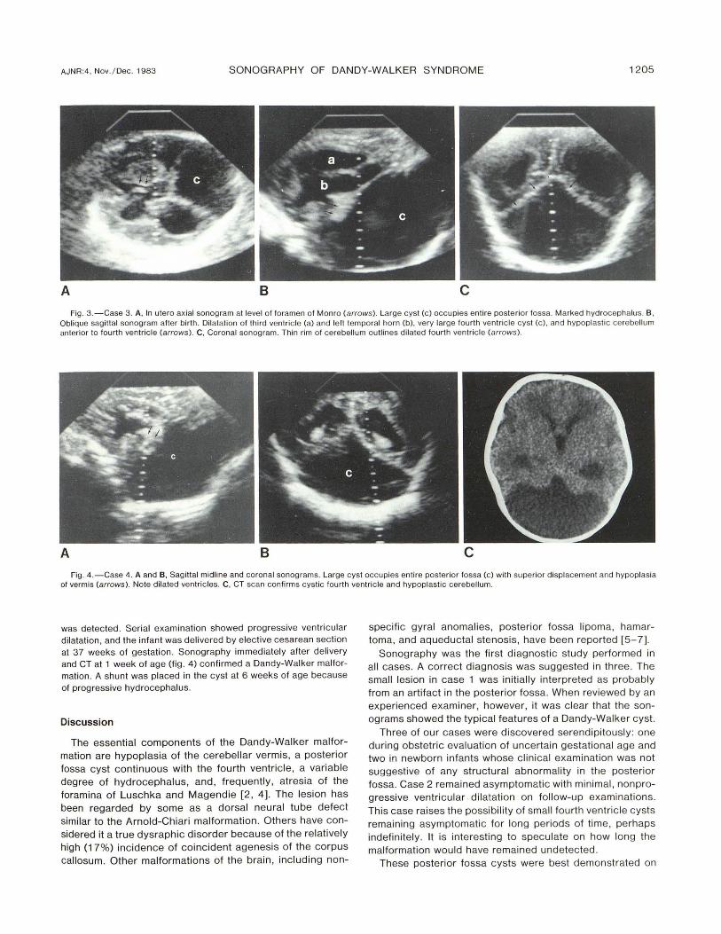
A 24-year-old gravida 1 with a normal pregnancy through the second trimester was examined by sonography because of uncertain dates. Moderate fetal hydrocephalus with a posterior fossa cyst
AJNR:4, Nov./Dec. 1983 SONOGRAPHY OF DANDY-WALKER SYNDROME 1205
A B c Fig. 3 .-Case 3 . A, In ulero axial sonogram al level of foramen of Monro (arrows). Large c yst (c) occupies ent ire posterior fossa. Marked hydrocephalus . B ,
Obl ique sag ittal sonogram after birth . Dilatat ion of third ventricle (a) and left temporal horn (b) , very large fourlh ventricl e cyst (c ), and hypoplastic cerebellum anteri or to fou rth ventric le (arrows) . C, Coronal sonogram. Thin rim of cerebellum outlines di lated fourth ventricle (arrows).
A B c Fig . 4.-Case 4. A and B, Sagi ttal midline and coronal sonograms. Large cyst occupies entire posteri or fossa (c ) with superior displacement and hypoplasia
of vermis (arrows). Note dilated ventric les. C, CT scan confirms cystic fourth ventric le and hypoplast ic cerebellum .
was detected . Serial examination showed progressive ventricular dilatation, and the infant was delivered by elective cesarean section at 37 weeks of gestation. Sonography immediately after delivery and CT at 1 week of age (fig. 4) con firmed a Dandy-Walker malformation. A shunt was placed in the cyst at 6 weeks of age because of progressive hydrocephalus.
Discussion
The essential components of the Dandy-Walker malformation are hypoplasia of the cerebellar vermis, a posterior fossa cyst continuous with the fourth ventricle , a variable degree of hydrocephalus, and, frequently, atresia of the foramina of Luschka and Magendie [2 , 4). The lesion has been regarded by some as a dorsal neural tube defect similar to the Arnold-Chiari malformation . Others have considered it a true dysraphic disorder because of the relatively high (17%) incidence of coincident agenesis of the corpus callosum. Other malformations of the brain , including non-
specific gyral anomalies, posterior fossa lipoma, hamartoma, and aqueductal stenosis, have been reported [5-7].
Sonography was the first diagnostic study performed in all cases. A correct diagnosis was suggested in three. The small lesion in case 1 was initially interpreted as probably from an artifact in the posterior fossa. When reviewed by an experienced examiner, however, it was clear that the sonograms showed the typical features of a Dandy-Walker cyst.
Three of our cases were discovered serendipitously: one during obstetri c evaluation of uncertain gestational age and two in newborn infants whose c linical examination was not suggestive of any structural abnormality in the posterior fossa. Case 2 remained asymptomatic with minimal , nonprogressive ventricular · dilatation on follow-up examinations . This case raises the possibility of small fourth ventri c le cysts remaining asymptomatic for long periods of time, perhaps indefinitely. It is interesting to speculate on how long the malformation would have remained undetected .
These posterior fossa cysts were best demonstrated on
1206 TAYLOR AND SANDERS AJNR:4, Nov. / Dec. 1983
coronal sonograms ang led posteriorly , and showed a characteristic triangular appearance. On midline sagittal sonograms, a dilated aqueduct of Sylvius in direct communication with the cyst was seen, even in cases with small cysts and minimal hydrocephalus. Small mounds of tissue representing hypoplastic cerebellar structures were well visualized, except in cases having marked dilatation of the fourth ventricle. In the latter cases, only a thin rim of tissue could be discerned. As expected, various degrees of associated hydrocephalus were detected .
Differentiation between a Dandy-Walker cyst and other cysti c lesions of the posterior fossa (such as arachnoid cysts or extraaxial posterior fossa cysts) may not always be possible with sonography. However, the latter two entities are not assoc iated with midline cerebe llar deformities and do not communicate directly with the ventricular system [1, 4 , 8]. Sonography is often the first diagnostic procedure performed on these patients in the neonatal period and can be very useful in recognizing the abnormality .
ACKNOWLEDGMENTS
We thank John Reeder and Mary Henciak for assistance in manuscript preparat ion.
REFERENCES
1. Roach ES, Laster OW, Summer TE , Volberg FM. Posterior fossa arachnoid cyst demonstrated by ultrasound . JCU 1982; 10 : 88-90
2. Newman GC, Buschi AI, Sugg NK, et al. Dandy-Walker syndrome diagnosed in utero by ultrasonography. Neurology (NY)
1982;32: 1 80-1 84 3. Sauerbrei EE, Cooperberg PL. Neonatal brain : sonography
of congenital anomalies. AJNR 1981 ;2: 1 25-1 28, AJR 1981;136 : 1167-1170
4 . Dempsey PJ , Koch HJ. In-utero diagnosis of the Dandy-Walker syndrome: differentiation from extra-axial posterior fossa cyst. JCU 1981 ;9: 403-405
5. Gardner E, O 'Rahilly R, Prolo D. The Dandy-Walker and Arnold-Chiari malformations: clinical, developmental and teratological considerations. Arch NeuroI1975 ;32 : 393-409
6. Padget DH . Development of so-called dysraphism: with embryological evidence of clinical Arnold-Chiari and DandyWalker malformation. Johns Hopkins Med J 1972; 130: 1 27-165
7. Sawaya R, McLaurin R. Dandy-Walker syndrome c linica l analysis of 23 cases. J Neurosurg 1981 ;55 : 89- 98
8. Vaquero J, Carrillo R, Cabezudo J, et al. Arachnoid cyst of the posterior fossa. Surg Neurol 1981 ; 16 : 11 7 -121


![DW.ppt [Mode de compatibilité] - pe.sfrnet.orgpe.sfrnet.org/Data/ModuleConsultationPoster/pdf/2010/1/cfaa9a31-c... · Malformation Dandy-Walker (DW): se définie par : ••Dilatation](https://img.pdfslide.us/doc/110x75/5bb2a8fc09d3f2622d8d0f61/dwppt-mode-de-compatibilite-pe-malformation-dandy-walker-dw-se-definie.jpg)