Embed Size (px)
Citation preview
NeuroImage 135 (2016) 273–286
Contents lists available at ScienceDirect
NeuroImage
j ourna l homepage: www.e lsev ie r .com/ locate /yn img
Dance and music training have different effects on white matterdiffusivity in sensorimotor pathways
Chiara Giacosa a,b,⁎, Falisha J. Karpati a,c, Nicholas E.V. Foster a,d, Virginia B. Penhune a,b, Krista L. Hyde a,c,d
a International Laboratory for Brain, Music and Sound Research (BRAMS), Pavillon 1420 Mont Royal, FAS, Département de psychologie, CP 6128 Succ. Centre Ville, Montreal, QC H3C 3J7, Canadab Department of Psychology, Concordia University, 7141 Sherbrooke Street West, Montreal, Quebec H4B 1R6, Canadac Faculty of Medicine, McGill University, 3655 Sir William Osler, Montreal, Quebec H3G 1Y6, Canadad Department of Psychology, University of Montreal, Pavillon Marie-Victorin, 90 avenue Vincent d'Indy, Montreal, Quebec H2V 2S9, Canada
⁎ Corresponding author.E-mail address: [email protected] (C. Giacosa).
http://dx.doi.org/10.1016/j.neuroimage.2016.04.0481053-8119/© 2016 Elsevier Inc. All rights reserved.
a b s t r a c t
a r t i c l e i n f oArticle history:Received 29 September 2015Revised 18 April 2016Accepted 20 April 2016Available online 22 April 2016
Dance and music training have shared and distinct features. Both demand long and intense physical training tomaster. Dance engages the whole body, and requires the integration of visual, auditory and motor information.In comparison, music engages specific parts of the body and primarily requires the integration of auditory andmotor information. Comparing these two forms of long-term training offers a unique way to investigate brainplasticity. Therefore, in the present study we compared the effects of dance and music training on white matter(WM) structure using diffusion tensor imaging (DTI), and examined the relationship between training-inducedbrain changes and specific measures of dance and music abilities. To this aim, groups of dancers and musiciansmatched for years of experience were tested on a battery of behavioural tasks and a range of DTI measures.Our findings show that dancers have increased diffusivity and reduced fibre coherence inWM regions, includingthe corticospinal tract, superior longitudinal fasciculus and the corpus callosum. In contrast, musicians showedreduced diffusivity and greater coherence of fibres in similar regions. Crucially, diffusivity measureswere relatedto performance on dance and music tasks that differentiated the groups. This suggests that dance and musictraining produce opposite effects on WM structure. We hypothesize that intensive whole-body dance trainingmay result in greater fanning of fibres connecting different brain regions, an increase in crossing fibres, or largeraxon diameter. In contrast, musical trainingmay result inmore focussed enhancements of effector-specific path-ways. These findings expand our understanding of brain plasticity by emphasizing that different types of trainingcan have different long-term effects on brain structure (Takeuchi et al., 2011; Baer et al., 2015).
© 2016 Elsevier Inc. All rights reserved.
Keywords:NeuroplasticityAction observation networkSensorimotor integrationMotor trainingCrossing fibresMultiple DTI measures
1. Introduction
Dance andmusic are universal forms of human expression that haveboth shared and distinct features. Both dance andmusic training requirelong, intense and quantifiable training to master. Dance training en-gages the whole body, and requires the integration of visual, auditoryand motor information. It focuses on perfectingmovement through ob-servation and imitation. In comparison, music engages specific parts ofthe body, typically the hands and fingers, and primarily requires the in-tegration of auditory andmotor information.Music training emphasizesperfecting sound through listening and refining movement. Thus, theneural systems relevant for long-term dance training likely includethose important forwhole-body control, visual and auditory sensorimo-tor integration and the “action observation network” (AON) (Crosset al., 2009b; Grafton, 2009; Keysers and Gazzola, 2009; Caspers et al.,2010). Conversely, the neural systems relevant for long-term musictraining likely include the regions important for control of specific
effectors and those engaged in auditory–motor integration (Bangertet al., 2006; Lahav et al., 2007; Zatorre et al., 2007). Therefore, compar-ing white matter (WM) structure between dancers and musicians pro-vides a new window to investigate the neural correlates of long-termtraining. Examination of long-term training in a variety of domainshas shown that it has lasting effects on brain structure and function(Maguire et al., 2000; Draganski et al., 2004; Driemeyer et al., 2008;Jäncke et al., 2009; Keller and Just, 2009; Taubert et al., 2010; Bezzolaet al., 2011). Among these disciplines, music training has received par-ticular attention (Hyde et al., 2009a, 2009b, Schlaug et al., 2009;Herholz and Zatorre, 2012; Groussard et al., 2014; Schlaug, 2015). Incontrast, research about the structural neural correlates of dance train-ing is still at a very early stage and very few studies have specifically ad-dressed this topic (Hänggi et al., 2010; Nigmatullina et al., 2015).However, these works compared dancers only to untrained controls,and there were no behavioural measures of dance performance. Thus,our goals in the present study were to characterize the specific effectsof dance training on WM structure in comparison with another groupof experts with similar long-term sensorimotor training, and to relatethose changes to their acquired skills. To do this, we directly compared

274 C. Giacosa et al. / NeuroImage 135 (2016) 273–286
expert dancers with equally well-trained musicians and untrained con-trols using both behavioural and a range of DTI measures.
1.1. Previous work on dance and music
The study of specialized training such as dance and music offers aunique way to investigate brain plasticity and its interaction with be-haviour. The literature about the neural correlates of music training ismuch richer than the one about dance and has been previouslyreviewed (Moore et al., 2014); therefore, here, our main focus will beon dance training.
Previous research about dance has been largely behavioural. Thesestudies have examined various components of dancers' acquired skills,such as postural, balance and motor control (Crotts et al., 1996;Golomer et al., 2010; Kiefer et al., 2011; Costa et al., 2013), timing, syn-chrony and choreography (Minvielle-Moncla et al., 2008; Waterhouseet al., 2014; Woolhouse and Lai, 2014), as well as memory (Poon andRodgers, 2000; Vicary et al., 2014) and imagery for sequences of move-ments (Golomer et al., 2008) (see (Bläsing et al., 2012) for a review).Further, dance expertise has been shown to improve skills that areclosely related to the training received, such as balance, posture andsensitivity to the biological motion of familiar actions (Calvo-Merinoet al., 2010; Kattenstroth et al., 2011).
Some recent research has investigated the functional correlates ofdance (see (Bläsing et al., 2012; Karpati et al., 2015a) for review). Alarge part of this literature has focussed on the AON, which includestemporo-parietal and frontal sensorimotor regions that are involved invisuo-motor integration and learning of actions performedwith specificeffectors (Caspers et al., 2010; Landmann et al., 2011; Krüger et al.,2014) or the whole body (Calvo-Merino et al., 2005; Cross et al., 2006;Cross et al., 2009b; Gardner et al., 2015). In both animal and humanstudies (Grèzes and Decety, 2001; Rizzolatti and Craighero, 2004;Hecht et al., 2013), these regions have been found to be engaged duringthe observation and performance of mouth or single limb actions(Fadiga et al., 1995; Gallese et al., 1996; Rizzolatti et al., 1996a;Rizzolatti et al., 1996b; Buccino et al., 2001; Gazzola and Keysers,2009), as well as of whole-body movements (Cross et al., 2009a;Sevdalis and Keller, 2011). It has been shown that this network is partic-ularly relevant for dance learning, which requires observing, simulatingand imitating others' whole-body movements (Calvo-Merino et al.,2005; Cross et al., 2006; Cross et al., 2009b). In addition to studies ofdance observation, a few experiments have examined lower limbdance-like movements which can be performed during brain imaging.Cortical, subcortical, and cerebellar regions have been shown to be in-volved in specific aspects of these dance-like movements (Brownet al., 2006; Tachibana et al., 2011; Ono et al., 2014). These studies arethe first ones that identified the regions that are functionally relevantfor dance execution as opposed to dance observation. However, theseparadigms have limited generalizability to realwhole-body dance train-ing, and the tested participants were not experts.
There are only few studies that have examined the structural neuralcorrelates of dance expertise (Hänggi et al., 2010; Nigmatullina et al.,2015). Hänggi et al. (2010) compared female ballet dancers and non-dancers using voxel-based morphometry (VBM) and diffusion tensorimaging (DTI). They found that dancers had decreased GM volumes incortical and subcortical structures critical for motor control and sensori-motor integration, along with decreases in WM volume and fractionalanisotropy (FA) in sensorimotor pathways and the corpus callosum.They hypothesised that reductions of WM volume and FA might bethe result of greater efficiency, or enhancements in crossing fibre path-ways. Although these changes in brain structure were found to be relat-ed to the age of commencement of training, no behaviouralmeasures ofdance performance were obtained. Further, the authors reported onlytwo global DTImeasures, which give little information about crossing fi-bres. Similar decreases in FAwere also found in fronto-parietal and sen-sorimotor pathways of professional gymnasts (Huang et al., 2013). Just
like dancers, gymnasts are experts in whole-bodymovements and theirtraining focuses on visual-motor integration and action observation.Therefore, their similar training might result in similar changes in WMstructure.
Structural brain imaging studies have shown that music training isassociated with enhancements of grey (GM) and white matter (WM)in motor regions associated with effector-specific motor control,the corpus callosum, and the auditory cortex (Schlaug et al., 1995;Sluming et al., 2002; Gaser and Schlaug, 2003; Bengtsson et al., 2005;Bermudez and Zatorre, 2005; Bermudez et al., 2009; Han et al., 2009;Hyde et al., 2009b; Abdul-Kareem et al., 2011; Groussard et al., 2014).Further, these brain differences have been linked to performance onmusic-related tasks (Foster and Zatorre, 2010a; Steele et al., 2013;Bailey et al., 2014; Elmer et al., 2014).
In summary, structural imaging studies in dancers and gymnastsshowed a reduction in theWMvolume and anisotropy localised in sen-sorimotor and commissural pathways, as well as fronto-parietal associ-ation fibres (Hänggi et al., 2010; Huang et al., 2013; Nigmatullina et al.,2015). In contrast, despite some inconsistencies (Schmithorst andWilke, 2002; Imfeld et al., 2009), studies in musicians suggest thatmusic training tends to increase fractional anisotropy, especially in thesensorimotor projection fibres (Bengtsson et al., 2005; Han et al.,2009) and in the corpus callosum (Schlaug et al., 1995; Steele et al.,2013).
1.2. DTI measures
Currently, DTI is the most widely used method to investigate themicro-structural properties of WM. It measures the characteristics ofdiffusion of water molecules in brain tissues. This motion is modelledas an ellipsoid characterised by three axes. Biological features, such asaxonal size, density, coherence and degree of myelination all constrainwater molecule motion, and thus influence diffusivity measures(Moseley et al., 1990; Basser et al., 1994; Neil et al., 1998; Gulani et al.,2001; Beaulieu, 2002; Assaf and Pasternak, 2008). Because no one-to-one relationship exists between any DTI measure and the architectureof WM structure (Wheeler-Kingshott and Cercignani, 2009; Joneset al., 2013; Walhovd et al., 2014), a multi-parametric combined analy-sis of diffusion data is helpful. Therefore, in the present study we exam-ined both non-directional and directional measures in order to have abetter understanding of the different possible underlying biological con-figurations (Alexander et al., 2007). Themost commonly used DTImea-sure is fractional anisotropy (FA), which gives a global estimate of theelongation of the ellipsoid or the linearity of diffusion. Other non-directional measures are: axial diffusivity (AD) which measures theamount of diffusion along the principal axis; radial diffusivity (RD)which measures the diffusion on the plane perpendicular to the princi-pal axis; mean diffusivity (MD) which quantifies the amount of diffu-sion in each voxel; and the mode of anisotropy (MO) which describeswhether diffusion is more planar or linear (Basser and Pierpaoli, 1996;Beaulieu, 2002; Ennis and Kindlmann, 2006; Assaf and Pasternak,2008). In addition, we assessed the partial volume fractions of primaryand secondary fibres (F1 and F2). Based on the “ball and stick model”(Behrens et al., 2003), these directional measures give an estimationof the relative proportion of the primary and secondary fibres in thevoxels where the co-existence of at least two fibre populations isdetected.
1.3. Objectives and predictions of this study
Taken together, previous literature suggests that long-term dancetraining can have specific effects on the sensorimotor and action obser-vation systems. The purpose of the present study is to investigate the ef-fects of long-term dance training on WM structure by comparingdancers to musicians and untrained controls. Musicians are a usefulcomparison group for dancers because music and dance training are

275C. Giacosa et al. / NeuroImage 135 (2016) 273–286
both long and intense, require similar integration of sensory and motorinformation, and the amount of training can be quantified. This canallow us to make more specific interpretations about any observed dif-ferences inWMstructure. In addition,wewanted to relate any training-induced brain modifications to specific measures of dance and musicabilities. Because previous DTI studies in dancers have found reductionsin FA that are difficult to interpret, in this study, we decided to analysemultiple diffusivity measures in order to better understand the biologi-cal underpinnings of diffusivity changes. We tested groups of highlytrained dancers and musicians who were matched for years of experi-ence, and also compared them to controls with limited dance or musictraining. Importantly, we tested all participants on a battery of danceand music-related tasks, as well as tests of global cognitive functionand auditoryworkingmemory. Based on previous research, we predict-ed that dancers would show reduced anisotropy in sensorimotor,fronto-parietal and callosal connections, whereas musicians wouldshow higher anisotropy in the corticospinal tract and corpus callosum.We also expected that these changes would be related to group differ-ences in behavioural performance on dance- and music-based tasks.
2. Materials and methods
2.1. Participants
Three groups of participants (age 18–40) were recruited for thisstudy: expert dancers (N= 20), expert musicians (N= 19) and a con-trol group of non-musician/non-dancers (N = 20). Dancers and musi-cians were either currently practising as professionals, or studentsinvolved in professional training. Their training was assessed via a de-tailed questionnaire developed in our lab: the Montreal Dance andMusic History Questionnaire (MDMHQ) (Karpati et al., 2015b), basedon (Bailey and Penhune, 2010; Coffey et al., 2011). Dancers and musi-cians had on average approximately 15 years of experience in their re-spective disciplines, controls had on average less than one year indance, music, figure skating, and aerobics. All participants were physi-cally active (biking, running or practising other physical exercises).Dancers were currently practising contemporary dance as their princi-pal style, but had a variety of training backgrounds, including ballet,tap, jazz, swing, and ballroom. Dancers whose main style was too simi-lar to the dance task used here (i.e., urban, street or hip-hop dance)were excluded. Analogously, musicians had various instrumental back-grounds, including keyboard instruments, strings, woodwinds, brass,and percussion. None of the musicians had absolute pitch. Since thedance task was based on a video-game, participants were also excludedif they had more than 2 years of experience with dance video-games.The groups did not differ in age, sex distribution, body-mass index(BMI) or years of education (See Table 1). Participants had no past orcurrent learning or developmental disorder, neurological or psychiatricconditions, or reported current or past alcohol or substance abuse. Oneparticipant in each group was excluded due to artefacts in the DTI data(see DTI data analysis section below). The experimental protocol wasapproved by the Research Ethics Board at the Montreal NeurologicalInstitute and Hospital, and a written informed consent was obtained
Table 1Participant characteristics.
Group NAge(yrs ± SD)
SexBMI(±SD)
Years of
Dancers 19 25.1 ± 3.9 13F, 6 M 21.6 ± 2.3 15.5 ± 5Musicians 18 22.9 ± 3.4 12F, 6 M 22.5 ± 3.2 1.04 ± 1Controls 19 25.4 ± 5.1 12F, 7 M 22.1 ± 3.1 0.4 ± 0.9
Comparison between groups 56D = M = Cns
D = M = Cns
D N M =P b 0.000
F = females, M = males, SD = standard deviation, and BMI = body mass index.Education levels for each group are calculated on a scale 1–5, where 1 is the lowest (complete
from all participants. All participants were compensated for theirparticipation.
2.2. General procedures
Participants took part in two testing sessions distributed over twonon-consecutive days: one behavioural and the other for MRI acquisi-tion, including DTI. The behavioural battery included dance- andmusic-related tasks as well as tests of global cognitive and memoryfunction. The Dance Imitation Task was developed in our laboratorybased on the video game Dance Central 1 for the console Xbox Kinect360 (Harmonix, http://www.harmonixmusic.com) using the Kinect in-frared light sensor (http://www.xbox.com/en-ca/Kinect; US Patent No.20100197399). Participants are required to imitate a selection ofseven dance routines of increasing levels of complexity. Scoring isbased on the percent of correctmoves provided by the video game scor-ing system. This task assesses the ability to observe and imitate in realtimewhole-body dancemovements synchronisedwithmusic. TheMel-ody Discrimination Task (Foster and Zatorre, 2010a, 2010b) requiresparticipants to detect subtle pitch changes in a series of melodies. Thistask assesses auditory processing and pitch discrimination. Finally, theRhythm Synchronisation Task has been used in a number of our previ-ous studies and requires participants to tap in synchrony with a seriesof musical rhythms. It has been previously used with musicians andnon-musicians (Chen et al., 2008; Bailey and Penhune, 2010; Baileyet al., 2014). This task assesses auditory–motor integration and finemotor response. Furthermore, a language task and three standardizedcognitive tests were given to all participants to examine any possiblegroup differences in global cognitive or memory function: the SyllableSequence Discrimination Task (Foster and Zatorre, 2010a, 2010b), thathas the same design as the Melody Discrimination Task but uses sylla-bles instead of tones, the Digit Span and Letter-Number Sequencingsubtasks from the Wechsler Adult Intelligence Scale III (Wechsler,1997) and the Matrix Reasoning subtask from theWechsler Abbreviat-ed Scale of Intelligence (Wechsler, 1999). Full behavioural results forthese groups are reported in a previous paper (Karpati et al., 2015b).For the purpose of this paper, only brief descriptions of the behaviouralresults and the relationships between behavioural performance andWM diffusivity measures will be reported.
2.3. Diffusion tensor imaging and analysis
2.3.1. Diffusion data acquisitionDiffusion-weighted images (DWI) were acquired for all participants
at the Montreal Neurological Institute (MNI) on a 3T Siemens Trio MRscannerwith a 32-channel head coil. The following parameterswere ap-plied: 99 diffusion-weighted gradient directions with a b-value of1000 s/mm2, 10 b0 non-weighted images, TE of 88 ms, TR of 9340 ms,EPI factor 128, isotropic voxels of 2 × 2 × 2 mm3, 72 slices, FOV of256 mm. Ear plugs and headphones, as well as foam pads were usedto reduce noise perception and head motion, respectively.
dance training (±SD) Years of music training (±SD) Level of education (±SD)
.2 1.7 ± 1.9 2.37 ± 0.6
.8 15 ± 3.6 2.39 ± 0.980.4 ± 1.0 2.58 ± 1.12
C1
M N D = CP b 0.0001
D = M = Cns
d high school) and 5 is the highest (completed PhD).

276 C. Giacosa et al. / NeuroImage 135 (2016) 273–286
2.3.2. Behavioural analysisBehavioural analyses for the full sample are reported in detail in
Karpati et al. (2015b). Here, similar analyses were conducted on thesubjects used for the DTI analysis. Briefly, to allow for between-taskcomparisons, overall scores for the dance, melody and rhythm taskswere converted to z-scores. A linear fixed-effects model was conductedon these data, with group as a between-subjects fixed factor and task asa within-subjects repeated measure using an unstructured covariancematrix.
Each control task was separately analysed with univariate analyses,with group as the between subject factor. Raw scores on each cognitivetask were converted to scaled scores using standard protocols. All anal-yses included age and sex as covariates of no interest.
2.3.3. Brain analysisAll the following analyses included age and sex as covariates of no
interest. Results were considered significant at p b 0.05, after family-wise error (FWE) correction for multiple comparisons.
2.3.3.1. Voxelwise group comparisons of diffusivity measures. Group com-parisons between dancers, musicians and controls were performed onboth non-directional (RD, AD, MD, FA and MO) and directional (F1and F2) diffusivity measures following the Tract-Based Spatial Statistics(TBSS) procedure, using the FMRIB Software Library (FSL v5.0) (Smithet al., 2004).
2.3.3.1.1. TBSS for non-directional measures. Each subject's raw datawere first corrected for eddy current distortions and head motionusing the FMRIB's Diffusion Toolbox (FDT); then, the Brain ExtractionToolbox (BET) (Smith, 2002) was used to exclude non-brain voxelsfrom the analyses, and the diffusion tensor model was applied, bymeans of FDT, to estimate the diffusivity measures in each voxel.From the three eigenvalues, axial (AD), radial (RD) and mean (MD)diffusivities were easily calculated as the highest eigenvalue (AD),the average between the other two eigenvalues (RD) and the tensortrace (MD). Fractional anisotropy (FA) and the mode of anisotropy(MO), representing the eccentricity and type of anisotropy respectively,were also calculated with FDT.
Individual data of each diffusivity measure were then averaged andcompared to the other subjects' data of the samemeasure. If one subjecthad at least 3 diffusivity measures that fell 3 SD out of their own groupdistributions, that subject was considered an outlier and excluded fromthe rest of the analyses. According to this rule, two subjects were ex-cluded, one dancer and one control. One musician had to be excludeddue to scanning artefacts. Other vibration artefacts (Gallichan et al.,2010), which occurred especially with large x-directed gradients, werecorrected by excluding the affected frames. After correction, all subjectshad more than 70 gradient directed frames of good quality.
Data that survived the above mentioned quality control wereanalysed with the TBSS method (Smith et al., 2006), implemented inFSL. First, individual FA images were non-linearly aligned to theFMRIB58_FA target template, and transformed into the 1 × 1 × 1 mmMNI152 standard space. The resulting FA images were averaged andthinned to obtain a study-specific mean FA skeleton, which representsthe centres of all fibre bundles that are common to all participants.Each subject's aligned FA image was then projected onto its individualFA skeleton, before entering the permutation-based non-parametricvoxelwise statistical analyses. The mean FA skeleton was thresholdedat the value of 0.25 in order to include only major tracts that existedin all individuals.
A similar procedure was then applied to the other diffusion mea-sures, namely RD, AD,MDandMO. For each diffusivitymeasure, nonlin-ear registration to the common space and skeletonisation procedurewere based on the steps accomplished for FA. Then, each individualmeasure map was projected onto the mean FA skeleton, before statisti-cal analyses were performed.
2.3.3.1.2. TBSS for directional measures (FSL). In order to increase theinterpretability of the diffusivity scalar measures obtained with TBSSin crossing-fibre regions, TBSS was performed also on scalars associatedwith a specific direction of fibres in each voxel. The model applied as-sumed that two fibre populations existed for each voxel. The modelwith three fibreswas tested, but gave an inconsistent distribution of ter-tiary fibre orientations across consecutive voxels (impossibility ofreconstructing smooth fibre pathways).
After the pre-processing steps, common to the previous TBSS analy-ses, partial volume estimates forfibre orientations 1 and 2 (F1 and F2 re-spectively) were calculated in each voxel for each subject. F1 and F2were reassigned within subject at each voxel in order to ensure consis-tency across voxels, such that adjacent voxels had the same labelassigned to the same fibre population. Then, F1 and F2 were reassignedagain in order to ensure that orientations were consistent across sub-jects. Both these steps were accomplished using the “tbss_x” software,part of FSL (Jbabdi et al., 2010).
2.3.3.1.3. Voxelwise statistical analyses. For all non-directional (RD,AD, MD, FA and MO) and directional (F1 and F2) diffusivity measures,non-parametric permutation tests (5000 permutations per analysis)were carried out using the FSL's tool Randomise (Winkler et al., 2014)for the general linearmodel (GLM). Three group comparisonswere per-formed: 1) dancers versus musicians, 2) dancers versus controls, and3) musicians versus controls.
2.3.3.2. Group comparisons of the subjectwise averaged extracted values ofdiffusivity measures. RD, FA and MO were further tested with univariateANOVAs including age and sex as covariates of no interest. For each DTImeasure, individual data were extracted and averaged either over thewhole WM skeleton or the ‘Dancers versus Musicians’ ROIs. The‘Dancers versus Musicians’ ROIs were selected to include all the voxelswhere each DTI measure differed between dancers and musicians,thus a different ROI was created for each measure. The resultingsubjectwise averaged values were then used in the ANOVAs to compareeach parameter between groups.
2.3.4. Brain–behaviour relationsAll analyses included age and sex as covariates of no interest. Results
were considered significant at p b 0.05. Family-wise error (FWE) correc-tion for multiple comparisons was applied to all voxelwise analyses.
2.3.4.1. Voxelwise regressions of diffusivity measures with behavioural per-formance. Independent linear regressions between each diffusivitymea-sure (dependent variables) and performance on each behavioural task(independent variables) were calculated by inserting the appropriategeneral linear model (GLM) matrix in the FSL's Randomise tool. Theseanalyses were performed over the whole WM skeleton and in the‘Dancers versus Musicians’ ROIs, across all groups together and in eachone separately.
2.3.4.2. Correlations of the subjectwise averaged extracted values with be-havioural performance. Each of the non-directional and directional diffu-sivity measures was also correlated with performance on behaviouraltasks. Partial correlations with age and sex as covariates of no interestwere performed in SPSS over the whole WM skeleton and the ‘Dancersversus Musicians’ ROIs.
3. Results
3.1. Behavioural results
3.1.1. Group characteristicsOne-way ANOVAs conducted on age and BMI with group (dancer,
musician or control) as the between-subjects factor did not reveal anysignificant differences between groups (see Table 1; Age: F(2,53) =2.12, p = 0.13; BMI: F(2,53) = 0.4, p = 0.67). One-way ANOVAs

Fig. 1. Behavioural results. Performance on the behavioural tasks (z-scores) across thethree groups.
277C. Giacosa et al. / NeuroImage 135 (2016) 273–286
conducted on years of dance and music training revealed significantgroup differences for both training types (Dance: F(2,53) N 131,p b 0.0001;Music: F(2,53) N 0.201, p b 0.0001). Post-hoc pair-wise com-parisons confirmed that both dancers and musicians had significantlylonger training in their own discipline compared to the other groups.Furthermore, both dancers and musicians had comparable training tocontrols in the other discipline, measured as years of music ordance training, respectively (see Table 1). Univariate analyses onyears of dance and music training, including age and sex as covari-ates of no interest, consistently revealed significant differencesfor both training types (Dance: F(2,51) N 136, p b 0.0001; Music:F(2,51) N 235, p b 0.0001).
3.1.2. Dance and music tasksBehavioural results for the full sample were reported previously
(Karpati et al., 2015b). As described above, three participants (onefromeach group)were excluded due to unreliableMRI data. For the cur-rent samplewe followed similar procedures and obtained consistent re-sultswith Karpati et al. (2015b). To compare task performance betweengroups, we performed a linear fixed-effects analysis on the z-scores ofthe dance, rhythm and melody tasks (see Fig. 1) with group as abetween-subjects factor and task as a within-subjects repeatedmeasure; age and sex were considered in the model as variables ofno interest. This analysis revealed a significant interaction between
Table 2Cognitive results.
Cognitive tasks
Group Letter number sequence
DancersMean ± SD(range)
10.05 ± 2.01(8–15)
MusiciansMean ± SD(Range)
12.50 ± 2.47(7–17)
ControlsMean ± SD(range)
10.00 ± 3.2(5–18)
TotalMean ± SD(range)
10.82 ± 2.82(5–18)
Comparison between groupsM N D = CP b 0.05
Mean values, standard deviations (SD) and ranges for each of the cognitive measures are showtween groups are applicable only for significant differences.
group and task (F(4,53) = 26.4, p b 0.0001). In addition, to comparebehavioural performance between groups on each task separately,one-way ANOVAs and Bonferroni post-hoc tests were performed.As expected, for the Dance Imitation Task, dancers outperformedboth musicians and controls (ps b 0.0001). Musicians also per-formed better than controls (p b 0.007). Analogously, on the mostmusic-relevant task, the Melody Discrimination Task, musiciansoutperformed both dancers and controls (ps b 0.0001), whereasdancers' performances were comparable to those of controls (p =1). Finally, on the Rhythm Synchronisation Task, musiciansoutperformed both dancers (p = 0.013) and controls (p b 0.0001)with no other significant differences between groups.
3.1.3. Control tasksIn order to compare cognitive abilities between groups, one-way
ANOVAs were performed for each control task separately with groupas the between subject factor (See Table 2). Main effects of groupwere found for Letter Number Sequencing (F(2, 53) = 0.5, p =0.007), where musicians outperformed both dancers (p = 0.019) andcontrols (p = 0.016), and Matrix Reasoning (F(2, 53) = 4.2, p =0.019), where dancers (p = 0.044) and musicians (p = 0.045) bothoutperformed controls. No significant effect was found in either theDigit Span (F(2, 53)=0.8, p=0.463) or the Syllable SequenceDiscrim-ination Task (F(2, 53) = 0.3, p = 0.729).
Univariate analyses including age and sex as covariates of no interestconfirmed the ANOVAs results (Letter Number Sequencing: F(2, 51) =3.675, p = 0.032; Matrix Reasoning: F(2, 51) = 4.076, p = 0.023; DigitSpan: F(2, 51) = 0.879, p = 0.421; Syllable Sequence DiscriminationTask: F(2, 51) = 0.333, p = 0.718).
Taken together, the groups performed relatively similarly on alltasks and all were in the normal range. This suggests that any differ-ences on the experimental tasks or in brain structure are not likely tobe related to differences in global cognitive function.
3.2. Diffusion tensor imaging results
3.2.1. Group comparisonsIn order to determine how dance training differently affects WM
structure in comparison to music training, three group analyseswere performed: dancers versus musicians, dancers versus controlsand musicians versus controls. For each analysis, five non-directional (RD, AD, MD, FA and MO) and two directional (F1 andF2) diffusivity measures were compared between groups. All signif-icant results have ps b 0.05, FWE corrected for multiple compari-sons (see Table 3 for specific peak p-values and localisation andSupplementary Table 1 for a summary).
Matrix reasoning Digit span Syllable sequence discrimination
13.37 ± 1.3(11–16)
9.05 ± 2.12(7–13)
69.65 ± 7.42(53.33–86.67)
13.39 ± 2.03(10–16)
10.11 ± 2.56(5–14)
67.96 ± 9.24(43.33–81.67)
11.58 ± 2.91(2–15)
9.89 ± 3.36(5–18)
67.60 ± 8.57(51.67–83.33)
12.77 ± 2.31(2–16)
9.68 ± 2.72(5–18)
68.41 ± 8.32(43.33–86.67)
D = M N CP b 0.05
D = M = Cns
D = M = Cns
n for each group separately and for all groups together. P-values for the comparisons be-

Table 3Localisation of TBSS results and statistical values.
Contrast ROI Diffusivity measurement WM region # of voxels Peakp-value
Peak t-value Peak coordinates(mm)
X Y Z
DancersN
musicians
Whole WM skeleton RD R SCR/CST 9661 0.003 5.26 27 −20 25L SCR/CST 2499 0.022 5.76 −25 −12 23R –/SLF 24 0.049 3.22 47 6 14R –/ATR-IFOF 19 0.050 2.52 28 30 20
AD
R PCR/– 654 0.034 4.26 20 −33 36L –/SLF 425 0.041 3.68 −22 −41 42R spl.CC/FM 223 0.046 4.2 6 −38 14R body CC/ATR 37 0.049 3.45 −6 −26 24
MDR SCR/– 18,495 0.004 6.43 21 −12 35R –/– 336 0.048 4.21 13 −16 7
Dancersb
musicians
Whole WM skeleton FA R plIC/SLF-CST 1634 0.027 4.43 27 −9 18
MO
R alIC/– 1892 0.009 4.68 22 −3 17R rlIC/ILF-IFOF 730 0.029 4.36 36 −32 8R ACR/IFOF-UF 466 0.013 5.26 21 28 −7L PCR/CST 222 0.037 3.66 −27 −24 25L plIC/ATR 204 0.041 4.08 −19 −5 11L rlIC/– 77 0.047 3.34 −27 −31 12L alIC/ATR 68 0.045 3.45 −21 8 17L ILF-IFOF/= 38 0.049 2.46 42 −32 −12
F1
R SCR/CST 5359 0.007 5.34 26 −19 34R body CC/ATR 712 0.034 4.84 12 −26 27R –/SLF 35 0.048 4.02 46 7 11R CST/CST 28 0.049 3.52 11 −21 −25L EC/SLF 25 0.046 5.04 −29 −11 16R SCR/CST 10 0.050 2.87 −26 −11 22
Dancersb
controls
Whole WM skeleton MO R alIC/ATR 2093 0.014 4.52 23 6 18L alIC/ATR 790 0.023 4.34 −22 16 12R –/SLF 27 0.047 4.02 48 −11 25
MusiciansN
controls
Whole WM skeleton F1 R plIC/CST 193 0.045 3.21 20 −12 4R rlIC/IFOF 140 0.048 3.43 31 −29 5R –/– 78 0.046 3.96 29 −18 −8R –/SLF 57 0.049 2.97 53 −22 −14R –/ILF 15 0.050 2.84 41 −10 −18R plIC/CST 11 0.050 2.86 26 −18 13
Regressions with dance performance Whole WM skeleton MO R plIC/CST 3047 0.002 −4.19 20 −8 6L alIC/ATR-IFOF-UF 1021 0.008 −4.6 −22 16 12
DvsM ROIs
RD R SLF/SLF 2935 0.002 5.1 31 −18 36L EC/CST 522 0.022 4.06 −28 −20 17
MD R –/SLF 2308 0.016 4.11 33 −15 38FA R SLF/SLF 950 0.003 −4.3 32 −15 34MO R plIC/CST 1168 0.007 −4.19 23 −12 9F1 R SCR/– 608 0.004 −4.06 26 0 34
R SCR/SLF 80 0.031 −2.93 29 −9 22R EC/SLF 62 0.020 −3.23 30 −9 15
Regressions with melody performance DvsM ROIs AD R SCR/– 14 0.045 −2.88 18 −7 39R PCR/– 11 0.041 −4.02 20 −34 38
FA R plIC/CST 434 0.008 3.92 26 −17 15R EC/IFOF 7 0.049 2.38 32 −18 5
MO R plIC/CST 511 0.004 4.57 21 −7 9R –/IFOF-ATR-UF 25 0.045 3.51 18 43 −8
WM = white matter; R = right; and L = left.Regions are labelled according to the JHU ICBM-DTI-81 White-Matter Labels/JHU White-Matter Tractography Atlas.– = no labels provided with the first (–/) or the second (/–) atlas.alIC= anterior limb of internal capsule; ATR=anterior thalamic radiations; CC= corpus callosum; CST= corticospinal tract; EC=external capsule; FM= forcepsmajor; IFOF= inferiorfronto-occipital fasciculus; ILF= inferior longitudinal fasciculus; PCR= posterior corona radiata; plIC= posterior limb of internal capsule; rlIC= retro-lenticular part of internal capsule;SCR = superior corona radiata; SLF = superior longitudinal fasciculus; spl. = splenium; and UF = uncinate fasciculus.
278 C. Giacosa et al. / NeuroImage 135 (2016) 273–286
3.2.1.1. TBSS voxelwise analysis of non-directional diffusivity measures.Over the whole WM skeleton and across all measures, dancers showedincreased diffusivity relative to musicians predominantly in the righthemisphere (Fig. 2A).More specifically, dancers had significantly higherRD values in bilateral projection fibres (corticospinal tract (CST), coronaradiata (CR), internal (IC) and external (EC) capsules), association fibres(bilateral superior longitudinal fasciculus (SLF) and right cingulum), thebody and spleniumof the corpus callosum (CC) and in right ventralWMregions of the temporal and prefrontal lobes, including the inferior-fronto-occipital fasciculus (IFOF) and the inferior longitudinal fasciculus(ILF). Peaks of significance were reached for RD in most of the voxels of
the right hemisphere (p b 0.01). Note that, except for the CC, all theabove-mentioned regions are particularly rich in crossing fibres. For in-stance, the projection fibres of the CST are crossed by the association fi-bres of the SLF and the IFOF. ADwas also increased in dancers, especiallyin the medial regions of the CC. MD values were increased in dancerscompared to musicians in the same WM regions as RD, although witha more symmetrical involvement of the left CST, SLF and body of theCC. Conversely, FA and MO were lower in dancers compared to musi-cians in the main WM regions where RD and MD were higher indancers. These findings are consistent since greater RD values reduceFA values. In particular, lower FA values were observed in similar

Fig. 2. Dancers versus musicians group comparisons and brain–behaviour correlations. The left column (A) illustrates the regions of significant difference between dancers andmusicians for axial (AD), radial diffusivities (RD), fractional anisotropy (FA) and mode of anisotropy (MO). The right column (B) illustrates the correlations between diffusivitymeasures and dance performance. (A) In comparison to musicians, dancers had higher AD and RD (rows 1 and 2) and lower FA and MO (rows 3 and 4). 1-p-values are presented overthe FA mean image, specifically calculated for the present study sample. Images are presented in neurological view. All images were taken at the same slice level (MNI coordinates).Colours show the voxels where groups statistically differ (p b 0.05, FWE corrected). The colour scale in the RD map (Ab) shows significant voxels ranging from dark blue (p b 0.05) tolight blue (most significant). (B) The extracted values of AD and RD, averaged over the ‘Dancers versus Musicians’ ROIs were positively correlated with dance performance (rows 1 and2); those of FA and MO were negatively correlated with dance performance (rows 3 and 4). L = left, R = right, D = dorsal, V = ventral. ATR = anterior thalamic radiations, CC =corpus callosum, CB = cingulum bundle, CR = corona radiata, CST = corticospinal tract, EC = external capsule, IC = internal capsule, IFOF = inferior fronto-occipital fasciculus,ILF = inferior longitudinal fasciculus, SLF = superior longitudinal fasciculus.
279C. Giacosa et al. / NeuroImage 135 (2016) 273–286

280 C. Giacosa et al. / NeuroImage 135 (2016) 273–286
regions of the right hemisphere, including projection fibres (CST, IC, andEC), association fibres (SLF) and, more ventrally, the crossing betweenthe CST, the ILF and IFOF. MO differed in the same, although more ex-tended, regions as FA, but involved also some homologous tracts inthe left hemisphere (CST and SLF).
In the group comparison of dancers versus controls, the onlysignificant differences were found for MO. Analogously to thecomparison with musicians, dancers had reduced MO in bothhemispheres, although predominantly in the right. More specifi-cally, lower MO was found in dancers' projection fibres (especiallythe CST), association fibres (SLF), and some ventral prefrontal re-gions. Consistently, compared to controls, dancers also showedtrends for higher values of RD (p b 0.12) and MD (p b 0.1) in theCST crossing the SLF, in the SLF more laterally, and in the CC, thesame regions of the right hemisphere where dancers differedfrom musicians.
Therewere no significant differences in any non-directional diffusiv-ity measures between musicians and controls.
3.2.1.2. Analysis of subjectwise averaged non-directional diffusivitymeasures. Univariate analyses were performed across all populationson the extracted values of RD, FA andMO over the whole WM skeletonand the ‘Dancers versus Musicians’ ROIs. Bonferroni-corrected betweengroup post-hoc tests not including age and sex allowed for pair-wisecomparisons between groups.
Consistent with the voxelwise analyses, over the whole WMskeleton, a main effect of group emerged for MO (F(2,51) =3.835; p = 0.028). Furthermore, over the ‘Dancers versus Musi-cians’ ROIs, all measures showed significant group differences (FA:F(2,51) = 10.49, p b 0.0005; MO: F(2,51) = 24.91, p b 0.0001;RD: F(2,51) = 10.22, p b 0.0005). Musicians had highest FA (musi-cians versus dancers: p b 0.0001; dancers versus controls: p =0.023; musicians versus controls: p = 0.2) and MO (musicians ver-sus dancers: p b 0.0001; dancers versus controls: p b 0.0005; musi-cians versus controls: p = 0.03), and lowest RD (musicians versusdancers: p b 0.0005; dancers versus controls: p = 0.05; musiciansversus controls: p = 0.154), whereas dancers occupied the oppositeextremes, with highest RD, and lowest FA and MO (see Supplemen-tary Table 2 for a summary).
3.2.1.3. TBSS voxelwise analysis of directional diffusivity measures. Consis-tent with the results of the non-directional measures, compared tomusicians, dancers showed reduced F1 in right projection (CR,CST) and association (SLF) fibres, as well as bilateral commissural fi-bres (CC), the same WM regions where these two groups differed innon-directional measures. This suggests that musicians show great-er coherence in these regions. In agreement with this finding, com-pared to controls, musicians had increased F1 (p b 0.05) in theinferior portions of the CST and in the ILF, where the two fibre bun-dles cross. These locations are included in the regions where diffu-sivity (especially RD and MD) was reduced and anisotropy (FAand MO) increased in musicians compared to dancers. The orienta-tions associated with F1 correctly follow the anatomical orientationof projection and association fibres, as shown in Fig. 3A and B. Onthe other hand, F2 was reduced in the musicians' right spleniumof CC (p b 0.05). The orientations of F2 are shown in Fig. 3C. No sig-nificant differences were found in F1 or F2 when comparing dancersto controls.
3.2.2. Brain–behaviour relationsTo further establish the relationship between brain structure
and dance or music expertise, voxelwise regressions were per-formed between all diffusivity measures and performance on be-havioural tasks (Dance Imitation, Melody Discrimination andRhythm Synchronisation). Moreover, correlations between thesubjectwise averaged extracted values of diffusivity measures and
behavioural performance were analysed. All analyses were accom-plished over the ‘Dancers versus Musicians’ ROIs and over thewhole WM skeleton, across all groups together and in each oneseparately.
3.2.2.1. Dance imitation task. Voxelwise regressions in the ‘Dancersversus Musicians’ ROIs revealed positive relations between danceperformance and RD (p b 0.01) or MD (p b 0.05), and negative as-sociations between dance task performance and FA, MO or F1(p b 0.01) (Fig. 2B). More precisely, better performance predictedhigher RD and MD, as well as lower FA and MO, in the right pro-jection (CR, CST) and association fibres (SLF). RD and MD valueswere also positively associated with dance performance in theright CC, whereas MO showed negative associations in some ante-rior projection and association tracts, including CR, anterior limbof IC and ATR. Similar trends were also observed over the wholeWM skeleton, although significant associations were found onlyfor MO.
Separate regressions within groups were not significant, indicatingthat the association is likely driven by group differences, especially be-tween dancers and musicians.
Correlation analyses with the subjectwise averaged extractedvalues were consistent with the analyses of regression, showingpositive relations between dance performance and diffusivity(RD, AD and MD) and negative ones with FA and MO. Significancewas reached in the ‘Dancers versus Musicians’ ROIs for allmeasures.
3.2.2.2. Melody discrimination task. Regressions with the melody dis-crimination performance over the ‘Dancers versus Musicians ROI’revealed opposite associations with diffusivity measures comparedto dance performance. Better melody performance predicted lowervalues of RD, AD and MD, and higher values of FA and MO. Morespecifically, melody performance was negatively associated withRD in projection fibres, especially including the CST, and with ADand MD in small regions of the CR. Furthermore, melody perfor-mance was positively associated with both FA and MO in the rightCST (with peaks of p b 0.01), also where it crosses the SLF, and,with MO only, in the anterior limb of IC. Results over the wholeWM skeleton were not significant, nor were any of the individualgroup analyses.
Regression analyses were also conducted across all groups in the re-gionswheremusicians and controls differed. Over these ‘Musicians ver-sus Controls’ ROIs, better melody performance predicted higher F1values in the right posterior limb of IC and lower F2 in the rightsplenium of CC. This suggests that music training particularly enhancesthese WM structures (right IC and CC).
The correlations with the subjectwise averaged values were consis-tentwith the voxelwise regressions.Melody performancewas negative-ly correlated with diffusivity measures (RD, AD andMD) and positivelywith FA and MO in the ‘Dancers versus Musicians’ ROIs. Conversely, notrends were found over the whole WM skeleton.
3.2.2.3. Rhythm Synchronisation Task. Regressions between diffusionmeasures and performance on the rhythm task in the ‘Dancers versusMusicians’ ROIs revealed that, inmusicians, better performance predict-ed higher FA values in the right SLF. No significant resultswere obtainedfor the whole WM skeleton.
However, the correlations with the subjectwise ROI averageddiffusivity measures revealed significant negative associations be-tween rhythm performance and FA or MO over the ‘Dancers versusMusicians’ ROIs; these results persisted for MO in the whole WMskeleton.
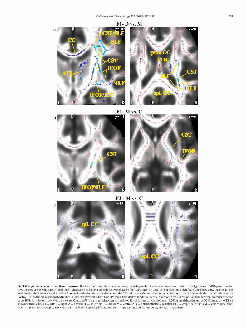
Fig. 3. Group comparisons of directionalmeasures. The left panels illustrate the coronal view; the right panels show the axial view. Coordinates on the figures are inMNI space. 3a— Toprow. Dancers versusMusicians, F1 (red lines). Musicians had higher F1: significant voxels range from dark blue (p b 0.05) to light blue (most significant). Red lines show the orientationsassociatedwith F1 at each voxel. Principal fibres follow the dorsal–ventral direction in the CST regions, and the anterior–posterior direction in the SLF. 3b—Middle row. Musicians versusControls, F1 (red lines).Musicianshad higher F1 (significant voxels in light blue). Principalfibres follow thedorsal–ventral direction in theCST regions, and the anterior–posterior directionin the IFOF. 3c— Bottom row. Musicians versus Controls, F2 (blue lines). Musicians had reduced F2 (pink, here thresholded at p b 0.06) in the right splenium of CC. Orientations of F2 areshown with blue lines. L = left; R = right; A = anterior; P = posterior; D = dorsal; V = ventral. ATR = anterior thalamic radiations; CC= corpus callosum; CST = corticospinal tract;IFOF = inferior fronto-occipital fasciculus; ILF = inferior longitudinal fasciculus; SLF = superior longitudinal fasciculus; and spl. = splenium.
281C. Giacosa et al. / NeuroImage 135 (2016) 273–286

282 C. Giacosa et al. / NeuroImage 135 (2016) 273–286
4. Discussion
4.1. Summary of main findings
This study is the first to examine the differential effects of long-termdance andmusic training onwhitematter (WM) structure. Ourfindingsshow that dancers have increased diffusivity and reduced anisotropy inWM regions, including the CST, the SLF and the CC. In contrast, musi-cians showed reduced diffusivity and a greater proportion of primary fi-bres in similar regions, particularly in the right hemisphere. Crucially,diffusivity measures were related to performance on dance and musictasks that differentiated the groups. Groups were well matched forage, sex, and body-mass index. Further, dancers and musicians hadequal years of experience in their respective disciplines. To better un-derstand the physiological underpinnings of the observed decreases inFA, we examined multiple concurrent diffusivity measures. Based onthese findings, we hypothesise that increased diffusivity in dancersmay be either related to greater heterogeneity of fibre orientationwith-in the above-mentioned tracts, or enhanced coherence of specific tractsin crossing pathways. It is also possible that our findings result from thecombined effects of enhanced and reduced diffusivity in overlappingpathways that are each related to specific aspects of dance training.For musicians, reduced diffusivity is more likely due to increased coher-ence of effector-specific fibre pathways. This suggests that dance andmusic training may produce opposite effects on WM structure. Whole-body dance training may result in greater fanning of fibres connectingdifferent brain regions and/or an increase in crossing fibres. In contrast,musical trainingmay result inmore focussed enhancements of effector-specific pathways.
4.2. The direction of diffusivity and anisotropy in dancers
In the present study, we found reduced anisotropy (FA, MO) and in-creased diffusivity (RD, AD and MD) in widespread WM regions ofdancers in comparison to musicians with similar trends in comparisonto controls. While most research in musicians has reported FA increases(Bengtsson et al., 2005; Han et al., 2009; Halwani et al., 2011; Rüberet al., 2013; Steele et al., 2013), studies in dance and other whole-bodymotor activities, such as gymnastics, have found reduced FA values(Hänggi et al., 2010; Huang et al., 2013; Hummel et al., 2014).
Decreases in FA, particularly in clinical studies, have beeninterpreted as indicating disruption of the organization and integrityof fibres, or damage to myelin sheaths (Werring et al., 1999; Filippiet al., 2001; Concha et al., 2006; Han et al., 2009). However, in the con-text of learning and expertise, other interpretations may be more rele-vant, such as changes in axon diameter, in the fanning of primaryfibres, or in the density and coherence of secondary fibres in crossingfibre regions (Beaulieu, 2002; Mori and Zhang, 2006; Douaud et al.,2009; Douaud et al., 2011; Zatorre et al., 2012). Large axon diametershave been associated with increased RD (Barazany et al., 2009), whichstudies in phantoms have shown can lead to reduced FA (Fieremanset al., 2008). Indeed, large axons are usually less densely packed thansmaller axons (LaMantia and Rakic, 1990; Alexander et al., 2010), leav-ingmore extracellular space between them,which could also contributeto increased RD (Barazany et al., 2009; Beaulieu, 2009). Similarly, highlyfanning tracts that make connections with broader, or even divergent(Kalil and Dent, 2014), cortical regions would have a less coherentorientation than more coherent tracts that connect narrower regions,potentially leading to lower FA and higher RD values (Budde andAnnese, 2013; Chiang et al., 2014; Pasternak et al., 2014; Teipel et al.,2014; Canese et al., 2015). Consistent with this idea, Taubert et al.(2010) attributed reductions in FA related to learning of a whole-bodybalance task to possible increases in crossing fibre. It might also be pos-sible that increased connections with broader cortical regions are phys-iologically underpinned by increased axonal branching. Duringdevelopment more frequent and intense activity between neurons
guides the axons to expand and intensify connections by sproutingand extending collateral branches (Cantallops and Routtenberg, 1999),especially in the CST (Carmel and Martin, 2014). Thus, it is possiblethat long-term intense training might create similar conditions, espe-cially when training starts at a young age.
Another reason why diffusivity might be greater in dancers com-pared to musicians is that many of the regions that differed betweenthe groups are rich in crossing fibres, particularly between the CSTand the SLF. The FA reductions associated with dance training were ac-companied by increases in RD andMD,with only limited changes in AD.This indicates that the observed increases in FA are not due to a decreasealong the principal axis of diffusion (AD), but rather to an increase alongthe transverse axis (RD). Because RD is a combinedmeasure of diffusiv-ity in the two orthogonal directions, it is impossible to know whetherdiffusion is increased in one or both. In regions where many fibrescross each other, MO can be used to discriminate between an effect in-volvingmultiple fibres lying on the same plane, and one occurring alonga specific direction (Douaud et al., 2011). In our study, we observed con-current decreases in MO and FA, suggesting that there is a preferentialplane of diffusivity (Ennis and Kindlmann, 2006). Therefore, the FA de-creases observed in dancers are unlikely to be due to general changes inmyelinisation of axons, which would have symmetrical effects aroundthe principal axis of diffusion, and may rather be attributed to the pro-liferation of secondary fibres.
4.3. Region specific findings
4.3.1. Corticospinal tractIn this study, two of the WM regions where groups diverged most
significantly included the corticospinal tract (CST) and the superior longi-tudinal fasciculus (SLF)where dancers showed increased diffusivity com-pared to musicians. Both of these tracts are part of the sensorimotorsystem. Thefibres of the CST project from the sensorimotor and premotorcortices to themotor-neurons in the spinal cord, and this pathway plays akey role in the control of voluntarymovement (Wakana et al., 2004). TheSLF connects posterior sensory to frontal regions, and is thought to be in-volved in the integration of sensory and motor information for action(Ptak, 2012; Hecht et al., 2013; Rodriguez-Herreros et al., 2015).
The finding of reduced anisotropy in the CST is consistent with theonly previous study in dancers (Hänggi et al., 2010) and with one ingymnasts (Huang et al., 2013). Both dance and gymnastics require theability to execute rapid andprecisewhole-bodymovements, and to rap-idly integrate proprioceptive information, crucially transmitted via theCST. We hypothesize that whole-body dance or gymnastics trainingcould generate intensified connections between widespread sensori-motor areas resulting in increased fibre branching and fanning, andthus higher RD and lower FA values. In contrast, music requires inten-sive training of specific body parts which would be more likely to leadto focal changes inWM structure that appear as local decreases in diffu-sivity. Long-term dance training might result in increased diffusivity inthe CST because of the proliferation of crossing fibres. Studies of expertgymnasts showed that FA was lower in part of the CST (Huang et al.,2013). It is possible that greater diffusivity in this region of the CSTwas due to fibre bundles crossing this region, such as the SLF.Supporting this hypothesis, we also observed reduced MO in the regionof the CST, which suggests that the increased diffusivity lies on the sameplane, such as the plane formed by the CST and the SLF (Douaud et al.,2011). While dance training might enhance crossing connections be-tween widespread cortical regions, music training might reinforce thecoherence of CST principal fibres. Higher FA values have been reportedin the CST of expert musicians which have been shown to be relatedto childhood piano practice and finger tapping performance(Bengtsson et al., 2005; Han et al., 2009; Rüber et al., 2013).
Thus, music and dance expertise might affect WM in two oppositedirections. Indeed, performance on the dance and melody tasks,reflecting expertise, showed opposite relationships with diffusivity

283C. Giacosa et al. / NeuroImage 135 (2016) 273–286
measures: while dance performance was positively associated with dif-fusivity, and negativelywith anisotropy and F1 (Fig. 2B);melody perfor-mance was negatively associated with diffusivity and positively withanisotropy.
4.3.2. Superior longitudinal fasciculusThe SLF is an associative fibre bundle that crosses the CST, travelling
perpendicularly to it for themajority of its length (Wakana et al., 2004).The SLF ismade up of short- and long-range bi-directional fibre bundles,linking the posterior sensory regions to parietal and frontal areas(Catani et al., 2002; Makris et al., 2005; Martino et al., 2013; Kamaliet al., 2014). In particular, three fibre bundles connect the occipitaland parietal lobes to the motor and prefrontal cortices; in addition,the arcuate fasciculus makes connections between the superior tempo-ral and prefrontal cortices (Makris et al., 2005). The first three bundlesare implicated in the regulation of higher aspects of motor behaviour(Makris et al., 2005; Koch et al., 2010), as well as the visuo-spatial as-pects of working memory (Olesen et al., 2003; Nagy et al., 2004;Klingberg, 2006; Vestergaard et al., 2011) and visuo-spatial attention(Hoeft et al., 2007; Chechlacz et al., 2012; Chechlacz et al., 2013; Vallaret al., 2014; Cerami et al., 2015). The arcuate fasciculus is particularlyrelevant for auditory–motor integration necessary for perception andproduction of speech and music (Catani et al., 2005; Oechslin et al.,2009; Lopez-Barroso et al., 2013). In the current study, several compo-nents of the SLF differed between dancers and musicians, especiallythe long-range fronto-parietal connections (Hua et al., 2008) andshort-range fibres within the frontal lobe.
Similar to our findings in dancers, gymnasts showed reduced FAvalues in the SLF compared with non-athletes (Huang et al., 2013).The authors attributed these FA changes to greater axonal diameter inSLF fibres. These changes in the SLF may be related to enhanced visuo-motor integration skills developed with dance training. In support ofthis interpretation, FA in the SLF has been linked to visuo-motor se-quence learning (Tomassini et al., 2011; Steele et al., 2012).
Part of the SLF that connects posterior with frontal GM regions hasbeen shown to be involved in the action observation-executionmatching,or “mirror neuron”, system in humans (Makris et al., 2005; Hecht et al.,2013; Kamali et al., 2014), which is part of the AON. This network is com-posed of sensorimotor regions of the occipital, temporal, parietal, as wellas frontal lobes that respond to the observation of others' actions (Buccinoet al., 2001; Cross et al., 2009b; Grafton, 2009; Caspers et al., 2010). Thissystem has been hypothesised to be critical for dance learning becausedancers typically observe and imitate others in order to learn newmove-ments (Cross et al., 2006). Therefore, greater efficiency of this network indancers might partially explain the reduction of FA that we observed inthe SLF. Indeed, the conduction velocity of fibres is facilitated by largeraxon calibres (Horowitz et al., 2015) that are detectable with DTI bylower values of FA (Fieremans et al., 2008).
Another part of the SLF might convey vestibular responses (Spenaet al., 2006), which are inhibited in dancers and other balance-trainedindividuals (Keller et al., 2012) to reduce destabilizing compensatorymovements in favour of increased visual (Hugel et al., 1999; Hufneret al., 2011; Costa et al., 2013) and proprioceptive information (Jolaet al., 2011;Hutt andRedding, 2014). The attenuationof the vertigo reflexindancers has been explained in termsof the uncoupling between vestib-ular perception and reflex involving an extended network, centredaround the SLF and temporo-parietal WM (Nigmatullina et al., 2015).
One possible explanation for these contrasting results of the FA di-rection in the SLF is that this fibre bundle contains various subcompo-nents that convey specific information (visuo-spatial, vestibular,audio-motor) between different cortical regions (Martino et al., 2013).Each subcomponentmay therefore be differently affected by the variousaspects of dance and, more generally, motor training. Future studieswith tractography may investigate this hypothesis, by subdividing theSLF into its subcomponents and linking them to specific behavioural as-pects of dance training.
4.3.3. Corpus callosumDancers andmusicians also differed in lateral andmedial portions of
the corpus callosum (CC), including the posterior body and splenium,which connect primary sensory and motor cortices (Hofer and Frahm,2006; Wahl et al., 2007). The body of the CC links premotor and senso-rimotor cortices, whereas the splenium links the visual, parietal and au-ditory cortices (Hofer and Frahm, 2006; Knyazeva, 2013). The lateralportions of the CC are crossed by other fibre tracts, including the SLFand the CST. Increased connectivity of these crossing fibres might re-duce the density of fibres in the lateral CC, explaining the reduced FAand augmented RD observed in dancers (Budde and Annese, 2013). Im-portantly, for dancers, in the lateral portions of the CC, RD was in-creased, whereas, in the medial CC, both RD and AD were increased.Because the lateral portions of CC connect many different corticalareas, fibres in this region may tend to fan and be less coherent than fi-bres in the medial CC. Therefore, the higher RD values observed indancers may be explained in terms of increased connections betweensensorimotor regions. Dance and music training differ crucially in theinvolvement of whole-body movements as opposed to effector-specific movements, respectively. Given the somatotopic organisationof sensorimotor cortices, it is possible that dance training enhances con-nections between widespread cortical regions that involve the repre-sentation of the whole body. Conversely, music training may increasethe density and coherence of the fibres that link more limited regionsrepresenting the trained effectors. Indeed, F1 was increased in our sam-ple of musicians, indicating increased coherence of fibres of the CC. Thisinterpretation is further supported by previous studieswheremusiciansshowed higher FA in the genu (Schmithorst andWilke, 2002), body andsplenium (Bengtsson et al., 2005; Steele et al., 2013) of CC.
The interpretation of our concomitant findings for increased RD andAD in themedial CC is more complex. The fact that FA did not differ be-tween groups means that the ratio between diffusivities along the lon-gitudinal direction (AD) and perpendicular to it (RD) is constant. Ithas been shown that anisotropy varies along the midsagittal course ofthe CC (Hofer and Frahm, 2006). Nonetheless, the medial fibres of CCare extremely packed and parallel to each other, thus constituting oneof the most coherent fibre bundles of the brain (Johansen-Berg et al.,2007). Therefore, while higher values of AD in experts are easily attrib-utable to increased myelination, coherence or packaging of fibres, theconcomitant findings for greater RD in dancers are less clear. One possi-bility might be that the enhanced connections between whole-bodycortical representations developed with dance training result in greaterheterogeneity of fibre orientation, and thus less coherent, and lessdensely packed, fibres. This would explain the global increase of theamount of diffusion (MD) that we observed in dancers, irrespective ofthe direction of motion.
4.4. Limitations and future directions
In this paper, we attributed the WM differences observed betweengroups to brain plasticity related to their specific training. However,with a cross-sectional design we cannot exclude the possibility thatthere were pre-existing differences in structure between groups thatmight underlie dance skills and the propensity to undertake training.Longitudinal studies of dance training could allow us to verify whetherWM changes were due to training or pre-existing differences; althoughexpertise is likely to result from the combination of both environmentaland genetic factors.
DTI findings must be interpreted with care due to the intrinsic limi-tations of this technique, especially in regions rich in overlapping path-ways (e.g., the multiple components of the SLF) or crossing fibres(e.g., CST and SLF).We have proposed several plausible macrostructuralfibre configurations that may explain the observed lower FA values indancers, such as reduced coherence due to fanning, crossing or in-creased axonal diameter. Additional analysesmayhelp to specify our re-sults and future studies using techniques, such as tractography

284 C. Giacosa et al. / NeuroImage 135 (2016) 273–286
(Tournier et al., 2004; Tuch, 2004), and the estimation of the axon diam-eter (Assaf et al., 2008), may be useful next steps to validate the inter-pretation of our findings. Furthermore, novel methodologies ofinvestigation might explore alternative physiological explanations,such as modifications of the axonal membrane permeability or of themyelin thickness, that cannot be excluded neither with DTI nortractography.
The present study revealed the most significant results in the con-trast between dancers and musicians rather than in comparison withuntrained subjects. This may be the result of the fact that the highly se-lected dancer andmusician groupsmay bemore homogeneous than thecontrol group, merely selected to have negligible training in both danceand music. Pure and uniform groups of experts, like our samples ofdancers and musicians, may have more extreme and localised values,which are more easily discernible than dispersed ones. In support ofthis interpretation, comparisons between specific groups of musiciansrevealed more evident differences than contrasts with controls (Steeleet al., 2013; Bailey et al., 2014; Vollmann et al., 2014).
5. Conclusions
This study is the first to examine the differential effects of long-termdance and music training on WM structure. Dancers showed increaseddiffusivity in sensorimotor pathways in comparison to musicians, whoshowed greater coherence in the same regions. We propose that inten-sive whole-body dance training may result in greater heterogeneity offibre orientation connecting various brain regions, an increase in cross-ing fibres, or larger axon diameter. In contrast, musical trainingmay re-sult in more focussed enhancements of effector-specific pathways.These findings expand our understanding of brain plasticity literatureby emphasizing that different types of training can have differentlong-term effects on brain structure (Takeuchi et al., 2011; Baer et al.,2015).
Acknowledgements
We would like to thank our participants for their time, Jennifer Bai-ley, Emily Coffey and Jamila Andoh for their assistance in the recruitingand testing process, and Ilana Leppert for technical counselling. Thiswork was funded by a grant from the Natural Sciences and EngineeringCouncil of Canada (NSERC) to Dr. Krista Hyde and Dr. Virginia Penhune(238670); NSERC CREATE in Auditory Cognitive Neuroscience andQuebec Bioimaging Network fellowships to Chiara Giacosa.
Appendix A. Supplementary data
Supplementary data to this article can be found online at http://dx.doi.org/10.1016/j.neuroimage.2016.04.048.
References
Abdul-Kareem, I.A., Stancak, A., Parkes, L.M., Sluming, V., 2011. Increased gray matter vol-ume of left pars opercularis in male orchestral musicians correlate positively withyears of musical performance. J. Magn. Reson. Imaging 33, 24–32.
Alexander, A.L., Lee, J.E., Lazar, M., Field, A.S., 2007. Diffusion tensor imaging of the brain.Neurotherapeutics 4, 316–329.
Alexander, D.C., Hubbard, P.L., Hall, M.G., Moore, E.A., Ptito, M., Parker, G.J., Dyrby, T.B.,2010. Orientationally invariant indices of axon diameter and density from diffusionMRI. NeuroImage 52, 1374–1389.
Assaf, Y., Pasternak, O., 2008. Diffusion tensor imaging (DTI)-based white matter mappingin brain research: a review. J. Mol. Neurosci. 34, 51–61.
Assaf, Y., Blumenfeld-Katzir, T., Yovel, Y., Basser, P.J., 2008. AxCaliber: a method for mea-suring axon diameter distribution from diffusion MRI. Magn. Reson. Med. 59,1347–1354.
Baer, L.H., Park, M.T., Bailey, J.A., Chakravarty, M.M., Li, K.Z., Penhune, V.B., 2015. Regionalcerebellar volumes are related to early musical training and finger tapping perfor-mance. NeuroImage 109, 130–139.
Bailey, J.A., Penhune, V.B., 2010. Rhythm synchronization performance and auditoryworking memory in early- and late-trained musicians. Exp. Brain Res. 204, 91–101.
Bailey, J.A., Zatorre, R.J., Penhune, V.B., 2014. Early musical training is linked to graymatterstructure in the ventral premotor cortex and auditory-motor rhythm synchronizationperformance. J. Cogn. Neurosci. 26, 755–767.
Bangert, M., Peschel, T., Schlaug, G., Rotte, M., Drescher, D., Hinrichs, H., Heinze, H.J.,Altenmuller, E., 2006. Shared networks for auditory and motor processing in profes-sional pianists: evidence from fMRI conjunction. NeuroImage 30, 917–926.
Barazany, D., Basser, P.J., Assaf, Y., 2009. In vivo measurement of axon diameter distribu-tion in the corpus callosum of rat brain. Brain 132, 1210–1220.
Basser, P.J., Pierpaoli, C., 1996. Microstructural and physiological features of tissues eluci-dated by quantitative-diffusion-tensor MRI. J. Magn. Reson. B 111, 209–219.
Basser, P.J., Mattiello, J., LeBihan, D., 1994. MR diffusion tensor spectroscopy and imaging.Biophys. J. 66, 259–267.
Beaulieu, C., 2002. The basis of anisotropic water diffusion in the nervous system — atechnical review. NMR Biomed. 15, 435–455.
Beaulieu C (2009) The biological basis of diffusion anisotropy. In: Diffusion MRI: FromQuantitative Measurement to In-vivo Neuroanatomy (H., J.-B. and E., B. T., eds), pp105–123 London, UK: Academic Press.
Behrens, T.E., Woolrich, M.W., Jenkinson, M., Johansen-Berg, H., Nunes, R.G., Clare, S.,Matthews, P.M., Brady, J.M., Smith, S.M., 2003. Characterization and propagation ofuncertainty in diffusion-weighted MR imaging. Magn. Reson. Med. 50, 1077–1088.
Bengtsson, S.L., Nagy, Z., Skare, S., Forsman, L., Forssberg, H., Ullen, F., 2005. Extensivepiano practicing has regionally specific effects on white matter development. Nat.Neurosci. 8, 1148–1150.
Bermudez, P., Zatorre, R.J., 2005. Differences in gray matter between musicians and non-musicians. Ann. N. Y. Acad. Sci. 1060, 395–399.
Bermudez, P., Lerch, J.P., Evans, A.C., Zatorre, R.J., 2009. Neuroanatomical correlates of mu-sicianship as revealed by cortical thickness and voxel-based morphometry. Cereb.Cortex 19, 1583–1596.
Bezzola, L., Merillat, S., Gaser, C., Jancke, L., 2011. Training-induced neural plasticity in golfnovices. J. Neurosci. 31, 12444–12448.
Bläsing, B., Calvo-Merino, B., Cross, E.S., Jola, C., Honisch, J., Stevens, C.J., 2012. Neurocognitivecontrol in dance perception and performance. Acta Psychol. 139, 300–308.
Brown, S., Martinez, M.J., Parsons, L.M., 2006. The neural basis of human dance. Cereb.Cortex 16, 1157–1167.
Buccino, G., Binkofski, F., Fink, G.R., Fadiga, L., Fogassi, L., Gallese, V., Seitz, R.J., Zilles, K.,Rizzolatti, G., Freund, H.J., 2001. Action observation activates premotor and parietalareas in a somatotopic manner: an fMRI study. Eur. J. Neurosci. 13, 400–404.
Budde, M.D., Annese, J., 2013. Quantification of anisotropy and fiber orientation in humanbrain histological sections. Front. Integr. Neurosci. 7, 3.
Calvo-Merino, B., Glaser, D.E., Grezes, J., Passingham, R.E., Haggard, P., 2005. Action obser-vation and acquired motor skills: an FMRI study with expert dancers. Cereb. Cortex15, 1243–1249.
Calvo-Merino, B., Ehrenberg, S., Leung, D., Haggard, P., 2010. Experts see it all: configuraleffects in action observation. Psychol. Res. 74, 400–406.
Canese, R., Zoratto, F., Altabella, L., Porcari, P., Mercurio, L., de Pasquale, F., Butti, E.,Martino, G., Lacivita, E., Leopoldo, M., Laviola, G., Adriani, W., 2015. Persistent modi-fication of forebrain networks and metabolism in rats following adolescent exposureto a 5-HT7 receptor agonist. Psychopharmacology 232, 75–89.
Cantallops, I., Routtenberg, A., 1999. Activity-dependent regulation of axonal growth:posttranscriptional control of the GAP-43 gene by the NMDA receptor in developinghippocampus. J. Neurobiol. 41, 208–220.
Carmel, J.B., Martin, J.H., 2014. Motor cortex electrical stimulation augments sprouting ofthe corticospinal tract and promotes recovery of motor function. Front. Integr.Neurosci. 8, 51.
Caspers, S., Zilles, K., Laird, A.R., Eickhoff, S.B., 2010. ALE meta-analysis of action observa-tion and imitation in the human brain. NeuroImage 50, 1148–1167.
Catani, M., Howard, R.J., Pajevic, S., Jones, D.K., 2002. Virtual in vivo interactive dissectionof white matter fasciculi in the human brain. NeuroImage 17, 77–94.
Catani, M., Jones, D.K., ffytche, D.H., 2005. Perisylvian language networks of the humanbrain. Ann. Neurol. 57, 8–16.
Cerami, C., Crespi, C., Della Rosa, P.A., Dodich, A., Marcone, A., Magnani, G., Coppi, E., Falini,A., Cappa, S.F., Perani, D., 2015. Brain changeswithin the visuo-spatial attentional net-work in posterior cortical atrophy. J. Alzheimers Dis. 43, 385–395.
Chechlacz, M., Rotshtein, P., Hansen, P.C., Riddoch, J.M., Deb, S., Humphreys, G.W., 2012.The neural underpinings of simultanagnosia: disconnecting the visuospatial attentionnetwork. J. Cogn. Neurosci. 24, 718–735.
Chechlacz, M., Rotshtein, P., Hansen, P.C., Deb, S., Riddoch, M.J., Humphreys, G.W., 2013.The central role of the temporo-parietal junction and the superior longitudinal fascic-ulus in supporting multi-item competition: evidence from lesion-symptom mappingof extinction. Cortex 49, 487–506.
Chen, J.L., Penhune, V.B., Zatorre, R.J., 2008. Listening to musical rhythms recruits motorregions of the brain. Cereb. Cortex 18, 2844–2854.
Chiang, C.W., Wang, Y., Sun, P., Lin, T.H., Trinkaus, K., Cross, A.H., Song, S.K., 2014. Quanti-fying white matter tract diffusion parameters in the presence of increased extra-fibercellularity and vasogenic edema. NeuroImage 101, 310–319.
Coffey, E.B.J., Herholz, S.C., Scala, S., Zatorre, R.J., 2011. The Montreal Music HistoryQuestionnaire: a tool for the assessment of music-related experience in musiccognition research. Neurosciences and Music IV: Learning and Memory, Edin-burgh, UK.
Concha, L., Gross, D.W., Wheatley, B.M., Beaulieu, C., 2006. Diffusion tensor imaging oftime-dependent axonal and myelin degradation after corpus callosotomy in epilepsypatients. NeuroImage 32, 1090–1099.
Costa, M.S.S., Ferreira, A.S., Felicio, L.R., 2013. Static and Dynamic Balance in BalletDancers: A Literature Review 20. Fisioterapia e Pesquisa, p. 7.
Cross, E.S., Hamilton, A.F., Grafton, S.T., 2006. Building a motor simulation de novo: obser-vation of dance by dancers. NeuroImage 31, 1257–1267.
285C. Giacosa et al. / NeuroImage 135 (2016) 273–286
Cross, E.S., Hamilton, A.F., Kraemer, D.J., Kelley,W.M., Grafton, S.T., 2009a. Dissociable sub-strates for body motion and physical experience in the human action observationnetwork. Eur. J. Neurosci. 30, 1383–1392.
Cross, E.S., Kraemer, D.J., Hamilton, A.F., Kelley, W.M., Grafton, S.T., 2009b. Sensitivity ofthe action observation network to physical and observational learning. Cereb. Cortex19, 315–326.
Crotts, D., Thompson, B., Nahom, M., Ryan, S., Newton, R.A., 1996. Balance abilities of pro-fessional dancers on select balance tests. J. Orthop. Sports Phys. Ther. 23, 12–17.
Douaud, G., Behrens, T.E., Poupon, C., Cointepas, Y., Jbabdi, S., Gaura, V., Golestani, N.,Krystkowiak, P., Verny, C., Damier, P., Bachoud-Levi, A.C., Hantraye, P., Remy, P.,2009. In vivo evidence for the selective subcortical degeneration in Huntington's dis-ease. NeuroImage 46, 958–966.
Douaud, G., Jbabdi, S., Behrens, T.E., Menke, R.A., Gass, A., Monsch, A.U., Rao, A., Whitcher,B., Kindlmann, G., Matthews, P.M., Smith, S., 2011. DTI measures in crossing-fibreareas: increased diffusion anisotropy reveals early white matter alteration in MCIand mild Alzheimer's disease. NeuroImage 55, 880–890.
Draganski, B., Gaser, C., Busch, V., Schuierer, G., Bogdahn, U.,May, A., 2004.Neuroplasticity:changes in grey matter induced by training. Nature 427, 311–312.
Driemeyer, J., Boyke, J., Gaser, C., Buchel, C., May, A., 2008. Changes in gray matter inducedby learning — revisited. PLoS One 3, e2669.
Elmer, S., Hanggi, J., Jancke, L., 2014. Interhemispheric transcallosal connectivity betweenthe left and right planum temporale predicts musicianship, performance in temporalspeech processing, and functional specialization. Brain Struct. Funct.
Ennis, D.B., Kindlmann, G., 2006. Orthogonal tensor invariants and the analysis of diffu-sion tensor magnetic resonance images. Magn. Reson. Med. 55, 136–146.
Fadiga, L., Fogassi, L., Pavesi, G., Rizzolatti, G., 1995. Motor facilitation during action obser-vation: a magnetic stimulation study. J. Neurophysiol. 73, 2608–2611.
Fieremans, E., De Deene, Y., Delputte, S., Ozdemir, M.S., Achten, E., Lemahieu, I., 2008. Thedesign of anisotropic diffusion phantoms for the validation of diffusion weightedmagnetic resonance imaging. Phys. Med. Biol. 53, 5405–5419.
Filippi, M., Cercignani, M., Inglese, M., Horsfield, M.A., Comi, G., 2001. Diffusion tensormagnetic resonance imaging in multiple sclerosis. Neurology 56, 304–311.
Foster, N.E., Zatorre, R.J., 2010a. Cortical structure predicts success in performing musicaltransformation judgments. NeuroImage 53, 26–36.
Foster, N.E., Zatorre, R.J., 2010b. A role for the intraparietal sulcus in transforming musicalpitch information. Cereb. Cortex 20, 1350–1359.
Gallese, V., Fadiga, L., Fogassi, L., Rizzolatti, G., 1996. Action recognition in the premotorcortex. Brain 119 (Pt 2), 593–609.
Gallichan, D., Scholz, J., Bartsch, A., Behrens, T.E., Robson,M.D., Miller, K.L., 2010. Addressing asystematic vibration artifact in diffusion-weighted MRI. Hum. Brain Mapp. 31, 193–202.
Gardner, T., Goulden, N., Cross, E.S., 2015. Dynamic modulation of the action observationnetwork by movement familiarity. J. Neurosci. 35, 1561–1572.
Gaser, C., Schlaug, G., 2003. Gray matter differences between musicians and nonmusi-cians. Ann. N. Y. Acad. Sci. 999, 514–517.
Gazzola, V., Keysers, C., 2009. The observation and execution of actions share motor andsomatosensory voxels in all tested subjects: single-subject analyses of unsmoothedfMRI data. Cereb. Cortex 19, 1239–1255.
Golomer, E., Bouillette, A., Mertz, C., Keller, J., 2008. Effects of mental imagery styles onshoulder and hip rotations during preparation of pirouettes. J. Mot. Behav. 40, 281–290.
Golomer, E., Mbongo, F., Toussaint, Y., Cadiou, M., Israel, I., 2010. Right hemisphere in vi-sual regulation of complex equilibrium: the female ballet dancers' experience.Neurol. Res. 32, 409–415.
Grafton, S.T., 2009. Embodied cognition and the simulation of action to understand others.Ann. N. Y. Acad. Sci. 1156, 97–117.
Grèzes, J., Decety, J., 2001. Functional anatomy of execution, mental simulation, observa-tion, and verb generation of actions: a meta-analysis. Hum. Brain Mapp. 12, 1–19.
Groussard, M., Viader, F., Landeau, B., Desgranges, B., Eustache, F., Platel, H., 2014. The ef-fects ofmusical practice on structural plasticity: the dynamics of greymatter changes.Brain Cogn. 90, 174–180.
Gulani, V., Webb, A.G., Duncan, I.D., Lauterbur, P.C., 2001. Apparent diffusion tensor mea-surements in myelin-deficient rat spinal cords. Magn. Reson. Med. 45, 191–195.
Halwani, G.F., Loui, P., Rüber, T., Schlaug, G., 2011. Effects of practice and experience onthe arcuate fasciculus: comparing singers, instrumentalists, and non-musicians.Front. Psychol. 2, 156.
Han, Y., Yang, H., Lv, Y.T., Zhu, C.Z., He, Y., Tang, H.H., Gong, Q.Y., Luo, Y.J., Zang, Y.F., Dong,Q., 2009. Graymatter density andwhitematter integrity in pianists' brain: a combinedstructural and diffusion tensor MRI study. Neurosci. Lett. 459, 3–6.
Hänggi, J., Koeneke, S., Bezzola, L., Jancke, L., 2010. Structural neuroplasticity in the senso-rimotor network of professional female ballet dancers. Hum. Brain Mapp. 31,1196–1206.
Hecht, E.E., Gutman, D.A., Preuss, T.M., Sanchez, M.M., Parr, L.A., Rilling, J.K., 2013. Processversus product in social learning: comparative diffusion tensor imaging of neural sys-tems for action execution-observation matching in macaques, chimpanzees, andhumans. Cereb. Cortex 23, 1014–1024.
Herholz, S.C., Zatorre, R.J., 2012. Musical training as a framework for brain plasticity: be-havior, function, and structure. Neuron 76, 486–502.
Hoeft, F., Barnea-Goraly, N., Haas, B.W., Golarai, G., Ng, D., Mills, D., Korenberg, J., Bellugi,U., Galaburda, A., Reiss, A.L., 2007. More is not always better: increased fractional an-isotropy of superior longitudinal fasciculus associated with poor visuospatial abilitiesin Williams syndrome. J. Neurosci. 27, 11960–11965.
Hofer, S., Frahm, J., 2006. Topography of the human corpus callosum revisited— compre-hensive fiber tractography using diffusion tensor magnetic resonance imaging.NeuroImage 32, 989–994.
Horowitz, A., Barazany, D., Tavor, I., Bernstein, M., Yovel, G., Assaf, Y., 2015. In vivo corre-lation between axon diameter and conduction velocity in the human brain. BrainStruct. Funct. 220, 1777–1788.
Hua, K., Zhang, J., Wakana, S., Jiang, H., Li, X., Reich, D.S., Calabresi, P.A., Pekar, J.J., van Zijl,P.C., Mori, S., 2008. Tract probability maps in stereotaxic spaces: analyses of whitematter anatomy and tract-specific quantification. NeuroImage 39, 336–347.
Huang, R., Lu, M., Song, Z., Wang, J., 2013. Long-term intensive training induced brainstructural changes in world class gymnasts. Brain Struct. Funct. 220, 625–644.
Hufner, K., Binetti, C., Hamilton, D.A., Stephan, T., Flanagin, V.L., Linn, J., Labudda, K.,Markowitsch, H., Glasauer, S., Jahn, K., Strupp, M., Brandt, T., 2011. Structural andfunctional plasticity of the hippocampal formation in professional dancers andslackliners. Hippocampus 21, 855–865.
Hugel, F., Cadopi, M., Kohler, F., Perrin, P., 1999. Postural control of ballet dancers: a spe-cific use of visual input for artistic purposes. Int. J. Sports Med. 20, 86–92.
Hummel, N., Hufner, K., Stephan, T., Linn, J., Kremmyda, O., Brandt, T., Flanagin, V.L., 2014.Vestibular loss and balance training cause similar changes in human cerebral whitematter fractional anisotropy. PLoS One 9, e95666.
Hutt, K., Redding, E., 2014. The effect of an eyes-closed dance-specific training program ondynamic balance in elite pre-professional ballet dancers: a randomized controlledpilot study. J. Dance Med. Sci. 18, 3–11.
Hyde, K.L., Lerch, J., Norton, A., Forgeard, M., Winner, E., Evans, A.C., Schlaug, G., 2009a.The effects of musical training on structural brain development: a longitudinalstudy. Ann. N. Y. Acad. Sci. 1169, 182–186.
Hyde, K.L., Lerch, J., Norton, A., Forgeard, M., Winner, E., Evans, A.C., Schlaug, G., 2009b.Musical training shapes structural brain development. J. Neurosci. 29, 3019–3025.
Imfeld, A., Oechslin, M.S., Meyer, M., Loenneker, T., Jancke, L., 2009. White matter plastic-ity in the corticospinal tract of musicians: a diffusion tensor imaging study.NeuroImage 46, 600–607.
Jäncke, L., Koeneke, S., Hoppe, A., Rominger, C., Hanggi, J., 2009. The architecture of thegolfer's brain. PLoS One 4, e4785.
Jbabdi, S., Behrens, T.E., Smith, S.M., 2010. Crossing fibres in tract-based spatial statistics.NeuroImage 49, 249–256.
Johansen-Berg, H., Della-Maggiore, V., Behrens, T.E., Smith, S.M., Paus, T., 2007. Integrity ofwhite matter in the corpus callosum correlates with bimanual co-ordination skills.NeuroImage 36 (Suppl. 2), T16–T21.
Jola, C., Davis, A., Haggard, P., 2011. Proprioceptive integration and body representation:insights into dancers' expertise. Exp. Brain Res. 213, 257–265.
Jones, D.K., Knosche, T.R., Turner, R., 2013. White matter integrity, fiber count, and otherfallacies: the do's and don'ts of diffusion MRI. NeuroImage 73, 239–254.
Kalil, K., Dent, E.W., 2014. Branchmanagement: mechanisms of axon branching in the de-veloping vertebrate CNS. Nat. Rev. Neurosci. 15, 7–18.
Kamali, A., Flanders, A.E., Brody, J., Hunter, J.V., Hasan, K.M., 2014. Tracing superior longi-tudinal fasciculus connectivity in the human brain using high resolution diffusiontensor tractography. Brain Struct. Funct. 219, 269–281.
Karpati, F.J., Giacosa, C., Foster, N.E., Penhune, V.B., Hyde, K.L., 2015a. Dance and the brain:a review. Ann. N. Y. Acad. Sci. 1337, 140–146.
Karpati, F.J., Giacosa, C., Foster, N.E., Penhune, V.B., Hyde, K.L., 2015b. Sensorimotor inte-gration is enhanced in dancers and musicians. Exp. Brain Res.
Kattenstroth, J.C., Kalisch, T., Kolankowska, I., Dinse, H.R., 2011. Balance, sensorimotor, andcognitive performance in long-year expert senior ballroom dancers. J. Aging Res.2011, 176709.
Keller, T.A., Just, M.A., 2009. Altering cortical connectivity: remediation-induced changesin the white matter of poor readers. Neuron 64, 624–631.
Keller, M., Pfusterschmied, J., Buchecker, M., Muller, E., Taube,W., 2012. Improved postur-al control after slackline training is accompanied by reduced H-reflexes. Scand.J. Med. Sci. Sports 22, 471–477.
Keysers, C., Gazzola, V., 2009. Expanding the mirror: vicarious activity for actions, emo-tions, and sensations. Curr. Opin. Neurobiol. 19, 666–671.
Kiefer, A.W., Riley, M.A., Shockley, K., Sitton, C.A., Hewett, T.E., Cummins-Sebree, S., Haas,J.G., 2011. Multi-segmental postural coordination in professional ballet dancers. GaitPosture 34, 76–80.
Klingberg, T., 2006. Development of a superior frontal-intraparietal network for visuo-spatial working memory. Neuropsychologia 44, 2171–2177.
Knyazeva, M.G., 2013. Splenium of corpus callosum: patterns of interhemispheric interac-tion in children and adults. Neural Plast. 2013, 639430.
Koch, G., Cercignani, M., Pecchioli, C., Versace, V., Oliveri, M., Caltagirone, C., Rothwell, J.,Bozzali, M., 2010. In vivo definition of parieto-motor connections involved in plan-ning of grasping movements. NeuroImage 51, 300–312.
Krüger, B., Bischoff, M., Blecker, C., Langhanns, C., Kindermann, S., Sauerbier, I., Reiser, M.,Stark, R., Munzert, J., Pilgramm, S., 2014. Parietal and premotor cortices: activation re-flects imitation accuracy during observation, delayed imitation and concurrent imita-tion. NeuroImage 100, 39–50.
Lahav, A., Saltzman, E., Schlaug, G., 2007. Action representation of sound: audiomotor rec-ognition network while listening to newly acquired actions. J. Neurosci. 27, 308–314.
LaMantia, A.S., Rakic, P., 1990. Axon overproduction and elimination in the corpuscallosum of the developing rhesus monkey. J. Neurosci. 10, 2156–2175.
Landmann, C., Landi, S.M., Grafton, S.T., Della-Maggiore, V., 2011. FMRI supports the sen-sorimotor theory of motor resonance. PLoS One 6, e26859.
Lopez-Barroso, D., Catani, M., Ripolles, P., Dell'Acqua, F., Rodriguez-Fornells, A., de Diego-Balaguer, R., 2013.Word learning ismediated by the left arcuate fasciculus. Proc. Natl.Acad. Sci. U. S. A. 110, 13168–13173.
Maguire, E.A., Gadian, D.G., Johnsrude, I.S., Good, C.D., Ashburner, J., Frackowiak, R.S., Frith,C.D., 2000. Navigation-related structural change in the hippocampi of taxi drivers.Proc. Natl. Acad. Sci. U. S. A. 97, 4398–4403.
Makris, N., Kennedy, D.N., McInerney, S., Sorensen, A.G., Wang, R., Caviness Jr., V.S.,Pandya, D.N., 2005. Segmentation of subcomponents within the superior longitudinalfascicle in humans: a quantitative, in vivo, DT-MRI study. Cereb. Cortex 15, 854–869.
Martino, J., De Witt Hamer, P.C., Berger, M.S., Lawton, M.T., Arnold, C.M., de Lucas, E.M.,Duffau, H., 2013. Analysis of the subcomponents and cortical terminations of the

286 C. Giacosa et al. / NeuroImage 135 (2016) 273–286
perisylvian superior longitudinal fasciculus: a fiber dissection and DTI tractographystudy. Brain Struct. Funct. 218, 105–121.
Minvielle-Moncla, J., Audiffren, M., Macar, F., Vallet, C., 2008. Overproduction timing er-rors in expert dancers. J. Mot. Behav. 40, 291–300.
Moore, E., Schaefer, R.S., Bastin, M.E., Roberts, N., Overy, K., 2014. Can musical training in-fluence brain connectivity? Evidence from diffusion tensorMRI. Brain Sci. 4, 405–427.
Mori, S., Zhang, J., 2006. Principles of diffusion tensor imaging and its applications to basicneuroscience research. Neuron 51, 527–539.
Moseley, M.E., Cohen, Y., Kucharczyk, J., Mintorovitch, J., Asgari, H.S., Wendland, M.F.,Tsuruda, J., Norman, D., 1990. Diffusion-weighted MR imaging of anisotropic waterdiffusion in cat central nervous system. Radiology 176, 439–445.
Nagy, Z., Westerberg, H., Klingberg, T., 2004. Maturation of white matter is associatedwith the development of cognitive functions during childhood. J. Cogn. Neurosci.16, 1227–1233.
Neil, J.J., Shiran, S.I., McKinstry, R.C., Schefft, G.L., Snyder, A.Z., Almli, C.R., Akbudak, E.,Aronovitz, J.A., Miller, J.P., Lee, B.C., Conturo, T.E., 1998. Normal brain in human new-borns: apparent diffusion coefficient and diffusion anisotropymeasured by using dif-fusion tensor MR imaging. Radiology 209, 57–66.
Nigmatullina, Y., Hellyer, P.J., Nachev, P., Sharp, D.J., Seemungal, B.M., 2015. The neuroan-atomical correlates of training-related perceptuo-reflex uncoupling in dancers. Cereb.Cortex 25, 554–562.
Oechslin, M.S., Imfeld, A., Loenneker, T., Meyer, M., Jancke, L., 2009. The plasticity of thesuperior longitudinal fasciculus as a function of musical expertise: a diffusion tensorimaging study. Front. Hum. Neurosci. 3, 76.
Olesen, P.J., Nagy, Z., Westerberg, H., Klingberg, T., 2003. Combined analysis of DTI andfMRI data reveals a joint maturation ofwhite and greymatter in a fronto-parietal net-work. Brain Res. Cogn. Brain Res. 18, 48–57.
Ono, Y., Nomoto, Y., Tanaka, S., Sato, K., Shimada, S., Tachibana, A., Bronner, S., Noah, J.A.,2014. Frontotemporal oxyhemoglobin dynamics predict performance accuracy ofdance simulation gameplay: temporal characteristics of top-down and bottom-upcortical activities. NeuroImage 85 (Pt 1), 461–470.
Pasternak, O., Koerte, I.K., Bouix, S., Fredman, E., Sasaki, T., Mayinger, M., Helmer, K.G.,Johnson, A.M., Holmes, J.D., Forwell, L.A., Skopelja, E.N., Shenton, M.E., Echlin, P.S.,2014. Hockey concussion education project, part 2. Microstructural white matter al-terations in acutely concussed ice hockey players: a longitudinal free-water MRIstudy. J. Neurosurg. 120, 873–881.
Poon, P.P., Rodgers, W.M., 2000. Learning and remembering strategies of novice and ad-vanced jazz dancers for skill level appropriate dance routines. Res. Q. Exerc. Sport71, 135–144.
Ptak, R., 2012. The frontoparietal attention network of the human brain: action, saliency,and a priority map of the environment. Neuroscientist 18, 502–515.
Rizzolatti, G., Craighero, L., 2004. The mirror-neuron system. Annu. Rev. Neurosci. 27,169–192.
Rizzolatti, G., Fadiga, L., Gallese, V., Fogassi, L., 1996a. Premotor cortex and the recognitionof motor actions. Brain Res. Cogn. Brain Res. 3, 131–141.
Rizzolatti, G., Fadiga, L., Matelli, M., Bettinardi, V., Paulesu, E., Perani, D., Fazio, F., 1996b.Localization of grasp representations in humans by PET: 1. Observation versus execu-tion. Exp. Brain Res. 111, 246–252.
Rodriguez-Herreros, B., Amengual, J.L., Gurtubay-Antolin, A., Richter, L., Jauer, P.,Erdmann, C., Schweikard, A., Lopez-Moliner, J., Rodriguez-Fornells, A., Munte, T.F.,2015. Microstructure of the superior longitudinal fasciculus predicts stimulation-induced interference with on-line motor control. NeuroImage 120, 254–265.
Rüber, T., Lindenberg, R., Schlaug, G., 2013. Differential adaptation of descending motortracts in musicians. Cereb. Cortex.
Schlaug, G., 2015. Musicians andmusicmaking as amodel for the study of brain plasticity.Prog. Brain Res. 217, 37–55.
Schlaug, G., Jancke, L., Huang, Y., Staiger, J.F., Steinmetz, H., 1995. Increased corpuscallosum size in musicians. Neuropsychologia 33, 1047–1055.
Schlaug, G., Forgeard, M., Zhu, L., Norton, A., Winner, E., 2009. Training-inducedneuroplasticity in young children. Ann. N. Y. Acad. Sci. 1169, 205–208.
Schmithorst, V.J., Wilke, M., 2002. Differences in white matter architecture between musi-cians and non-musicians: a diffusion tensor imaging study. Neurosci. Lett. 321, 57–60.
Sevdalis, V., Keller, P.E., 2011. Captured by motion: dance, action understanding, and so-cial cognition. Brain Cogn. 77, 231–236.
Sluming, V., Barrick, T., Howard, M., Cezayirli, E., Mayes, A., Roberts, N., 2002. Voxel-basedmorphometry reveals increased gray matter density in Broca's area in male sympho-ny orchestra musicians. NeuroImage 17, 1613–1622.
Smith, S.M., 2002. Fast robust automated brain extraction. Hum. BrainMapp. 17, 143–155.Smith, S.M., Jenkinson, M., Woolrich, M.W., Beckmann, C.F., Behrens, T.E., Johansen-Berg,
H., Bannister, P.R., De Luca, M., Drobnjak, I., Flitney, D.E., Niazy, R.K., Saunders, J.,Vickers, J., Zhang, Y., De Stefano, N., Brady, J.M., Matthews, P.M., 2004. Advances infunctional and structural MR image analysis and implementation as FSL. NeuroImage23 (Suppl. 1), S208–S219.
Smith, S.M., Jenkinson, M., Johansen-Berg, H., Rueckert, D., Nichols, T.E., Mackay, C.E.,Watkins, K.E., Ciccarelli, O., Cader, M.Z., Matthews, P.M., Behrens, T.E., 2006. Tract-based spatial statistics: voxelwise analysis of multi-subject diffusion data. NeuroImage31, 1487–1505.
Spena, G., Gatignol, P., Capelle, L., Duffau, H., 2006. Superior longitudinal fasciculus sub-serves vestibular network in humans. Neuroreport 17, 1403–1406.
Steele, C.J., Scholz, J., Douaud, G., Johansen-Berg, H., Penhune, V.B., 2012. Structural corre-lates of skilled performance on a motor sequence task. Front. Hum. Neurosci. 6, 289.
Steele, C.J., Bailey, J.A., Zatorre, R.J., Penhune, V.B., 2013. Early musical training and white-matter plasticity in the corpus callosum: evidence for a sensitive period. J. Neurosci.33, 1282–1290.
Tachibana, A., Noah, J.A., Bronner, S., Ono, Y., Onozuka, M., 2011. Parietal and temporal ac-tivity during a multimodal dance video game: an fNIRS study. Neurosci. Lett. 503,125–130.
Takeuchi, H., Taki, Y., Sassa, Y., Hashizume, H., Sekiguchi, A., Fukushima, A., Kawashima, R.,2011.Working memory training using mental calculation impacts regional gray mat-ter of the frontal and parietal regions. PLoS One 6, e23175.
Taubert, M., Draganski, B., Anwander, A., Muller, K., Horstmann, A., Villringer, A., Ragert,P., 2010. Dynamic properties of human brain structure: learning-related changes incortical areas and associated fiber connections. J. Neurosci. 30, 11670–11677.
Teipel, S.J., Grothe,M.J., Filippi,M., Fellgiebel, A., Dyrba,M., Frisoni, G.B.,Meindl, T., Bokde, A.L.,Hampel, H., Kloppel, S., Hauenstein, K., 2014. Fractional anisotropy changes inAlzheimer's disease depend on the underlyingfiber tract architecture: amultiparametricDTI study using joint independent component analysis. J. Alzheimers Dis. 41, 69–83.
Tomassini, V., Jbabdi, S., Kincses, Z.T., Bosnell, R., Douaud, G., Pozzilli, C., Matthews, P.M.,Johansen-Berg, H., 2011. Structural and functional bases for individual differencesin motor learning. Hum. Brain Mapp. 32, 494–508.
Tournier, J.D., Calamante, F., Gadian, D.G., Connelly, A., 2004. Direct estimation of the fiberorientation density function from diffusion-weighted MRI data using sphericaldeconvolution. NeuroImage 23, 1176–1185.
Tuch, D.S., 2004. Q-ball imaging. Magn. Reson. Med. 52, 1358–1372.Vallar, G., Bello, L., Bricolo, E., Castellano, A., Casarotti, A., Falini, A., Riva, M., Fava, E.,
Papagno, C., 2014. Cerebral correlates of visuospatial neglect: a direct cerebral stimu-lation study. Hum. Brain Mapp. 35, 1334–1350.
Vestergaard, M., Madsen, K.S., Baare, W.F., Skimminge, A., Ejersbo, L.R., Ramsoy, T.Z.,Gerlach, C., Akeson, P., Paulson, O.B., Jernigan, T.L., 2011.White matter microstructurein superior longitudinal fasciculus associated with spatial working memory perfor-mance in children. J. Cogn. Neurosci. 23, 2135–2146.
Vicary, S.A., Robbins, R.A., Calvo-Merino, B., Stevens, C.J., 2014. Recognition of dance-likeactions: memory for static posture or dynamic movement? Mem. Cogn. 42, 755–767.
Vollmann, H., Ragert, P., Conde, V., Villringer, A., Classen, J., Witte, O.W., Steele, C.J., 2014.Instrument specific use-dependent plasticity shapes the anatomical properties of thecorpus callosum: a comparison between musicians and non-musicians. Front. Behav.Neurosci. 8, 245.
Wahl, M., Lauterbach-Soon, B., Hattingen, E., Jung, P., Singer, O., Volz, S., Klein, J.C., Steinmetz,H., Ziemann, U., 2007. Human motor corpus callosum: topography, somatotopy, andlink between microstructure and function. J. Neurosci. 27, 12132–12138.
Wakana, S., Jiang, H., Nagae-Poetscher, L.M., van Zijl, P.C., Mori, S., 2004. Fiber tract-basedatlas of human white matter anatomy. Radiology 230, 77–87.
Walhovd, K.B., Johansen-Berg, H., Karadottir, R.T., 2014. Unraveling the secrets of whitematter–bridging the gap between cellular, animal and human imaging studies. Neu-roscience 276, 2–13.
Waterhouse, E., Watts, R., Blasing, B.E., 2014. Doing duo — a case study of entrainment inWilliam Forsythe's choreography “duo”. Front. Hum. Neurosci. 8, 812.
Wechsler, D., 1997. Wechsler Adult Intelligence Scale. 3rd Edition. Harcourt Assessment,San Antonio, TX.
Wechsler, D., 1999. Wechsler Abbreviated Scale of Intelligence. The Psychological Corpo-ration: Harcourt Brace & Company, New York, NY.
Werring, D.J., Clark, C.A., Barker, G.J., Thompson, A.J., Miller, D.H., 1999. Diffusion tensorimaging of lesions and normal-appearing white matter in multiple sclerosis. Neurol-ogy 52, 1626–1632.
Wheeler-Kingshott, C.A., Cercignani, M., 2009. About “axial” and “radial” diffusivities.Magn. Reson. Med. 61, 1255–1260.
Winkler, A.M., Ridgway, G.R., Webster, M.A., Smith, S.M., Nichols, T.E., 2014. Permutationinference for the general linear model. NeuroImage 92, 381–397.
Woolhouse, M.H., Lai, R., 2014. Traces across the body: influence of music-dance synchro-ny on the observation of dance. Front. Hum. Neurosci. 8, 965.
Zatorre, R.J., Chen, J.L., Penhune, V.B., 2007. When the brain plays music: auditory–motorinteractions in music perception and production. Nat. Rev. Neurosci. 8, 547–558.
Zatorre, R.J., Fields, R.D., Johansen-Berg, H., 2012. Plasticity in gray and white: neuroimag-ing changes in brain structure during learning. Nat. Neurosci. 15, 528–536.

Reproduced with permission of the copyright owner. Further reproduction prohibited withoutpermission.