Embed Size (px)
Citation preview

Damage to White Matter Pathways in Subacute and Chronic Spatial Neglect: A GroupStudy and 2 Single-Case Studies with Complete Virtual “In Vivo” Tractography Dissection
Michel Thiebaut de Schotten1,2, Francesco Tomaiuolo3, Marilena Aiello5, Sheila Merola5, Massimo Silvetti5,7, Francesca Lecce5,Paolo Bartolomeo2,4 and Fabrizio Doricchi5,6
1Natbrainlab, Department of Forensic and Neurodevelopmental Sciences, Institute of Psychiatry, King’s College London, London,UK, 2Inserm U975; UPMC-Paris6, UMR_S 975; CNRS UMR 7225, Centre de Recherche de l'Institut du Cerveau et de la Moelleépinière, Groupe Hospitalier Pitié-Salpêtrière, 75013 , Paris, France, 3Auxilium Vitae, USL 5, Pisa, Italy, 4Department ofPsychology, Catholic University, Milan, Italy, 5Fondazione Santa Lucia IRCCS, Roma, Italy, 6Dipartimento di Psicologia 39,Università degli Studi di Roma “La Sapienza”, Roma, Italy and 7Gent University, Gent, Belgium
Address correspondence to Fabrizio Doricchi, PhD, Dipartimento di Psicologia 39, Università degli Studi di Roma “La Sapienza”, Via dei Marsi78, 00185 Roma, Italy; Fondazione Santa Lucia IRCCS, Via Ardeatina 306, 00179 Roma, Italy. Email: [email protected]
The exact anatomical localization of right hemisphere lesions thatlead to left spatial neglect is still debated. The effect of confound-ing factors such as acute diaschisis and hypoperfusion, visual fielddefects, and lesion size may account for conflicting results thathave been reported in the literature. Here, we present a comprehen-sive anatomical investigation of the gray- and white matter lesioncorrelates of left spatial neglect, which was run in a sample 58patients with subacute or chronic vascular strokes in the territoryof the right middle cerebral artery. Standard voxel-based correlatesconfirmed the role played by lesions in the posterior parietal cortex(supramarginal gyrus, angular gyrus, and temporal–parietal junc-tion), in the frontal cortex (frontal eye field, middle and inferiorfrontal gyrus), and in the underlying parietal–frontal white matter.Using a new diffusion tensor imaging-based atlas of the humanbrain, we were able to run, for the first time, a detailed analysis ofthe lesion involvement of subcortical white matter pathways. Theresults of this analysis revealed that, among the different pathwayslinking parietal with frontal areas, damage to the second branch ofthe superior longitudinal fasciculus (SLF II) was the best predictorof left spatial neglect. The group study also revealed a subsampleof patients with neglect due to focal lesion in the lateral–dorsalportion of the thalamus, which connects the premotor cortex withthe inferior parietal lobule. The relevance of fronto-parietal discon-nection was further supported by complete in vivo tractography dis-section of white matter pathways in 2 patients, one with and theother without signs of neglect. These 2 patients were studied both inthe acute phase and 1 year after stroke and were perfectly matchedfor age, handedness, stroke onset, lesion size, and for cortical lesioninvolvement. Taken together, the results of the present study supportthe hypothesis that anatomical disconnections leading to a functionalbreakdown of parietal–frontal networks are an important pathophy-siological factor leading to chronic left spatial neglect. Here, wepropose that different loci of SLF disconnection on the rostro-caudalaxis can also be associated with disconnection of short-range whitematter pathways within the frontal or parietal areas. Such differentlocal disconnection patterns can play a role in the important clinicalvariability of the neglect syndrome.
Keywords: anatomy, diffusion tensor imaging, human, spatial neglect, stroke
Introduction
In humans, spatial neglect is a debilitating neurological con-dition that is frequently associated with stroke in the righthemisphere (Gainotti et al. 1972; Oxbury et al. 1974;
Weintraub and Mesulam 1987). Patients with neglect havereduced or no attention to events occurring in the contrale-sional side of extrapersonal (i.e. visual and auditory), per-sonal (i.e. somatosensory), or representational (i.e. imageryspace, Mesulam 1981) space and may fail to produce move-ments toward the unattended space (i.e. motor neglect). Thecognitive resources of neglect patients are pathologicallybiased toward the ipsilesional right side of space, so thatevents taking place in the contralesional left side of space failto receive full cognitive processing and to reach patients’awareness. Based on this acquired lack of awareness for oneside of space, the study of the anatomical correlates of spatialneglect is considered an important source of evidence for theunderstanding of the anatomical foundation of consciousnessand attention.
Since original seminal case reports (Brain 1941; Patersonand Zangwill 1944; Mcfie et al. 1950), spatial neglect has beenclassically interpreted as a “parietal sign” (Critchley 1953;Vallar and Perani 1986). However, ensuing investigationspointed out that spatial neglect can also arise from damage tofrontal areas (Heilman and Valenstein 1972; Damasio et al.1980; Mesulam 1981; Stein and Volpe 1983; Husain andKennard 1996 1997; Husain et al. 2000) to the thalamus(Watson and Heilman 1979; Cambier et al. 1980; Watson et al.1981; Graff-Radford et al. 1985; Hirose et al. 1985; Bogous-slavsky et al. 1986; Motomura et al. 1986; Vallar and Perani1986; Waxman et al. 1986; Rafal and Posner 1987; Kumralet al. 1995; Chung et al. 1996; Leibovitch et al. 1998; Karussiset al. 2000; Karnath et al. 2002; De Witte et al. 2008) and tosubcortical gray matter nuclei. Due to the diffuse connectionsof subcortical gray matter nuclei with the cortical mantle, inthis latter case, spatial neglect has been interpreted as orig-inating from widespread reduction in the functioning of parie-tal and frontal areas connected with damaged subcorticalnuclei. (Hier et al. 1977; Damasio et al. 1980; Healton et al.1982; Stein and Volpe 1983; Ferro et al. 1987; Perani et al.1987; Caplan et al. 1990; Weiller et al. 1990; Donnan et al.1991; Weiller et al. 1993; Kumral et al. 1995; Chung et al.1996; Leibovitch et al. 1998; Karnath et al. 2002).
More recently, anatomical (Leibovitch et al. 1998; Doricchiand Tomaiuolo 2003; Bartolomeo et al. 2007), neurosurgical(Thiebaut de Schotten et al. 2005; Shinoura et al. 2009) andfunctional magnetic resonance imaging (fMRI) investigations(He et al. 2007) have provided converging evidence,suggesting that disconnection of long-range white matterfiber bundles connecting parietal with frontal areas has a
© The Author 2012. Published by Oxford University Press. All rights reserved. For Permissions, please e-mail: [email protected]
Cerebral Cortexdoi:10.1093/cercor/bhs351
Cerebral Cortex Advance Access published November 15, 2012 at IN
SER
M on D
ecember 17, 2012
http://cercor.oxfordjournals.org/D
ownloaded from
relevant role in engendering severe and chronic signs ofspatial neglect both in humans and in monkeys (Gaffan andHornak 1997). These findings provided new vigor to pioneer-ing views by Critchley (1953), Geschwind (1965), andMesulam (1981), who suggested that enduring lack of aware-ness for the left side of space can be influenced in a substan-tial way by damage to white matter connections in the righthemisphere.
One relevant and well-known clinical feature of the spatialneglect syndrome is its reduction in severity and incidence inthe chronic when compared with the acute phase of thestroke. Although in the acute phase up to two-thirds of theright brain damaged patients can show spatial neglect signs,the majority of these patients show spontaneous recovery inthe post-acute and chronic phase (Stone et al. 1992; Corbettaet al. 2005). In a recent report, using a cytoarchitectonic atlasbased on postmortem staining of the myelin of coronal slicesof the human brain (Bürgel et al. 2006), Karnath et al. (2009)concluded that damage of gray matter structures is a strongerpredictor of acute spatial neglect than associative tract discon-nection. However, these authors also commented that, in theacute phase, even small lesions can lead to temporary pro-found cerebral dysfunctions of large brain areas throughmechanisms such as hypometabolism (Yasaka et al. 1998),hypoperfusion (Hillis et al. 2000), or acute diaschisis(Monakow 1914; Kempler et al. 1988; Price et al. 2001; Fingeret al. 2004). Thus, the authors concluded that their studycannot decide whether or not spatial neglect should best beinterpreted as a “disconnection syndrome” (Geschwind 1965;Watson et al. 1974; Mesulam and Geschwind 1978; Watsonet al. 1978; Bartolomeo 2007; Doricchi et al. 2008) andfurther recommended conducting the same investigation witha large group of patients who have chronic injury.
A few case studies run with direct diffusion tensor imaging(DTI) tracking of white matter pathways have alreadysuggested that fronto-parietal (Shinoura et al. 2009; Ciaraffaet al. 2012) or fronto-occipital disconnection (Urbanski et al.2008 2011) can be a strong predictor of spatial neglect.However, the role of white matter disconnection in chronicspatial neglect has never been formally tested in large groupof patients by using adequate statistical procedures to mapthe lesion involvement of relevant white matter tracts. Theaim of the present study was therefore to evaluate and con-trast the respective contributions of gray- and white matterdamage to chronic spatial neglect in an adequately largesample of right brain damaged patients.
Studies of the effect of brain lesions on behavior frequentlyassume that discrete anatomical modules handle specific cog-nitive functions (Rorden and Karnath 2004). Consequently,analyses to determine the behavioral implications of brainlesions are performed via statistical tests completed indepen-dently at each of the intersubjects aligned voxel (voxel-wiseor topological statistics), without any attempt to capture corre-lations across them (Catani 2007; Husain and Nachev 2007).However, 2 voxels should be considered as correlated if theyshare common anatomical features. While 2 distant voxelscannot share the same neuronal soma, several voxels canpertain to the same axon. Therefore, while the architecture ofthe cortex and the subcortical nuclei is well suited for voxel-wise statistics, the study of white matter organization requiresadapted statistics at the tract level (track-wise or hodologicalstatistics; Catani and Mesulam 2008; Rudrauf et al. 2008;
Thiebaut de Schotten et al. 2008). Track-wise statistics requireanatomical knowledge on how voxels in the white matter areassociated in common pathways.
Several atlases make this anatomical information now avail-able, but each of them has both advantages and disadvan-tages. Tracing studies in monkeys provide astonishing detailsof the white matter organization (Schmahmann and Pandya2006); however, the precise identification of the homologousstructures in human is not straightforward (Thiebaut de Schot-ten et al. 2012). The human postmortem atlas (Bürgel et al.2006), used by Karnath et al. (2009), offers high-quality resol-ution of white matter tracts running parallel to coronal slicesused for myelin staining such as the cortico-spinal tract;however, as stressed by Bürgel et al. (2006), this techniquestrongly underestimates parietal–frontal association pathwaysthat run orthogonally to coronal slices (Bürgel et al. 2006;Thiebaut de Schotten, ffytche, et al. 2011). Importantly, suchtracts include some of the crucial pathways whose damagemight be important to spatial neglect, such as the superiorlongitudinal fasciculus (SLF) and the inferior fronto-occipitalfasciculus (IFOF). To circumvent these limitations, in thepresent study, we used an atlas of white matter connectionsbased on DTI dissection of the human brain. Despite somelimitation in the resolution of tracts that cross between eachother (Jones 2008; Dell’acqua et al. 2010), DTI atlases providea representative description of the anatomy and variability ofthe association tracts in the human living brain, giving the op-portunity to measure in vivo white matter pathways location,asymmetry and intersubject variability on large populations ofpatients (Catani et al. 2007; Lawes et al. 2008; Mori et al.2008; Thiebaut de Schotten, ffytche, et al. 2011).
To investigate the role of gray- and white matter damage inspatial neglect, we used voxel-wise and track-wiseregressions. Since it has been suggested that the chronicityand severity of spatial neglect can increase with the size ofthe brain damage (Levine et al. 1986; Perani et al. 1987), aswell as with the presence of an associated visual field defect(Halligan et al. 1990; Doricchi and Angelelli, 1999; Doricchiet al. 2005), we covaried both of these factors out from theanalyses.
To summarize, in a first step, we used a classical voxellesion symptom mapping (VLSM) approach in order to allowa direct comparison with data previously gathered fromsamples of acute patients (Karnath et al. 2009). In a secondstep, we employed regression to reveal the location of thebrain lesions and the pattern of disconnection that correlatewith the occurrence of chronic spatial neglect; right deviationin line bisection and omissions in cancellation tasks covaryingout the effect of the lesion size and the presence of an associ-ated visual field defect. Gray matter correlates were studiedusing a voxel-wise approach that better fits the architecture ofthe cerebral cortex and the subcortical nuclei. White mattercorrelates were studied by using a track-wise statistical ap-proach that takes into consideration how different voxels areassociated along the same white matter pathway.
During the completion of the group study, we had therather unique opportunity of performing a complete tractogra-phy dissection of white matter fiber tracts in 2 patients whowere perfectly matched on all relevant clinical variables (i.e.lesion size, lesion location, stroke onset, sex, age, handed-ness, educational level), except for the presence or absence ofspatial neglect both in the acute and chronic phase. As it will
2 Damage to White Matter Pathways in Spatial Neglect • Thiebaut de Schotten et al.
at INSE
RM
on Decem
ber 17, 2012http://cercor.oxfordjournals.org/
Dow
nloaded from

be shown in the following, direct and complete tractographydissection of these 2 cases added further evidence convergingwith the main results from the group study.
Materials and Methods
Group Study
ParticipantsFifty-eight patients with a vascular stroke in the territory of themiddle cerebral artery (MCA) in the right hemisphere participated inthis study. Written and informed consent to participate to this re-search was obtained. The ethics committee of the Santa Lucia Hospi-tal in Rome, Italy approved the study. Chronic visual field defectswere assessed by Goldmann perimetry. Seven patients were found tohave chronic visual field defects. Left spatial neglect was assessedwith the line bisection test (5 trials, line length = 200 mm, cut-offscore +6.5 mm; Azouvi et al. 2002) and the letter cancellation task(Diller et al. 1974; cut-off score left minus right omissions = >4; Pizza-miglio et al. 1989). All task stimuli were presented on a horizontallyoriented A4 paper sheet. Thirty-eight patients were found to havevarying degrees of left unilateral neglect (N+) on both tasks, whereas20 patients were free from neglect signs (N−). The 2 groups werecomparable for age (t(56) = 0.9, P = 0.3) and time of stroke onset(t(56) = 1.8, P = 0.18). As expected, N+ had a larger ipsilesional biasboth in line bisection (t(56) = 5.1, P < 0.0001) and letter cancellation(t(56) = 7.6, P < 0.0001) than N−. On average, lesion size was higher inN+ when compared with N− (t(56) = 4, P < 0.001). Clinical and demo-graphic data are reported in Table 1.
Individual Lesion MappingTen of the 38 N+ patients and 5 of the 20 N− patients had CT scanexamination. The remaining patients underwent radiological 1.5-TMRI examination, including T1, T2, fluid attenuated inversion recov-ery, and diffusion images. Radiological examination was run within10 days from the time of neuropsychological testing. Mapping oflesions was performed using DISPLAY (http://packages.bic.mni.mcgill.ca). For each patient, we rotated the MNI template (MontrealNeurological Institute, MNI) from the MNI space to the orientation ofthe patients individual clinical MRI or CT slices scan. The lesion wasthen drawn on the reoriented template by an expert neuroanatomistand subsequently taken back to the MNI space using the inverserotation (Doricchi and Tomaiuolo 2003).
General Voxel Lesion Symptom Mapping Analysis (Classic Approach)In a first step, we used a classical VLSM approach in order to allow adirect comparison with data previously gathered from samples ofacute patients (Karnath et al. 2009). We used MRIcron (www.sph.sc.edu/comd/rorden/mricron) to compute a Brunner Munzel testbetween the lesions of the spatial neglect group and the group ofpatient without spatial neglect for each voxel of the brain (Rordenet al. 2007). The results are corrected for multiple comparisons with afalse discovery rate threshold (Genovese et al. 2002) and projected ona high-resolution template (Holmes et al. 1998) in the MNI usingMRIcron (www.sph.sc.edu/comd/rorden/mricron).
Voxel-wise “Topological” Lesion-Deficit AnalysisWe used MRIcron to compute regressions (Karnath et al. 2004;Rorden and Karnath 2004) in order to identify cortical areas that pre-dicted whether a patient belonged or not to the N+ group (i.e. spatialneglect present or absent on both line bisection and letter cancellationtasks: A binary measure), cortical areas that predicted the degree ofipsilesional-rightward deviation in the line bisection task (a continu-ous measure of spatial neglect severity), and areas that predicted thenumber of omissions in the letter cancellation task (a continuousmeasure of spatial neglect severity). This approach identifies predic-tive regions by covarying out confounding factors such as, forexample, the lesion size or the presence of chronic visual field defect.Time from stroke onset, that was not statistically different between N+and N−, though numerically longer by 50 days in the N+ group(Table 1), did not need to be considered as a covariate in the analysis.This is because: 1) using stroke onset as a covariate would reveal theanatomical correlates of neglect independently of chronicity, contraryto the aims of our study; 2) differences in stroke onset time cannotaccount for our results, because it is logical to assume that N−patients, who did not show signs of neglect at an average of 70 daysfrom onset, would not have turned into N+ patients at a longer timeinterval.
A first logistic regression used 3 independent variables: Lesionvolume (a continuous measure), the presence or absence of visualfield defects (a binary measure), and whether or not each single voxel(1 × 1 ×mm) was damaged in each patient (a binary measure). Wethen calculated whether these 3 variables were able to predict thepresence of chronic spatial neglect (the dependent variable and abinary measure).
A second linear regression used 3 independent variables: Lesionvolume, the presence or absence of chronic visual field defects andwhether or not the target voxel was damaged in each individual. Wethen calculated whether these 3 variables were able to predict thedegree of ipsilesional-rightward deviation in line bisection (a continu-ous measure).
For the cancellation task, we used the same approach to identifycortical areas that have a predictive value for the number of omissionsin the letter cancellation task (a continuous measure).
Track-wise “Hodological” Lesion-Deficit AnalysisWe used a recently published DTI atlas (Thiebaut de Schotten,ffytche, et al. 2011) to describe the pattern of disconnection inducedby each lesion at the individual level. The atlas provides a probabilityin the MNI for each voxel belonging to a specific track. When apatient’s lesion overlapped on a voxel with a probability superior to50% to contain a given tract (above the chance level), we consideredthis tract to be disconnected. We used SPSS software (SPSS, Inc.,Chicago, IL, United States of America) to compute regressions toidentify the tracks whose lesion had a predictive value, after exclud-ing confounding factors such as the presence of chronic visual fielddefects and the lesion size. Stroke onset was not considered as acovariate.
A first logistic regression used 3 independent variables: Lesionvolume (a continuous measure), the presence or absence of chronicvisual field defects (a binary measure), and whether or not the targettract was damaged in each patient (a binary measure). We then calcu-lated whether these 3 variables were able to predict the presenceof chronic spatial neglect (the dependent variable and a binarymeasure).
As a control analysis, we performed a second logistic regression byusing the presence or absence of spatial neglect, lesion size, andwhether or not the target tract was damaged in each individual as in-dependent variables to calculate whether these variables were able topredict the presence of chronic visual field defects.
We then used linear regression to identify the tracks that had a pre-dictive value for right deviation in line bisection and omissions inletter cancellation, after excluding confounding factors such as thepresence of chronic visual field defects and the lesion size.
All track-wise hodological lesion-deficit results were subjected toBonferroni correction for multiple comparisons (α level, P = 0.004).
Table 1Clinical and demographic data of the groups of patients with (N+) and without (N−) left spatialneglect
Age (years) Strokeonset (days)
Linebisection (mm)
Letter cancellation(omissions)
Lesionsize (cc)
N+ (n= 38)Mean 61.1 120 9.8 55.2 144.1SD 12.3 113.7 7.1 29.5 116.4
N− (n= 20)Mean 57.6 70.4 1.1 3.4 34.8-SD 13.7 59.3 3.1 3 37.9
SD, standard deviations.
Cerebral Cortex 3
at INSE
RM
on Decem
ber 17, 2012http://cercor.oxfordjournals.org/
Dow
nloaded from

Role of the Different SLF Branches in Spatial NeglectWe took advantage of a recently published atlas of the 3 branches ofthe SLF I, II, III (Thiebaut de Schotten, Dell’Acqua, et al. 2011) toreport the pattern of disconnection induced by each lesion at the indi-vidual level by following the same track-wise hodological lesion-deficit described above. Results are presented after Bonferroni correc-tion for multiple comparison (α level, P = 0.0167).
Single-Case StudyWe had the opportunity to run standard and advanced DTI tractogra-phy in 2 patients who were perfectly matched on all clinical relevantvariables, except for the presence or absence of spatial neglect signsboth in the acute and chronic phase of their cerebrovascular stroke(detailed clinical data are reported in Table 2).
Standard Diffusion Tensor TractographyWe used a 3-T whole-body system (Siemens Allegra) to acquire 60diffusion-weighted volume directions and height volumes with no dif-fusion gradient applied covering the whole head of the patient withan isotropic resolution of 2 mm3. At each slice raw diffusion-weighteddata were simultaneously registered and corrected for subject motionand geometrical distortions using ExploreDTI (http://www.exploredti.com; Leemans and Jones 2009). The tensor model was fitted to thedata using the Levenberg–Marquardt nonlinear regression (Marquardt1963). The fractional anisotropy (FA) was estimated in each voxel byscaling the water diffusion orientation from zero (random diffusion)to one (one direction only; Basser and Pierpaoli 1996). Whole-braintractography was performed using an interpolated streamline algor-ithm that propagates from voxel to voxel following a step length of0.5 mm and a maximum angle threshold of 35°. Voxel showing an FAvalue inferior to 0.2 was excluded from the tractography (Jones et al.2002; Jones 2003, 2004). The whole-brain tractography was importedto “TrackVis” (Wedeen et al. 2008; http://www.trackvis.org) using ahome-made software written in Matlab 2009b (http://www.matworks.com). Region of interest (ROIs) were defined on the axial FA imagesand were used as starting regions for tracking. Unlike other methodsthat use cortical masks as starting regions, the approach adopted heredefines ROIs around the areas of white matter that represent “obliga-tory passages” along the course of each tract. Hence, the use of obli-gatory passages as starting seed points for tracking allows to visualizeall fibers of a single tract without constraining its cortical projections,which may vary from one subject to the other. Using the ROIs pre-viously described in Catani and Thiebaut de Schotten (2008), we per-formed single dissections of commissural pathways (anterior commissureand the corpus callosum), projection pathways (cortico-spinal, cortico-ponto-cerebellar, and the fornix) and associative pathways (fronto-temporal, fronto-parietal, and parieto-temporal segments of the arcuate,cingulum, inferior longitudinal fasciculus, inferior occipito-frontal fascicu-lus, and uncinate fasciculus).
Advanced Spherical Deconvolution TractographyStandard diffusion tensor tractography does not allow reconstructingthe 3 branches of the SLF I, II and III (Thiebaut de Schotten, Dell’Ac-qua, et al. 2011) because of the crossing of the dorsal associationfibers with commissural and projection fibers (Thiebaut de Schotten,Dell’Acqua, et al. 2011). Therefore, spherical deconvolution waschosen to estimate multiple orientations in voxels containing different
populations of crossing fibers (Alexander 2006). A modified(damped) version of the Richardson-Lucy algorithm for spherical de-convolutions (Dell’acqua et al. 2010) was employed using the Soft-ware StarTrack (http://www.natbrainlab.com). Algorithm parameterswere chosen as described before (Dell’Acqua et al. 2012). A fixedfiber response corresponding to a shape factor of α = 2 × 10⁻³ mm2/swas chosen (Dell’acqua et al. 2012). Fiber orientation estimates wereobtained by selecting the orientation corresponding to the peaks(local maxima) of the fiber orientation distribution (FOD) profiles. Toexclude spurious local maxima, we applied an absolute and a relativethreshold. A first “absolute” threshold was used to exclude small localmaxima due to noise or isotropic tissue. This threshold is 3 times theamplitude of a spherical FOD obtained from a gray matter isotropicvoxel. A second “relative” threshold of 8% of the maximum amplitudeof the FOD was applied to remove the remaining local maxima withvalues greater than the absolute threshold (Dell’Acqua et al. 2009).Tractography dissection of the SLF I, II and III was performed using amultiple ROIs approach as previously described in Thiebaut de Schot-ten, Dell’Acqua, et al. (2011).
Results
Group Study
General Voxel Lesion Symptom Mapping analysisThe Brunner Munzel test between the lesions of the spatialneglect group (N+) and the group of patient without signs ofspatial neglect (N−) revealed a significant difference(P < 0.05) localized mainly in the perisylvian white matter, butalso involving other areas such as the posterior part of themiddle and the inferior frontal gyri, the pre- and postcentralgyri, the temporo-parietal junction (TPJ; traditionally includ-ing supramarginal, angular, and posterior part of the superiortemporal gyri), and the white matter just below the frontal eyefields (Fig. 1).
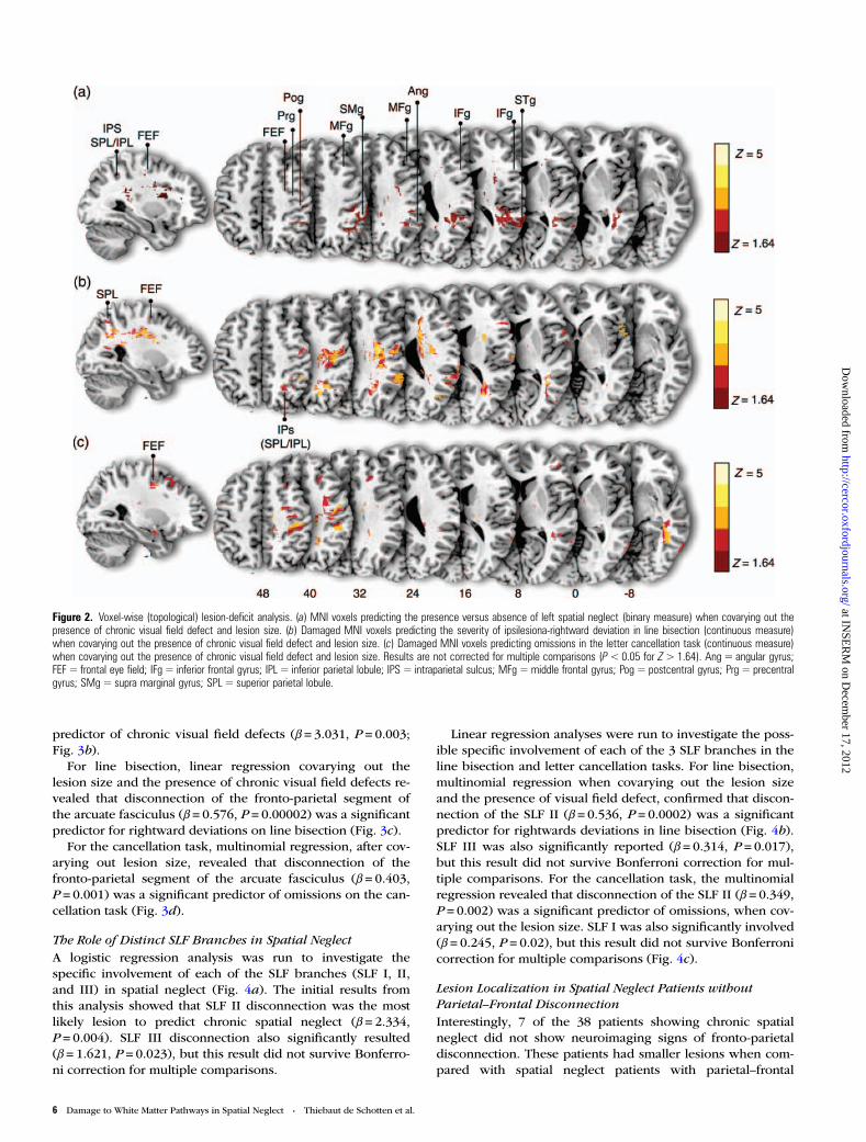
Voxel-wise Topological Lesion-Deficit AnalysisPrevious voxel-wise investigations have rarely controlled forthe effect of lesion size. A notable exception is the study byKarnath et al. (2004) who, however, showed that when lesionsize is used as a regressor in the voxel-wise analysis, resultsdo not survive correction for multiple comparisons. In linewith this finding, also in our study, results lost most of theirsignificance when lesion size was used as a covariate and cor-rection for multiple comparisons was applied. To allow com-parison with previous studies in the following, we reportresults not corrected for multiple comparisons. These anatom-ical findings were projected on a high-resolution template(Holmes et al. 1998) in the MNI by using MRIcron and areshowed in Figure 2.
A logistic regression was performed to identify lesion locicrucial to the presence versus absence of spatial neglect (i.e.
Table 2Study of the 2 single cases. Demographic and clinical data
N+ N-
Sex M MAge (years) 55 56Handedness R REducational level (years) 13 13Lesion size 130.92 145.48Time Testing post-stroke 20 days 13 months 20 days 12 monthsLine Cancellation L 2/11 - R 10/10 L 5/11 - R 10/10 L 11/11 - R 10/10 L 11/11 - R 10/10Letter Cancellation L 0/53 - R 40/51 L 0/53 - R 41/51 L 53/53 - R 49/51 L 53/53 - R 49/51
4 Damage to White Matter Pathways in Spatial Neglect • Thiebaut de Schotten et al.
at INSE
RM
on Decem
ber 17, 2012http://cercor.oxfordjournals.org/
Dow
nloaded from

binary measure) when covarying out the lesion size and thepresence of visual field defect. The results (Fig. 2a) revealedthat especially lesions in the white matter are significant pre-dictors of spatial neglect. However, other areas were also criti-cal (Fig. 2a): The supramarginal gyrus and the adjacent mostposterior part of the superior temporal gyrus at the temporal–parietal junction [this latter temporal area should not be con-founded with the most anterior–central sector of the superiortemporal gyrus advocated by Karnath et al.(2001, 2004, 2011)], the intraparietal sulcus adjacent to theangular gyrus, and the superior parietal lobe and a smalllesion spot in the frontal eye fields.
In agreement with previous evidence (Binder et al. 1992;Verdon et al. 2010; Aiello et al. 2012), the separate linearregressions performed, respectively, on the continuousmeasures of rightwards deviations in line bisection and omis-sions in the letter cancellation task, provided slightly differentresults. For the line bisection task, there was a significant in-volvement of several areas in the white matter and corticalareas such as the precentral gyrus (“hand-knob” region), thesupramarginal gyrus, the middle and inferior frontal gyri(partes opercularis and triangularis), the temporal–parietaljunction, the intraparietal sulcus between the angular gyrusand the superior parietal lobule, and 2 small lesion spots, thatis, one in the superior parietal lobule and another in thefrontal eye fields (Fig. 2b). The linear regressions performed
on the continuous measure of omissions in the letter cancella-tion task revealed no relevant involvement of parietal areasand a significant implication of the dorsal sectors of themiddle frontal gyrus, the white matter just below the frontaleye field, the precentral gyrus (hand-knob region), the adja-cent postcentral area, a lesion spot including the posteriorportion of middle and inferior temporal gyri (extrastriatecortex), and the underlying white matter.
Track-Wise or Hodological Lesion-Deficit AnalysisHere, except when specifically indicated, we report resultsthat survived the Bonferroni correction for multiple compari-sons. A logistic regression was performed to identify trackscrucial to left spatial neglect when covarying out the lesionsize and the presence of chronic visual field defect. The analy-sis revealed that disconnection of the fronto-parietal segmentof the arcuate fasciculus (β = 3.782, P = 0.002) was a signifi-cant predictor of spatial neglect (Fig. 3a). The analysis alsodisclosed an involvement of the fronto-temporal segment ofthe arcuate fasciculus (β = 2.4, P = 0.007), although this resultdid not survive to Bonferroni correction. As a control, we per-formed an identical analysis to identify tracks dedicated tochronic visual field defect, after covarying out the lesion sizeand the presence of spatial neglect. As anticipated, disconnec-tion of the optic radiations resulted to be a significant
Figure 1. Overlay of lesions in ( a) patients without left spatial neglect (N−; n=20); (b) patients with left spatial spatial neglect (N+; n= 38); ( c) statistical analysiscomparing the 2 populations of patients (N+ vs. N−; results are corrected for multiple comparisons, P< 0.05 for Z>2.1). Ang = angular gyrus; FEF = frontal eye field;IFg = inferior frontal gyrus; MFg =middle frontal gyrus; Pog = postcentral gyrus; Prg = precentral gyrus; SMg= supramarginal gyrus; STg = superior temporal gyrus.
Cerebral Cortex 5
at INSE
RM
on Decem
ber 17, 2012http://cercor.oxfordjournals.org/
Dow
nloaded from
predictor of chronic visual field defects (β = 3.031, P = 0.003;Fig. 3b).
For line bisection, linear regression covarying out thelesion size and the presence of chronic visual field defects re-vealed that disconnection of the fronto-parietal segment ofthe arcuate fasciculus (β = 0.576, P = 0.00002) was a significantpredictor for rightward deviations on line bisection (Fig. 3c).
For the cancellation task, multinomial regression, after cov-arying out lesion size, revealed that disconnection of thefronto-parietal segment of the arcuate fasciculus (β = 0.403,P = 0.001) was a significant predictor of omissions on the can-cellation task (Fig. 3d).
The Role of Distinct SLF Branches in Spatial NeglectA logistic regression analysis was run to investigate thespecific involvement of each of the SLF branches (SLF I, II,and III) in spatial neglect (Fig. 4a). The initial results fromthis analysis showed that SLF II disconnection was the mostlikely lesion to predict chronic spatial neglect (β = 2.334,P = 0.004). SLF III disconnection also significantly resulted(β = 1.621, P = 0.023), but this result did not survive Bonferro-ni correction for multiple comparisons.
Linear regression analyses were run to investigate the poss-ible specific involvement of each of the 3 SLF branches in theline bisection and letter cancellation tasks. For line bisection,multinomial regression when covarying out the lesion sizeand the presence of visual field defect, confirmed that discon-nection of the SLF II (β = 0.536, P = 0.0002) was a significantpredictor for rightwards deviations in line bisection (Fig. 4b).SLF III was also significantly reported (β = 0.314, P = 0.017),but this result did not survive Bonferroni correction for mul-tiple comparisons. For the cancellation task, the multinomialregression revealed that disconnection of the SLF II (β = 0.349,P = 0.002) was a significant predictor of omissions, when cov-arying out the lesion size. SLF I was also significantly involved(β = 0.245, P = 0.02), but this result did not survive Bonferronicorrection for multiple comparisons (Fig. 4c).
Lesion Localization in Spatial Neglect Patients withoutParietal–Frontal DisconnectionInterestingly, 7 of the 38 patients showing chronic spatialneglect did not show neuroimaging signs of fronto-parietaldisconnection. These patients had smaller lesions when com-pared with spatial neglect patients with parietal–frontal
Figure 2. Voxel-wise (topological) lesion-deficit analysis. (a) MNI voxels predicting the presence versus absence of left spatial neglect (binary measure) when covarying out thepresence of chronic visual field defect and lesion size. (b) Damaged MNI voxels predicting the severity of ipsilesiona-rightward deviation in line bisection (continuous measure)when covarying out the presence of chronic visual field defect and lesion size. (c) Damaged MNI voxels predicting omissions in the letter cancellation task (continuous measure)when covarying out the presence of chronic visual field defect and lesion size. Results are not corrected for multiple comparisons (P<0.05 for Z>1.64). Ang = angular gyrus;FEF = frontal eye field; IFg = inferior frontal gyrus; IPL = inferior parietal lobule; IPS = intraparietal sulcus; MFg =middle frontal gyrus; Pog = postcentral gyrus; Prg = precentralgyrus; SMg= supra marginal gyrus; SPL = superior parietal lobule.
6 Damage to White Matter Pathways in Spatial Neglect • Thiebaut de Schotten et al.
at INSE
RM
on Decem
ber 17, 2012http://cercor.oxfordjournals.org/
Dow
nloaded from

disconnection (t(36) = 3.421, P = 0.001). Six of these 7 patientshad no concomitant visual field defect. There were no signifi-cant differences between this subgroup and the fronto-
parietal one in performance on line bisection (t(36) = 1.205,P = 0.235) and cancellation tasks (t(36) < 1). In this smallgroup, we performed a simple overlap of individual lesions
Figure 3. Track-wise (hodotopic) lesion-deficit analysis. (a) Percentages of patients with and without left spatial neglect, with disconnection of each of the examined whitematter tracts. (b) Percentage of hemianopic patients and patients without visual field defects, with disconnection of each of the examined white matter tracts. (c) Theperformance on the line bisection task (mean deviation with 95% confidence intervals) for patients with or without disconnection of each of the examined white matter tracts.(d) The performance on the letter cancellation task (mean omissions with 95% confidence intervals) for patients with or without disconnection of each of the examined whitematter tracts. *Tracts significantly involved when covarying out the presence of chronic neglect and the lesion size. Only results that survived Bonferroni correction are reported.
Figure 4. SLF disconnection. (a) Percentage of patients with and without left spatial neglect with disconnection of SLF I, II, and III. (b) The performance on the line bisectiontask (mean deviation with 95% confidence intervals) for patients with or without disconnection of each of the 3 different branches of the SLF (I, II, and III). (c) The performancein the letter cancellation task (mean omissions with 95% confidence intervals) in patients with or without disconnection of each of the 3 different branches of the SLF (I, II, andIII). Asterisks indicate SLF branches showing significant lesion involvement after covarying out the presence of visual field defect and lesion size. Only results that survivedBonferroni correction are presented.
Cerebral Cortex 7
at INSE
RM
on Decem
ber 17, 2012http://cercor.oxfordjournals.org/
Dow
nloaded from

(Bates et al. 2003). Based on a tractography atlas ofconnectivity-based segmentation of the thalamus (Behrenset al. 2003; FSL 4.1 software package; http://www.fmrib.ox.ac.uk/fsl), we found that individual lesions overlapped withinthe latero-dorsal portion of the thalamus (Fig. 5). This part ofthe thalamus has strong connections with the premotor cortex(Behrens et al. 2003).
Single-Case StudyThe 2 patients considered in this study showed minor differ-ences in the pattern of cortical damage, with the superior tem-poral and the inferior frontal gyri being partially preserved inthe spatial neglect patient and almost entirely damaged in thenon-neglect patient (Fig. 6). Lesion volume was slightly higherin the non-neglect patient (Table 2). Figure 7 shows the recon-struction of the posterior segment, the inferior longitudinal fas-ciculus, and the cingulum for the 2 patients. Both patients hadcomparable partial disconnection of the corpus callosum and acomplete disconnection of the IFOF and the uncinate fascicu-lus. The patient with spatial neglect had an additional discon-nection of the fronto-parietal segment and the fronto-temporalsegment of the arcuate fasciculus and disconnection of thecortico-ponto-cerebellar tract, which were instead normally re-constructed in the non-neglect patient. Advanced spherical trac-tography of the SLF I, II, and III revealed that both patient had
preserved SLF I and damaged SLF III. SLF II was solelydamaged in the spatial neglect patient (Fig. 8).
Discussion
In this study, we used a recently published atlas of humanbrain connections (Thiebaut de Schotten, ffytche, et al.2011; Catani and Thiebaut de Schotten 2012) to formallytest, in a relatively large sample of the right brain damagedpatients, whether lesion of white matter parietal–frontalpathways is a relevant anatomical determinant in the per-sistence of left spatial neglect in the chronic phase of cer-ebrovascular stroke. Three main findings emerged from ourinvestigation. First, the most reliable predictor of chronicspatial neglect was the disconnection of one of thebranches of the SLF, that is, the SLF II. Additional supportto this group study result came by the complete whitematter tractography dissection run in 2 clinically matchedpatients, one with and the other without signs spatialneglect. Although both of these patients had lesions ofsimilar cortical location and size, only the patient with sub-cortical parietal–frontal disconnection showed signs ofspatial neglect both in the acute and in the chronic phase.Secondly, the most reliable predictor of chronic spatialneglect in the gray matter was damage to the middlefrontal gyrus and/or the TPJ (i.e. the ventral attentionalsystem) although, in some cases, the lesioned area ex-tended to or was close to areas belonging to the dorsal at-tentional system, such as the frontal eye field and thesuperior parietal lobule (Shomstein et al. 2010). Thirdly, inthe small subgroup of chronic spatial neglect patients whodid not have neuroimaging signs of parietal–frontal discon-nection, most lesions affected the latero-dorsal portion ofthe thalamus.
Spatial Neglect as Both a “Disconnective Breakdown”and a “Disconnective Syndrome”The role of fronto-parietal white matter disconnection con-firms, at an extended group level, evidence gathered fromneurosurgical studies in the monkey (Gaffan and Hornak1997) and from more recent neurosurgical, fMRI, and DTIinvestigations run in relatively small samples of humanpatients (Thiebaut de Schotten et al. 2005; He et al. 2007; Ur-banski et al. 2008, 2011; Shinoura et al. 2009; Ciaraffa et al.2012). Altogether, the present evidence supports the con-clusions of previous lesion studies that, without beneficiatingof the later development of tractography-based track-wisestatistics, indicated a role of selective SLF disconnection (Dor-icchi and Tomaiuolo 2003) in human spatial neglect.
This converging evidence from different studies andmethods suggests, rather homogenously, that cerebrovasculardamage to long-range white matter parietal–frontal tracts is animportant cause of spatial neglect. It is therefore important todiscuss the possible pathophysiological mechanisms throughwhich this type of lesion can engender chronic signs ofspatial neglect in human patients. A neurological syndrome isusually interpreted as being purely disconnective, when corti-cal areas that get reciprocally disconnected by brain damageremain functionally and anatomically unaffected whereas anyother function depending on their interaction is disrupted(Catani and Mesulam 2008). One might wonder whether this
Figure 5. (a) Overlay of lesions in neglect patients without fronto-parietaldisconnection. (b) Tractography atlas of the connectivity-based segmentation of thethalamus (Behrens et al. 2003).
8 Damage to White Matter Pathways in Spatial Neglect • Thiebaut de Schotten et al.
at INSE
RM
on Decem
ber 17, 2012http://cercor.oxfordjournals.org/
Dow
nloaded from

is the case for spatial neglect due to damage of parietal–frontal white matter pathways in the MCA territory. It hasbeen suggested (Doricchi and Tomaiuolo 2003; Thiebaut deSchotten et al. 2005; Bartolomeo et al. 2007) that a lesion cen-tered in the tightly packed parietal–frontal white matter fiberscan be more disrupting than a cortical lesion of equivalentvolume, because it can provoke widespread hypofunctioningof the whole network of interconnected cortical areas. In thissense, MCA spatial neglect could be better interpreted as adisconnective breakdown rather than a purely disconnectivesyndrome, that is, as resulting from dysfunction of large-scalecortical networks. Further converging evidence supportingthis hypothesis comes from recent fMRI studies, demonstrat-ing that the temporal correlation (i.e. functional connectivity)between the blood oxygenation level-dependent (BOLD)signals in ventral parietal (TPJ) and frontal areas (IFG–MFG)is crucially disrupted in spatial neglect patients (He et al.2007).
It is noteworthy, however, that there are other cases inwhich neglect can be properly interpreted as a purely
disconnective syndrome. Lesions in the right posterior cerebralartery (PCA) territory that cause disconnection of the spleniumof the corpus callosum and concomitant damage of the adja-cent right striate cortex with accompanying left hemianopiaprovoke severe left side visual neglect both in humans and inmonkeys (Gaffan and Hornak 1997; Bird et al. 2006; Parket al. 2006; Tomaiuolo et al. 2010). Based on a pioneeringneurosurgical modeling study run in the monkey, Gaffan andHornak (1997) have proposed that in this case visual neglectis severe because splenial disconnection confines the visualinput arriving from the seeing right visual field in the lefthemisphere. Thus, this information cannot reach the anatomi-cally intact attentional parietal–frontal network in the righthemisphere. As a consequence, no exploration of the leftunseen hemifield is possible. This pathophysiological mechan-ism mirrors in the right hemisphere the anatomical–functionalimpairment that in the left hemisphere produces “pure alexia”without agraphia, a prototypical example of pure disconnec-tive syndrome (Dejerine 1891, 1892; Catani and ffytche 2005),where interruption of callosal fibers conveying visual inputs
Figure 6. Study of the 2 single cases. (a) MRI-based reconstructions of the lesion for the patient with left spatial neglect (N+, top left) and for the patient without signs ofneglect (N−, top right). The blue and red lines superimposed on the lateral views of the 3-dimensional reconstructions of the right hemisphere indicate the lateral (sylvian)sulcus and the superior temporal sulcus, respectively. The light blue and yellow arrows indicate the intraparietal sulcus and the vertical ramus of the sylvian fissure, respectively.(b) Axial sections showing the localization and extent of brain damage in the 2 patients. Red arrows indicate subcortical areas where the damage produced disconnection ofparietal–frontal pathways in N+; green arrows indicate corresponding spared areas in N−. (c) Coronal sections passing at Y=−28 and Y=−32 (MNI coordinates) in N+ andN− patients. Red arrows indicate corresponding point of disconnection in coronal and transversal slices in N+ patient. On the rightmost column, the lesion of N+ is drawn inred and that of the N− patient in green. The overlap between the 2 lesions is shown in blue.
Cerebral Cortex 9
at INSE
RM
on Decem
ber 17, 2012http://cercor.oxfordjournals.org/
Dow
nloaded from

from the right hemisphere to language centers in the lefthemisphere impairs reading with preserved visual and verbalfunctions. In the case of PCA neglect due to right splenial dis-connection, the purely disconnective nature of the syndrome
is further confirmed by the finding that in these patientsneglect is restricted to the visual domain with no accompany-ing signs of personal, representational-imagery, or motorneglect (Tomaiuolo et al. 2010).
Figure 7. Complete DTI dissection of white matter fiber tracts in the 2 matched cases with (N+) and without (N−) visuospatial neglect. Disconnected tracts were manuallydrawn in gray.
Figure 8. Spherical deconvolution dissection of white matter fiber tracts in the 2 matched cases with (N+) and without (N−) visuospatial neglect. Disconnected tracts weremanually drawn in gray.
10 Damage to White Matter Pathways in Spatial Neglect • Thiebaut de Schotten et al.
at INSE
RM
on Decem
ber 17, 2012http://cercor.oxfordjournals.org/
Dow
nloaded from

The Contribution of “Long-Range” and “Short-Range”Disconnections in MCA Spatial NeglectThere is shared agreement that spatial neglect is not a unitarysyndrome made up of a homogenous collection of symptoms(Doricchi et al. 2008; Verdon et al. 2010). Spatial neglectcannot only selectively affect different sectors of space (extra-personal vs. personal vs. representational space) or differentspatial reference frames (egocentric vs. allocentric), but alsodifferent modes of spatial attentional processing as, forexample, the simultaneous versus sequential analysis ofspatial positions that are entailed by the line bisection andmultiple item cancellation tasks, respectively (Binder et al.1992). Based on this clinical evidence, recent research on theanatomical correlates of spatial neglect has moved from thestudy of the general anatomical correlates of the syndrome,toward the more focused study of the correlates of the specificclinical features characterizing different forms of the syn-drome, or the different types of attentional disturbancesassociated with the spatial neglect syndrome (i.e. top-downvs. bottom up attention; Shomstein et al. 2010). Expanding onthe original findings by Binder et al. (1992), Verdon et al.(2010) used the VLSM technique to demonstrate that differentbehavioral components of chronic spatial neglect are associ-ated with lesion of different sectors of the right parietal–frontal attentional network. Consistent with original fMRIfindings in healthy participants (Fink et al. 2000) and with theresults from the present study, Verdon et al. (2010) identifieda perceptual visuospatial component revealed by the perform-ance on line bisection, text, and word reading tasks, andassociated with lesions of the parietal lobe. By contrast, anexploratory visuomotor component revealed by the perform-ance in sequential multi-item cancellations tasks and wasassociated with lesion of frontal areas. However, in each ofthese cases, lesion of the parietal and frontal components ofthe attentional network was associated with lesion involve-ment of the underlying white matter. This finding suggeststhat lesions encroaching upon long white matter connectionsdo not only have diffuse disruptive effects on the entire righthemispheric network and that, depending on their relativeposterior or anterior location (Doricchi et al. 2008), they maycontribute to the selective functional disruption of more local
circuits supported by short-range white matter fibers withinthe parietal or the frontal lobe (Fig. 9). In keeping with thishypothesis, spatial neglect patients with lesions centered onthe supramarginal gyrus show significant changes intask-related BOLD activity in the adjacent visual cortex (Cor-betta et al. 2005), whereas those with lesions centered in theinferior frontal gyrus display a breakdown of functional con-nectivity in more dorsal frontal areas (He et al. 2007). Note,however, that neither lesions limited to the associative visualcortex, nor those restricted to the dorsal frontal regions, typi-cally produce spatial neglect. In summary, spatial neglectpatients might demonstrate more perceptual or visuomotorsymptoms depending on loss of function induced, by localanatomical diaschisis or functional breakdown, in the corticalareas surrounding the white matter lesion (Fig. 9). This con-clusion received support by the recent finding (Aiello et al.2012) that the severity of spatial neglect in a multiple itemletter cancellation task correlates with frontal white matterdamage producing both disconnection of the parietal–frontalpathways (i.e. SLF and arcuate fasciculus) and disconnectionof a local white matter pathway linking the supplementarymotor area and the superior frontal gyrus with the inferiorfrontal gyrus (Lawes et al. 2008; Oishi et al. 2008; Catani et al.2012; Thiebaut de Schotten et al. 2012).
It is worth of note that this pathophysiological mechanismcan produce chronic effects having the same behavioral selec-tivity of transitory effects described by Hillis et al. (2000) inacute spatial neglect patients (Khurshid et al. 2012). Usingdiffusion- and perfusion-weighted MRI, these authors showedthat, in the hyperacute phase, neglect limited to specificspatial coordinates (egocentric vs. object-centered neglect) isassociated with local hypoperfusion of specific cortical areas,caused by temporary stenosis of blood vessels. Neglect disap-peared when pharmacological intervention restored properblood perfusion.
Gray Matter Components of Spatial NeglectThe voxel-based analyses run in the present study, when takinginto account the lesion size as a regressor did not survive cor-rection for multiple comparisons. This result, similarly to
Figure 9. A diaschisis model of the symptomatological dissociation observed between left spatial neglect deriving from parietal or frontal strokes. (a) Parietal and (b) frontalstrokes interrupting long-range parietal–frontal connections, and producing general hypoactivation of the parietal–frontal attentional network, can be associated with interruptionof short-range connections causing local dysfunction of the neighboring areas in the parietal or in the frontal lobe.
Cerebral Cortex 11
at INSE
RM
on Decem
ber 17, 2012http://cercor.oxfordjournals.org/
Dow
nloaded from

preliminary evidences reported by Karnath et al. (2004),confirm that lesion-deficit analyses focused on the severityspatial neglect are frequently biased by the effect of the lesionsize. This result also strongly supports the idea of a distributedcortical origin for spatial neglect underpowering standardvoxel-wise topological lesion-deficit analysis. Nevertheless, un-corrected analyses systematically showed that gray matterlesions predicting signs of chronic spatial neglect concernregions reciprocally interconnected through the arcuate fascicu-lus and SLF, such as the TPJ and the middle frontal gyrus(Catani et al. 2005, 2007; Catani and Thiebaut de Schotten 2008;Thiebaut de Schotten et al. 2008; Tsang et al. 2009). Thisfinding is consistent with earlier observations that were focusedon the role of localized gray matter damage in spatial neglect(Vallar and Perani 1986; Husain and Kennard 1996, 1997; Mortet al. 2003; Corbetta et al. 2005; Committeri et al. 2007) and, atthe same time, discloses a fine anatomical and functional con-gruency between the white matter hodological and the gray-matter topological lesional components of spatial neglect, thusreaffirming the crucial role played by the integrated functioningof the right hemispheric parietal–frontal network in spatial or-ienting and awareness.
The TPJ and the middle frontal gyrus belong to a ventralattentional network that shows an increase in the BOLDwhen attention is dragged to unexpected visual events(Downar et al. 2000; Corbetta and Shulman 2002; Serenceset al. 2005; Asplund et al. 2010). Based on the finding of aselective BOLD activation of the right TPJ and the middlefrontal gyrus in response to targets presented at attentionallyunattended-invalid spatial locations, Corbetta and Shulman(2002) have proposed that both the attentional disturbancesand the higher frequency of spatial neglect after right hemi-spheric stroke depend on the anatomical and functional dis-ruption of this ventral attentional network. Because ofanatomical proximity and reciprocal connectivity, damage ofthe right ventral network would produce hypoactivation of anadjacent dorsal attentional system composed by the intrapar-ietal sulcus and the frontal eye field areas. Due to reciprocalcallosal inhibition, this hypoactivation would release hyperac-tivation in homologous dorsal areas in the left hemispherewith a pathologically increased rightward attentional bias.This theoretical approach establishes coherence betweenfMRI and anatomo-clinical findings and opens up new inter-esting points of discussion and investigation. As an example,one might still argue that since in this model only the role ofthe right temporo-parietal junction and the middle frontalgyrus is emphasized for attentional disengagement and reor-ienting, then lesion in the right ventral network shouldproduce a generalized slowing of attentional disengagement.This seems not entirely congruent with the behavior of spatialneglect patients that are well characterized by prevalent defec-tive disengagement from ipsi- to contralesional space ratherthan from contra- to ipsilesional space. However, residual dis-engagement abilities in spatial neglect patients could be ex-plained by fMRI findings, showing that also the left TPJ–IFGhas an important BOLD response to invalid targets and stimu-lus driven orienting (Doricchi et al. 2010). This might suggestthat in spatial neglect patients slowed reorienting towardinvalid targets presented in the left visual field might be dueto preferred response of the left ventral network to invalidtargets in the right visual space, or to the fact that right braindamage precludes or slows down the processing of invalid
targets presented in the left visual space to the spared reor-ienting network in the left hemisphere.
It is finally important to stress that different cytoarchitec-tonic areas form the region that is usually labeled temporalparietal junction: Brodmann area (BA) 40 (corresponding toareas PF, PFt, PFop, PFcm in Economo and Koskinas’ atlas;Economo and Koskinas 1925; see also Tomaiuolo and Pet-rides 2010), BA 39 (corresponding to PG), the caudal of BA22 (corresponding to TA1), and BA 37 (corresponding to PH,PHP, PHT, PHO). Future studies should clarify whether thedifferent cytoarchitectonic areas forming the temporal parietaljunction have different roles in attentional orienting andwhether, in case of brain damage, they equally contribute ornot to the occurrence of the spatial neglect syndrome.
Subcortical Thalamic Spatial NeglectA small percentage of spatial neglect patients showed a smalllesion sparing the fronto-parietal segment of the arcuate fasci-culus and its projections. These patients showed similar clini-cal signs as the more typical spatial neglect patients withfronto-parietal disconnection. Our analysis revealed thatlesions in these patients were centered on the thalamus andits adjacent white matter. Spatial neglect has previously beenreported in humans with a lesion in the thalamus (Watsonand Heilman 1979; Cambier et al. 1980; Schott et al. 1981;Rafal and Posner 1987; Mesulam 1999) and has been associ-ated with a target engagement deficit in the contralesionalhemifield (Rafal and Posner 1987). Nearly all incoming infor-mation to the cortex is directed through the thalamus(Behrens et al. 2003). However, in this small percentage, thelesion was found on the latero-dorsal portion of the thalamus,including the posterior and lateral dorsal thalamic nuclei andthe anterior part of the pulvinar (Morel et al. 1997; Nieuwen-huys et al. 2008). The lateral posterior nucleus and the pulvi-nar receive their mains afferent fibers from the superiorcolliculus and pretectum, known as the visuomotor complexof the midbrain (Trojanowski and Jacobson 1975; Bender1981; Benevento and Standage 1983; Huerta and Harting1983; Lysakowski et al. 1986). Apart from the above, thelateral posterior nucleus and the pulvinar also receive inputfrom the motor and premotor cortex (Romansky et al. 1997).Lateral posterior nucleus and pulvinar send efferent fiberstoward the superior and inferior parietal lobules (Burton andJones 1976), while the lateral dorsal nucleus is reciprocallyconnected with the entire prefrontal cortex including, in par-ticular, the frontal eye field (Tanaka 1976; Huerta and Harting1983; Schell and Strick 1984; Goldman-Rakic and Porrino1985; Russchen et al. 1987). The posterior and lateral dorsalthalamic nuclei and the anterior part of the pulvinar can beconsidered as association nuclei characterized by strong reci-procal connections with the association areas (Nieuwenhuyset al. 2008). It is suggested that these nuclei relay high-ordercortico-cortical communication and modulate functions re-quiring visual–sensory–motor integration (Sherman and Guil-lery 2002). A disconnection of these nuclei might thus inducean indirect fronto-parietal dysfunction with consequentspatial neglect.
Caveats and ConclusionsSome notes of caution must be considered for the presentstudy. First, the white matter atlas that we have used is based
12 Damage to White Matter Pathways in Spatial Neglect • Thiebaut de Schotten et al.
at INSE
RM
on Decem
ber 17, 2012http://cercor.oxfordjournals.org/
Dow
nloaded from
on anatomical information gathered from a population ofnormal subjects aged from 18 to 22 years, while the strokepopulation we studied was much older. A decrease in the sizeof the pathways reconstructed with tractography in relation toage has been previously reported (Stadlbauer et al. 2008).White matter pathways also show a descending gradient of in-tersubjects variability going from the stem portion (>90% ofthe population studied) of the white matter pathways to themost peripheral zones (<50% of the population studied; Thie-baut de Schotten, ffytche, et al. 2011). In our analysis, wechose a probability >50% in order to consider only the almostinvariable anatomical core of each single tract and not its per-iphery (Thiebaut de Schotten, ffytche, et al. 2011).
A second note of caution concerns the possible contri-bution of interhemispheric callosal disconnection to MCAspatial neglect. At present, DTI tractography poorly recon-structs crossing fibers, with a consequent underestimation ofthe lateral projections of the cortico-spinal tract and thecorpus callosum in the atlas we used. Therefore, the presentstudy does not allow excluding that disconnection of callosalfibers also contributes to spatial neglect following vasculardamage in the MCA (Bartolomeo et al. 2007).
In conclusion, this study confirms that lesion of whitematter pathways is a relevant determinant of chronic MCAspatial neglect. When the integrated interplay between theright frontal and parietal lobes is interrupted by disconnection(direct anatomical–functional damage) or by thalamic infarc-tion (indirect functional damage), chronic spatial neglect islikely to occur. In addition, we were able to provide advancesin the understanding of the role of disconnection in spatialneglect, by pinpointing the role of distinct SLF branches. Atvariance with the proposals made by Karnath et al. (2001,2004, 2009, 2011), stressing the importance of purely corticaldamage, we conclude that chronicity of left spatial neglect inthe post-acute phase of MCA stroke is linked to the anatomicaland functional disruption of parietal–frontal white matter con-nections and not linked to damage of the central sector of thesuperior temporal gyrus.
NotesWe thank Dr Mara Cercignani and Dr Marco Bozzali for help in theacquisition of DTI data and Flavio Dell’Acqua for the help with theSpherical Deconvolution analysis. Conflict of Interest: None declared.
Funding
F.D. was supported by a grant from the Fondazione SantaLucia IRCCS. M.T.d.S. was supported by the Marie CurieIntra-European Fellowships for Career Development (FP7)and the Agence Nationale de la Recherche (ANR; project CA-FORPFC, number ANR-09-RPDOC-004-01 and project HM-TC,number ANR-09-EMER-006).
ReferencesAiello M, Jacquin-Courtois S, Merola S, Ottaviani T, Tomaiuolo F,
Bueti D, Rossetti Y, Doricchi F. 2012. No inherent left and rightside in human ‘mental number line’: evidence from right braindamage. Brain. doi:10.1093/brain/aws114.
Alexander DC. 2006. An introduction to computational diffusion MRI:the diffusion tensor and beyond. In: Visualization and processingof tensor fields. Berlin: Springer. p. 83–106.
Asplund CL, Todd JJ, Snyder AP, Marois R. 2010. A central role for thelateral prefrontal cortex in goal-directed and stimulus-driven atten-tion. Nat Neurosci. 13:507–514.
Azouvi P, Samuel C, Louis-Dreyfus A, Bernati T, Bartolomeo P, BeisJM, Chokron S, Leclercq M, Marchal F, Martin Y et al. (GEREN/GRECO) FCSGoAoUN. 2002. Sensitivity of clinical and behaviouraltests of spatial neglect after right hemisphere stroke. J Neurol Neu-rosurg Psychiatr. 73:160–166.
Bartolomeo P. 2007. Visual neglect. Curr Opin Neurol. 20:381–386.Bartolomeo P, Thiebaut de Schotten M, Doricchi F. 2007. Left unilat-
eral neglect as a disconnection syndrome. Cereb Cortex.17:2479–2490.
Basser PJ, Pierpaoli C. 1996. Microstructural and physiological fea-tures of tissues elucidated by quantitative-diffusion-tensor MRI. JMagn Reson B. 111:209–219.
Bates E, Wilson S, Saygin A, Dick F, Sereno MI, Knight RT, DronkersNF. 2003. Voxel-based lesion-symptom mapping. Nat Neurosci.6:448–450.
Behrens TE, Johansen-Berg H, Woolrich MW, Smith SM, Wheeler-Kingshott CA, Boulby PA, Barker GJ, Sillery EL, Sheehan K, Cic-carelli O et al. 2003. Non-invasive mapping of connectionsbetween human thalamus and cortex using diffusion imaging. NatNeurosci. 6:750–757.
Bender DB. 1981. Retinotopic organization of macaque pulvinar. JNeurophysiol. 46:672–693.
Benevento LA, Standage GP. 1983. The organization of projections ofthe retinorecipient and nonretinorecipient nuclei of the pretectalcomplex and layers of the superior colliculus to the lateral pulvi-nar and medial pulvinar in the macaque monkey. J Comp Neurol.217:307–336.
Binder J, Marshall R, Lazar R, Benjamin J, Mohr JP. 1992. Distinct syn-dromes of hemineglect. Arch Neurol. 49:1187–1194.
Bird C, Malhotra P, Parton A, Coulthard EJ, Rushworth MFS, HusainM. 2006. Visual neglect after right posterior cerebral artery infarc-tion. J Neurol Neurosurg Psychiatr. 77:1008–1012.
Bogousslavsky J, Regli F, Assal G. 1986. The syndrome ofunilateral tuberothalamic artery territory infarction. Stroke.17:434–441.
Brain RW. 1941. Visual disorientation with special reference to lesionof the right brain hemisphere. Brain. 64:244–272.
Bürgel U, Amunts K, Hoemke L, Mohlberg H, Gilsbach JM, Zilles K.2006. White matter fiber tracts of the human brain: three-dimensional mapping at microscopic resolution, topography andintersubject variability. Neuroimage. 29:1092–1105.
Burton H, Jones EG. 1976. The posterior thalamic region and its corti-cal projection in New World and Old World monkeys. J CompNeurol. 168:249–302.
Cambier J, Elghozi D, Strube E. 1980. Lésion du thalamus droit avecsyndrome de l’hémisphère mineur. Discussion du concept de nég-ligence thalamique. Rev Neurol. 136:105–116.
Caplan LR, Schmahmann JD, Kase CS, Feldmann E, Baquis G, Green-berg JP, Gorelick PB, Helgason C, Hier DB. 1990. Caudate infarcts.Arch Neurol. 47:133–143.
Catani M. 2007. From hodology to function. Brain. 130:602–605.Catani M, Allin MP, Husain M, Pugliese L, Mesulam MM, Murray RM,
Jones DK. 2007. Symmetries in human brain language pathwayscorrelate with verbal recall. Proc Natl Acad Sci USA.104:17163–17168.
Catani M, Dell’acqua F, Vergani F, Malik F, Hodge H, Roy P, Valabre-gue R, Thiebaut de Schotten M. 2012. Atlas of the short frontallobe connections: proposal for a new nomenclature. Cortex.48:273–291.
Catani M, ffytche DH. 2005. The rises and falls of disconnection syn-dromes. Brain. 128:2224–2239.
Catani M, Jones DK, ffytche DH. 2005. Perisylvian language networksof the human brain. Ann Neurol. 57:8–16.
Catani M, Mesulam M. 2008. The arcuate fasciculus and the discon-nection theme in language and aphasia: history and current state.Cortex. 44:953–961.
Catani M, Mesulam MM. 2008. What is a disconnection syndrome?Cortex. 44:911–913.
Cerebral Cortex 13
at INSE
RM
on Decem
ber 17, 2012http://cercor.oxfordjournals.org/
Dow
nloaded from

Catani M, Thiebaut de Schotten M. 2012. Atlas of human brain con-nections. Oxford: Oxford University Press.
Catani M, Thiebaut de Schotten M. 2008. A diffusion tensor imagingtractography atlas for virtual in vivo dissections. Cortex.44:1105–1132.
Chung CS, Caplan LR, Han W, Pessin MS, Lee KH, Kim JM. 1996.Thalamic haemorrhage. Brain. 119(Pt 6):1873–1886.
Ciaraffa F, Castelli G, Parati EA, Bartolomeo P, Bizzi A. 2012. Visualneglect as a disconnection syndrome? A confirmatory case report.Neurocase. [Epub ahead of print]. http://dx.doi.org/10.1080/13554794.2012.667130.
Committeri G, Pitzalis S, Galati G, Patria F, Pelle G, Sabatini U,Castriota-Scanderbeg A, Piccardi L, Guariglia C, Pizzamiglio L.2007. Neural bases of personal and extrapersonal neglect inhumans. Brain. 130:431–441.
Corbetta M, Kincade M, Lewis C, Snyder A, Sapir A. 2005. Neuralbasis and recovery of spatial attention deficits in spatial neglect.Nat Neurosci. 8:1603–1610.
Corbetta M, Shulman GL. 2002. Control of goal-directed andstimulus-driven attention in the brain. Nat Rev Neurosci.3:201–215.
Critchley M. 1953. The Parietal lobes. London: Edward Arnold.Damasio AR, Damasio H, Chui HC. 1980. Neglect following
damage to frontal lobe or basal ganglia. Neuropsychologia.18:123–132.
Dejerine J. 1892. Contribution a l’étude anatomo-pathologique et clin-ique des differentes variétés de cécité-verbale. Mem Soc Biol.4:61–90.
Dejerine J. 1891. Sur un cas de cécité verbale avec agraphie suivied’autopsie. Mem Soc Biol. 3:197–201.
Dell’Acqua F, Coward J, Simmons A, Murphy DGM, Williams S, CataniM. 2009. Mapping crossing fibres of the human brain with spheri-cal deconvolution: towards an atlas for clinico-anatomical corre-lation studies. Proc Int Soc Magn Reson Med. 17:3562.
Dell’acqua F, Scifo P, Rizzo G, Catani M, Simmons A, Scotti G, Fazio F.2010. A modified damped Richardson-Lucy algorithm to reduceisotropic background effects in spherical deconvolution. Neuro-image. 49:1446–1458.
Dell’acqua F, Simmons A, Williams S, Catani M. 2012. Can sphericaldeconvolution provide more information than fiber orientations?Hindrance modulated orientational anisotropy, a true-tract specificindex to characterize white matter diffusion. Hum Brain Mapp.doi:10.1002/hbm.22080.
De Witte L, Verhoeven J, Engelborghs S, De Deyn PP, Mariën P. 2008.Crossed aphasia and visuo-spatial neglect following a right thal-amic stroke: a case study and review of the literature. BehavNeurol. 19:177–194.
Diller L, Ben-Yishay Y, Gerstman L, Goodkin R, Gordon W, WeinbergJ. 1974. Studies in cognition and rehabilitation in hemiplegia.New York: University Medical Center.
Donnan GA, Bladin PF, Berkovic SF, Longley WA, Saling MM. 1991.The stroke syndrome of striatocapsular infarction. Brain. 114(Pt1A):51–70.
Doricchi F, Angelelli P. 1999. Misrepresentation of horizontal space inleft unilateral neglect: role of hemianopia. Neurology. 52(9):1845–1852.
Doricchi F, Guariglia P, Figliozzi F, Silvetti M, Bruno G, Gasparini M.2005. Causes of cross-over in unilateral neglect: between-groupcomparisons, within-patient dissociations and eye movements.Brain. 128(Pt 6):1386–1406.
Doricchi F, Macci E, Silvetti M, Macaluso E. 2010. Neural correlates ofthe spatial and expectancy components of endogenous andstimulus-driven orienting of attention in the Posner task. CerebCortex. 20:1574–1585.
Doricchi F, Thiebaut de Schotten M, Tomaiuolo F, Bartolomeo P.2008. White matter (dis)connections and gray matter (dys)func-tions in visual neglect: gaining insights into the brain networks ofspatial awareness. Cortex. 44:983–995.
Doricchi F, Tomaiuolo F. 2003. The anatomy of neglect without hemi-anopia: a key role for parietal-frontal disconnection. Neuroreport.14:2239–2243.
Downar J, Crawley AP, Mikulis DJ, Davis KD. 2000. A multimodalcortical network for the detection of changes in the sensoryenvironment. Nat Neurosci. 3:277–283.
Economo C, Koskinas GN. 1925. Die Cytoarchitektonik der Hirnrindedes erwachsenen Menschen. Wien, Berlin:Springer.
Ferro JM, Kertesz A, Black SE. 1987. Subcortical neglect: quantitation,anatomy, and recovery. Neurology. 37:1487–1492.
Finger S, Koehler PJ, Jagella C. 2004. The Monakow conceptof diaschisis: origins and perspectives. Arch Neurol. 61:283–288.
Gaffan D, Hornak J. 1997. Visual neglect in the monkey. Represen-tation and disconnection. Brain. 120(Pt 9):1647–1657.
Gainotti G, Messerli P, Tissot R. 1972. Qualitative analysis of unilateralspatial neglect in relation to laterality of cerebral lesions. J NeurolNeurosurg Psychiatry. 35:545–550.
Genovese CR, Lazar NA, Nichols T. 2002. Thresholding of statisticalmaps in functional neuroimaging using the false discovery rate.Neuroimage. 15:870–878.
Geschwind N. 1965. Disconnexion syndromes in animals and man.Brain. 88:585–644.
Goldman-Rakic PS, Porrino LJ. 1985. The primate mediodorsal (MD)nucleus and its projection to the frontal lobe. J Comp Neurol.242:535–560.
Graff-Radford NR, Damasio H, Yamada T, Eslinger PJ, Damasio AR.1985. Nonhaemorrhagic thalamic infarction. Clinical, neuropsy-chological and electrophysiological findings in four anatomicalgroups defined by computerized tomography. Brain. 108(Pt 2):485–516.
Halligan PW, Marshall JC, Wade DT. 1990. Do visual field deficitsexacerbate visuo-spatial neglect? J Neurol Neurosurg Psychiatry.53:487–491.
He BJ, Snyder A, Vincent JL, Epstein A, Shulman GL, Corbetta M.2007. Breakdown of functional connectivity in frontoparietal net-works underlies behavioral deficits in spatial neglect. Neuron.53:905–918.
Healton EB, Navarro C, Bressman S, Brust JC. 1982. Subcorticalneglect. Neurology. 32:776–778.
Heilman KM, Valenstein E. 1972. Frontal lobe neglect in man. Neurol-ogy. 22:660–664.
Hier DB, Davis KR, Richardson EP, Mohr JP. 1977. Hypertensive pu-taminal hemorrhage. Ann Neurol. 1:152–159.
Hillis AE, Barker PB, Beauchamp NJ, Gordon B, Wityk RJ. 2000. MRperfusion imaging reveals regions of hypoperfusion associatedwith aphasia and neglect. Neurology. 55:782–788.
Hirose G, Kosoegawa H, Saeki M, Kitagawa Y, Oda R, Kanda S, Mat-suhira T. 1985. The syndrome of posterior thalamic hemorrhage.Neurology. 35:998–1002.
Holmes CJ, Hoge R, Collins L, Woods R, Toga AW, Evans AC. 1998.Enhancement of MR images using registration for signal averaging.J Comput Assist Tomogr. 22:324–333.
Huerta MF, Harting JK. 1983. Sublamination within the superficialgray layer of the squirrel monkey: an analysis of the tectopulvinarprojection using anterograde and retrograde transport methods.Brain Res. 261:119–126.
Husain M, Kennard C. 1997. Distractor-dependent frontal neglect.Neuropsychologia. 35:829–841.
Husain M, Kennard C. 1996. Visual neglect associated with frontallobe infarction. J Neurol. 243:652–657.
Husain M, Mattingley JB, Rorden C, Kennard C, Driver J. 2000. Dis-tinguishing sensory and motor biases in parietal and frontalneglect. Brain. 123(Pt 8):1643–1659.
Husain M, Nachev P. 2007. Space and the parietal cortex. TrendsCogn Sci. 11:30–36.
Jones DK. 2003. Determining and visualizing uncertainty in estimatesof fiber orientation from diffusion tensor MRI. Magn Reson Med.49:7–12.
Jones DK. 2004. The effect of gradient sampling schemes onmeasures derived from diffusion tensor MRI: a Monte Carlo study.Magn Reson Med. 51:807–815.
Jones DK. 2008. Studying connections in the living human brain withdiffusion MRI. Cortex. 44:936–952.
14 Damage to White Matter Pathways in Spatial Neglect • Thiebaut de Schotten et al.
at INSE
RM
on Decem
ber 17, 2012http://cercor.oxfordjournals.org/
Dow
nloaded from
Jones DK, Griffin LD, Alexander DC, Catani M, Horsfield MA, HowardR, Williams SC. 2002. Spatial normalization and averaging of diffu-sion tensor MRI data sets. Neuroimage. 17:592–617.
Karnath H-O, Ferber S, Himmelbach M. 2001. Spatial awareness is afunction of the temporal not the posterior parietal lobe. Nature.411:950–963.
Karnath H-O, Himmelbach M, Rorden C. 2002. The subcorticalanatomy of human spatial neglect: putamen, caudate nucleus andpulvinar. Brain. 125:350–360.
Karnath H-O, Rennig J, Johannsen L, Rorden C. 2011. The anatomyunderlying acute versus chronic spatial neglect: a longitudinalstudy. Brain. 134:903–912.
Karnath HO, Fruhmann Berger M, Küker W, Rorden C. 2004. Theanatomy of spatial neglect based on voxelwise statistical analysis:a study of 140 patients. Cereb Cortex. 14:1164–1172.
Karnath HO, Rorden C, Ticini LF. 2009. Damage to white matter fibertracts in acute spatial neglect. Cereb Cortex. 19:2331–2337.
Karussis D, Leker RR, Abramsky O. 2000. Cognitive dysfunction fol-lowing thalamic stroke: a study of 16 cases and review of the lit-erature. J Neurol Sci. 172:25–29.
Kempler D, Metter EJ, Jackson CA, Hanson WR, Riege WH, MazziottaJC, Phelps ME. 1988. Disconnection and cerebral metabolism. Thecase of conduction aphasia. Arch Neurol. 45:275–279.
Kumral E, Kocaer T, Ertübey NO, Kumral K. 1995. Thalamichemorrhage. A prospective study of 100 patients. Stroke.26:964–970.
Khurshid S, Trupe LA, Newhart M, Davis C, Molitoris JJ, Medina J,Leigh R, Hillis AE. 2012. Reperfusion of specific cortical areas isassociated with improvement in distinct forms of hemispatialneglect. Cortex. 48:530–539.
Lawes INC, Barrick TR, Murugam V, Spierings N, Evans DR, Song M,Clark CA. 2008. Atlas-based segmentation of white matter tracts ofthe human brain using diffusion tensor tractography and compari-son with classical dissection. Neuroimage. 39:62–79.
Leemans A, Jones DK. 2009. The B-matrix must be rotated when cor-recting for subject motion in DTI data. Magn Reson Med.61:1336–1349.
Leibovitch FS, Black SE, Caldwell CB, Ebert PL, Ehrlich LE, Szalai JP.1998. Brain-behavior correlations in hemispatial neglect usingCT and SPECT: the Sunnybrook Stroke Study. Neurology. 50:901–908.
Levine DN, Warach JD, Benowitz LI, Calvanio R. 1986. Left spatialneglect: effects of lesion size and premorbid brain atrophy on se-verity and recovery following right cerebral infarction. Neurology.36:362–366.
Lysakowski A, Standage GP, Benevento LA. 1986. Histochemical andarchitectonic differentiation of zones of pretectal and collicularinputs to the pulvinar and dorsal lateral geniculate nuclei in themacaque. J Comp Neurol. 250:431–448.
Marquardt D. 1963. An algorithm for least-squares estimation of non-linear parameters. Siam J Appl Math. 11:431–441.
Mcfie J, Piercy MF, Zangwill OL. 1950. Visual-spatial agnosia associ-ated with lesions of the right cerebral hemisphere. Brain.73:167–190.
Mesulam MM. 1981. A cortical network for directed attention and uni-lateral neglect. Ann Neurol. 10:309–325.
Mesulam MM. 1999. Spatial attention and neglect: parietal, frontal andcingulate contributions to the mental representation and atten-tional targeting of salient extrapersonal events. Philos Trans R SocLond B Biol Sci. 354:1325–1346.
Mesulam MM, Geschwind N. 1978. On the possible role of neocortexand its limbic connections in the process of attention and schizo-phrenia: clinical cases of inattention in man and experimentalanatomy in monkey. J Psychiatr Res. 14:249–259.
Monakow CV. 1914. Die Lokalisation im Grosshirn und der Funktiondurch kortikale Herde. Wiesbaden: Verlag von J.F. Bergmann.
Morel A, Magnin M, Jeanmonod D. 1997. Multiarchitectonic andstereotactic atlas of the human thalamus. J Comp Neurol.387:588–630.
Mori S, Oishi K, Jiang H, Li X, Akhter K, Hua K, Faria AV, MahmoodA, Woods R, Toga AW et al. 2008. Stereotaxic white matter atlas
based on diffusion tensor imaging in an ICBM template. Neuro-image. 40:570–582.
Mort DJ, Malhotra P, Mannan SK, Rorden C, Pambakian A, KennardC, Husain M. 2003. The anatomy of visual neglect. Brain.126:1986–1997.
Motomura N, Yamadori A, Mori E, Ogura J, Sakai T, Sawada T. 1986.Unilateral spatial neglect due to hemorrhage in the thalamicregion. Acta Neurol Scand. 74:190–194.
Nieuwenhuys R, Voogd J, van Huijzen C. 2008. The human centralnervous system. Berlin (NY): Springer.
Oishi K, Zilles K, Amunts K, Faria A, Jiang H, Li X, Akhter K, Hua K,Woods R, Toga AW et al. 2008. Human brain white matter atlas:identification and assignment of common anatomical structures insuperficial white matter. Neuroimage. 43:447–57.
Oxbury JM, Campbell DC, Oxbury SM. 1974. Unilateral spatial neglectand impairments of spatial analysis and visual perception. Brain.97:551–564.
Park KC, Lee BH, Kim EJ, Shin MH, Choi KM, Yoon SS, Kwon SU,Chung CS, Lee KH, Heilman KM et al. 2006. Deafferentation-disconnection neglect induced by posterior cerebral artery infarc-tion. Neurology. 66:56–61.
Paterson A, Zangwill O. 1944. Disorders of visual space perceptionassociated with lesions of the right cerebral hemisphere. Brain.67:331–358.
Perani D, Vallar G, Cappa S, Messa C, Fazio F. 1987. Aphasia andneglect after subcortical stroke. A clinical/cerebral perfusion corre-lation study. Brain. 110:1211–1229.
Pizzamiglio L, Judica A, Razzano C, Zoccolotti P. 1989. Toward a com-prehensive diagnosis of visuo-spatial disorders in unilateral brain-damaged patients. Psychol Assess. 5:199–218.
Price CJ, Warburton EA, Moore CJ, Frackowiak RSJ, Friston KJ. 2001.Dynamic diaschisis: anatomically remote and context-sensitivehuman brain lesions. J Cogn Neurosci. 13:419–429.
Rafal RD, Posner MI. 1987. Deficits in human visual spatial attentionfollowing thalamic lesions. Proc Natl Acad Sci USA. 84:7349–7353.
Romansky LM, Giguere M, Bates JF, Goldman-Rakic PS. 1997. Topo-graphic organization of medial pulvinar connections with the pre-frontal cortex in the rhesus monkey. J Comp Neurol. 379:313–332.
Rorden C, Bonilha L, Nichols TE. 2007. Rank-order versus meanbased statistics for neuroimaging. Neuroimage. 35:1531–1537.
Rorden C, Karnath HO. 2004. Using human brain lesions to inferfunction: a relic from a past era in the fMRI age? Nat Rev Neurosci.5:813–819.
Rudrauf D, Mehta S, Grabowski TJ. 2008. Disconnection’s renaissancetakes shape: formal incorporation in group-level lesion studies.Cortex. 44:1084–1096.
Russchen FT, Amaral DG, Price JL. 1987. The afferent input to themagnocellular division of the mediodorsal thalamic nucleus in themonkey, Macaca fascicularis. J Comp Neurol. 256:175–210.
Schell GR, Strick PL. 1984. The origin of thalamic inputs to thearcuate premotor and supplementary motor areas. J Neurosci.4:539–560.
Schmahmann JD, Pandya DN. 2006. Fiber pathways of the brain.Oxford: Oxford University Press.
Schott B, Laurent B, Mauguiere F, Chazot G. 1981. Motor negligencein a case of right thalamic hematoma. Rev Neurol. 137:447–455.
Serences JT, Shomstein S, Leber AB, Golay X, Egeth HE, Yantis S.2005. Coordination of voluntary and stimulus-driven attentionalcontrol in human cortex. Psychol Sci. 16:114–122.
Sherman SM, Guillery RW. 2002. The role of the thalamus in the flowof information to the cortex. Philos Trans R Soc Lond B Biol Sci.357:1695–1708.
Shinoura N, Suzuki Y, Yamada R, Tabei Y, Saito K, Yagi K. 2009.Damage to the right superior longitudinal fasciculus in the inferiorparietal lobe plays a role in spatial neglect. Neuropsychologia.47:2600–2603.
Shomstein S, Lee J, Behrmann M. 2010. Top-down and bottom-upattentional guidance: investigating the role of the dorsal andventral parietal cortices. Exp Brain Res. 206:197–208.
Stadlbauer A, Salomonowitz E, Strunk G, Hammen T, Ganslandt O.2008. Age-related degradation in the central nervous system:
Cerebral Cortex 15
at INSE
RM
on Decem
ber 17, 2012http://cercor.oxfordjournals.org/
Dow
nloaded from

assessment with diffusion-tensor imaging and quantitative fibertracking. Radiology. 247:179–188.
Stein S, Volpe BT. 1983. Classical “parietal” neglect syndrome aftersubcortical right frontal lobe infarction. Neurology. 33:797–799.
Stone SP, Patel P, Greenwood RJ, Halligan PW. 1992. Measuringvisual neglect in acute stroke and predicting its recovery: thevisual neglect recovery index. J Neurol Neurosurg Psychiatr.55:431–436.
Tanaka D. 1976. Thalamic projections of the dorsomedial prefrontalcortex in the rhesus monkey (Macaca mulatta). Brain Res.110:21–38.
Thiebaut de Schotten M, Dell’Acqua F, Forkel SJ, Simmons A, VerganiF, Murphy DGM, Catani M. 2011. A lateralized brain network forvisuospatial attention. Nat Neurosci. 14:1245–1246.
Thiebaut de Schotten M, Dell’acqua F, Valabregue R, Catani M. 2012.Monkey to human comparative anatomy of the frontal lobe associ-ation tracts. Cortex. 48:82–96.
Thiebaut de Schotten M, ffytche DH, Bizzi A, Dell’Acqua F, Allin M,Walshe M, Murray R, Williams SC, Murphy DG, Catani M. 2011.Atlasing location, asymmetry and inter-subject variability of whitematter tracts in the human brain with MR diffusion tractography.Neuroimage. 54:49–59.
Thiebaut de Schotten M, Kinkingnehun S, Delmaire C, Lehericy S,Duffau H, Thivard L, Volle E, Levy R, Dubois B, Bartolomeo P.2008. Visualization of disconnection syndromes in humans.Cortex. 44:1097–1103.
Thiebaut de Schotten M, Urbanski M, Duffau H, Volle E, Levy R,Dubois B, Bartolomeo P. 2005. Direct evidence for a parietal-frontal pathway subserving spatial awareness in humans. Science.309:2226–2228.
Tomaiuolo F, Petrides M. 2010. Citoarchitettonica cerebrale umana: Ilcontributo di un vecchio atlante di anatomia all’acquisizione dellenuove conoscenze sul cervello. Italia: Springer-Verlag.
Tomaiuolo F, Voci L, Bresci M, Cozza S, Posteraro F, Oliva M, DoricchiF. 2010. Selective visual neglect in right brain damaged patientswith splenial interhemispheric disconnection. Exp Brain Res.206:209–217.
Trojanowski JQ, Jacobson S. 1975. A combined horseradishperoxidase-autoradiographic investigation of reciprocal connec-tions between superior temporal gyrus and pulvinar in squirrelmonkey. Brain Res. 85:347–353.
Tsang JM, Dougherty RF, Deutsch GK, Wandell BA, Ben-Shachar M.2009. Frontoparietal white matter diffusion properties predictmental arithmetic skills in children. Proc Natl Acad Sci USA.106:22546–22551.
Urbanski M, Thiebaut de Schotten M, Rodrigo S, Catani M, Oppen-heim C, Touzé E, Chokron S, Méder JF, Levy R, Dubois Bet al. 2008. Brain networks of spatial awareness: evidence fromdiffusion tensor imaging tractography. J Neurol Neurosurg Psy-chiatr. 79:598–601.
Urbanski M, Thiebaut de Schotten M, Rodrigo S, Oppenheim C,Touzé E, Méder J-F, Moreau K, Loeper-Jeny C, Dubois B,Bartolomeo P. 2011. DTI-MR tractography of white matterdamage in stroke patients with neglect. Exp Brain Res.208:491–505.
Vallar G, Perani D. 1986. The anatomy of unilateral neglect after right-hemisphere stroke lesions. A clinical/CT-scan correlation study inman. Neuropsychologia. 24:609–622.
Verdon V, Schwartz S, Lovblad KO, Hauert CA, Vuilleumier P. 2010.Neuroanatomy of hemispatial neglect and its functional com-ponents: a study using voxel-based lesion-symptom mapping.Brain. 133:880–894.
Watson RT, Heilman KM. 1979. Thalamic neglect. Neurology. 29:690–694.Watson RT, Heilman KM, Miller BD, King FA. 1974. Neglect after me-
sencephalic reticular formation lesions. Neurology. 24:294–298.Watson RT, Miller BD, Heilman KM. 1978. Nonsensory neglect. Ann
Neurol. 3:505–508.Watson RT, Valenstein E, Heilman KM. 1981. Thalamic neglect. Poss-
ible role of the medial thalamus and nucleus reticularis in behav-ior. Arch Neurol. 38:501–506.
Waxman SG, Ricaurte GA, Tucker SB. 1986. Thalamichemorrhage with neglect and memory disorder. J Neurol Sci.75:105–112.
Wedeen VJ, Wang RP, Schmahmann JD, Benner T, Tseng WY, Dai G,Pandya DN, Hagmann P, D’arceuil HE, De Crespigny AJ. 2008.Diffusion spectrum magnetic resonance imaging (DSI) tractogra-phy of crossing fibers. Neuroimage. 41:1267–1277.
Weiller C, Ringelstein EB, Reiche W, Thron A, Buell U. 1990. Thelarge striatocapsular infarct. A clinical and pathophysiologicalentity. Arch Neurol. 47:1085–1091.
Weiller C, Willmes K, Reiche W, Thron A, Isensee C, Buell U, Ring-elstein EB. 1993. The case of aphasia or neglect after striatocapsu-lar infarction. Brain. 116:1509–1525.
Weintraub S, Mesulam MM. 1987. Right cerebral dominance in spatialattention. Further evidence based on ipsilateral neglect. ArchNeurol. 44:621–625.
Yasaka M, Read SJ, O’Keefe GJ, Egan GF, Pointon O, McKay WJ,Donnan GA. 1998. Positron emission tomography in ischaemicstroke: cerebral perfusion and metabolism after stroke onset. JClin Neurosci. 5:413–416.
16 Damage to White Matter Pathways in Spatial Neglect • Thiebaut de Schotten et al.
at INSE
RM
on Decem
ber 17, 2012http://cercor.oxfordjournals.org/
Dow
nloaded from