Embed Size (px)
Citation preview
69
DAFTAR PUSTAKA
1. U.S. Cancer Statistics Working Group. CDC - about USCS - NPCR -
cancer. [homepage on the Internet]. 1999-2011 [cited 2013 Apr 17].
Available from : Department of Health and Human Services, Centers for
Disease Control and Prevention. Web site: http://www.cdc.gov/
cancer/npcr/uscs/about.htm
2. Pusat Data dan Informasi Departemen Kesehatan Profil kesehatan
Indonesia 2008. Jakarta: Departemen Kesehatan Republik Indonesia; 2009.
3. Dinas kesehatan kota Semarang Profil kesehatan kota Semarang tahun
2009. Semarang: DKK Semarang; 2009.
4. Sarjadi, Trihartini, Miranti IP, Insiden kanker penduduk Semarang tahun
1990-1999. Media Medika Indonesiana 2001; 87-92
5. National Comprehensive Cancer Network NCCN clinical practice
guidelines in oncology: Colon cancer. 3rd ver. Washington: NCCN Inc.;
2013.
6. Pallis AG, Mouzas IA, Adjuvant chemotherapy for colon cancer. Anticancer
Research 2006; 4809-4816
7. Johnson PW, Thomson PI, Seymour MT, Deasy NP, Thuraisingham RC,
ML Slevin et al, A less toxic regimen of 5-fluorouracil and high-dose folinic
acid for advanced gastrointestinal adenocarcinomas. Br. J. Cancer 1991;
603-605
70
8. Committee on The Use of Complementary and Alternative Medicine by The
American Public Complementary and alternative medicine in the United
States. Washington: The National Academies Press; 2005.
9. Tim Editorial PT. Sidomuncul. Smallcrab online. [homepage on the
internet]. 2011 [cited 2012 Apr 12]. Available from : Aneka Resep Mahkota
Dewa. Nutrend mahkota dewa. Web site : http://smallcrab.com/
index.php?option=com/content&task=view&id=67&Itemid=2
10. Gangga E, Asriani H, Novita L, Analisis pendahuluan metabolit sekunder
dari kalus mahkota dewa (Phaleria macrocarpa [Scheff.] Boerl.). Jurnal
Ilmu Kefarmasian Indonesia 2007 April; 17-22
11. Harmanto N Mahkota dewa obat pusaka para dewa: A medicine the legacy
of the gods. 6th ed. Jakarta: Agro Media Pustaka; 2003.
12. Lisdawati V. Phalerindofarma network [homepage on the internet]. 2010
[cited 2012 Apr 18]. Available from : Mahkota dewa: toksisitas, efek anti
oksidan, dan efek antikanker berdasarkan uji penapisan farmakologi. Web
site : http://www.indonetwork.phalerindofarma/34716.htm
13. Faried A, Kurnia D, Faried LS, Usman, N, Miyazaki T, Kato H, Kuwano H,
Anticancer effects of gallic acid isolated from Indonesian herbal medicine
Phaleria macrocarpa (Scheff.) Boerl on human cancer cell lines. Int J
Oncol 2007; 605-613
14. Hiroko D, Teruhiko F, Shino N, Toshihiro K, Kazuo S, Analysis of cell
growth inhibitory effect of cathechin through MAPK in human breast
cancer cell line T47D. Int J Oncol 2002; 1301-1305
71
15. Kusmardiyani S, Nawawi A, Rahmi K, Isolasi benzofenon dari daun
mahkota dewa [Phaleria macrocarpa (Scheff.) Boerl.]. Acta Pharmaceutica
Indonesia 2004; 150-152
16. Ladish H, Baltimore D, Berk A, Zipursky SL, Matsudaira P & Darnell J
Molecular cell biology. 6th ed. New York: Scientific American Books;
2000.
17. Bullwinkel J, Baron-Lühr B, Lüdemann A, Wohlenberg C, Gerdes J,
Scholzen T, Ki-67 protein is associated with ribosomal RNA transcription in
quiescent and proliferating cells. J Cell Physiol 2006 March; 624–635
18. Jemal A, Bray F, Center MM, Ferlay J, Ward E, Forman D, Global cancer
statistics 2008. CA Cancer J Clin 2011; 69-90
19. Parkin DM, Bray F, Ferlay J, Global cancer statistics 2002. CA Cancer J
Clin 2011; 74-76
20. Sjamsuhidajat R, Natawijana HA & Kartowisastro H Faktor penyebab dan
resiko kanker usus besar: Simposium upaya penemuan keganasan usus
besar secara dini. Semarang: Ikabdi press; 1986.
21. Sinicrope FA, Hart J, Hsu H, Apoptotic and mitotic indices predict survival
rates in lymph node-negative colon carcinoma. Clin Cancer Res 1999; 1793-
1804
22. Gordon PH & Nivatvongs S. Principles and practice of surgery for the
colon, rectum, and anus. 3rd ed. New York: Informa Healthcare USA;
2007.
23. Center MM, Jemal A, Smith RA, Ward E, Worldwide variations in
colorectal center. CA cancer J clin 2009; 366-378
72
24. Mescher AL. Junqueira’s basic histology. 12th ed. Indiana: McGraw-Hill
education; 2010.
25. Zinner MJ & Ashley SW. Maingot’s abdominal operations. 11th ed.
Boston: McGraw-Hill; 2011.
26. Brunicardi FC, Andersen DK, Billiar TR, Dunn DL, Hunter JG, Matthews
JB & et al Schwartz’s principles of surgery. 9th ed. Houston: McGraw-Hill;
2010.
27. Rosai J Rosai and Ackerman’s surgical pathology. 10th ed. New York:
Mosby Elsevier; 2011.
28. Hamilton SR & Aaltonen LA World Health Organization classification of
tumours: Pathology and genetics of tumors of the digestive system. Lyon:
IARC Press; 2000.
29. Sobin LH & Wittekind CH International union against cancer UICC :
Classification of malignant tumors TNM. 7th ed. Oxford: A John Wiley &
Sons Inc; 2009.
30. National Comprehensive Cancer Network NCCN clinical practice
guidelines in oncology: Colon cancer. 3rd ver. Washington: NCCN Inc.;
2013.
31. Kelompok Kerja Adenokarsinoma Kolorektal Indonesia Pengelolaan
karsinoma kolorektal : Suatu panduan klinis nasional. Jakarta: Peraboi;
2004.
32. Buyse M, Zeleniuch-Jacquotte A, Chalmers TC, Adjuvant therapy of
colorectal cancer: Why we still don’t know. JAMA 1988; 3571-3578
73
33. Wilkinson NW, Adjuvant chemotherapy for colorectal cancer: New
perspective for effective treatment. US gastroenterology review 2007; 91-93
34. Naito Y, Saito K, Shiiba K, Ohuchi A, Saiqenji K, Naqura H et al, CD8+ T
cells infiltrated within cancer cells nests as prognostic factor in human
colorectal cancer. Cancer Res 1998; 3491-3494
35. Ali AA, McMillan DC, Matulka II, McNicol AM, McArdel CS, Tumor T-
lymphocytes subset infiltration and tumor recurrence following curative
resection for colorectal cancer. Eur J Surg Oncol 2004; 292-295
36. Cotran RS, Kumar V & Robbins SL Robin pathologic basis of disease. 7th
ed. Philadelpia: WB Saunders; 2007.
37. Ladish H, Baltimore D, Berk A, Zipursky SL, Matsudaira P & Darnell J
Molecular cell biology. 6th ed. New York: Scientific American Books;
2000.
38. Vermeulen K, Van Bockstaele DR, Berneman ZN, The cell cycle : A review
of regulation, deregulation and therapeutic targets in cancer. J Cell Prolif
2003; 131–149
39. Thomas DP & Earnshaw WC Cell biology. 2nd ed. Philadelphia: Saunders
Elsevier; 2008.
40. Ladstein RG, Bachmann IM, Straume O, Akslen LA, Ki-67 expression is
superior to mitotic count and novel proliferation marker PHH3, MCM4 and
mitosin as a prognostic factor in thick cutaneus melanoma. BMC Cancer
2010; 140-154
41. Oshima CT, Iriya K, Forones NM, Ki-67 as a prognostic marker in
colorectal cancer but not in gastric cancer. Neoplasma 2005; 420-424
74
42. Hendra R, Ahmad S, Oskoueian E, Sukari A, Shukor MY, Antioxidant, anti-
inflammatory and cytotoxicity of Phaleria macrocarpa (Boerl.) scheff fruit.
BMC Complementary and Alternative Medicine 2011; 110-119
43. Boyd M, The NCI invitro anticancer drug discovery screen: Concept,
implementation, and operation. In BA Teicher editor. Anticancer drug
development guide: preclinical screening, clinical trials and approval. New
York: Humana Press; 1975. pp.23-42
44. De Avezedo WF, Dickmann HM, Gahmen US, Worland PJ, Sausville E,
Kim S, Structural basis for specifity and potency of a flavonoid inhibitor of
human CDK2, a cell cycle kinase. Proc Natl Acad Sci USA 1996; 2735-
2740
45. Khan M, Rasul A, Yi F, Zhong L, Ma T, Jaceosidin induces p53-dependent
G2/M phase arrest in U87 glioblastoma cells. Asian Pacific J Cancer Prev
2011; 3235-3238
46. Fisher PM, Lane DP, Inhibitors of cyclin-dependent kinases as anti-cancer
therapeutics. Current medicinal chemistry 2000; 1213-1245
47. Landis SH, Murray T, Bolden S, Cancer statistics 2000. CA Cancer J Clin
2000; 1398-1424
48. Jonker D, Spithoff K, Maroun J, Gastrointestinal cancer disease site group
of cancer care Ontario’s program in evidence-based care, Adjuvant systemic
chemotherapy for stage II and III colon cancer following complete
resection: An update practice guideline. Clin Oncol (R Coll Radiol) 2011;
314-322

75
49. Andre T, O’neil BH, Meyerhardt JA, Stage III colon cancer : What works,
what doesn’t and why, and what’s next. American Society of Clinical
Oncology 2012; 223-230
50. Govindan R & Arquette MA The Washington manual of oncology.
Washington: Lippincott Williams & Wilkins; 2002.
51. Chu E & DeVita VT Physicians’ cancer chemotherapy drug manual 2010.
Massachusetts: Jones and Bartlett Publishers; 2010.
52. De Gramont A, Figer A, Seymour M, Homerin M, Hmissi A, Cassidy J, et
al, Leucovorin and fluorouracil without oxaliplatin as first-line treatment in
advanced colorectal cancer. Journal of Clinical Oncology 2000; 2938-2947
53. Anonymous 1,2 Dimethylhydrazin. IARC Monographs Supplement. vol 7.
London: Academic Press; 1987.
54. Joseph CA, Woo YT & Argus MF Chemical of Cancer. London: Academic
Press; 1982.
55. Nauss KM, Lockniskir M, Pavlina T, Newbere PM, Morphology and
distribution of 1,2-dimethylhidrazine-induced colon tumor and their
relationship to gut-associated lymphoid tissue in the rat. J Natl Cancer Inst
1984; 915-924
56. Samiasih A Perbedaan ekspresi VEGF sel adenokarsinoma kolorektal tikus
Sprague dawley dengan dan tanpa pemberian ekstrak Phyllantus niruri.
Semarang: Magister Ilmu Biomedik Program Pasca Sarjana Universitas
Diponegoro; 2010.
76
57. Dani V, Goel A, Vaiphei K, Dhawa K, Chemopreventive potential of zinc in
experimentally induced colon carcinogenesis. Toxicollogy Letters 2007; 10-
18.
58. World Health Organization Research guidelines for evaluating the safety
and efficacy of herbal medicine. Manila: WHO Regional Office for the
Western Pacific; 1993.
59. Laurence DR & Bacharach AL Evaluation of drug activities :
Pharmacometrics. New York: Academic Press; 1964
60. Boyiadzis MM, Lebowitz PF, Frame JN & Fojo T Hematology-oncology
therapy. New York: McGraw-Hill; 2006.
61. Tanaka T, Colorectal carcinogenesis : Review of human and experimental
animal studies. Journal of Carcinogenesis 2009; 1-19
62. Johnson CR, Thames HD, Huang DT, Schmidt-Ullrich RK, The tumor
volume and clonogen number relationship : Tumor control predictions based
upon tumor volume estimates derived from computed tomography. Int J
Radiat Oncol Biol Phys 1995; 281–287.
63. Wanders J, Kaplan RS, Rubinstein L, Verweij J, Van Glabbeke M, Van
Oosterom AT et al, New guidelines to evaluate the response to treatment in
solid tumors. J Natl Cancer Inst 2000; 205–216.
64. Balta AZ, Ozdemir Y, Sucullu I, Derici ST, Bagci M, Demirel D et al, Can
horizontal diameter of colorectal tumor help predict prognosis?. Ulusal Cer
Derg 2014; 115-119
65. Riwanto I, Budijitno S, Dharmana E, Handojo D, Prasetyo SA, Eko A et al,
Effect of Phaleria macrocarpa supplementation on apoptosis and tumor
77
growth of C3h mice with breast cancer under treatment with adriamycin-
cyclophosphamide. Int Surg 2011 Apr-Jun; 164-170
66. Budijitno S Pengaruh ekstrak mahkota dewa terhadap skor ekspresi
perforin CTL dan sel NK serta indeks apoptosis pada adenokarsinoma
mamma mencit C3H. Semarang: Magister Ilmu Biomedik Program Pasca
Sarjana Universitas Diponegoro; 2006.
67. Guzinska-ustymowicz K, Pien’ ES, Kemona A, MCM-2, Ki-67 and PCNA
protein expressions in pT3G2. Colorectal cancer indicated lymph node
involvement. Anticancer Research 2008; 451-458.
68. Saleh HA, Jackson H, Khatib G, Banerjee M, Correlation of Bcl-2
oncoprotein immunohistochemical expression with proliferation index and
histopathologic parameters in colorectal neoplasia. Pathol Oncol Res 1999;
273-279.
78
LAMPIRAN
Lampiran 1. Perhitungan Dosis Konversi
Konversi dosis antara jenis hewan dengan manusia
(Laurence and Bacharach, 1964).59
Dosis manusia ̴ berat 70 kg
Dosis tikus ̴ berat 200 g
Konstanta = 0.018
Konstanta hasil ekstrak Phaleria macrocarpa adalah 0,55%, dan hasil ekstrak
diencerkan dengan aquabidest sampai tercapai konsentrasi 0,5 mg/ml. Maka dosis
pemberian Phaleria macrocarpa pada hewan coba tikus yaitu
Dosis pemberian ekstrak Phaleria macrocarpa
5000 (gram) x 0,018 x 0,0055
= 0,495 mg/hr,
= 0,99 ml/hr
Mencit
20 g Tikus 200 g
Marmot 400 g
Kelinci 1,5 kg
Kera 4 kg
Anjing 12 kg
Manusia 70 kg
Mencit 20 g
1.0 7.0 12.25 27.8 64.1 124.3 387.9
Tikus 200 g
0.14 1.0 1.74 3.0 9.2 17.8 56.0
Marmot 400 g
0.008 0.57 1.0 2.25 5.2 10.2 31.5
Kelinci 1,5 kg
0.04 0.25 0.44 1.0 2.4 4.5 14.2
Kera 4 kg
0.016 0.11 0.19 0.42 1.0 1.9 6.1
Anjing 12 kg
0.008 0.06 0.10 0.22 0.52 1.0 3.1
Manusia 70 kg
0.0026 0.018 0.031 0.07 0.16 0.32 1.0
79
Lampiran 2. Hasil Pengukuran Penelitian
Kelompok Label Jumlah
tumor
Diameter
tumor (cm)
Proliferasi sel
(%)
Kontrol
K 1 2 1,832 28
K 2 4 1,414 53
K 3 2 1,036 38
K 4 3 1,521 29.6
K 5 2 1,282 38
K 6 2 1,332 36.3
Perlakuan 1
P1 1 2 1,323 16.6
P1 2 2 1,014 31.6
P1 3 4 1,182 41.6
P1 4 2 1,245 25
P1 5 3 0,903 43.3
P1 6 4 1,144 13.3
Perlakuan 2
P2 1 2 1,156 31.6
P2 2 3 1,340 35
P2 3 2 1,423 50
P2 4 3 1,314 41.6
P2 5 3 1,481 25
P2 6 2 1,318 30
Perlakuan 3
P3 1 1 1,258 25.6
P3 2 2 1,220 19.3
P3 3 4 0,827 17.6
P3 4 3 0,973 22.6
P3 5 2 1,019 26
P3 6 4 1,146 24.3
80
Lampiran 3. Foto Penelitian
Gambar 10. Pemberian induksi 1,2 Dimethylhidrazine secara subkutan.(kanan)
Gambar 11. Gambaran makroskopis tumor kolon. (kiri)
Gambar 12. Pengukuran massa tumor kolon
81
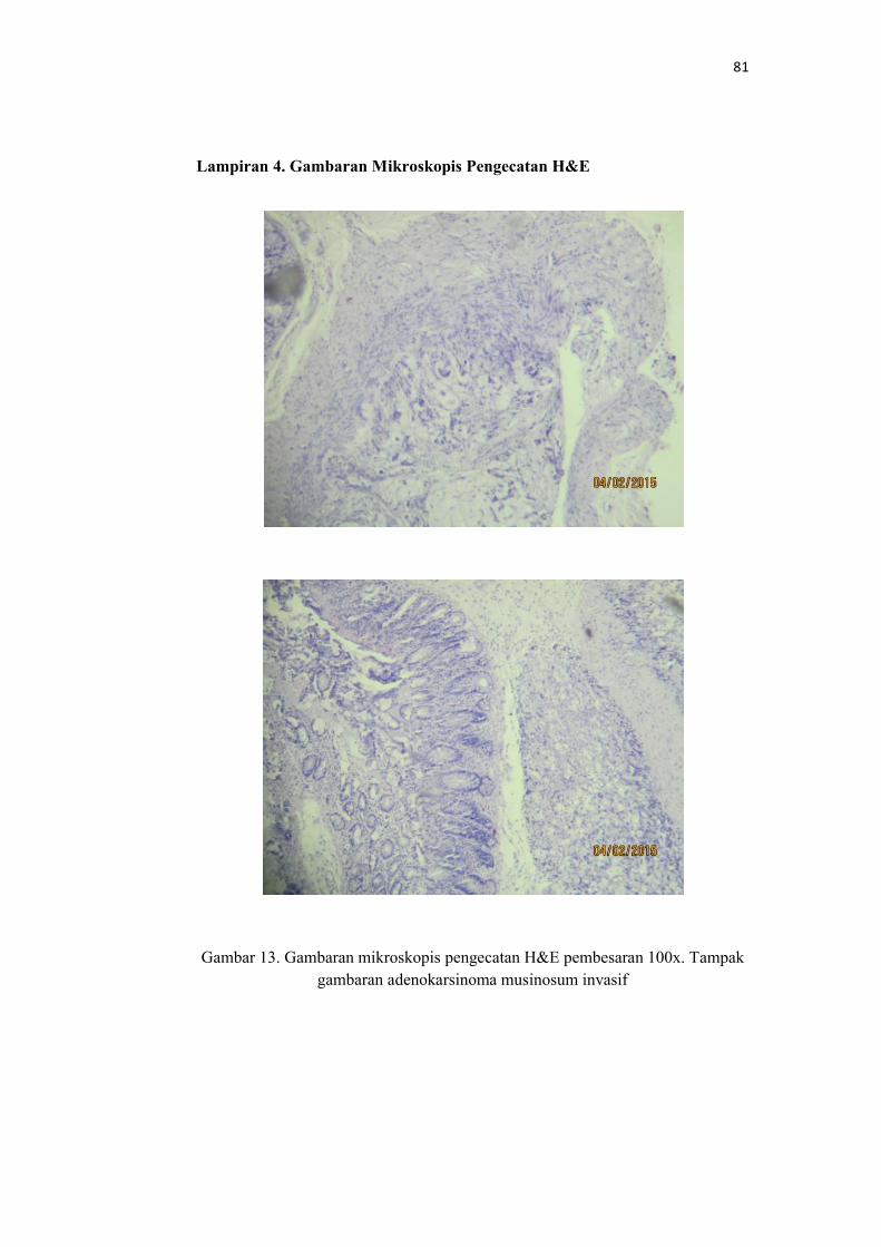
Lampiran 4. Gambaran Mikroskopis Pengecatan H&E
Gambar 13. Gambaran mikroskopis pengecatan H&E pembesaran 100x. Tampak
gambaran adenokarsinoma musinosum invasif
82
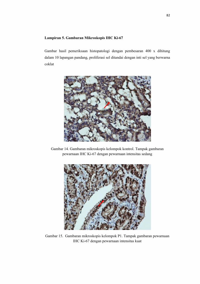
Lampiran 5. Gambaran Mikroskopis IHC Ki-67 Gambar hasil pemeriksaan histopatologi dengan pembesaran 400 x dihitung
dalam 10 lapangan pandang, proliferasi sel ditandai dengan inti sel yang berwarna
coklat
Gambar 14. Gambaran mikroskopis kelompok kontrol. Tampak gambaran
pewarnaan IHC Ki-67 dengan pewarnaan intensitas sedang
Gambar 15. Gambaran mikroskopis kelompok P1. Tampak gambaran pewarnaan
IHC Ki-67 dengan pewarnaan intensitas kuat
83
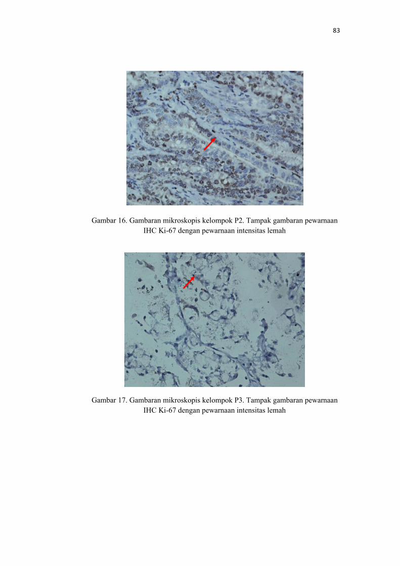
Gambar 16. Gambaran mikroskopis kelompok P2. Tampak gambaran pewarnaan
IHC Ki-67 dengan pewarnaan intensitas lemah
Gambar 17. Gambaran mikroskopis kelompok P3. Tampak gambaran pewarnaan
IHC Ki-67 dengan pewarnaan intensitas lemah
84
Lampiran 6. Data Statistik
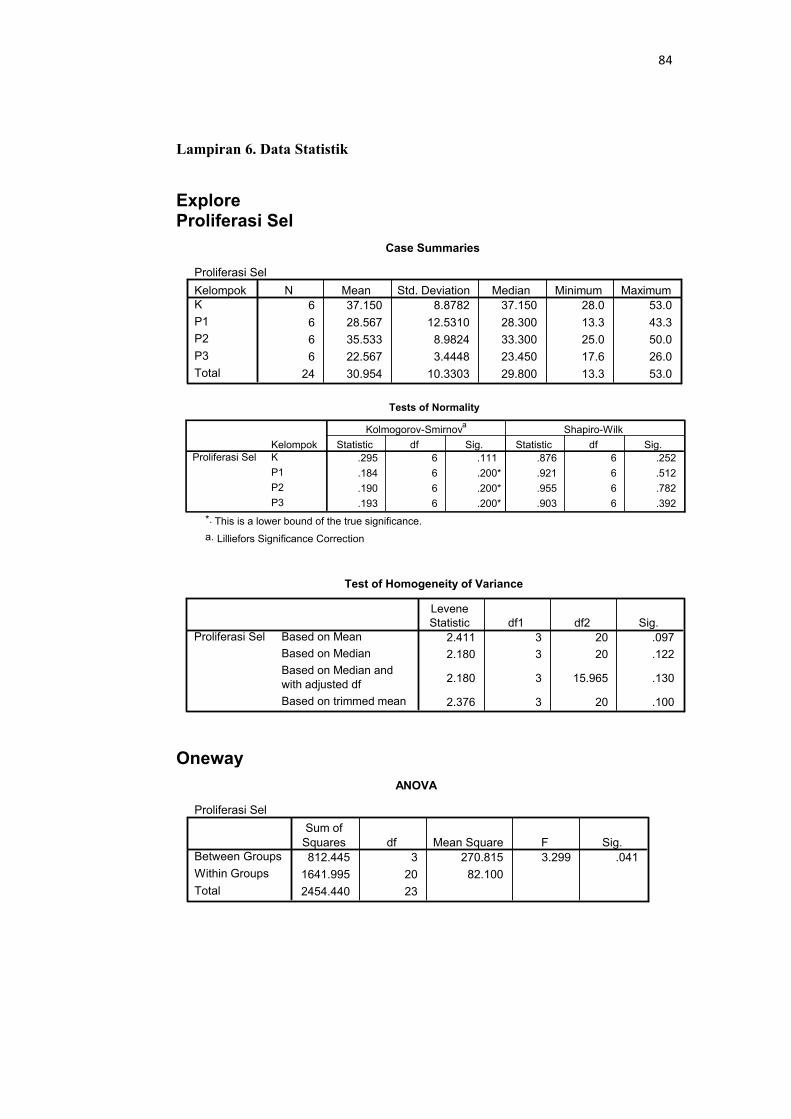
Explore Proliferasi Sel
Oneway
Case Summaries
Proliferasi Sel
6 37.150 8.8782 37.150 28.0 53.0
6 28.567 12.5310 28.300 13.3 43.3
6 35.533 8.9824 33.300 25.0 50.0
6 22.567 3.4448 23.450 17.6 26.0
24 30.954 10.3303 29.800 13.3 53.0
KelompokK
P1
P2
P3
Total
N Mean Std. Deviation Median Minimum Maximum
Tests of Normality
.295 6 .111 .876 6 .252
.184 6 .200* .921 6 .512
.190 6 .200* .955 6 .782
.193 6 .200* .903 6 .392
KelompokK
P1
P2
P3
Proliferasi SelStatistic df Sig. Statistic df Sig.
Kolmogorov-Smirnova
Shapiro-Wilk
This is a lower bound of the true significance.*.
Lilliefors Significance Correctiona.
Test of Homogeneity of Variance
2.411 3 20 .097
2.180 3 20 .122
2.180 3 15.965 .130
2.376 3 20 .100
Based on Mean
Based on Median
Based on Median andwith adjusted df
Based on trimmed mean
Proliferasi Sel
LeveneStatistic df1 df2 Sig.
ANOVA
Proliferasi Sel
812.445 3 270.815 3.299 .041
1641.995 20 82.100
2454.440 23
Between Groups
Within Groups
Total
Sum ofSquares df Mean Square F Sig.
85
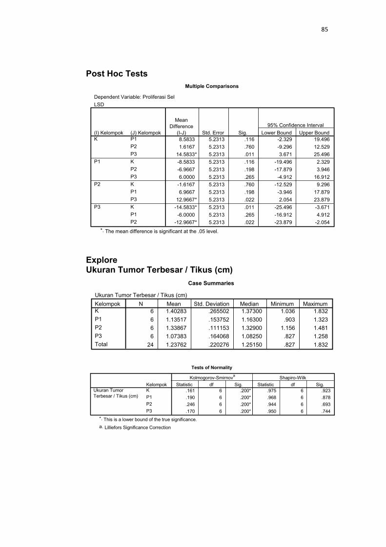
Post Hoc Tests
Explore Ukuran Tumor Terbesar / Tikus (cm)
Multiple Comparisons
Dependent Variable: Proliferasi Sel
LSD
8.5833 5.2313 .116 -2.329 19.496
1.6167 5.2313 .760 -9.296 12.529
14.5833* 5.2313 .011 3.671 25.496
-8.5833 5.2313 .116 -19.496 2.329
-6.9667 5.2313 .198 -17.879 3.946
6.0000 5.2313 .265 -4.912 16.912
-1.6167 5.2313 .760 -12.529 9.296
6.9667 5.2313 .198 -3.946 17.879
12.9667* 5.2313 .022 2.054 23.879
-14.5833* 5.2313 .011 -25.496 -3.671
-6.0000 5.2313 .265 -16.912 4.912
-12.9667* 5.2313 .022 -23.879 -2.054
(J) KelompokP1
P2
P3
K
P2
P3
K
P1
P3
K
P1
P2
(I) KelompokK
P1
P2
P3
MeanDifference
(I-J) Std. Error Sig. Lower Bound Upper Bound
95% Confidence Interval
The mean difference is significant at the .05 level.*.
Case Summaries
Ukuran Tumor Terbesar / Tikus (cm)
6 1.40283 .265502 1.37300 1.036 1.832
6 1.13517 .153752 1.16300 .903 1.323
6 1.33867 .111153 1.32900 1.156 1.481
6 1.07383 .164068 1.08250 .827 1.258
24 1.23762 .220276 1.25150 .827 1.832
KelompokK
P1
P2
P3
Total
N Mean Std. Deviation Median Minimum Maximum
Tests of Normality
.161 6 .200* .975 6 .923
.190 6 .200* .968 6 .878
.246 6 .200* .944 6 .693
.170 6 .200* .950 6 .744
KelompokK
P1
P2
P3
Ukuran TumorTerbesar / Tikus (cm)
Statistic df Sig. Statistic df Sig.
Kolmogorov-Smirnova
Shapiro-Wilk
This is a lower bound of the true significance.*.
Lilliefors Significance Correctiona.
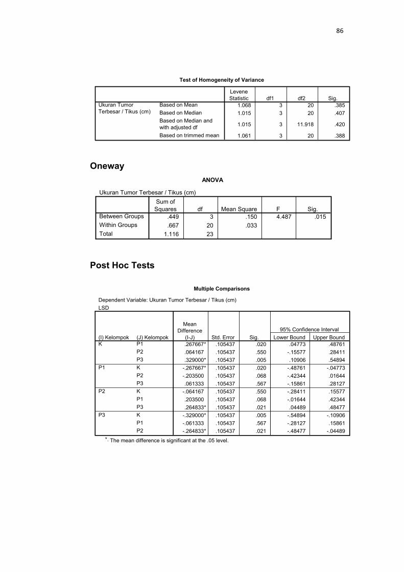
86
Oneway
Post Hoc Tests
Test of Homogeneity of Variance
1.068 3 20 .385
1.015 3 20 .407
1.015 3 11.918 .420
1.061 3 20 .388
Based on Mean
Based on Median
Based on Median andwith adjusted df
Based on trimmed mean
Ukuran TumorTerbesar / Tikus (cm)
LeveneStatistic df1 df2 Sig.
ANOVA
Ukuran Tumor Terbesar / Tikus (cm)
.449 3 .150 4.487 .015
.667 20 .033
1.116 23
Between Groups
Within Groups
Total
Sum ofSquares df Mean Square F Sig.
Multiple Comparisons
Dependent Variable: Ukuran Tumor Terbesar / Tikus (cm)
LSD
.267667* .105437 .020 .04773 .48761
.064167 .105437 .550 -.15577 .28411
.329000* .105437 .005 .10906 .54894
-.267667* .105437 .020 -.48761 -.04773
-.203500 .105437 .068 -.42344 .01644
.061333 .105437 .567 -.15861 .28127
-.064167 .105437 .550 -.28411 .15577
.203500 .105437 .068 -.01644 .42344
.264833* .105437 .021 .04489 .48477
-.329000* .105437 .005 -.54894 -.10906
-.061333 .105437 .567 -.28127 .15861
-.264833* .105437 .021 -.48477 -.04489
(J) KelompokP1
P2
P3
K
P2
P3
K
P1
P3
K
P1
P2
(I) KelompokK
P1
P2
P3
MeanDifference
(I-J) Std. Error Sig. Lower Bound Upper Bound
95% Confidence Interval
The mean difference is significant at the .05 level.*.
87
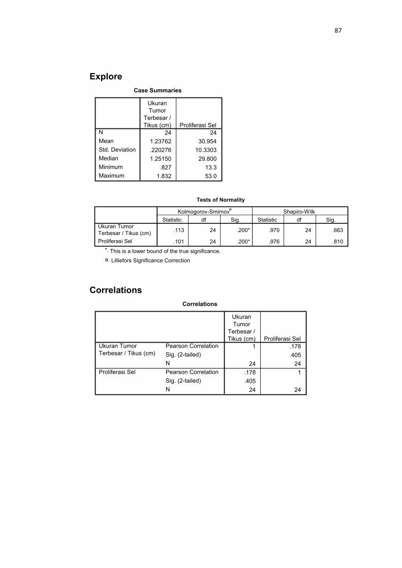
Explore
Correlations
Case Summaries
24 24
1.23762 30.954
.220276 10.3303
1.25150 29.800
.827 13.3
1.832 53.0
N
Mean
Std. Deviation
Median
Minimum
Maximum
UkuranTumor
Terbesar /Tikus (cm) Proliferasi Sel
Tests of Normality
.113 24 .200* .970 24 .663
.101 24 .200* .976 24 .810
Ukuran TumorTerbesar / Tikus (cm)
Proliferasi Sel
Statistic df Sig. Statistic df Sig.
Kolmogorov-Smirnova
Shapiro-Wilk
This is a lower bound of the true significance.*.
Lilliefors Significance Correctiona.
Correlations
1 .178
.405
24 24
.178 1
.405
24 24
Pearson Correlation
Sig. (2-tailed)
N
Pearson Correlation
Sig. (2-tailed)
N
Ukuran TumorTerbesar / Tikus (cm)
Proliferasi Sel
UkuranTumor
Terbesar /Tikus (cm) Proliferasi Sel