Embed Size (px)
Citation preview

54
DAFTAR PUSTAKA
1. Pawankar R, Canonica GW, Holgate ST, Lockey RF. Introduction and
Executive Summary: Establishing the need to treat Allergic Diseases as a
Global Public Health issue. In: WAO White Book on Allergy. ; 2011:11-20.
2. Pawankar RS, Sánchez-Borges M, Bonini S, Kaliner MA. The Burden of
Allergic Diseases. In: WAO White Book on Allergy. ; 2011:27-38.
3. Riset Kesehatan Dasar 2013 [Internet]. Jakarta: Riset Kesehatan Dasar;
2013 [cited 2016 Jan 23]. Available from: http://www.depkes.go.id.
4. Levinson W. Hypersensitivity (Allergy). In: Review of Medical
Microbiology and Immunology. Vol 1. 13th ed. San Francisco: McGraw-
Hill Education; 2014:1195-202.
5. Abbas AK, Lichtmann AH, Pillai S. Allergy. In: Cellular and Molecular
Immunology. 8th ed. Elsevier Inc.; 2015:417-34.
6. Lötvall J, Pawankar R, Wallace DV, Akdis CA, Rosenwasser LJ, Weber
RW. We call for iCAALL: International Collaboration in Asthma, Allergy
and Immunology. J Allergy Clin Immunol. 2012; 129(4):904-5.
7. Meltzer EO, Gross GN, Katial R, Storms WW. Allergic rhinitis
substantially impacts patient quality of life: findings from the Nasal Allergy
Survey Assessing Limitations. J Fam Pract. 2012; 61(2 Suppl):S5—10.
8. Portnoy JM, Partridge MR. Health economics, medical education and cost-
effective health care in allergy. In: WAO White Book on Allergy. ;
2011:139-52.
9. Feng CH, Miller MD, Simon RA. The united allergic airway: connections
between allergic rhinitis, asthma, and chronic sinusitis. Am J Rhinol
Allergy. 2012; 26(3):187-90.
10. Cingi C, Muluk NB, Cobanoglu B, Catli T. Nasobronchial interaction.
World J Clin cases. 2015; 3(6):499-503.
11. Castillo JA, Navarro A, Quirce S, Jesús M, Luis G-M, Berta, et al.
Prevalence and characteristics of rhinitis in adult asthmatic patients
55
attending allergist, primary care and pulmonologist settings in Spain (AIR
study). Med Clin (Barc). 2011; 136(7):284-9.
12. Domínguez-Ortega J, Quirce S, Delgado J, Dávila I, Martí-Guadaño E,
Valero A. Diagnostic and therapeutic approaches in respiratory allergy are
different depending on the profile of aeroallergen sensitisation. Allergol
Immunopathol (Madr). 2014; 42(1):11-8.
13. Linneberg A, Henrik Nielsen N, Frølund L, Madsen F, Dirksen A,
Jørgensen T. The link between allergic rhinitis and allergic asthma: a
prospective population-based study. The Copenhagen Allergy Study.
Allergy. 2002; 57(11):1048-52.
14. Barlianto W, Slamet M, Kusuma C, Karyono S, Mintaroem K.
Pengembangan Model Mencit Alergi dengan Paparan Kronik Ovalbumin. J
Kedokt Brawijaya. 2007;XXV(6).
15. Lin Y Te, Wu C Te, Huang JL, Cheng JH, Yeh KW. Correlation of
ovalbumin of egg white components with allergic diseases in children. J
Microbiol Immunol Infect. 2014:1-7.
16. Baratawidjaja KG, Rengganis I. Alergi Dasar. 1st ed. Jakarta: Interna
Publishing; 2009.
17. Amini-Vaughan ZJ, Martinez-Moczygemba M, Huston DP. Therapeutic
strategies for harnessing human eosinophils in allergic inflammation,
hypereosinophilic disorders, and cancer. Curr Allergy Asthma Rep. 2012;
12(5):402-12.
18. Braunstahl GJ, Overbeek SE, KleinJan A, Prins JB, Hoogsteden HC,
Fokkens WJ. Nasal allergen provocation induces adhesion molecule
expression and tissue eosinophilia in upper and lower airways. J Allergy
Clin Immunol. 2001;107:469-76.
19. Min Y-G. The Pathophysiology, Diagnosis and Treatment of Allergic
Rhinitis. Allergy, Asthma Immunol Res. 2010; 2(2):65-76.
20. Chattopadhyay I, Biswas K, Bandyopadhyay U, Banerjee RK. Turmeric and
curcumin: Biological actions and medicinal applications. Curr Sci.
2004;87(1):44-53.

56
21. Ahmad K, Road K, Pradesh U. Curcuma longa: Boon for Health Care
System with its Biomedical Application. 2015; 6(10):4168-73.
22. Zhang N, Li H, Jia J, He M. Anti-inflammatory effect of curcumin on mast
cell-mediated allergic responses in ovalbumin-induced allergic rhinitis
mouse. Cell Immunol. 2015.
23. Bermawie N, Rahardjo M. Status Teknologi Budidaya dan Pasca Panen
Tanaman Kunyit dan Temu Lawak sebagai Penghasil Kurkumin. 2013:5-6.
24. Shin HS, See H-J, Jung SY, Choi DW, KwonD-A, Bae M-J, et al. Turmeric
(Curcuma longa) attenuates food allergy symptoms by regulating type
1/type 2 helper T cells (Th1/Th2) balance in a mouse model of food allergy.
J Ethnopharmacol. 2015; 175:21-9.
25. Subhashini, Chauhan PS, Kumari S, Kumar JP, Chawla R, Dash D, et al.
Intranasal curcumin and its evaluation in murine model of asthma. Int
Immunopharmacol. 2013; 17(3):733-43.
26. Kay AB. Overview of “allergy and allergic diseases: with a view to the
future”. Br Med Bull. 2000; 56(4):843-64.
27. Bousquet J, Khaltaev N, Cruz AA, Denburg J, Fokkens WJ, Togias A, et al.
Allergic Rhinitis and its Impact on Asthma (ARIA) 2008 update (in
collaboration with the World Health Organization, GA2LEN and AllerGen).
Allergy Eur J Allergy Clin Immunol. 2008; 63:8-160.
28. Wang D-Y. Risk factors of allergic rhinitis: genetic or environmental? Ther
Clin Risk Manag. 2005; 1(2):115-23.
29. Calderón MA, Linneberg A, Kleine-Tebbe J, Blay FD, Rojas DHF, Virchow
JC, et al. Respiratory allergy caused by house dust mites: What do we really
know? J Allergy Clin Immunol. 2015; 136(1):38-48.
30. Galli SJ. Allergy. Curr Biol. 2000; 10(3):R93-R95.
31. Koplin J, Allen K, Gurrin L, Peters RL, Lowe AJ, Tang MLK, et al. The
Impact of Family History of Allergy on Risk of Food Allergy: A
Population-Based Study of Infants. Int J Environ Res Public Health. 2013;
10(11):5364-77.
32. Bantz SK, Zhu Z, Zheng T. The Atopic March: Progression from Atopic
57
Dermatitis to Allergic Rhinitis and Asthma. NIH Public Access. 2014; 5(2).
33. Saarinen KM, Pelkonen AS, Mäkelä MJ, Savilahti E. Clinical course and
prognosis of cow’s milk allergy are dependent on milk-specific IgE status. J
Allergy Clin Immunol. 2005; 116(4):869-75.
34. Wuthrich B, Schmid-Grendelmeier P. Natural course of AEDS. 2002;
45(2):267-8.
35. Warm K, Hedman L, Lindberg A, Lötvall J, Lundbäck B, Rönmark E.
Allergic sensitization is age-dependently associated with rhinitis, but less so
with asthma. J Allergy Clin Immunol. 2015; 136(6):1559-65.
36. Dave ND, Xiang L, Rehm KE, Marshall GD. Stress and allergic diseases.
Immunol Allergy Clin North Am. 2011; 31(1):55-68.
37. Maio S, Cerrai S, Simoni M, Sarno G, Baldacci S, Viegi G. Risk factors for
allergic disease. In: WAO White Book on Allergy.; 2011:84-90.
38. Abbas AK, Lichtmann AH, Pillai S. Leukocyte Circulation and Migration
into Tissues. In: Cellular and Molecular Immunology. 8th ed. Elsevier Inc.;
2015:35-49.
39. Abbas AK, Lichtmann AH, Pillai S. Cytokines. In: Cellular and Molecular
Immunology. 8th ed. Elsevier Inc.; 2015:493-5.
40. Mandhane SN, Shah JH, Thennati R. Allergic rhinitis: An update on
disease, present treatments and future prospects. Int Immunopharmacol.
2011;11(11):1646-62.
41. Bousquet J, Van Cauwenberge P, Khaltaev N. Allergic Rhinitis and Its
Impact on Asthma. Allergy Eur J Allergy Clin Immunol. 2001; 108(5):147-
334.
42. Bjerg A, Eriksson J, Ólafsdóttir IS, Middelveld R, Franklin K, Forsberg B,
et al. The association between asthma and rhinitis is stable over time despite
diverging trends in prevalence. Respir Med. 2015; 109(3):312-9.
43. Lack G. Pediatric allergic rhinitis and comorbid disorders. J Allergy Clin
Immunol. 2001; 108(1):S9-S15.
44. Braunstahl G-J. United airways concept: what does it teach us about

58
systemic inflammation in airways disease? Proc Am Thorac Soc. 2009;
6(18):652-4.
45. Komisi Etik Penelitian Kesehatan Badan Litbangkes Pedoman Operasional
Komisi Etik Penelitian Kesehatan (PO KEPK). Jakarta: Departemen
Kesehatan Republik Indonesia; 2007.
46. Ridwan E. Etika Pemanfaatan Hewan Percobaan dalam Penelitian
Kesehatan. J Indon Med Assos. 2013; 63(3):112-6.
47. Boverhof DR, Billington R, Gollapudi BB, Hotchkiss JA, Krieger SM,
Poole A, et al. Respiratory sensitization and allergy: Current research
approaches and needs. Toxicol Appl Pharmacol. 2008; 226(1):1-13.
48. Epstein MM. Are mouse models of allergic asthma useful for testing novel
therapeutics? Exp Toxicol Pathol. 2006; 57(SUPPL. 2):41-4.
49. Nials AT, Uddin S. Mouse models of allergic asthma: acute and chronic
allergen challenge. Dis Model Mech. 2012; 1(4-5):213–20.
50. Kumar R, Herbert C, Foster P. The “Classical” Ovalbumin Challenge Model
of Asthma in Mice. Curr Drug Targets. 2008; 9(6):485-94.
51. Huntington JA, Stein PE. Structure and properties of ovalbumin. J
Chromatogr B Biomed Sci Appl. 2001; 756(1-2):189-98.
52. Gettins PGW. Serpin structure, mechanism, and function. Chem Rev. 2002;
102(12):4751-803.
53. Young B, Lowe JS, Stevens A, Heath JW, Deakin PJ. Wheater’s Functional
Histology. 5th ed. Elsevier Health Sciences; 2006.
54. Lowe JS, Anderson PG. Stevens & Lowe’s Human Histology. 4th ed.
Elsevier Health Sciences; 2014.
55. Blanchard C, Rothenberg ME. Biology of the Eosinophil. In: Advances in
Immunology. Vol 101. 1st ed. Elsevier Inc.; 2009:81-121.
56. Abbas AK, Lichtmann AH, Pillai S. Leukocyte Circulation and Migration
into Tissues. In: Cellular and Molecular Immunology. 8th ed. Elsevier Inc.;
2015:35-51.
57. Compalati E, Ridolo E, Passalacqua G, Braido F, Villa E, Canonica GW.
The link between allergic rhinitis and asthma: the united airways disease.

59
Expert Rev Clin Immunol. 2010; 6(3):413-23.
58. National Plant Germplams System. Taxon: Curcuma Longa L [Internet].
2008 [updated 2008 May 12; cited 2016 Jan 22]. Available from:
https://npgsweb.ars-grin.gov/gringlobal/taxonomydetail.aspx?12676.56.
59. Mohiuddin E, Asif M, Sciences A. Curcuma longa and Curcumin : a Review
Article. Rom J Biol – Plant Biol. 2010; 55(2):65-70.
60. Rodrigues JL, Prather KLJ, Kluskens LD, Rodrigues LR. Heterologous
production of curcuminoids. Microbiol Mol Biol Rev. 2015; 79(1):39-60.
61. Kumar N, Sakhya SK. Ethnopharmacological Properties of Curcuma longa:
A Review. Int J Pharm Sci Res. 2013; 4(1):103-12.
62. Laurence D, Bacharach J. Evaluation of Drug Activities. 1st ed. London:
Academic Press.; 1964.117
63. World Health Organization. Research Guidelines for Evaluating the Safety
and Efficacy of Herbal Medicines. Manila: WHO Regional Office for the
Western Pacific.; 1993:35

60
LAMPIRAN
Lampiran 1. Ethical Clearance

61
Lampiran 2. Surat Ijin Penelitian
62

63
Lampiran 3. Surat Keterangan Telah Melakukan Penelitian

64
Lampiran 4. Perhitungan Jumlah Eosinofil di Jaringan Peribronkhial Paru
Kelompok Mencit
Ke-
Lapangan Pandang
I II III IV V
Kontrol Negatif
1 0 1 0 0 1
2 2 0 0 1 0
3 0 1 1 1 0
4 0 1 0 1 0
5 1 0 1 0 0
Kontrol Positif
1 1 2 1 3 2
2 2 2 1 2 1
3 1 0 2 3 2
4 3 4 3 2 5
5 5 1 2 1 2
Perlakuan
1 1 0 2 1 1
2 1 1 2 1 0
3 2 2 1 2 1
4 1 3 0 2 1
5 1 0 1 1 0

65
Lampiran 5. Hasil Analisis Statistik
Case Processing Summary
Kelompok
Cases
Valid Missing Total
N Percent N Percent N Percent
Eosinofil Kontrol Negatif 5 100,0% 0 0,0% 5 100,0%
Kontrol Positif 5 100,0% 0 0,0% 5 100,0%
Perlakuan 5 100,0% 0 0,0% 5 100,0%
Descriptives
Kelompok Statistic Std. Error
Eosinofil Kontrol Negatif
Mean 0,4800 ,04899
95% Confidence Interval for Mean
Lower Bound 0,3440
Upper Bound 0,6160
5% Trimmed Mean 0,4778
Median 0,4000
Variance 0,012
Std. Deviation 0,10954
Minimum 0,40
Maximum 0,60
Range 0,20
Interquartile Range 0,20
Skewness 0,609 0,913
Kurtosis -3,333 2,000
Kontrol Positif
Mean 2,1200 0,33823
95% Confidence Interval for Mean
Lower Bound 1,1809
Upper Bound 3,0591
5% Trimmed Mean 2,0778
Median 1,8000
Variance 0,572
Std. Deviation 0,75631
Minimum 1,60
Maximum 3,40
Range 1,80
Interquartile Range 1,20
Skewness 1,718 0,913
Kurtosis 2,854 2,000
Perlakuan Mean 1,1200 0,17436
95% Confidence Interval for Mean
Lower Bound 0,6359
Upper Bound 1,6041
5% Trimmed Mean 1,1222
Median 1,0000
Variance 0,152
Std. Deviation 0,38987
Minimum 0,60
Maximum 1,60
Range 1,00
Interquartile Range 0,70

66
Skewness -,081 0,913
Kurtosis -,817 2,000
Tests of Normality
Kolmogorov-Smirnova Shapiro-Wilk
Statistic df Sig. Statistic df Sig.
Eosinofil ,178 15 ,200* ,871 15 ,035
*. This is a lower bound of the true significance.
a. Lilliefors Significance Correction
Test of Homogeneity of Variances
Eosinofil
Levene Statistic df1 df2 Sig.
3,154 2 12 ,079
Transformasi Data
Tests of Normality
Kolmogorov-Smirnova Shapiro-Wilk
Statistic df Sig. Statistic df Sig.
sqrt_Eosinofil ,184 15 ,181 ,920 15 ,191
a. Lilliefors Significance Correction
Test of Homogeneity of Variances
sqrt_Eosinofil
Levene Statistic df1 df2 Sig.
1,628 2 12 ,237

67
ANOVA
sqrt_Eosinofil
Sum of Squares df Mean Square F Sig.
Between Groups 1,409 2 ,705 20,882 ,000
Within Groups ,405 12 ,034
Total 1,814 14

68
Post Hoc Tests
Multiple Comparisons
Dependent Variable: sqrt_Eosinofil
Bonferroni
(I) Kelompok (J) Kelompok Mean
Difference
(I-J)
Std. Error Sig. 95% Confidence
Interval
Lower
Bound
Upper
Bound
Kontrol Negatif Kontrol Positif -,75041
* ,11617 ,000 -1,0733 -,4275
Perlakuan -,35523* ,11617 ,030 -,6781 -,0323
Kontrol Positif Kontrol Negatif ,75041
* ,11617 ,000 ,4275 1,0733
Perlakuan ,39518* ,11617 ,016 ,0723 ,7181
Perlakuan Kontrol Negatif ,35523
* ,11617 ,030 ,0323 ,6781
Kontrol Positif -,39518* ,11617 ,016 -,7181 -,0723
*. The mean difference is significant at the 0.05 level.

69
Lampiran 6. Metode Pembuatan Ekstrak Kunyit
PROSEDUR PEMBUATAN EKSTRAK RIMPANG KUNYIT DENGAN
PELARUT ETANOL 70% (METODE MASERASI)
A. ALAT
1. Erlenmeyer 1 Liter
2. Neraca Analitik
3. Cawan Porselin
4. Waterbath
5. Pipet Tetes
6. Corong
7. Beker glass
8. Kertas saring
9. Gelas ukur
B. BAHAN
1. Rimpang Kunyit
2. Pelarut Etanol
3. Aquadest
C. CARA KERJA
1. Kunyit dicuci bersih hingga hilang kotorannya kemudian dipotong-potong
tipis dan keringkan dibawah sinar matahari tetapi tidak terkena langsung
sinarnya. Tujuan mengeringkan adalah untuk mengurangi/menghilangkan
kandungan air yang ada dalam kunyit
2. Timbang seksama sampel (I) ± 85 gram dan sampel (II) ± 32 gram kunyit
yang telah kering, kemudian masukkan keduanya dalam dua erlenmeyer
ukuran 1 liter
3. Tambahkan keduanya pelarut 70%, sampai sampel terendam semua
4. Tutup dan gojok rendaman, simpan ditempat gelap lalu biarkan selama 24
jam

70
5. Setelah 24 jam, saring rendaman tersebut dengan kertas saring dan
tuangkan dalam cawan porselin
6. Uapkan diatas water bath dengan suhu 60-70o sampai air dan pelarutnya
hilang dan didapat ekstraknya
7. Sisa rendaman ditambah pelarut lagi dengan volume yang lebih kecil dari
yang pertama dan dilakukan percobaan yang sama
8. Proses rendaman keduanya dilakukan 3 kali dengan volume pelarut
masing-masing rendaman untuk sampel pertama (I) 600 ml, rendaman (II)
500 ml, dan rendaman (III) 400 ml. Untuk sampel kedua (I) 350 ml,
rendaman (II) 300 ml, rendaman (III) 200 ml
9. Tambahkan n-heksana untuk mengambil minyak dalam ekstrak
10. Keringkan kurang lebih 5 – 6 hari untuk mendapatkan ekstrak dalam
bentuk serbuk
11. Timbang Ekstrak yang diperoleh

71
Lampiran 7. Metode Pembuatan Preparat Histopatologi
PROSEDUR PEMBUATAN PREPARAT HISTOPATOLOGI DI RSDK
1. Terminasi dan Pembedahan
Mencit BALB/c diterminasi dengan metode dislokasi sendi atlantooccipital
kemudian direbahkan dan ekstremitas difiksasi dengan jarum. Ruang
peritoneum dibuka dengan insisi pada abdomen. Ruang dada dibuka dengan
memotong tulang rusuk pada bagian sternum, kemudian organ paru
dikeluarkan
2. Cara pengiriman bahan pemeriksaan
Organ paru yang dikirim untuk pemeriksaan Patologi Anatomi adalah
jaringan dalam cairan pengawet dan formulir permintaan pemeriksaan.
Jaringan ukuran kecil langsung masuk ke dalam pot plastik atau botol kecil,
kemudian tambahkan cairan pengawet formalin 10% atau Buffered Neutral
Formalin. Perlu diperhatikan bahwa jumlah cairan pengawet harus
mencukupi, sehingga seluruh jaringan terendam. Yang sempurna adalah
jumlah cairan pengawet kira-kira 10 kali volume jaringan yang diawetkan,
minimal 5 kali volume jaringan.
2. Penerimaan bahan pemeriksaan
Memberi nama dan nomor urut PA pada botol yang berisi bahan
pemeriksaan, kemudian mengirimkan bahan dan formulir permintaan
pemeriksaan tersebut ke kamar potong basah.
3. Pengolahan jaringan dan pembuatan slide mikroskopik
Menyiapkan jaringan yang akan dipotong, kemudian dipilih dan dipotong
jaringan untuk dibuat sediaan makroskopik, selanjutnya dimasukkan
kaset metal
Mengolah jaringan yang telah dipilih dengan prinsip dehidrasi, clearing
dan embedding secara manual dan selanjutnya dibuat blok parafin.
Memotong blok parafin dengan mempergunakan Rotary Microtom
setebal 5 micron dan menaruh potongan tersebut pada air panas (50C).

72
Potongan jaringan diletakkan pada obyek glass
Memasuk ke inkubator untuk mencairkan parafin
Mewarnai dengan pewarnaan Hematoksilin – Eosin (HE),
Menetesi preparat dengan entelan dan ditutup dengan deckglass
selanjutnya diberi label
Menyerahkan sediaan beserta formulir permintaan pemeriksaan kepada
dokter spesialis PA untuk dianalisis
Pengolahan Jaringan Secara Manual
Nama reagen Lama perlakuan
1. Penyempurnaan fiksasi : Formalin 10% sampai 24/48 jam
secara total
2. Dehidrasi : Alkohol 70% ½ jam
Alkohol 95% ½ jam
Alkohol 100% ½ jam
Alkohol 100% 1 jam
Alkohol 100% 1 jam
Alkohol 100% 1 jam
Alkohol 100%/xylol ½ jam
3. Clearing : Xylol 1 jam
Xylol 2 jam
4. Impregnasi : Parafin 2 ½
Parafin 4 jam
5. Jaringan masuk dalam blok parafin
73
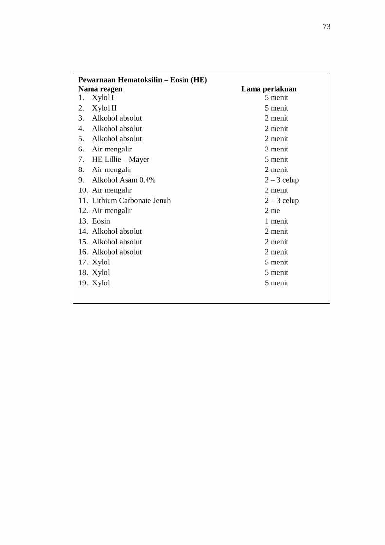
Pewarnaan Hematoksilin – Eosin (HE)
Nama reagen Lama perlakuan
1. Xylol I 5 menit
2. Xylol II 5 menit
3. Alkohol absolut 2 menit
4. Alkohol absolut 2 menit
5. Alkohol absolut 2 menit
6. Air mengalir 2 menit
7. HE Lillie – Mayer 5 menit
8. Air mengalir 2 menit
9. Alkohol Asam 0.4% 2 – 3 celup
10. Air mengalir 2 menit
11. Lithium Carbonate Jenuh 2 – 3 celup
12. Air mengalir 2 me
13. Eosin 1 menit
14. Alkohol absolut 2 menit
15. Alkohol absolut 2 menit
16. Alkohol absolut 2 menit
17. Xylol 5 menit
18. Xylol 5 menit
19. Xylol 5 menit

74
Lampiran 8. Dokumentasi
Mencit dikelompokkan ke dalam tiga kandang
Injeksi ovalbumin intraperitoneal Pemberian ekstrak kunyit
menggunakan sonde lambung

75
Inhalasi ovalbumin Pembedahan mencit
Organ paru yang dibenamkan dalam larutan Buffered Neutral Formalin

76
Pembuatan blok parafin
Pemotongan blok jaringan dengan Potongan jaringan diletakkan dalam
mikrotom air panas dan diletakkan pada objekglass

77
Pengecatan HE
Pembuatan preparat selesai dan siap di periksa melalui mikroskop

78
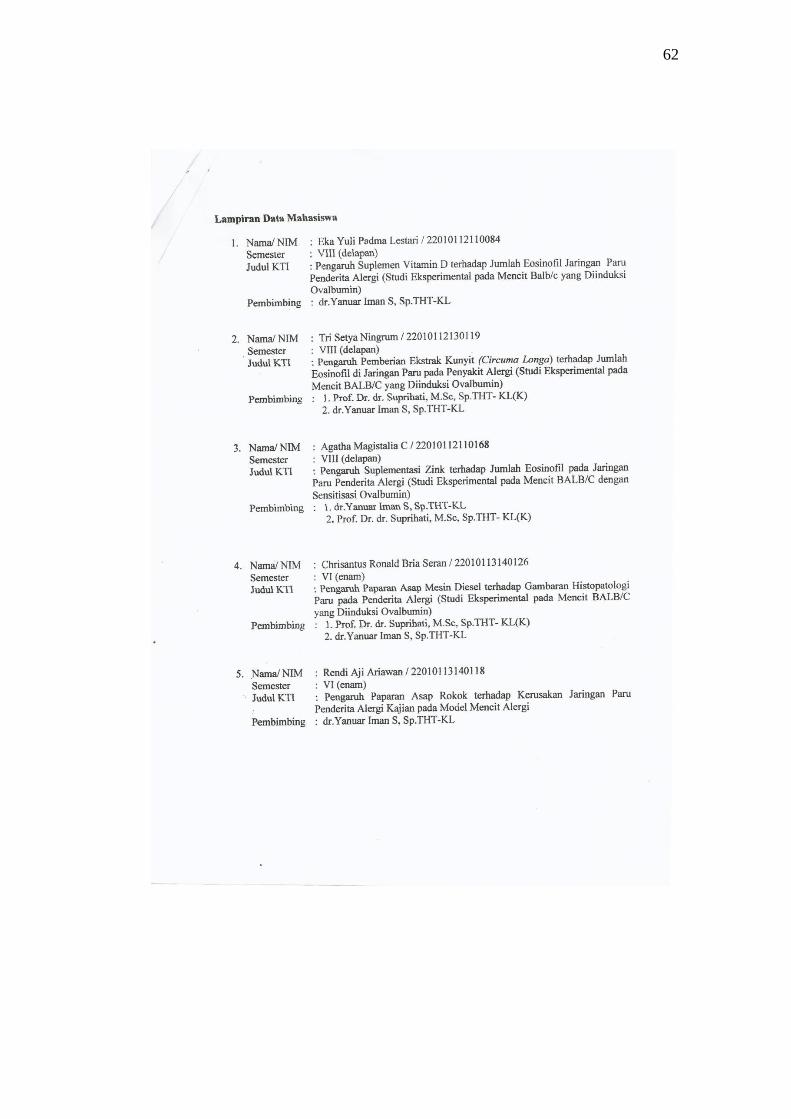
Lampiran 9. Biodata Mahasiswa
Identitas
Nama : Tri Setya Ningrum
NIM : 22010112130119
Tempat/Tanggal Lahir : Banyumas, 17 Januari 1994
Jenis Kelamin : Perempuan
Alamat : Desa Karangduren RT 02 RW 02, Sokaraja, Banyumas
Jl. Menoreh Utara V No.7, Sampangan, Semarang
Nomor Telepon : -
Nomor HP : 085727927898
Email : [email protected]
Riwayat Pendidikan Formal
1. SD : SD Negeri 1 Karangduren Lulus Tahun : 2006
2. SMP : SMP Negeri 1 Purwokerto Lulus Tahun : 2009
3. SMA : SMA Negeri 1 Purwokerto Lulus Tahun : 2012
4. S1 : Fakultas Kedokteran Universitas Diponegoro Masuk Tahun: 2012


![Daftar Pustaka [799.8 KB]](https://img.pdfslide.us/doc/110x75/5879e7431a28abd0398c04b0/daftar-pustaka-7998-kb.jpg)
![Daftar Pustaka [1724 KB]](https://img.pdfslide.us/doc/110x75/586771db1a28ab17578b5daa/daftar-pustaka-1724-kb.jpg)


![DAFTAR PUSTAKA - etd.repository.ugm.ac.idetd.repository.ugm.ac.id/downloadfile/96937/potongan/S1-2016... · 90 DAFTAR PUSTAKA DAFTAR PUSTAKA [1] Badan Standardisasi Nasional. “SNI](https://img.pdfslide.us/doc/110x75/5ccf188d88c99385278e02a1/daftar-pustaka-etd-90-daftar-pustaka-daftar-pustaka-1-badan-standardisasi.jpg)