Embed Size (px)
Citation preview

© 2015 Wu et al. This work is published by Dove Medical Press Limited, and licensed under Creative Commons Attribution – Non Commercial (unported, v3.0) License. The full terms of the License are available at http://creativecommons.org/licenses/by-nc/3.0/. Non-commercial uses of the work are permitted without any further
permission from Dove Medical Press Limited, provided the work is properly attributed. Permissions beyond the scope of the License are administered by Dove Medical Press Limited. Information on how to request permission may be found at: http://www.dovepress.com/permissions.php
International Journal of Nanomedicine 2015:10 5219–5235
International Journal of Nanomedicine Dovepress
submit your manuscript | www.dovepress.com
Dovepress 5219
O r I g I N a l r e s e a r c h
open access to scientific and medical research
Open access Full Text article
http://dx.doi.org/10.2147/IJN.S82847
d-α-tocopherol polyethylene glycol succinate-based derivative nanoparticles as a novel carrier for paclitaxel delivery
Yupei Wu1,*Qian chu2,*songwei Tan1
Xiangting Zhuang1
Yuling Bao1
Tingting Wu1
Zhiping Zhang1,3,4
1Tongji school of Pharmacy, 2Department of Oncology, Tongji hospital, Tongji Medical school, 3hubei engineering research center for NDDs, 4National engineering research center for Nanomedicine, huazhong University of science and Technology, Wuhan, People’s republic of china
*These authors contributed equally to this work
Abstract: Paclitaxel (PTX) is one of the most effective antineoplastic drugs. Its current clinical
administration Taxol® is formulated in Cremophor EL, which causes serious side effects.
Nanoparticles (NP) with lower systemic toxicity and enhanced therapeutic efficiency may be
an alternative formulation of the Cremophor EL-based vehicle for PTX delivery. In this study,
novel amphipathic 4-arm-PEG-TPGS derivatives, the conjugation of d-α-tocopherol poly-
ethylene glycol succinate (TPGS) and 4-arm-polyethylene glycol (4-arm-PEG) with different
molecular weights, have been successfully synthesized and used as carriers for the delivery
of PTX. These 4-arm-PEG-TPGS derivatives were able to self-assemble to form uniform NP
with PTX encapsulation. Among them, 4-arm-PEG5K
-TPGS NP exhibited the smallest particle
size, highest drug-loading efficiency, negligible hemolysis rate, and high physiologic stability.
Therefore, it was chosen for further in vitro and in vivo investigations. Facilitated by the effec-
tive uptake of the NP, the PTX-loaded 4-arm-PEG5K
-TPGS NP showed greater cytotoxicity
compared with free PTX against human ovarian cancer (A2780), non-small cell lung cancer
(A549), and breast adenocarcinoma cancer (MCF-7) cells, as well as a higher apoptotic rate and
a more significant cell cycle arrest effect at the G2/M phase in A2780 cells. More importantly,
PTX-loaded 4-arm-PEG5K
-TPGS NP resulted in a significantly improved tumor growth inhibitory
effect in comparison to Taxol® in S180 sarcoma-bearing mice models. This study suggested that
4-arm-PEG5K
-TPGS NP may have the potential as an anticancer drug delivery system.
Keywords: 4-arm-PEG, TPGS, paclitaxel, nanoparticles, antitumor
IntroductionNanotechnology has been widely applied to anticancer drug delivery with the advan-
tages of high drug loading and encapsulation efficiency, enhanced cellular uptake, as
well as improved therapeutic effects and reduced side effects of the formulated drugs.1–3
Many nanosized formulations, including nanoparticles (NP), liposomes, microspheres,
polymer conjugates, dendritic polymers, and water-soluble prodrugs,4–9 have been
investigated and shown remarkable therapeutic efficiency. Nanomedicine can penetrate
through capillaries and be taken up by cells, leading to efficient drug accumulation
at target sites. Moreover, sustained and controlled release of drugs at target sites can
last over a period of days or even weeks, thereby offering the following enormous
advantages, such as reduction of dosage, improvement on the pharmacokinetic/dynamic
properties, protection of drugs against degradation, reduced side effects, etc.10 The
developments of nanomedicine have the potential to solve many of modern medicine’s
intractable problems, as evidenced from the fact that over 200 nanomedicine products
are approved or in different stages of clinical trials.11
correspondence: Zhiping ZhangNational engineering research center for Nanomedicine, huazhong University of science and Technology, Wuhan 430030, People’s republic of chinaTel +86 27 8360 1832email [email protected]
Journal name: International Journal of NanomedicineArticle Designation: Original ResearchYear: 2015Volume: 10Running head verso: Wu et alRunning head recto: 4-arm-PEG-TPGS nanoparticles for paclitaxel deliveryDOI: http://dx.doi.org/10.2147/IJN.S82847

International Journal of Nanomedicine 2015:10submit your manuscript | www.dovepress.com
Dovepress
Dovepress
5220
Wu et al
Paclitaxel (PTX), one of the internationally acknowledged
anticancer drugs, has excellent therapeutic activities against a
wide spectrum of cancers, including breast, brain, pancreatic,
ovarian, and non-small cell lung cancers.12 However, PTX
shows limitations in clinical application due to its poor aque-
ous solubility.13 Its current clinical administration, Taxol®, is
formulated in Cremophor EL and dehydrated alcohol (1:1, v/v),
which is diluted 5–20-fold in normal saline or glucose injection
before administration. Unfortunately, Cremophor EL is not well
tolerated and is associated with various severe side effects, such
as hypersensitivity reactions, gastrointestinal toxicity, cardio-
toxicity, and neurotoxicity.14 Hence, it is essential to develop a
new carrier to solve the formulation problem of PTX.
D-α-tocopheryl polyethylene glycol succinate (vitamin E
TPGS or simply TPGS), which has been approved by the
Food and Drug Administration as a pharmaceutical ingredi-
ent, is a water-soluble derivative of natural vitamin E. As
a PEGylated vitamin E, TPGS has an amphiphilic structure
of lipophilic alkyl tail and hydrophilic polar head with a
relatively low critical micelle concentration of 0.02% w/w.15
Its bulky structure and large surface area make it a safe phar-
maceutic adjuvant such as absorption enhancer, emulsifier,
solubilizer, and stabilizer.16 In addition, TPGS has also been
utilized as a P-glycoprotein inhibitor to overcome multidrug
resistance and to greatly improve the oral bioavailability of
anticancer drugs.17–20 In the past decade, TPGS-based deriva-
tives, which can significantly enhance the solubility and sta-
bility of the formulated drug and realize sustained, controlled,
and targeted drug delivery, have been widely investigated.21
Nevertheless, the application of independent TPGS micelles
for drug delivery is limited by the disadvantage that they
were not stable enough in physiological environments.22
Furthermore, the polyethylene glycol (PEG) chain of TPGS
is not long enough to ensure the micelles to prevent opsonin
bindings and realize the extended blood circulation time.23
PEGylation is a well-used technology in the pharmaceuti-
cal industry due to the aqueous solubility, biocompatibility,
and non-immunogenicity of PEG.24 Several new PEGylated
TPGS-based micelles with improved physiological stability
have been reported including TPGS2K
, PLV2K
, and PEG5K
-
VE2.23,25,26 Recently, 4-armed copolymers have been receiving
great attention because of their unique properties.27,28 It has
been reported that 4-armed copolymers presented a lower
surface tension, greater stability, and higher drug entrapment
efficiency.4,29 Hence, we designed and synthesized TPGS-
based derivatives – 4-arm-PEG-TPGS – as nanoplatforms
for hydrophobic drug PTX delivery.
In this study, novel derivatives based on 4-arm-PEG of
different molecular weights and TPGS were synthesized and
investigated. PTX-loaded 4-arm-PEG-TPGS NP (PTX-NP)
were prepared and characterized by particle size, morphol-
ogy, and drug loading efficiency. The release behavior
and stability in vitro of the NP were also investigated. The
cell cytotoxicity was carefully evaluated in human ovarian
cancer A2780, non-small cell lung cancer A549, human
breast cancer cells MCF-7, and mouse sarcoma tumor cell
line S180. The cellular uptake, induction of apoptosis, and
retardation of cell cycle of NP were studied against A2780
cells. The tumor inhibition effect was further evaluated in
S180 sarcoma-bearing mice models.
Materials and methodsMaterialsPTX of purity 99% was obtained from Jinhe Limited,
People’s Republic of China. 4-arm-PEG (molecular weight
of 5, 10, 20 kDa) were purchased from Sinopeg Biotech
Co., People’s Republic of China. TPGS, succinic anhy-
dride (SA), dicyclohexylcarbodiimide, propidium iodide
(PI), RNase A, and trypsin-ethylenediaminetetraacetic acid
(EDTA) were supplied by Sigma-Aldrich (St Louis, MO,
USA). 4-dimethylamino pyridine (DMAP) was purchased
from Aladdin, People’s Republic of China. RPMI-1640
medium was from Gibco BRL (Gaithersburg, MD, USA).
Taxol® was obtained from Bristol-Myers Squibb Caribbean
Company. Penicillin-streptomycin, fetal bovine serum (FBS),
and trypsin without EDTA were obtained from Hyclone
(Waltham, MA, USA). MTT (3-[4,5-dimethylthiazol-2-
yl]-2,5 diphenyltetrazolium bromide) and Hoechst 33342
were purchased from Biosharp, South Korea. The annexin
V-fluorescein isothiocyanate (FITC)/PI double staining
assay kit was supplied by KeyGEN, People’s Republic of
China. All of the solvents used were of analytical grade
and were procured from Sinopharm, People’s Republic of
China. Human ovarian cancer cell line A2780, human breast
adenocarcinoma cell line MCF-7, non-small cell lung cancer
cell line A549, and mouse sarcoma tumor cell line S180 were
provided by the Shanghai Institute of Biochemistry and Cell
Biology, Shanghai Institute for Biological Sciences, Chinese
Academy of Sciences, People’s Republic of China. Kunming
mice (female, 5–7 weeks old, 18–20 g) were obtained from
Laboratory Animal Resources of Huazhong University of
Science and Technology (Certificate No SCXK 2010-0009).
The animals were maintained at 25°C±1°C and 60%±10%
humidity under a 12-hours light–dark cycle during the
experiments. All animals were maintained under the spe-
cific pathogen-free (SPF) condition in the Animal Center of
Huazhong University of Science and Technology, People’s
Republic of China. All animals were treated according to the

International Journal of Nanomedicine 2015:10 submit your manuscript | www.dovepress.com
Dovepress
Dovepress
5221
4-arm-Peg-TPgs nanoparticles for paclitaxel delivery
regulations of Chinese law and the study was approved by
the local Ethical Committee Quantita.
synthesis and characterization of derivatives4-arm-PEG
5K-TPGS, 4-arm-PEG
10K-TPGS, and 4-arm-
PEG20K
-TPGS were synthesized by a two-step conjugation
method, shown in Figure 1. TPGS was first functionalized
with a carboxylic acid group by esterification with SA,
according to our previous work.30 The second step was
the formation of ester bond between the primary groups
hydroxy of 4-arm-PEG and carboxylic acid functions of
activated TPGS-SA. Briefly, TPGS-SA (1.64 g, 1.0 mmol),
4-dimethylamino pyridine (0.12 g, 1.2 mmol), and dicyclo-
hexylcarbodiimide (0.20 g, 1.2 mmol) were co-dissolved
in 5 mL anhydrous dichloromethane (DCM) and reacted at
room temperature for 24 hours. The turbid liquid was filtered
to remove N,N-dicyclohexylurea and mixed with a 5 mL
solution containing 0.1 mmol 4-arm-PEG. After 24 hours,
the products were precipitated in diethyl ether and washed
three times and dried under vacuum.
The structure of resultant TPGS-based derivatives
4-arm-PEG-TPGS were characterized by 1H-NMR spec-
tra (Bruker AVANCE III 400 MHz NMR spectrometer,
CDCl3) and Fourier transform infrared spectroscopy (FTIR)
(Bruker VERTEX 70 FTIR spectrophotometer). Gel per-
meation chromatography (GPC) (Waters-2410 system)
was also carried out to measure the molecular weights
of 4-arm-PEG-TPGS. Tetrahydrofuran was used as the
mobile phase at a flow rate of 1.0 mL/min. The molecular
weights were calculated by using a calibration curve con-
structed using polystyrene as the standard. The solubility
of the materials in water was estimated simply by visual
determination.
Preparation and characterization of NPThe PTX-loaded NP were prepared by a solid dispersion
method. Typically, PTX (1, 1.5, or 2 mg) and 4-arm PEG-
TPGS (10 mg) were dissolved in 2 mL of DCM by sonication.
The organic solvent was evaporated on a rotary evaporator
under reduced pressure at 37°C to obtain a homogenous
coevaporation PTX/copolymer film. Subsequently, the film
was hydrated with 5 mL phosphate buffered saline (PBS),
incubated at 37°C for 30 minutes. The resultant mixture
was centrifuged at 3,000 rpm for 10 minutes to remove the
nanoparticles and free PTX. The blank NP were prepared in
a similar manner without PTX added.
The average size, size distribution, and ζ potential of the
obtained NP were determined by dynamic light scattering
(DLS) (ZetaPlus, Brookhaven Instruments, USA). Data were
Figure 1 synthetic route of 4-arm-Peg-TPgs.Abbreviations: Peg, polyethylene glycol; TPgs, d-α-tocopherol polyethylene glycol succinate; sa, succinic anhydride; DMaP, 4-dimethylamino pyridine; Dcc, dicyclohexylcarbodiimide; rT, room temperature.

International Journal of Nanomedicine 2015:10submit your manuscript | www.dovepress.com
Dovepress
Dovepress
5222
Wu et al
displayed as the mean value of at least three measurements ±
standard deviation. The morphology of the NP was observed
by transmission electron microscope (JEM-1230, Japan).
The NP were diluted with distilled water and placed on a
copper grid covered with nitrocellulose, and dried at room
temperature before measurement.
A steady-state pyrene fluorescence method was used to
determine the critical aggregate concentration (CAC) of the
TPGS-based derivatives. Steady-state fluorescence spectra
were obtained on a Hitachi F-4600 luminescence spectrometer.
Fifty microliter of 4.8×10⁻⁵ M solution of pyrene in acetone was
added in the centrifuge tube. Acetone was then evaporated and
replaced with 4 mL solution of 4-arm PEG-TPGS with concen-
trations ranging from 0.01 to 1,000 μg/mL to get a final pyrene
concentration of 6×10⁻⁷ M. The solution was incubated over-
night. Excitation spectra of the sample solutions were obtained
at an emission wavelength of 372 nm with excitation spectra
(300–350 nm). The change of the fluorescence intensity ratio
(I339
/I335
) was analyzed as a function of the CAC value.
The stability of the NP was investigated by measuring
the sizes of samples at different time points. To examine
the effect of serum on particle stability, the PTX-loaded
NP samples were prepared with PBS at a concentration of
10 mg/mL, and then diluted with FBS or PBS by the ratio of
1:9. The changes in NP size were monitored by DLS.
Encapsulation efficiency and drug loading contentThe amount of PTX encapsulated in the NP was measured by
high-performance liquid chromatography (HPLC) (Hitachi
2000, Japan) equipped with an L-2130 pump, an L-2400 UV
detector, and an Inertsil® ODS-3 C18 reversed phase column
(150 mm ×4.6 mm, 5 μm) (Agilent, Santa Clara, CA, USA).
Briefly, 2 mg of the freeze-dried NP powder was dissolved
in 1 mL DCM in order to disrupt the NP structure and then
the solution was dried under nitrogen. Three milliliter mobile
phase (acetonitrile/water, 50:50, v/v) was added to dissolve
the drugs. The solution was then filtered by 0.45 μm filter for
HPLC analysis. The column effluent was detected at 227 nm
with a UV detector. The mobile phase was pumped at a flow
rate of 1.0 mL/min. The drug encapsulation efficacy (EE)
was obtained by the following equations:
EE (Weight of PTX in NP
Weight of feeding PTX%) %.= ×100
hemolytic effect of 4-arm-Peg-TPgsFresh blood from Sprague Dawley rat was collected in
heparinized tubes and washed three times with ice-cold
0.9% sodium chloride (NaCl) by centrifugation at 3,000 rpm
for 5 minutes at 4°C. The obtained red blood cells (RBCs)
were diluted to 2% (w/v) by ice-cold 0.9% NaCl containing
various concentrations (0.001, 0.01, 0.1, 1.0, and 5.0 mg/mL)
of 4-arm-PEG-TPGS and polyethylenimine (PEI) (25 kDa),
respectively, and then incubated at 37°C in an incubator
shaker for 4 hours. The samples were then centrifuged at
3,000 rpm for 10 minutes at 4°C, and 100 μL of supernatant
from each sample was transferred into a 96-well plate. The
absorbance of the supernatant was determined at 540 nm
using a microplate reader (Multiskan MK3; Thermo Sci-
entific, USA). RBCs treated with distilled water and 0.9%
NaCl were considered as the positive (100% hemolysis) and
negative (0% hemolysis) controls, respectively. The hemo-
lytic effect of Cremophor EL-based vehicle (Cremophor
EL and dehydrated alcohol, 1:1, v/v) was also assessed.
The degree of hemolysis was determined by the following
equation:
Hem (%) 1Sample
1
=−
−×
A A0
00 0
00A A
%
where A100
and A0 were the absorbances of the solution at
100% and 0% hemolysis, respectively.
In vitro release studyThe drug release behavior of NP was investigated by using
a dialysis method. Four milliliters of PTX-loaded 4-arm-
PEG5K
-TPGS NP (PTX-NP5K
) was placed in a dialysis bag
(Snakeskin, Pierce, USA) with a molecular weight cut-off of
2,000 Da. The dialysis bag was suspended in 50 mL of PBS
(pH 7.4) or FBS and placed in a shaking water bath at 37°C
with a shaking speed of 120 rpm. At every predetermined
time, 10 mL of the solution was removed followed by an
addition of 10 mL fresh PBS. PTX of the collected incuba-
tion medium was extracted by DCM. The drug concentration
was determined by HPLC as described earlier.
cell cultureAll cell lines were cultured in RPMI-1640 containing 10%
FBS and 1% penicillin-streptomycin in humidified environ-
ment at 37°C with 5% carbon dioxide (CO2). After the cells
grew to 80%–90% confluence, they were trypsinized with
0.25% trypsin-EDTA.
In vitro cellular uptakeCellular uptake was analyzed by confocal laser scanning
microscopy (CLSM) (Leica TCSNT1, Germany) and
coumarin-6, a widely used replacement fluorescent marker
International Journal of Nanomedicine 2015:10 submit your manuscript | www.dovepress.com
Dovepress
Dovepress
5223
4-arm-Peg-TPgs nanoparticles for paclitaxel delivery
of hydrophobic drug, was used as the probe. Coumarin-6
loaded NP of 4-arm-PEG5K
-TPGS (coumarin-6-NP5K
) were
prepared. A2780 cells were seeded onto a 24-well plate at
a density of 1.0×10⁴ cells/well. After 24 hours attachment,
they were incubated with coumarin-6-M at a concentration
of 25 μg/mL for 2 hours at 37°C. The wells were then rinsed
carefully three times with cold PBS and fixed with 4%
paraformaldehyde for 15 minutes. After being washed twice
again with cold PBS, the cells were stained with Hoechst
33342 for 8 minutes and then mounted on a glass slide for
observation by CLSM.
The cellular uptake was further studied by a flow cytom-
eter (Becton Dickinson, San Jose, CA, USA). A2780 cells
were seeded into six-well black plates at 5×10⁵ cells/well;
after the cells reached 80% confluence, the medium was
changed to the suspension of coumarin-6-M at an NP concen-
tration of 25 μg/mL and incubated for 0.5, 1, 2, and 4 hours,
respectively. Cells treated with only medium were used as
control. After incubation, the wells were rinsed three times
with cold PBS, and then cells were collected by centrifuga-
tion and resuspended in 0.5 mL PBS. The amount of uptake
was analyzed by flow cytometry.
In vitro cytotoxicityThe cytotoxicity of PTX formulated in 4-arm-PEG
5K-TPGS
NP was assessed with four types of cancer cell lines (A2780,
MCF-7, A549, and S180) and compared to Taxol® formula-
tion and free PTX (DMSO-dissolved, final DMSO concentra-
tion #0.1%). Briefly, A2780, MCF-7, and A549 cells in their
logarithmic growth were seeded in 96-well plates at a density
of 5,000 cells/well. Following overnight attachment, the cul-
ture medium in each well was carefully replaced with 100 μL
of medium containing Taxol®, PTX-NP5K
, or free PTX at PTX
concentrations ranging from 0.025 to 100 μg/mL (n=8). Cells
treated with only RPMI-1640 medium were tested as controls.
S180 cell was promptly seeded with a density approximately
5,000 cells/well before assay. After treatment for 24, 48, and
72 hours, respectively, the relative cell viability was assessed
by an MTT assay as described in our previous work.31 IC50
(concentration resulting in 50% inhibition of cell growth)
value was determined by SPSS software (version 19.0). The
experiment was repeated thrice.
apoptosis analysisThe qualitative apoptosis of the A2780 cell line treated
with different PTX formulation was determined by the
Hoechst 33342 staining method. Specifically, A2780 cells
were seeded onto a 24-well plate (104 cells/well). Following
overnight attachment, the cells were then treated with
medium containing Taxol®, PTX-NP5K
, or free PTX at the
same PTX concentration of 2.5 μg/mL. The control group
was incubated with drug-free culture medium. After incuba-
tion for 24 hours, the wells were rinsed three times with cold
PBS and then fixed with 200 μL of 4% paraformaldehyde
for 15 minutes. The cells were further washed three times
with 500 μL PBS, followed by staining with 200 μL Hoechst
33342 (10 μg/mL) for 8 minutes in the dark. After being
triple-washed with PBS, the cells were finally observed by a
fluorescence microscope (IX71; Olympus, Tokyo, Japan).
Annexin V-FITC/PI double staining is a sensitive method
for detecting quantitative apoptosis. A2780 cells were
seeded onto six-well plates at a density of 5×10⁵ cells/well,
followed by attachment for 24 hours. The cells were then
incubated with Taxol®, PTX-NP5K
, or free PTX at the PTX
concentration of 2.5 μg/mL in culture medium; untreated
cells were used as the control. After 24 hours incubation, the
cells were trypsinized, collected, and resuspended in 300 μL
of binding buffer. Thereafter, 3 μL of annexin V-FITC and
3 μL of PI were added and mixed for 30 minutes in the dark.
The stained cells were analyzed using a flow cytometer. The
quantitative apoptosis of S180 cells were detected in a similar
way except that the cell was promptly seeded before assay.
cell cycle distribution analysisCell cycle distribution analysis was further investigated.
A2780 cells were seeded onto six-well plates (1.0×10⁵ cells/
well). After attachment overnight, the cells were exposed
to Taxol®, PTX-NP5K
, or free PTX (drug concentration of
2.5 μg/mL). Cells treated with only medium were used
as controls. After 24 hours of incubation, the cells were
washed twice with cold PBS and fixed overnight with 70%
precooled alcohol at 4°C. The cells were washed twice
with cold PBS to eliminate alcohol and then incubated with
RNase A (100 μg/mL) for 15 minutes at 37°C, followed by
staining with PI solution (50 μg/mL) for 30 minutes in the
dark. The distribution of DNA content was analyzed by the
flow cytometry method and the percentage of cells in each
phase of the cell cycle was calculated using ModFit software
(Verity Software House, Topsham, ME, USA).
In vivo therapeutic studyTumor inhibition activity against a solid tumor model was
evaluated using female Kunming mice. Kunming mice were
subcutaneously injected at the right forelimb axilla with
0.2 mL S180 cell suspension containing 1×10⁷ cells. After
48 hours of transplantation, all the tumor-bearing mice were

International Journal of Nanomedicine 2015:10submit your manuscript | www.dovepress.com
Dovepress
Dovepress
5224
Wu et al
divided randomly into four groups (n=5). Treatment started
when the tumor volume of the mice reached 100–150 mm3
on average, and this was designated as day 1. Each group
was treated by tail vein injection on days 1, 3, 5, and 7 with
saline, PTX-NP5K
(at a dosage of 10 mg/kg), PTX-NP5K
(30 mg/kg), and Taxol® (10 mg/kg), respectively. Tumor
sizes were measured every day to evaluate the antitumor
efficiency. When the tumor length in the saline group was
greater than 20 mm, all the mice were sacrificed and the
tumors were extirpated and weighed. The tumor was finally
cut into small histological sections and stained with hema-
toxylin and eosin for histological analysis by light micros-
copy with a CAD system.
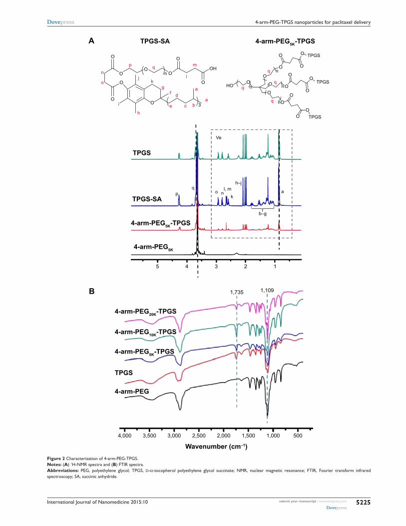
Results and discussionsynthesis and characterization of 4-arm-Peg-TPgsThree types of 4-arm-PEG-TPGS derivatives were syn-
thesized with various molecular weights of 4-arm-PEG (5,
10, 20 kDa). The products were investigated by 1H-NMR,
FTIR, and GPC analysis to confirm the successful conjuga-
tion. As shown in Figure 2A, the newly appearing signals at
2.65–2.72 ppm were assigned to the -CH2CH
2- part of the
succinyl group of TPGS-SA, verifying the esterification of
TPGS as compared to the TPGS spectrum.30 Taking 4-arm-
PEG5K
-TPGS as an example of the 4-arm-PEG-TPGS copo-
lymers, the intensity of 3.65 ppm ascribed to -OCH2CH
2- in
the PEG chain was significantly increased compared to that
of TPGS, proving the conjugation of TPGS with 4-arm-
PEG. The TPGS contents in 4-arm-PEG-TPGS derivative
were calculated on the basis of the peak area of 0.86 ppm
and 3.65 ppm, which were 78%, 68%, and 57% (denoted
as 4-arm-PEG5K
-TPGS, 4-arm-PEG10K
-TPGS, and 4-arm-
PEG20K
-TPGS), respectively. The structure of 4-arm-PEG-
TPGS was further studied by FTIR (Figure 2B). There were
no obvious differences among them. The characteristic peaks
of TPGS exhibited in the derivative, such as the vibration
peak of the C=O bond (νC=O
) of the ester bond at 1,735 cm−1
and the C–O stretching vibration (νC–O
) of PEG at 1,109 cm−1.
However, the enhancement of the peak at 1,109 cm−1 can
indicate the formation of 4-arm-PEG-TPGS.
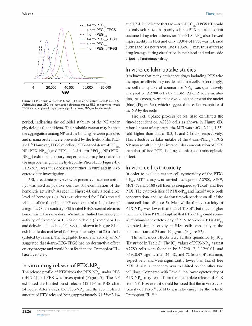
GPC was also performed. As shown in Figure 3, the
4-arm-PEG-TPGS exhibited increased molecular weight
with narrow molecular weight distribution and a significant
peak shift compared with 4-arm-PEG. This proved the suc-
cessful grafting of TPGS onto 4-arm-PEG. It should be noted
that the 4-arm-PEG-TPGS derivatives showed drastic dif-
ference in solubility. The solubility of 4-arm-PEG5K
-TPGS
was .10 mg/mL, while for 4-arm-PEG10K
-TPGS and 4-arm-
PEG20K
-TPGS, they were only approximately 1 mg/mL,
which might limit their applications.
Preparation and characterization of NPThe PTX-loaded NP were fabricated by a solid dispersion
method in this study. The 4-arm-PEG-TPGS NP were pre-
pared with spherical morphology and narrow size distribution
(as shown in Figure 4A–F). The diameter observed from DLS
was a little larger compared with the transmission electron
microscope result. This may be attributed to the fact that the
particle size measured by DLS was hydrodynamic diameter
with a solvation layer on the surface of the particles.32 As
seen from Table 1, the ζ potentials of 4-arm-PEG-TPGS NP
were all negative, and higher than that of TPGS micelles.
This may be caused by the different PEG densities on the
NP surface.
The CAC of 4-arm-PEG-TPGS was tested by the
pyrene fluorescence probe method. The CAC values were
obtained by plotting the I339
/I335
ratio of each curve in the
excitation spectra versus log concentration of the polymer.
The CAC values were 3.3×10⁻³, 4.9×10⁻³, and 5.6×10⁻³ g/L
for 4-arm-PEG5K
-TPGS, 4-arm-PEG10K
-TPGS, and 4-arm-
PEG20K
-TPGS, respectively (Figure 4G), which were
approximately similar to that of surfactant TPGS.17 The
CAC value was essential to evaluate the formation of NP.
It was anticipated that the NP with low CAC value would
be intact even on high dilution by a much larger volume of
blood in vivo.23
The relationship between the drug encapsulation effi-
ciency and the drug feeding ratio of TPGS and the three types
of NP were further studied. As seen in Table 1, the EE of the
NP were decreased along with the drug feeding ratio rising.
TPGS micelles exhibited EE as low as 9.9% for 20% drug
feeding ratio. The EE of 4-arm-PEG5K
-TPGS could be up to
91.7%±6.5% for 10% drug feeding ratio and was much higher
than 32.6%±3.2% for 4-arm-PEG10K
-TPGS and 43.1%±2.5%
for 4-arm-PEG20K
-TPGS. This may be caused by the differ-
ence of the binding affinity between hydrophobic PTX and
the hydrophobic core region from 4-arm-PEG-TPGS.33 It is
worth noting that the PTX concentration in PTX-NP5K
injec-
tion could be as high as 0.7 mg/mL. To summarize, 4-arm-
PEG5K
-TPGS was expected to be a better drug carrier with
smaller particle size and higher drug loading capacity.
DLS was also used to assess the colloidal stability of
the NP. As shown in Figure 4H, the mean diameter of PTX-
NP5K
did not remarkably change both in PBS and FBS.
Moreover, no drug precipitation was observed during this
International Journal of Nanomedicine 2015:10 submit your manuscript | www.dovepress.com
Dovepress
Dovepress
5225
4-arm-Peg-TPgs nanoparticles for paclitaxel delivery
OH
O
p
pq
o nk
l, m
b–g
a
h–j
q
q
q
kg
l
m
h
i
f d
e c ba
a
jo
nO
O
O
OO
O
O
O
3
n
HOO
O
O
O
O
O
OO
O
OO
O
OO
Ve
5 4 3
1,735
4,000 3,500 3,000 2,500
Wavenumber (cm–1)1,500 1,000 5002,000
1,109
2 1
O
On
n
n
n
TPGS
TPGS
TPGS
4-arm-PEG5K-TPGS
4-arm-PEG5K-TPGS
4-arm-PEG5K
TPGS-SA
TPGS
TPGS-SA
4-arm-PEG5K-TPGS
4-arm-PEG10K-TPGS
4-arm-PEG20K-TPGS
4-arm-PEG
TPGS
A
B
Figure 2 characterization of 4-arm-Peg-TPgs.Notes: (A) 1h-NMr spectra and (B) FTIr spectra.Abbreviations: Peg, polyethylene glycol; TPgs, d-α-tocopherol polyethylene glycol succinate; NMr, nuclear magnetic resonance; FTIr, Fourier transform infrared spectroscopy; sa, succinic anhydride.
International Journal of Nanomedicine 2015:10submit your manuscript | www.dovepress.com
Dovepress
Dovepress
5226
Wu et al
period, indicating the colloidal stability of the NP under
physiological conditions. The probable reason may be that
the aggregation among NP and the binding between particles
and plasma protein were prevented by the hydrophilic PEG
shell.34 However, TPGS micelles, PTX-loaded 4-arm-PEG10K
NP (PTX-NP10K
), and PTX-loaded 4-arm-PEG20K
NP (PTX-
NP20K
) exhibited contrary properties that may be related to
the improper length of the hydrophilic PEG chain (Figure 4I).
PTX-NP5K
was thus chosen for further in vitro and in vivo
cytotoxicity investigation.
PEI, a cationic polymer with potent cell surface activ-
ity, was used as positive contrast for examination of the
hemolytic activity.26 As seen in Figure 4J, only a negligible
level of hemolysis (,1%) was observed for RBCs treated
with all of the three blank NP even exposed to high dose of
5 mg/mL. On the contrary, PEI treated RBCs exerted obvious
hemolysis in the same dose. We further studied the hemolytic
activity of Cremophor EL-based vehicle (Cremophor EL
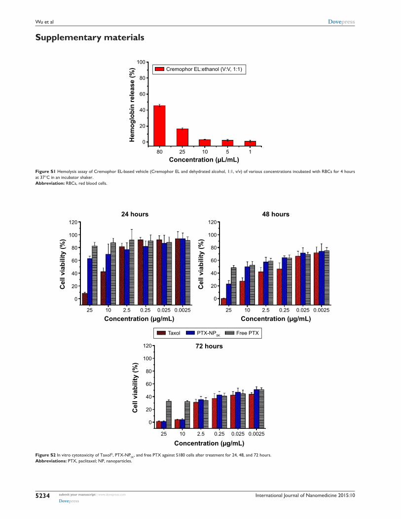
and dehydrated alcohol, 1:1, v/v), as shown in Figure S1, it
exhibited a distinct level (.10%) of hemolysis at 25 μL/mL
(diluted by saline). The negligible hemolytic activity of NP
suggested that 4-arm-PEG-TPGS had no destructive effect
on erythrocyte and would be safer than the Cremophor EL-
based vehicles.
In vitro drug release of PTX-NP5KThe release profile of PTX from the PTX-NP
5K under PBS
(pH 7.4) and FBS was investigated (Figure 5). The NP
exhibited the limited burst release (12.1%) in PBS after
24 hours. After 7 days, the PTX-NP5K
had the accumulated
amount of PTX released being approximately 31.5%±2.1%
at pH 7.4. It indicated that the 4-arm-PEG5K
-TPGS NP could
not only solubilize the poorly soluble PTX but also exhibit
sustained drug release behavior. The PTX-NP5K
also showed
high stability in FBS and only 18.8% of PTX was released
during the 168 hours test. The PTX-NP5K
may thus decrease
drug leakage during circulation in the blood and reduce side
effects of anticancer drug.
In vitro cellular uptake studiesIt is known that many anticancer drugs including PTX take
therapeutic effects only inside the tumor cells. Accordingly,
the cellular uptake of coumarin-6-NP5K
was qualitatively
analyzed on A2780 cells by CLSM. After 2 hours incuba-
tion, NP (green) were intensively located around the nuclei
(blue) (Figure 6A), which suggested the effective uptake of
the NP by the cells.
The cell uptake process of NP also exhibited the
time-dependent on A2780 cells as shown in Figure 6B.
After 4 hours of exposure, the MFI was 4.03-, 2.11-, 1.55-
fold higher than that of 0.5, 1, and 2 hours, respectively.
This effective cellular uptake of the 4-arm-PEG5K
-TPGS
NP may result in higher intracellular concentration of PTX
than that of free PTX, leading to enhanced antineoplastic
effect.
In vitro cell cytotoxicityIn order to evaluate cancer cell cytotoxicity of the PTX-
NP5K
, MTT assay was carried out against A2780, A549,
MCF-7, and S180 cell lines as compared to Taxol® and free
PTX. The cytotoxicities of PTX-NP5K
and Taxol® were both
concentration- and incubation time-dependent on all of the
three cell lines (Figure 7). Meanwhile, the cytotoxicity of
PTX-NP5K
was lower than that of Taxol®, but much higher
than that of free PTX. It implied that PTX-NP5K
could some-
what enhance the cytotoxicity of PTX. Moreover, PTX-NP5K
exhibited similar activity on S180 cells, especially in the
concentrations of 25 and 10 μg/mL (Figure S2).
The anticancer effects were further quantified by IC50
(illustrated in Table 2). The IC50
values of PTX-NP5K
against
A2780 cells were found to be 3.97±0.12, 1.12±0.01, and
0.19±0.07 μg/mL after 24, 48, and 72 hours of treatment,
respectively, and were significantly lower than that of free
PTX. A similar tendency was exhibited on the other two
cell lines. Compared with Taxol®, the lower cytotoxicity of
PTX-NP5K
may result from the incomplete release of PTX
from NP. However, it should be noted that the in vitro cyto-
toxicity of Taxol® could be partially caused by the vehicle
Cremophor EL.34–36
0 80,00060,00040,000
MW20,000
4-arm-PEG5K-TPGS4-arm-PEG5K
4-arm-PEG10K-TPGS4-arm-PEG10K
4-arm-PEG20K
4-arm-PEG20K-TPGS
Figure 3 gPc results of 4-arm-Peg and TPgs-based derivative 4-arm-Peg-TPgs.Abbreviations: gPc, gel permeation chromatography; Peg, polyethylene glycol; TPgs, d-α-tocopherol polyethylene glycol succinate; MW, molecular weight.

International Journal of Nanomedicine 2015:10 submit your manuscript | www.dovepress.com
Dovepress
Dovepress
5227
4-arm-Peg-TPgs nanoparticles for paclitaxel delivery
Figu
re 4
cha
ract
eriz
atio
n of
4-a
rm-P
eg-T
Pgs
nano
part
icle
s.N
otes
: (A
and
B)
Dls
res
ult
and
TeM
imag
e of
4-a
rm-P
eg5K
-TPg
s na
nopa
rtic
les,
(C
and
D)
Dls
res
ult
and
TeM
imag
e of
4-a
rm-P
eg10
K-T
Pgs
nano
part
icle
s, (
E a
nd F
) D
ls r
esul
t an
d T
eM im
age
of 4
-arm
-Peg
20K-T
Pgs
nano
part
icle
s,
(G)
plot
of t
he in
tens
ity r
atio
I 339/I
335 a
s a
func
tion
of lo
g c
for
TPg
s, 4
-arm
-Peg
5K-T
Pgs,
4-a
rm-P
eg10
K-T
Pgs,
and
4-a
rm-P
eg20
K-T
Pgs
nano
part
icle
s, (
H a
nd I
) th
e st
abili
ty o
f TPg
s m
icel
les
and
PTX
-NP
disp
erse
d in
PBs
and
FBs
, (J)
he
mol
ysis
ass
ay o
f bla
nk 4
-arm
-Peg
-TPg
s na
nopa
rtic
les
of v
ario
us c
once
ntra
tions
incu
bate
d w
ith r
Bcs
for
4 ho
urs
at 3
7°c
in a
n in
cuba
tor
shak
er.
Abb
revi
atio
ns: P
eg, p
olye
thyl
ene
glyc
ol; T
Pgs,
d-α
-toc
ophe
rol p
olye
thyl
ene
glyc
ol s
ucci
nate
; Dls
, dyn
amic
ligh
t sc
atte
ring
; TeM
, tra
nsm
issi
on e
lect
ron
mic
rosc
ope;
PT
X, p
aclit
axel
; NP,
nan
opar
ticle
s; P
Bs, p
hosp
hate
buf
fere
d sa
line;
FB
s, fe
tal b
ovin
e se
rum
; rBc
s, r
ed b
lood
cel
ls; P
eI, p
olye
thyl
enim
ine.

International Journal of Nanomedicine 2015:10submit your manuscript | www.dovepress.com
Dovepress
Dovepress
5228
Wu et al
00
10
20
30
40
50
60
40 80Time (hours)
Cum
ulat
ive
rele
ase
(%)
120 160
PBS pH 7.4FBS
Figure 5 In vitro release of PTX from PTX-NP5K in PBs (ph 7.4) and FBs.Abbreviations: PTX, paclitaxel; NP, nanoparticles; PBs, phosphate buffered saline; FBs, fetal bovine serum.
Table 1 characterization of PTX-loaded 4-arm-Peg-TPgs nanoparticles
Polymers Drug feeding concentration (wt %)
Particle sizea (nm)
PDIa ζ potential (mV)
EEb (%) PTX concentration (mg/mL)
TPgs 10 134.8±3.7 0.22±0.07 −3.44±0.04 18.2±0.3 0.090TPgs 15 158.2±9.3 0.25±0.04 −3.65±0.21 12.7±0.6 0.095TPgs 20 205.0±5.7 0.23±0.05 −3.78±0.39 9.9±0.5 0.104-arm-Peg5K-TPgs 10 260.0±1.1 0.24±0.06 −10.02±1.1 91.7±6.5 0.454-arm-Peg5K-TPgs 15 283.6±1.1 0.16±0.03 −9.81±0.22 79.8±2.4 0.604-arm-Peg5K-TPgs 20 307.3±3.7 0.21±0.03 −9.72±1.20 71.1±3.4 0.704-arm-Peg10K-TPgs 10 295.2±0.9 0.18±0.02 −13.02±1.5 32.6±3.2 0.164-arm-Peg10K-TPgs 15 305.4±3.7 0.22±0.01 −12.02±1.1 32.5±1.4 0.154-arm-Peg10K-TPgs 20 316.9±4.4 0.19±0.02 −11.99±1.1 29.4±3.6 0.304-arm-Peg20K-TPgs 10 313.7±4.7 0.15±0.02 −14.03±2.2 43.1±2.5 0.224-arm-Peg20K-TPgs 15 317.1±4.8 0.14±0.01 −13.67±1.7 35.3±1.6 0.27
4-arm-Peg20K-TPgs 20 329.7±5.5 0.20±0.02 −13.50±1.6 31.6±2.3 0.32
Notes: aMeasured by Dls. bMeasured by hPlc.Abbreviations: PTX, paclitaxel; Peg, polyethylene glycol; TPgs, d-α-tocopherol polyethylene glycol succinate; PDI, polydispersity index; EE, encapsulation efficacy; Dls, dynamic light scattering; hPlc, high-performance liquid chromatography.
cell apoptosis assaysIt has been widely reported that PTX kills cancer cells
through the induction of apoptosis.37 The apoptosis-inducing
ability of PTX-NP5K
was qualitatively evaluated via Hoechst
33342 staining nuclei of A2780 cells. As observed under
fluorescence microscopy, the cell nuclei showed a good integ-
rity in the control group. However, some typical apoptotic
features appeared in the PTX-NP5K
and Taxol® groups, such
as cell shrinkage, chromatin condensation, fragmentation of
the nucleus, and apoptosis bodies (Figure 8A). Moreover,
PTX-NP5K
and Taxol® induced more cell apoptosis than free
PTX, in accordance with the results of MTT assay.
Annexin V-FITC/PI staining assay was carried out to
quantitatively verify the cell apoptosis rate induced by dif-
ferent treatments. As shown in Figure 8B, after 24 hours
treatment, the percentages of early apoptotic cells (Q4,
annexin positive and PI negative) for Taxol®, PTX-NP5K
,
and free PTX were 22.3%, 9.6%, and 7.3%, while those of
late apoptotic cells (Q2, annexin, and PI double positive)
were 12.9%, 22.0%, and 6.2%, respectively. The quanti-
tative apoptosis of S180 cells showed a similar tendency
(Figure S3). Both the quantitative and qualitative results
demonstrated that PTX-NP5K
enhanced PTX-induced apop-
tosis compared with free PTX.
cell cycle arrest assaysThe antitumor efficacy of PTX is associated with mitosis
inhibition and cell arrest in the G2/M phase. Increased G2/M
phase arrest indicates the inhibition on cell division and
restraint on cell growth.38 The cell cycle of A2780 cells treated
with various formulation of PTX was examined to evaluate the
therapeutic effects of PTX. As seen from Figure 9, the G2/M
phase treated with PTX-NP5K
for 24 hours was significantly
increased to 70.8% compared with that of free PTX (43.7%).
The cell cycle arrest effect in the G2/M phase from PTX-NP5K
appeared to be consistent with the cell apoptosis analysis,
demonstrating strong antitumor efficacy.
antitumor activityThe in vivo antitumor efficiency of PTX-NP
5K was evaluated
in tumor-bearing mice. The mice were treated every other
day with saline, PTX-NP5K
(10 mg/kg and 30 mg/kg), and
Taxol® (10 mg/kg), respectively. Both PTX-NP5K
and Taxol®
demonstrated tumor growth inhibition (Figure 10A–C).
Tumors of saline, Taxol®, PTX-NP5K
(10 mg/kg), and PTX-
NP5K
(30 mg/kg) groups were 0.59±0.26 g, 0.38±0.19 g,
0.29±0.11 g, and 0.21±0.04 g, respectively. Clearly, PTX-NP5K

International Journal of Nanomedicine 2015:10 submit your manuscript | www.dovepress.com
Dovepress
Dovepress
5229
4-arm-Peg-TPgs nanoparticles for paclitaxel delivery
Figure 6 cellular uptake of coumarin-6-NP5K by a2780 cells.Notes: (A) clsM images after 2 hours incubation and (B) MFI value analyzed by flow cytometry.Abbreviations: NP, nanoparticles; CLSM, confocal laser scanning microscopy; MFI, mean fluorescence intensity; DAPI, 4′,6-diamidino-2-phenylindole.
Figure 7 In vitro cytotoxicity of Taxol®, PTX-NP5K, and free PTX against (A) a2780, (B) a549, and (C) McF-7 cells after treatment for 24, 48, and 72 hours.Abbreviations: PTX, paclitaxel; NP, nanoparticles.

International Journal of Nanomedicine 2015:10submit your manuscript | www.dovepress.com
Dovepress
Dovepress
5230
Wu et al
Figure 8 cell apoptosis analysis of a2780 cells with Taxol®, PTX-NP5K, and free PTX after 24 hours treatment.Notes: (A) Nucleus apoptosis assay and (B) annexin V-FITC/PI double staining by flow cytometry.Abbreviations: PTX, paclitaxel; NP, nanoparticles; V-FITC, V-fluorescein isothiocyanate; PI, propidium iodide.
Table 2 Ic50 values (μg/ml) of Taxol®, PTX-NP5K, and free PTX after 24, 48, and 72 hours incubation with a2780, a549, and McF-7 cells at 37°c
Incubation time
Taxol® PTX-NP5K Free PTX
A2780 A549 MCF-7 A2780 A549 MCF-7 A2780 A549 MCF-7
24 hours 2.40±0.34 7.83±0.46 1.35±0.05 3.97±0.12 20.69±1.94 17.0±1.52 .100 .100 .10048 hours 0.50±0.08 4.86±0.60 0.13±0.12 1.12±0.01 5.05±0.24 0.31±0.06 .100 .100 .10072 hours 0.05±0.01 0.11±0.02 0.03±0.01 0.19±0.07 0.46±0.08 0.20±0.06 .25 .25 .25
Abbreviations: PTX, paclitaxel; NP, nanoparticles; Ic50, half maximal inhibitory concentration.
exhibited better therapeutic efficiency than Taxol® at the dose
of 10 mg/kg. The tumor inhibition rates of Taxol® and PTX-
NP5K
were 36.4% and 50.8%, respectively. It is also worth not-
ing that although PTX-NP5K
showed a higher therapy property
than Taxol® at the dose of 10 mg/kg, their tumor-inhibition
result was not statistically significant. Another noteworthy fact
is that the mice treated with Taxol® at a dosage above 20 mg/
kg showed apathy and died 1 hour after injection. However,
for PTX-NP5K
, the dosage can be higher than 30 mg/kg with
the inhibition rate of 71.2%, which is 1.57-fold higher than
that treated with Taxol® (10 mg/kg). These results indicate
that the NP offer advantages of decreased side effects and
improved drug tolerance. It may suggest that the PTX-NP5K
is a promising platform for safe and efficient cancer chemo-
therapy. The body weight of the mice was also monitored
every day. As shown in Figure 10D, no significant variations
in body weight were noticed in saline and the treatment groups
with PTX dose of 10 mg/kg. The hematoxylin and eosin stain-
ing was further investigated (Figure 10E). In saline group, the
tumor cells were polykaryocytes with large irregular karyons,
rich cytoplasm, and more nuclear division. Nuclei apoptosis
and spotty necrosis was observed in the tumor section after
PTX treatment. These in vivo antitumor effects proved that
4-arm-PEG5K
-TPGS was a good vehicle of PTX and could
improve the chemotherapeutic efficacy of PTX. This might
be accounted for the reason that 4-arm-PEG5K
-TPGS NP
increased the local accumulation concentration of PTX in
the tumor tissue.
Conclusion4-arm-PEG-TPGS copolymers with different PEG molecu-
lar weights were successfully synthesized and they could
readily self-assemble into spherical nanosized NP. Among
the three, 4-arm-PEG5K
-TPGS drew our attention for its
CAC value, solubility, and drug loading efficiency. The
PTX-loaded 4-arm-PEG5K
-TPGS NP showed good stability
and a well-sustained drug release behavior in vitro. The NP
could be effectively uptaken by the A2780 cell line with a

International Journal of Nanomedicine 2015:10 submit your manuscript | www.dovepress.com
Dovepress
Dovepress
5231
4-arm-Peg-TPgs nanoparticles for paclitaxel delivery
0
100
200
300
Num
ber
Num
ber
Channels (PI-A)
400
500G0–G1: 60.0%S: 33.9%
G0–G1: 15.3%S: 3.0%
00
50
50
100
100
150
150
200
200
250Channels (PI-A)
0 50 100 150 200 250
Num
ber
G0–G1: 27.6%S: 28.6%
0
50
100
150
200
Channels (PI-A)0 50 100 150 200 250
Num
ber
G0–G1: 14.1%S: 15.1%
0
40
80
120
160
Channels (PI-A)0 50 100 150 200 250
G2-M: 6.1% G2-M: 81.7%
G2-M: 70.8% G2-M: 43.7%
A
C
B
D
Figure 9 cell cycle distribution in a2780 cells treated with various formulations.Notes: (A) control, (B) Taxol®, (C) PTX-NP5K, and (D) free PTX for 24 hours.Abbreviations: PTX, paclitaxel; NP, nanoparticles; PI, propidium iodide.
Saline
Taxol(10 mg/kg)
PTX-NP5K(10 mg/kg)
PTX-NP5K(30 mg/kg)
Saline Taxol(10 mg/kg)
PTX-NP5K(10 mg/kg)
PTX-NP5K(30 mg/kg)
Saline Taxol (10 mg/kg) PTX-NP5K (10 mg/kg) PTX-NP5K (30 mg/kg)
Tum
or w
eigh
t (g)
Rel
ativ
e bo
dy
wei
ght (
%)
Time (day)
60
80
100
120
140
2 4 6 8 10
0.0
0.2
0.4
0.6
0.8
1.0A B
C
E
D
800
600
400
Rel
ativ
e tu
mor
volu
me
(%)
Time (day)
200
02 4 6 8 10
*
SalineTaxol (10 mg/kg)PTX-NP5K (10 mg/kg)PTX-NP5K (30 mg/kg)
SalineTaxol (10 mg/kg)PTX-NP5K (10 mg/kg)PTX-NP5K (30 mg/kg)
Figure 10 In vivo antitumor efficacy in tumor-bearing Kunming mice treated with Taxol® (10 mg/kg), PTX-NP5K (10 mg/kg), and PTX-NP5K (30 mg/kg) (n=5).Notes: (A) relative tumor growth ratio (*P,0.05), (B) tumor weight, (C) images of tumor tissues, (D) relative body weight and (E) he staining assay of the tumor sections.Abbreviations: PTX, paclitaxel; NP, nanoparticles; he, hematoxylin and eosin.

International Journal of Nanomedicine 2015:10submit your manuscript | www.dovepress.com
Dovepress
Dovepress
5232
Wu et al
time-dependent manner. Besides, PTX-NP5K
could induce
cell death via the apoptosis pathway and G2/M phase cell
cycle arrest, in harmony with the results of the in vitro cyto-
toxicity assay. More importantly, the NP exhibited enhanced
therapeutic efficacy. These findings indicate that 4-arm-
PEG5K
-TPGS may be an appropriate carrier for anticancer
drug delivery in tumor.
AcknowledgmentsThis work was supported by the National Basic Research
Program of China (2012CB932501), the National Natural
Science Foundation of China (81373360), the Doctoral Fund
of Ministry of Education of China (20120142120093), the
Fundamental Research Funds for the Central Universities
(2014TS091 and 2014QN134), Chutian Scholar Award,
and 2013 Youth Scholar Award of HUST. We thank Prof
Li-Qun Wang and Mr Fang Yuan in the Department of
Polymer Science and Engineering, Zhejiang University, for
their assistance in GPC measurement. The authors thank the
Analytical and Testing Center of HUST for facilitating TEM
and FTIR measurements.
DisclosureThe authors report no conflicts of interest in this work.
References 1. Calixto G, Fonseca-Santos B, Chorilli M, Bernegossi J. Nanotechnolo-
gy-based drug delivery systems for treatment of oral cancer: a review. Int J Nanomedicine. 2014;9:3719–3735.
2. Javad S, Zohre Z. Advanced drug delivery systems: nanotechnology of health design: a review. J Saudi Chem Soc. 2014;18(2):85–99.
3. Steichen SD, Caldorera-Moore M, Peppas NA. A review of current nanoparticle and targeting moieties for the delivery of cancer thera-peutics. Eur J Pharm Sci. 2013;48(3):416–427.
4. Wang K, Guo L, Xiong W, Sun L, Zheng Y. Nanoparticles of star-like copolymer mannitol-functionalized poly(lactide)-vitamin E TPGS for delivery of paclitaxel to prostate cancer cells. J Biomater Appl. 2014; 29(3):329–340.
5. Chen M-X, Li B-K, Yin D-K, Liang J, Li S-S, Peng D-Y. Layer-by-layer assembly of chitosan stabilized multilayered liposomes for paclitaxel delivery. Carbohydr Polym. 2014;111:298–304.
6. Park I-K, Ki YJ, Tran TH, Huh KM, Lee YK. Water-soluble heparin-PTX conjugates for cancer targeting. Polymer. 2010;51:3387–3393.
7. Kojima C, Watanabe K, Nagayasu T, Nishio Y, Makiura R, Nakahira A. Preparation of hydroxyapatite-decorated poly(lactide-co-glycolide) microspheres for paclitaxel delivery. J Nanopart Res. 2013; 15(12):1.
8. Ooya T. Effects of ethylene glycol-based graft, star-shaped, and den-dritic polymers on solubilization and controlled release of paclitaxel. J Control Release. 2003;93(2):121–127.
9. Mi Y, Zhao J, Feng S-S. Vitamin E TPGS prodrug micelles for hydrophilic drug delivery with neuroprotective effects. Int J Pharm. 2012;438(1–2):98–106.
10. Liu Y, Miyoshi H, Nakamura M. Nanomedicine for drug delivery and imaging: a promising avenue for cancer therapy and diagnosis using targeted functional nanoparticles. Int J Cancer. 2007;120(12): 2527–2537.
11. Etheridge ML, Campbell SA, Erdman AG, Haynes CL, Wolf SM, McCullough J. The big picture on nanomedicine: the state of investi-gational and approved nanomedicine products. Nanomedicine. 2013; 9(1):1–14.
12. Zhang Z, Lee SH, Gan CW, Feng S-S. In vitro and in vivo investiga-tion on PLA-TPGS nanoparticles for controlled and sustained small molecule chemotherapy. Pharm Res. 2008;25(8):1925–1935.
13. Huh KM, Lee SC, Cho YW, Lee J, Jeong JH, Park K. Hydrotropic polymer micelle system for delivery of paclitaxel. J Control Release. 2005;101(1–3):59–68.
14. Liggins RT, Burt HM. Polyether-polyester diblock copolymers for the preparation of paclitaxel loaded polymeric micelle formulations. Adv Drug Deliv Rev. 2002;54:191–202.
15. Zhang Z, Tan S, Feng S-S. Vitamin E TPGS as a molecular biomaterial for drug delivery. Biomaterials. 2012;33(19):4889–4906.
16. Zhang Z, Lin M, Feng S-S. Vitamin E d-a-tocopheryl polyethyl-ene glycol 1000 succinate-based nanomedicine. Nanomedicine. 2012;7(11):1645–1647.
17. Bao Y, Guo Y, Zhuang X, et al. d-α-tocopherol polyethylene glycol succinate-based redox-sensitive paclitaxel prodrug for overcoming mul-tidrug resistance in cancer cells. Mol Pharm. 2014;11(9):3196–3209.
18. Tang J, Fu Q, Wang Y, Racette K, Wang D, Liu F. Vitamin E reverses multidrug resistance in vitro and in vivo. Cancer Lett. 2013;336(1): 149–157.
19. Zhao S, Tan S, Guo Y, et al. pH-sensitive docetaxel-loaded d-α-tocopheryl polyethylene glycol succinate-poly(β-amino ester) copolymer nanoparticles for overcoming multidrug resistance. Biomac-romolecules. 2013;14(8):2636–2646.
20. Zhao L, Feng S-S. Enhanced oral bioavailability of paclitaxel formu-lated in vitamin E-TPGS emulsified nanoparticles of biodegradable polymers: in vitro and in vivo studies. J Pharm Sci. 2010;99(8): 3552–3560.
21. Guo Y, Luo J, Tan S, Otieno BO, Zhang Z. The applications of vitamin E TPGS in drug delivery. Eur J Pharm Sci. 2013;49(2):175–186.
22. Zhang J, Li Y, Fang X, Zhou D, Wang Y, Chen M. TPGS-g-PLGA/Pluronic F68 mixed micelles for tanshinone IIA delivery in cancer therapy. Int J Pharm. 2014;476(1–2):185–198.
23. Mi Y, Liu Y, Feng S-S. Formulation of docetaxel by folic acid-conjugated d-α-tocopheryl polyethylene glycol succinate 2000 (vitamin E TPGS2k) micelles for targeted and synergistic chemotherapy. Biomaterials. 2011;32(16):4058–4066.
24. Jain A, Jain SK. PEGylation: an approach for drug delivery. A review. Crit Rev Ther Drug Carrier Syst. 2008;25(5):403–407.
25. Wang J, Sun J, Chen Q, et al. Star-shape copolymer of lysine-linked di-tocopherol polyethylene glycol 2000 succinate for doxorubi-cin delivery with reversal of multidrug resistance. Biomaterials. 2012;33(28):6877–6888.
26. Lu J, Huang Y, Zhao W, et al. Design and characterization of PEG-derivatized vitamin E as a nanomicellar formulation for delivery of paclitaxel. Mol Pharm. 2013;10(8):2880–2890.
27. Ding H, Yong KT, Roy I, et al. Bioconjugated PLGA-4-arm-PEG branched polymeric nanoparticles as novel tumor targeting carriers. Nanotechnology. 2011;22(16):165101.
28. Lv L, Shen Y, Li M, et al. Novel 4-arm poly(ethylene glycol)-block-poly(anhydride-esters) amphiphilic copolymer micelles loading cur-cumin: preparation, characterization, and in vitro evaluation. Biomed Res Int. 2013;2013:1–11.
29. Zhang X, Cheng J, Wang Q, Zhong Z, Zhuo R. Miktoarm copolymers bearing one poly(ethylene glycol) chain and several poly(ε-caprolactone) chains on a hyperbranched polyglycerol core. Macromolecules. 2010;43(16):6671–6677.
30. Guo Y, Chu M, Tan S, et al. Chitosan-g-TPGS nanoparticles for anti-cancer drug delivery and overcoming multidrug resistance. Mol Pharm. 2014;11(1):59–70.
31. Song Q, Tan S, Zhuang X, et al. Nitric oxide releasing d-α-tocopheryl polyethylene glycol succinate for enhancing antitumor activity of doxorubicin. Mol Pharm. 2014;11(11):4118–4129.

International Journal of Nanomedicine 2015:10 submit your manuscript | www.dovepress.com
Dovepress
Dovepress
5233
4-arm-Peg-TPgs nanoparticles for paclitaxel delivery
32. Du N, Song LP, Li XS, et al. Novel pH-sensitive nanoformulated docetaxel as a potential therapeutic strategy for the treatment of cho-langiocarcinoma. J Nanobiotechnology. 2015;13(1):17.
33. Zhang W, Sun J, Fang W, et al. Nanomicelles based on X-shaped four-armed PEGylated distearylglycerol as long circulating system for doxorubicin delivery. Eur J Pharm Sci. 2015;66:96–106.
34. Xin H, Chen L, Gu J, et al. Enhanced anti-glioblastoma efficacy by PTX-loaded PEGylated poly(ε-caprolactone) nanoparticles: in vitro and in vivo evaluation. Int J Pharm. 2010;402(1–2):238–247.
35. Wei Z, Hao J, Yuan S, et al. Paclitaxel-loaded Pluronic P123/F127 mixed polymeric micelles: formulation, optimization and in vitro characterization. Int J Pharm. 2009;376:176–185.
36. Liebmann J, Cook JA, Teague CLD, Fisher J, Mitchell JB. The influence of Cremophor EL on the cell cycle effects of paclitaxel (Taxol) in human tumor cell lines. Cancer Chemother Pharmacol. 1994;33:331–339.
37. Wang T-H, Wang H-S, Soong Y-K. Paclitaxel-induced cell death-where the cell cycle and apoptosis come together. Am Cancer Soc. 2000;88:2619–2628.
38. Shen J, Yin Q, Chen L, Zhang Z, Li Y. Co-delivery of paclitaxel and survivin shRNA by pluronic P85-PEI/TPGS complex nano-particles to overcome drug resistance in lung cancer. Biomaterials. 2012;33(33):8613–8624.
International Journal of Nanomedicine 2015:10submit your manuscript | www.dovepress.com
Dovepress
Dovepress
5234
Wu et al
Supplementary materials
Figure S1 hemolysis assay of cremophor el-based vehicle (cremophor el and dehydrated alcohol, 1:1, v/v) of various concentrations incubated with rBcs for 4 hours at 37°c in an incubator shaker.Abbreviation: rBcs, red blood cells.
0
20
25 10 2.5
Concentration (µg/mL)
24 hours
Cel
l via
bilit
y (%
)
0.25 0.025 0.0025
40
60
80
100
120
0
20
25 10 2.5
Concentration (µg/mL)
48 hours
Cel
l via
bilit
y (%
)
0.25 0.025 0.0025
40
60
80
100
120
0
20
25 10 2.5
Concentration (µg/mL)
72 hours
Cel
l via
bilit
y (%
)
0.25 0.025 0.0025
40
60
80
100
120
Taxol PTX-NP5K Free PTX
Figure S2 In vitro cytotoxicity of Taxol®, PTX-NP5K, and free PTX against s180 cells after treatment for 24, 48, and 72 hours.Abbreviations: PTX, paclitaxel; NP, nanoparticles.

International Journal of Nanomedicine
Publish your work in this journal
Submit your manuscript here: http://www.dovepress.com/international-journal-of-nanomedicine-journal
The International Journal of Nanomedicine is an international, peer-reviewed journal focusing on the application of nanotechnology in diagnostics, therapeutics, and drug delivery systems throughout the biomedical field. This journal is indexed on PubMed Central, MedLine, CAS, SciSearch®, Current Contents®/Clinical Medicine,
Journal Citation Reports/Science Edition, EMBase, Scopus and the Elsevier Bibliographic databases. The manuscript management system is completely online and includes a very quick and fair peer-review system, which is all easy to use. Visit http://www.dovepress.com/testimonials.php to read real quotes from published authors.
International Journal of Nanomedicine 2015:10 submit your manuscript | www.dovepress.com
Dovepress
Dovepress
Dovepress
5235
4-arm-Peg-TPgs nanoparticles for paclitaxel delivery
105 0.7%
Q1 Q2
Q3 Q4
7.5%
87.1% 4.7%
105
104PI
-A
FITC-A
Control
104
103
103
102
102
105 3.5%
Q1 Q2
Q3 Q4
34.5%
59.9% 2.2%
105
104
PI-A
FITC-A
Taxol
104
103
103
102
102
105 1.3%
Q1 Q2
Q3 Q4
17.2%
78.5% 3.0%
105
104
PI-A
FITC-A
PTX-NP5K
104
103
103
102
102
105 1.1%
Q1 Q2
Q3 Q4
12.0%
84.1% 2.9%
105
104
PI-A
FITC-A
Free PTX
104
103
103
102
102
Figure S3 cell apoptosis analysis of s180 cells with Taxol®, PTX-NP5K, and free PTX after 24 hours treatment.Abbreviations: PTX, paclitaxel; NP, nanoparticles; FITC, fluorescein isothiocyanate; PI, propidium iodide.