Embed Size (px)
Citation preview
[CANCER RESEARCH 50. 7.107-7317. November 15, 1990|
Cytotoxic Effects of Cell Cycle Phase Specific Agents: Result of CellCycle Perturbation1
Andrew L. Kung, Anders Zetterberg, Steven W. Sherwood, and Robert T. Schimke2Department of Biological Sciences, Stanford University, Stanford, California 94305 [A. L. A'., 5. W. S., R. T. S.J, and Department of Tumor Pathology; Karolinska
Institute!, S-104 01 Stockholm, Sweden [A. Z.]
ABSTRACT
Although agents which act in a cell cycle phase specific manner arecommonly used in the clinic and in basic research, it is as yet unclearwhy these agents are cytotoxic. In this paper, we examine the cellularevents associated with the cytotoxicity of aphidicolin and vincristine inCHO strain AA8 cells. Cell killing resulting from aphidicolin treatmentwas found to require a period of inhibition-free growth following removal
of the drug and was associated with characteristic aberrant mitoticprocesses. The cytotoxic effects of aphidicolin could be antagonized bythe concomitant inhibition of protein synthesis with cycloheximide in theperiod of DNA synthesis inhibition. Cell killing resulting from treatmentwith vincristine was associated with the aberrant segregation of nuclearmaterial and the formation of multiple partial nuclei. Vincristine cytotoxicity was found to be antagonized by concomitant administration ofcycloheximide or cytochalasin D. These data support a hypothesis thatthe cytotoxic effects of cell cycle phase specific agents do not derivedirectly from their biochemical actions per se. We propose that cell deathresults from processes that are evoked by dissociation of normallyintegrated cell cycle events, and that dissociation involves replicative/mitotic events in the case of aphidicolin and karyokinetic/nuclear reformation events in the case of vincristine.
INTRODUCTION
Agents which inhibit specific cellular processes in proliferating cells are commonly used as chemotherapeutics as well as inbasic cell research. The antimetabolites, e.g., methotrexate andnucleotide analogues (1, 2), as well as agents such as hydrox-yurea (1, 2) and aphidicolin (3) are inhibitors of DNA synthesis.The Vinca alkaloids (vincristine and vinblastine) and other plantalkaloids (colchicine and colcemid) are inhibitors of mitosis (1,2). Although the biochemical actions of these agents are generally well understood, it remains unclear as to why these agentsare cytotoxic.
Previous studies from this laboratory have shown that thetransient inhibition of DNA synthesis by hydroxyurea or aphidicolin results in "unbalanced growth" (an increase in cell size/
DNA ratio) which, subsequent to a period of recovery, leads tocell killing, extensive chromosomal aberrations, and enhancedmethotrexate resistance. Further, if protein synthesis is con-comitantly inhibited during the period of DNA synthesis inhibition, all of the above described consequences of drug treatment are greatly diminished (4). These results suggest that thecytotoxic effects of such agents are complex and require thecontinuation of certain cellular processes (e.g., protein synthesis) during the period in which the progression of other cellularprocesses (e.g., DNA synthesis) are inhibited.
In this paper we further examine the cellular events associatedwith the cytotoxicity of two classes of cell cycle phase specificagents: aphidicolin, which inhibits DNA synthesis, and vincris-
Reeeived 6/1/90; accepted 8/9/90.The costs of publication of this article were defrayed in part by the payment
of page charges. This article must therefore be hereby marked advertisement inaccordance with 18 U.S.C. Section 1734 solely to indicate this fact.
1This work was supported in part by NIH Grant GM-14931.2To whom requests for reprints should be addressed.
tine, which disrupts assembly of the mitotic spindle apparatus.We examined the fate of individual cells by time lapse photomicrography for a period prior to, during, and subsequent to drugtreatment. Cytotoxicity was measured by the exclusion of vitaldyes within the period of time during and subsequent to drugtreatment, as well as by clonogenic assays subsequent to treatment. Our results provide further support for the hypothesisthat the cytotoxicity of these agents does not derive directlyfrom the specific biochemical action of the drug per se, butrather results from the disparate inhibition of certain cell cycleprocesses relative to the progression of other processes, i.e., adissociation of normally coupled cell cycle progression events.
MATERIALS AND METHODS
Cell Culture. Chinese hamster ovary cells strain AA8 were maintainedas a monolayer in «-minimal essential medium, supplemented with10% fetal bovine serum, 50 ¿ig/mlgentamicin (GIBCO), buffered with20 m.M4-(2-hydroxyethyl)-l-piperazineethanesulfonic acid (Sigma) atpH 7.3-7.4, and incubated at 37°Cin 5% CO2. Cells were passaged
with trypsin/EDTA (GIBCO) as necessary to maintain subconfluency.Clonogenic Assays. Known numbers of exponentially growing cells
were plated in duplicate into 100-mm dishes and allowed to attach for2 h. Concentrated stock solutions of the drugs (lOOOx) were then addeddirectly to the medium in the dishes and gently mixed to achieve thespecified final concentrations. The cells were incubated 18 h with thedrugs, then gently washed 3 times with large volumes ( 10 ml) of warmedPBS1 and replenished with fresh medium. After 6 to 8 days of incuba
tion, the cultures were washed once with PBS, fixed with 70% ethanol,stained with crystal violet, and the colonies (>50 cells) were counted.Cell survival percentages were corrected for control plating efficiency(88%) or for survival in 10 Mg/ml cycloheximide (63%) or cytochalasinD (48%) alone.
Cell Viability. Cells were seeded into 35-mm dishes and incubated24-48 h. Concentrated stock solutions of drugs were then added directlyto the medium as described above. At the specific time points duringdrug treatment and following removal of the drugs, dishes of cells wereassayed for viability by exclusion of propidium iodide (5).
An aliquot of a concentrated stock solution (lOOOx) of PI was addeddirectly to the medium to achieve a final concentration of 5 Mg/ml andincubated for 5 min. The medium was then removed, centrifuged, andthe nonadherent cells were resuspended in a small volume of PBScontaining 5 /ig/ml PI. The number of cells was determined microscopically in a hemacytometer, and the proportion of dead cells was determined by fluorescence microscopy. The adherent cells were assayeddirectly in the dishes by fluorescence microscopy, then detached bytrypsinization and counted. The overall proportion of dead cells, bothadherent and nonadherent, was then calculated.
Chromosome Preparation. Cells were seeded on 22- x 22-mm cover-slips (No. 2, Clay Adams, Gold Seal) in 35-mm dishes and incubatedovernight. Drugs were added as detailed above, and at specific timesduring and following drug treatment, metaphase spreads were prepareddirectly on the coverslips by standard procedures. Briefly, the mediumwas removed from the dishes, 2 ml of 75 mM potassium chloride wereadded, and the medium was then incubated 10 min at room temperature. An equal volume of 3:1 methanol-acetic acid was then added.
3The abbreviations used are: PBS, phosphate-buffered saline: PI, propidium
iodide: CHO AA8. Chinese hamster ovary strain AA8.
7307
on June 5, 2018. © 1990 American Association for Cancer Research. cancerres.aacrjournals.org Downloaded from
CYTOTOXICITY OF CELL CYCLE PHASE SPECIFIC AGENTS
After 10 min, the mixture was replaced with pure 3:1 methanol-aceticacid for 10 min. The coverslips were then air dried overnight andstained with Giemsa.
Time Lapse Photomicrography. Cells were photographed in sealed25-cm2 flasks during and following drug treatment at 15- or 30-minintervals by an automatic time-lapse system attached to an invertedmicroscope (Nikon). This interval was sufficient to accurately followindividual cells throughout the course of treatment. The temperaturewas stably maintained at 37'C by an incubator which surrounded the
microscope stage, and the pH remained constant for the duration ofeach experiment. The microscope light was automatically turned offbetween exposures and the cells were further protected by a heatshielding filter.
RESULTS
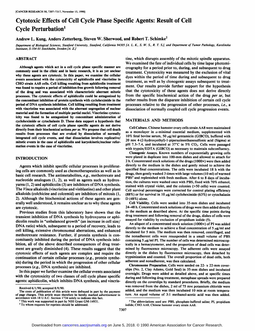
Cytotoxicity of Aphidicolin. The treatment of CHO AA8 cellswith aphidicolin for 18 h resulted in a concentration dependentreduction in clonal survival (Fig. 1/4). To determine the timecourse of cell killing by aphidicolin, cell viability, as assayed bythe exclusion of PI, was determined at various times during andfollowing treatment with aphidicolin (Fig. 2A). The proportionof viable cells remained near control levels (>95%) throughoutthe 18-h course of aphidicolin treatment and for the first 6 h
10-
Aphidicolin (ug ml)
100
>1_3
0 25 50 75 100
Vincristine (nM)Fig. 1. Effect of concomitant drug treatment on the clonal survival of cells.
Known numbers of CHO AA8 cells were plated in duplicate into 100-mm dishesand exposed 18 h to the specific concentrations of aphidicolin (A) or vincristine(B) in the absence (•)or presence of 10 /jg/ml cycloheximide (Ü)or 1 ng/mlcytochalasin D (A). Colonies »erecounted 7-9 days later. The percentages shownwere the averages of two separate experiments; bars. SD.
^ 95-
È 90O)
Oo>5 85CO
80-
0 6
| Block12 18 6 12
ARelease
Time (Hours)
ta 24
100-
^ 90-
rr sodiOO) .S 'co>
500 6^ Block
18 6
^ReleaseTime (Hours)
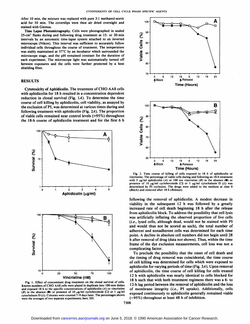
12 18
Fig. 2. Time course of killing of cells exposed to 18 h of aphidicolin orvincristine. The percentage of viable cells during and following an 18-h treatmentwith 5 fig/ml aphidicolin (A) or 100 nM vincristine (B) in the absence (•)orpresence of 10 ng/ml cycloheximide (D) or 1 jig/ml cytochalasin D (A) wasdetermined by PI exclusion. The drugs were added to the medium at time 0(Block) and removed after 18 h (Release).
following the removal of aphidicolin. A modest decrease inviability in the subsequent 12 h was followed by a greatlyincreased rate of cell death beginning 18 h after the releasefrom aphidicolin block. To address the possibility that cell lysiswas artificially inflating the observed proportion of live cells(i.e., lysed cells, although dead, would not be stained with PIand would thus not be scored as such), the total number ofadherent and nonadherent cells was determined for each timepoint. A decline in absolute cell numbers did not begin until 28h after removal of drug (data not shown). Thus, within the timeframe of the dye exclusion measurements, cell loss was not acomplicating factor.
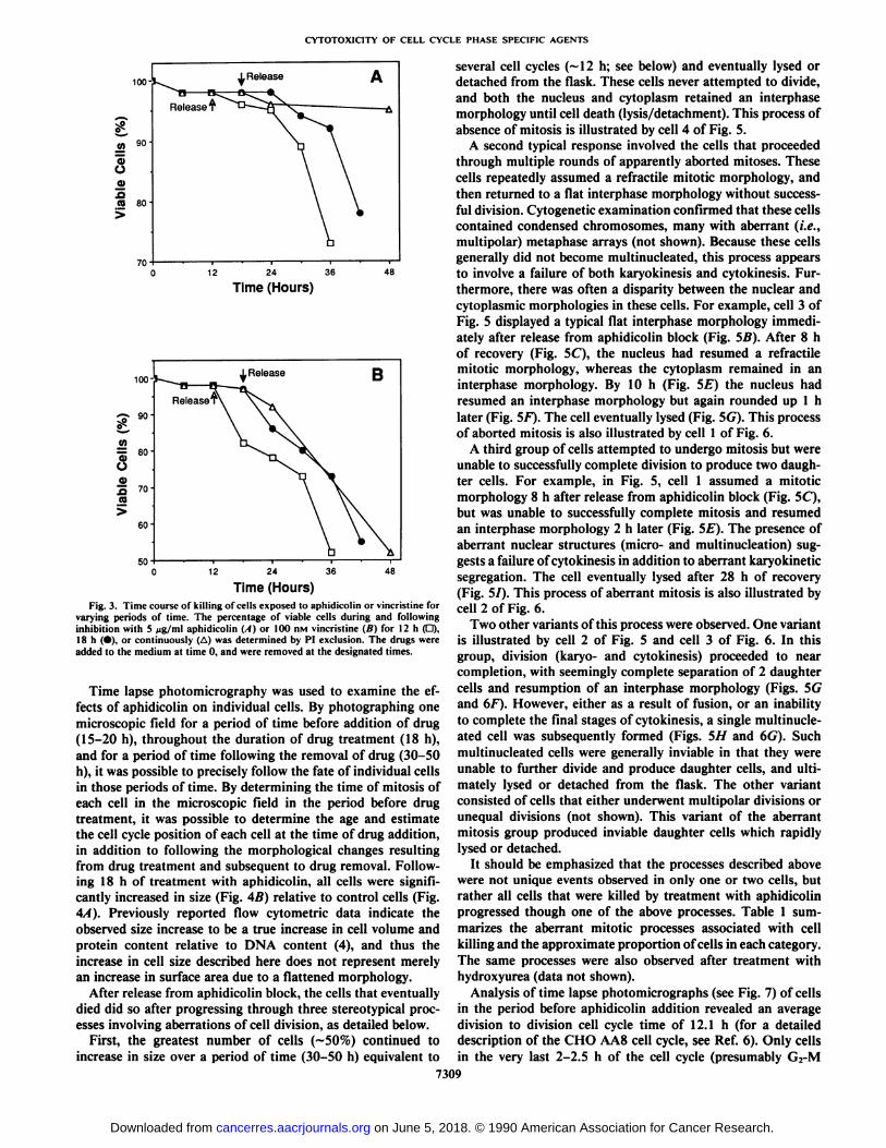
To preclude the possibility that the onset of cell death andthe timing of drug removal was coincidental, the time courseof cell killing was determined for cells which were exposed toaphidicolin for varying periods of time (Fig. 3A). Upon removalof aphidicolin, the time course of cell killing for cells treated12 h with aphidicolin was nearly identical to cells blocked for18 h such that with both treatment regimens there was a 6- to12-h lag period between the removal of aphidicolin and the lossof membrane integrity (i.e., PI uptake). Additionally, cellsexposed continuously to aphidicolin generally remained viable(—95%)throughout at least 48 h of inhibition.
7308
on June 5, 2018. © 1990 American Association for Cancer Research. cancerres.aacrjournals.org Downloaded from
CYTOTOXICITY OF CELL CYCLE PHASE SPECIFIC AGENTS
36
Time (Hours)
100
£ «H
OO
so
¿Release B
12 36 48
Time (Hours)Fig. 3. Time course of killing of cells exposed to aphidicolin or vincristine for
varying periods of time. The percentage of viable cells during and followinginhibition with 5 ng/ml aphidicolin (A) or 100 nM vincristine (B) for 12 h (O),18 h (•),or continuously (A) was determined by PI exclusion. The drugs »ereadded to the medium at time 0, and were removed at the designated times.
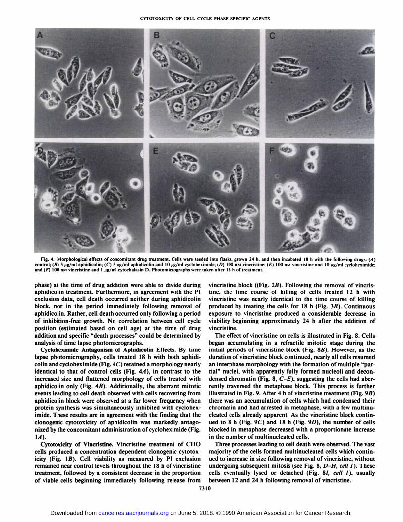
Time lapse photomicrography was used to examine the effects of aphidicolin on individual cells. By photographing onemicroscopic field for a period of time before addition of drug(15-20 h), throughout the duration of drug treatment (18 h),and for a period of time following the removal of drug (30-50h), it was possible to precisely follow the fate of individual cellsin those periods of time. By determining the time of mitosis ofeach cell in the microscopic field in the period before drugtreatment, it was possible to determine the age and estimatethe cell cycle position of each cell at the time of drug addition,in addition to following the morphological changes resultingfrom drug treatment and subsequent to drug removal. Following 18 h of treatment with aphidicolin, all cells were significantly increased in size (Fig. 4B) relative to control cells (Fig.4A). Previously reported flow cytometric data indicate theobserved size increase to be a true increase in cell volume andprotein content relative to DNA content (4), and thus theincrease in cell size described here does not represent merelyan increase in surface area due to a flattened morphology.
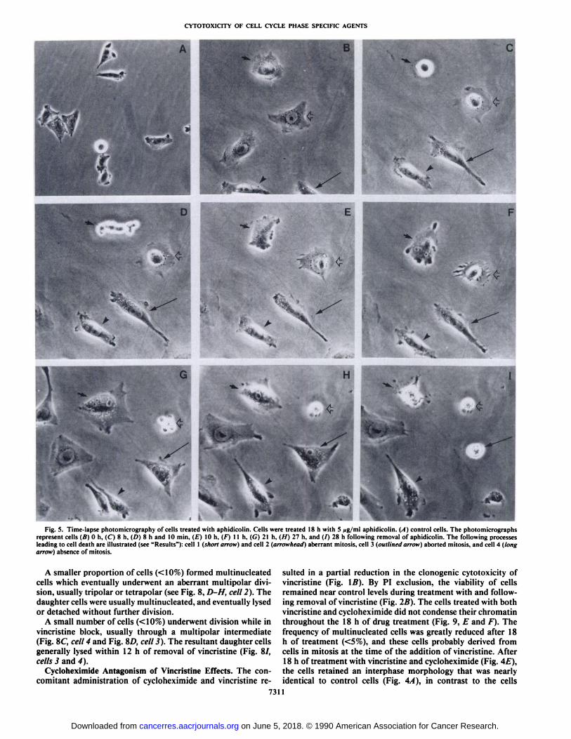
After release from aphidicolin block, the cells that eventuallydied did so after progressing through three stereotypical processes involving aberrations of cell division, as detailed below.
First, the greatest number of cells (~50%) continued toincrease in size over a period of time (30-50 h) equivalent to
several cell cycles (~12 h; see below) and eventually lysed ordetached from the flask. These cells never attempted to divide,and both the nucleus and cytoplasm retained an interphasemorphology until cell death (lysis/detachment). This process ofabsence of mitosis is illustrated by cell 4 of Fig. 5.
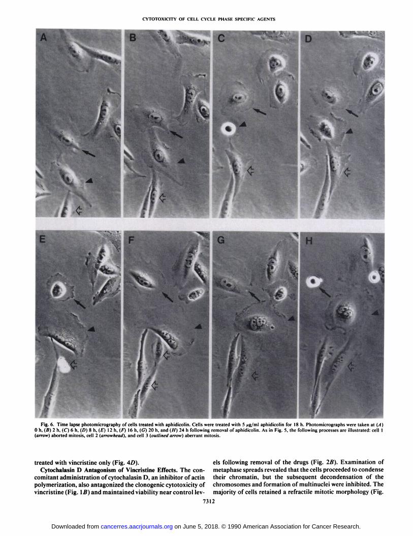
A second typical response involved the cells that proceededthrough multiple rounds of apparently aborted mitoses. Thesecells repeatedly assumed a refractile mitotic morphology, andthen returned to a flat interphase morphology without successful division. Cytogenetic examination confirmed that these cellscontained condensed chromosomes, many with aberrant (i.e.,multipolar) metaphase arrays (not shown). Because these cellsgenerally did not become multinucleated, this process appearsto involve a failure of both karyokinesis and cytokinesis. Furthermore, there was often a disparity between the nuclear andcytoplasmic morphologies in these cells. For example, cell 3 ofFig. 5 displayed a typical flat interphase morphology immediately after release from aphidicolin block (Fig. SB). After 8 hof recovery (Fig. 5C), the nucleus had resumed a refractilemitotic morphology, whereas the cytoplasm remained in aninterphase morphology. By 10 h (Fig. 5E) the nucleus hadresumed an interphase morphology but again rounded up 1 hlater (Fig. 5F). The cell eventually lysed (Fig. 5G). This processof aborted mitosis is also illustrated by cell 1 of Fig. 6.
A third group of cells attempted to undergo mitosis but wereunable to successfully complete division to produce two daughter cells. For example, in Fig. 5, cell 1 assumed a mitoticmorphology 8 h after release from aphidicolin block (Fig. 5C),but was unable to successfully complete mitosis and resumedan interphase morphology 2 h later (Fig. 5E). The presence ofaberrant nuclear structures (micro- and multinucleation) suggests a failure of cytokinesis in addition to aberrant karyokineticsegregation. The cell eventually lysed after 28 h of recovery(Fig. 51). This process of aberrant mitosis is also illustrated bycell 2 of Fig. 6.
Two other variants of this process were observed. One variantis illustrated by cell 2 of Fig. 5 and cell 3 of Fig. 6. In thisgroup, division (karyo- and cytokinesis) proceeded to nearcompletion, with seemingly complete separation of 2 daughtercells and resumption of an interphase morphology (Figs. 5Gand 6F). However, either as a result of fusion, or an inabilityto complete the final stages of cytokinesis, a single multinucleated cell was subsequently formed (Figs. 5H and 6G). Suchmultinucleated cells were generally inviable in that they wereunable to further divide and produce daughter cells, and ultimately lysed or detached from the flask. The other variantconsisted of cells that either underwent multipolar divisions orunequal divisions (not shown). This variant of the aberrantmitosis group produced inviable daughter cells which rapidlylysed or detached.
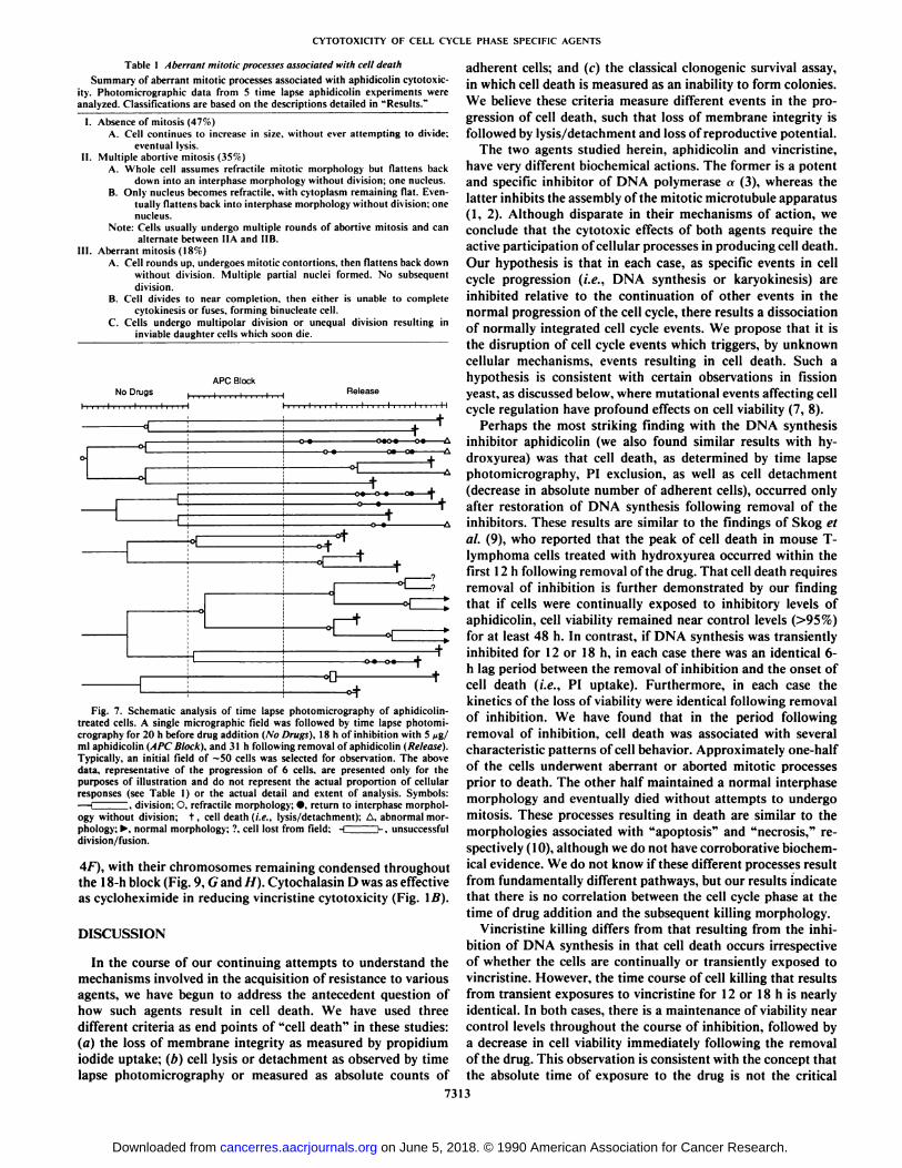
It should be emphasized that the processes described abovewere not unique events observed in only one or two cells, butrather all cells that were killed by treatment with aphidicolinprogressed though one of the above processes. Table 1 summarizes the aberrant mitotic processes associated with cellkilling and the approximate proportion of cells in each category.The same processes were also observed after treatment withhydroxyurea (data not shown).
Analysis of time lapse photomicrographs (see Fig. 7) of cellsin the period before aphidicolin addition revealed an averagedivision to division cell cycle time of 12.1 h (for a detaileddescription of the CHO AA8 cell cycle, see Ref. 6). Only cellsin the very last 2-2.5 h of the cell cycle (presumably G2-M
7309
on June 5, 2018. © 1990 American Association for Cancer Research. cancerres.aacrjournals.org Downloaded from
CYTOTOXICITY OF CELL CYCLE PHASE SPECIFIC AGENTS
Fig. 4. Morphological effects of concomitant drug treatment. Cells were seeded into flasks, grown 24 h. and then incubated 18 h with the following drugs: (A)control; (B) 5 Aig/ml aphidicolin; (C) 5 fig/ml aphidicolin and 10 >ig/ml cycloheximide; (D) 100 n\i vincristine; (£)100 nvi vincristine and 10 ng/ml cycloheximide;and (f~) 100 HMvincristine and 1 jig/ml cytochalasin D. Photomicrographs were taken after 18 h of treatment.
phase) at the time of drug addition were able to divide duringaphidicolin treatment. Furthermore, in agreement with the PIexclusion data, cell death occurred neither during aphidicolinblock, nor in the period immediately following removal ofaphidicolin. Rather, cell death occurred only following a periodof inhibition-free growth. No correlation between cell cycleposition (estimated based on cell age) at the time of drugaddition and specific "death processes" could be determined by
analysis of time lapse photomicrographs.Cycloheximide Antagonism of Aphidicolin Effects. By time
lapse photomicrography, cells treated 18 h with both aphidicolin and cycloheximide (Fig. 4C) retained a morphology nearlyidentical to that of control cells (Fig. 44), in contrast to theincreased size and flattened morphology of cells treated withaphidicolin only (Fig. 4B). Additionally, the aberrant mitoticevents leading to cell death observed with cells recovering fromaphidicolin block were observed at a far lower frequency whenprotein synthesis was simultaneously inhibited with cycloheximide. These results are in agreement with the finding that theclonogenic cytotoxicity of aphidicolin was markedly antagonized by the concomitant administration of cycloheximide (Fig.IA).
Cytotoxicity of Vincristine. Vincristine treatment of CHOcells produced a concentration dependent clonogenic cytotoxicity (Fig. IB). Cell viability as measured by PI exclusionremained near control levels throughout the 18 h of vincristinetreatment, followed by a consistent decrease in the proportionof viable cells beginning immediately following release from
vincristine block ((Fig. 2B). Following the removal of vincristine, the time course of killing of cells treated 12 h withvincristine was nearly identical to the time course of killingproduced by treating the cells for 18 h (Fig. 3B). Continuousexposure to vincristine produced a considerable decrease inviability beginning approximately 24 h after the addition ofvincristine.
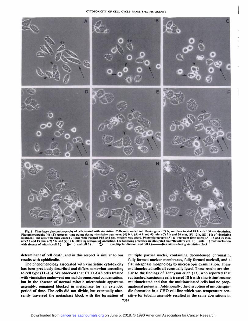
The effect of vincristine on cells is illustrated in Fig. 8. Cellsbegan accumulating in a refractile mitotic stage during theinitial periods of vincristine block (Fig. SB). However, as theduration of vincristine block continued, nearly all cells resumedan interphase morphology with the formation of multiple "partial" nuclei, with apparently fully formed nucleoli and decon-
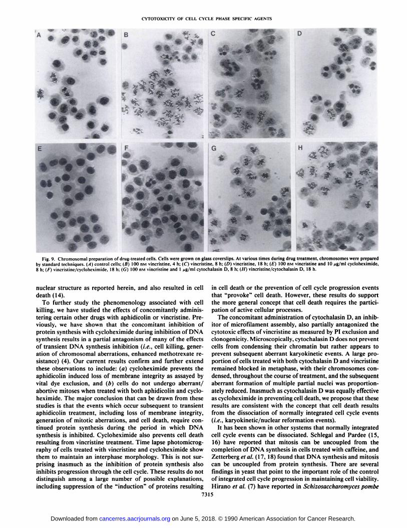
densed chromatin (Fig. 8, C-E), suggesting the cells had aber-rently traversed the metaphase block. This process is furtherillustrated in Fig. 9. After 4 h of vincristine treatment (Fig. 9B)there was an accumulation of cells which had condensed theirchromatin and had arrested in metaphase, with a few multinu-cleated cells already apparent. As the vincristine block continued to 8 h (Fig. 9C) and 18 h (Fig. 9D), the number of cellsblocked in metaphase decreased with a proportionate increasein the number of multinucleated cells.
Three processes leading to cell death were observed. The vastmajority of the cells formed multinucleated cells which continued to increase in size following removal of vincristine, withoutundergoing subsequent mitosis (see Fig. 8, D-H, cell 1). Thesecells eventually lysed or detached (Fig. 8/, cell /), usuallybetween 12 and 24 h following removal of vincristine.
7310
on June 5, 2018. © 1990 American Association for Cancer Research. cancerres.aacrjournals.org Downloaded from
CYTOTOXICITY OF CELL CYCLE PHASE SPECIFIC AGENTS
•SS
B
Fig. 5. Time-lapse photomicrography of cells treated with aphidicolin. Cells were treated 18 h with 5 /ig/ml aphidicolin. (A} control cells. The photomicrographsrepresent cells (B) 0 h, (C) 8 h. (D) 8 h and 10 min. (E) 10 h, (F) 11 h, (C) 21 h, (H) 27 h. and (/) 28 h following removal of aphidicolin. The following processesleading to cell death are illustrated (see "Results"): cell 1 (short arrow) and cell 2 (arrowhead) aberrant mitosis, cell 3 (outlined arrow) aborted mitosis, and cell 4 (long
arrow) absence of mitosis.
A smaller proportion of cells (<10%) formed multinucleatedcells which eventually underwent an aberrant multipolar division, usually tripolar or tetrapolar (see Fig. 8, D-H, cell 2). Thedaughter cells were usually multinucleated, and eventually lysedor detached without further division.
A small number of cells (<10%) underwent division while invincristine block, usually through a multipolar intermediate(Fig. 8C, cell 4 and Fig. 8D, cell 3). The resultant daughter cellsgenerally lysed within 12 h of removal of vincristine (Fig. 87,cells 3 and 4).
Cycloheximide Antagonism of Vincristine Effects. The concomitant administration of cycloheximide and vincristine re-
731
suited in a partial reduction in the clonogenic cytotoxicity ofvincristine (Fig. IB). By PI exclusion, the viability of cellsremained near control levels during treatment with and following removal of vincristine (Fig. 2B). The cells treated with bothvincristine and cycloheximide did not condense their chromatinthroughout the 18 h of drug treatment (Fig. 9, E and F). Thefrequency of multinucleated cells was greatly reduced after 18h of treatment (<5%), and these cells probably derived fromcells in mitosis at the time of the addition of vincristine. After18 h of treatment with vincristine and cycloheximide (Fig. 4E),the cells retained an interphase morphology that was nearlyidentical to control cells (Fig. 4A), in contrast to the cells1
on June 5, 2018. © 1990 American Association for Cancer Research. cancerres.aacrjournals.org Downloaded from
CYTOTOXICITY OF CELL CYCLE PHASE SPECIFIC AGENTS
Fig. 6. Time lapse photomicrography of cells treated with aphidicolin. Cells were treated with 5 fig/ml aphidicolin for 18 h. Photomicrographs »eretaken at (A)0 h, (B) 2 h, (C) 6 h, (D) 8 h, (E) 12 h. (F) 16 h, (G) 20 h, and (H) 24 h following removal of aphidicolin. As in Fig. 5. the following processes are illustrated: cell 1(arrow) aborted mitosis, cell 2 (arrowhead), and cell 3 (outlined arrow) aberrant mitosis.
treated with vincristine only (Fig. 4D).Cytochalasin D Antagonism of Vincristine Effects. The con
comitant administration of cytochalasin D, an inhibitor of actinpolymerization, also antagonized the clonogeniccytotoxicity ofvincristine (Fig. IB) and maintained viability near control lev
els following removal of the drugs (Fig. 2B). Examination ofmetaphase spreads revealed that the cells proceeded to condensetheir chromatin, but the subsequent decondensation of thechromosomes and formation of mull ¡nucleiwere inhibited. Themajority of cells retained a refractile mitotic morphology (Fig.
7312
on June 5, 2018. © 1990 American Association for Cancer Research. cancerres.aacrjournals.org Downloaded from
CYTOTOXICITY OF CELL CYCLE PHASE SPECIFIC AGENTS
Table 1 Aberrant mÃtotÃcprocesses associated with cell deathSummary of aberrant mitotic processes associated with aphidicolin cytotoxic-
ity. Photomicrographic data from 5 time lapse aphidicolin experiments wereanalyzed. Classifications are based on the descriptions detailed in "Results."
1. Absence of mitosis (47%)A. Cell continues to increase in size, without ever attempting to divide:
eventual lysis.II. Multiple abortive mitosis (35%)
A. Whole cell assumes refráctalemitotic morphology but flattens backdown into an interphase morphology without division; one nucleus.
B. Only nucleus becomes refráctale,with cytoplasm remaining flat. Eventually flattens back into ¡nterphasemorphology without division: onenucleus.
Note: Cells usually undergo multiple rounds of abortive mitosis and canalternate between IIA and IIB.
III. Aberrant mitosis (18%)A. Cell rounds up. undergoes mitotic contortions, then flattens back down
without division. Multiple partial nuclei formed. No subsequentdivision.
B. Cell divides to near completion, then either is unable to completecytokinesis or fuses, forming binucleate cell.
C. Cells undergo multipolar division or unequal division resulting ininviable daughter cells which soon die.
APC BlockNo Drugs
i I i i i i I i i i i
Release
Fig. 7. Schematic analysis of time lapse photomicrography of aphidicolin-treated cells. A single micrographie field was followed by time lapse photomicrography for 20 h before drug addition (.\o Drugs), 18 h of inhibition with 5 ^g/ml aphidicolin (APC Block), and 31 h follow ing removal of aphidicolin (Release).Typically, an initial field of ~50 cells was selected for observation. The abovedata, representative of the progression of 6 cells, are presented only for thepurposes of illustration and do not represent the actual proportion of cellularresponses (see Table 1) or the actual detail and extent of analysis. Symbols:—l division: O, refráctalemorphology: •.return to interphase morphology without division; t. cell death (i.e., lysis/detachment); A. abnormal morphology;^, normal morphology; ?, cell lost from field: H h. unsuccessfuldivision/fusion.
4F), with their chromosomes remaining condensed throughoutthe 18-h block (Fig. 9, G and //). Cytochalasin D was as effectiveas cycloheximide in reducing vincristine cytotoxicity (Fig. \B).
DISCUSSION
In the course of our continuing attempts to understand themechanisms involved in the acquisition of resistance to variousagents, we have begun to address the antecedent question ofhow such agents result in cell death. We have used threedifferent criteria as end points of "cell death" in these studies:
(a) the loss of membrane integrity as measured by propidiumiodide uptake; (b) cell lysis or detachment as observed by timelapse photomicrography or measured as absolute counts of
7313
adherent cells; and (c) the classical clonogenic survival assay,in which cell death is measured as an inability to form colonies.We believe these criteria measure different events in the progression of cell death, such that loss of membrane integrity isfollowed by lysis/detachment and loss of reproductive potential.
The two agents studied herein, aphidicolin and vincristine,have very different biochemical actions. The former is a potentand specific inhibitor of DNA polymerase «(3), whereas thelatter inhibits the assembly of the mitotic microtubule apparatus(1, 2). Although disparate in their mechanisms of action, weconclude that the cytotoxic effects of both agents require theactive participation of cellular processes in producing cell death.Our hypothesis is that in each case, as specific events in cellcycle progression (i.e., DNA synthesis or karyokinesis) areinhibited relative to the continuation of other events in thenormal progression of the cell cycle, there results a dissociationof normally integrated cell cycle events. We propose that it isthe disruption of cell cycle events which triggers, by unknowncellular mechanisms, events resulting in cell death. Such ahypothesis is consistent with certain observations in fissionyeast, as discussed below, where mutational events affecting cellcycle regulation have profound effects on cell viability (7, 8).
Perhaps the most striking finding with the DNA synthesisinhibitor aphidicolin (we also found similar results with hy-droxyurea) was that cell death, as determined by time lapsephotomicrography, PI exclusion, as well as cell detachment(decrease in absolute number of adherent cells), occurred onlyafter restoration of DNA synthesis following removal of theinhibitors. These results are similar to the findings of Skog etal. (9), who reported that the peak of cell death in mouse T-lymphoma cells treated with hydroxyurea occurred within thefirst 12 h following removal of the drug. That cell death requiresremoval of inhibition is further demonstrated by our findingthat if cells were continually exposed to inhibitory levels ofaphidicolin, cell viability remained near control levels (>95%)for at least 48 h. In contrast, if DNA synthesis was transientlyinhibited for 12 or 18 h, in each case there was an identical 6-h lag period between the removal of inhibition and the onset ofcell death (i.e., PI uptake). Furthermore, in each case thekinetics of the loss of viability were identical following removalof inhibition. We have found that in the period followingremoval of inhibition, cell death was associated with severalcharacteristic patterns of cell behavior. Approximately one-halfof the cells underwent aberrant or aborted mitotic processesprior to death. The other half maintained a normal interphasemorphology and eventually died without attempts to undergomitosis. These processes resulting in death are similar to themorphologies associated with "apoptosis" and "necrosis," re
spectively (10), although we do not have corroborative biochemical evidence. We do not know if these different processes resultfrom fundamentally different pathways, but our results indicatethat there is no correlation between the cell cycle phase at thetime of drug addition and the subsequent killing morphology.
Vincristine killing differs from that resulting from the inhibition of DNA synthesis in that cell death occurs irrespectiveof whether the cells are continually or transiently exposed tovincristine. However, the time course of cell killing that resultsfrom transient exposures to vincristine for 12 or 18 h is nearlyidentical. In both cases, there is a maintenance of viability nearcontrol levels throughout the course of inhibition, followed bya decrease in cell viability immediately following the removalof the drug. This observation is consistent with the concept thatthe absolute time of exposure to the drug is not the critical
on June 5, 2018. © 1990 American Association for Cancer Research. cancerres.aacrjournals.org Downloaded from
CYTOTOXICITY OF CELL CYCLE PHASE SPECIFIC AGENTS
Fig. 8. Time lapse photomicrography of cells treated with vincristinc. Cells were seeded into flasks, grown 24 h. and then treated 18 h with 100 nsi vincristine.Photomicrographs (A)-(E) represent time points during vincristine treatment: (A) 0 h, (fi) 6 h and 45 min, (C) 7 h and 24 min. (D) 10 h. (E) 18 h of vincristinetreatment. The cells were then washed 3 times with warmed PBS and new medium was added. Photomicrographs (F)-(l) represent time points (/•")I h and 30 min,(G) 2 h and 15 min. (//) 6 h. and (/) 12 h following removal of vincristine. The following processes are illustrated (see "Results"): cell I ( ' ^ ) mullinucleation
with absence of mitosis, cell 2 ( ^ ) and cell 3 ( ^> ), mullipolar division, and cell 4 ( > ) mitosis during vincrislinc block.
determinant of cell death, and in this respect is similar to ourresults with aphidicolin.
The phenomenology associated with vincristine cytotoxicityhas been previously described and differs somewhat accordingto cell type (11-13). We observed that CHO AA8 cells treatedwith vincristine underwent normal chromosomal condensation,but in the absence of normal mitotic microtubule apparatusassembly, remained blocked in metaphase for an extendedperiod of time. The cells did not divide, but eventually aberrantly traversed the metaphase block with the formation of
multiple partial nuclei, containing decondensed chromatin,fully formed nuclear membranes, fully formed nucleoli, and aflat interphase morphology by microscopic examination. Thesemultinucleated cells all eventually lysed. These results are similar to the findings of Tennyson et al. (13), who reported thatrat trachéalcarcinoma cells treated 18 h with vincristine becamemultinucleated and that the multinucleated cells had no prop-agational potential. Additionally, the disruption of mitotic spindle formation in a CHO cell line which was temperature sensitive for tubulin assembly resulted in the same aberrations in
7314
on June 5, 2018. © 1990 American Association for Cancer Research. cancerres.aacrjournals.org Downloaded from
CYTOTOXICITY OF CELL CYCLE PHASE SPECIFIC AGENTS
*<•*«t
* .*•*'
-f
Â̂¿A^p.
H -a*a* ^•RA
- •> 'if *'
4 * / v «<:jji **^' ^ *v "*
Fig. 9. Chromosomal preparation of drug-treated cells. Cells were grown on glass coverslips. At various times during drug treatment, chromosomes were preparedby standard techniques. (A) control cells; (B) 100 n\i vincristine. 4 h: (C) vincristine. 8 h: (D) vincristine. 18 h; (£)100 nM vincristine and 10 ng/ml cycloheximide,8 h; (F) vincristine/cycloheximide. 18 hi (G) 100 nM vincristine and 1 Mg/ml cytochalasin D, 8 h; (//) vincristine/cytochalasin D. 18 h.
nuclear structure as reported herein, and also resulted in celldeath (14).
To further study the phenomenology associated with cellkilling, we have studied the effects of concomitantly administering certain other drugs with aphidicolin or vincristine. Previously, we have shown that the concomitant inhibition ofprotein synthesis with cycloheximide during inhibition of DNAsynthesis results in a partial antagonism of many of the effectsof transient DNA synthesis inhibition (i.e., cell killing, generation of chromosomal aberrations, enhanced methotrexate resistance) (4). Our current results confirm and further extendthese observations to include: (a) cycloheximide prevents theaphidicolin induced loss of membrane integrity as assayed byvital dye exclusion, and (b) cells do not undergo aberrant/abortive mitoses when treated with both aphidicolin and cycloheximide. The major conclusion that can be drawn from thesestudies is that the events which occur subsequent to transientaphidicolin treatment, including loss of membrane integrity,generation of mitotic aberrations, and cell death, require continued protein synthesis during the period in which DNAsynthesis is inhibited. Cycloheximide also prevents cell deathresulting from vincristine treatment. Time lapse photomicrography of cells treated with vincristine and cycloheximide showthem to maintain an interphase morphology. This is not surprising inasmuch as the inhibition of protein synthesis alsoinhibits progression through the cell cycle. These results do notdistinguish among a large number of possible explanations,including suppression of the "induction" of proteins resulting
in cell death or the prevention of cell cycle progression eventsthat "provoke" cell death. However, these results do support
the more general concept that cell death requires the participation of active cellular processes.
The concomitant administration of cytochalasin D, an inhibitor of microfilament assembly, also partially antagonized thecytotoxic effects of vincristine as measured by PI exclusion andclonogenicity. Microscopically, cytochalasin D does not preventcells from condensing their chromatin but rather appears toprevent subsequent aberrant karyokinetic events. A large proportion of cells treated with both cytochalasin D and vincristineremained blocked in metaphase, with their chromosomes condensed, throughout the course of treatment, and the subsequentaberrant formation of multiple partial nuclei was proportionately reduced. Inasmuch as cytochalasin D was equally effectiveas cycloheximide in preventing cell death, we propose that theseresults are consistent with the concept that cell death resultsfrom the dissociation of normally integrated cell cycle events(i.e., karyokinetic/nuclear reformation events).
It has been shown in other systems that normally integratedcell cycle events can be dissociated. Schlegal and Pardee (15,16) have reported that mitosis can be uncoupled from thecompletion of DNA synthesis in cells treated with caffeine, andZetterberg et al. (17. 18) found that DNA synthesis and mitosiscan be uncoupled from protein synthesis. There are severalfindings in yeast that point to the important role of the controlof integrated cell cycle progression in maintaining cell viability.Mirano et al. (7) have reported in Schizosaccharomyces pombe
7315
on June 5, 2018. © 1990 American Association for Cancer Research. cancerres.aacrjournals.org Downloaded from
CYTOTOXICITV OF CELL CYCLE PHASE SPECIFIC AGENTS
that the lethality of a topoisomerase II mutant, which fails toallow normal chromosomal segregation at mitosis, is preventedby the presence of a mutation in the cdcl 1 gene which controlsseptation (cytokinesis). The double mutants remain viable andbecome polyploid. Additionally, Enoch and Nurse (8) havereported in 5. pombe that certain cdc25 mutants, which aredefective in the control of mitosis, are highly susceptible to thelethal effects of DNA synthesis inhibition by hydroxyurea inthat they undergo mitosis without completing replication. Incontrast, wild type cells do not undergo mitosis when DNAsynthesis is inhibited and thus do not die. However, if suchcdc25 mutants also contain a mutation in the cdc\Q gene (thuspreventing cells from entering S phase, i.e., START), then onlythose cells which have entered S phase prior to the addition ofhydroxyurea are killed. These results clearly support the concept of the importance of an ordered progression of cell cycleevents in maintaining cellular viability, and further support thatregulation events involved in mitosis are critical. Further support for the major role of mitotic control comes from studiesof the Rad9 gene in Saccharomyces cerevisiae (see Ref. 19 forreview). This gene identifies a mechanism which "senses" DNA
damage and is critical for preventing mitosis from occurringprior to the repair of damaged DNA.
Our results do not address the fundamental question of whycells die (i.e., lose membrane integrity/lyse) when treated withthese and other agents. Although various studies concernedwith cell death, as primarily studied in lymphocyte derived celllines, have centered on the hypothesis that death results fromthe rapid digestion of chromatin by newly induced endonu-cleases as part of a "death program" (20, 21), more recent
studies suggest that it is alterations in chromatin/nuclear structure that render DNA susceptible to the action of constitutivelypresent endonucleases (22). Based on our results with vincris-tine and aphidicolin, and taking into account the studies inyeast as discussed above, we suggest that it is the disorderedstate of nuclear structure that results in a susceptibility to eitherconstitutively expressed or induced enzymes which ultimatelyresult in cell death, and that this occurs subsequent to aberrantkaryokinetic processes. This hypothesis is consistent with ourresults that: (a) one-half of the cells that die with aphidicolinundergo aberrant mitotic processes before losing membraneintegrity; (b) all cells that die from vincristine treatmentundergo an aberrant traversal of metaphase with the disorderedreformation of multiple partial nuclei; and (c) with all treatmentmodalities, cell membrane integrity is maintained as long asaberrantly segregated nuclear structures are not generated (e.g.,continual S-phase block with aphidicolin and concomitant administration of vincristine and cytochalasin D). However, anysuggestion that nuclear structure abnormalities are necessaryfor cell death is clearly too simplistic in light of the fact thatone-half of the cells killed by aphidicolin do not undergo anygross changes in nuclear structure.
The finding that cell killing requires the continuation ofcertain cellular processes may have potential implications forvarious types of chemotherapy. Our results, most clearly evidentwith aphidicolin, demonstrate that in certain cases cell killingrequires a period of drug-free growth following a period ofinhibition. This suggests that the pharmacokinetics and dosingschedule of drugs may be important in maximizing cell killing.Maintenance of continual long-term inhibitory levels of drugsmay be less effective than regimens alternating periods ofinhibition followed by periods of drug-free growth.
The concept that cell killing requires active participation of
cellular processes may also assist in understanding some typesof drug resistance phenomena. Current understanding of acquired resistances, as studied in cell culture systems, has beencentered on processes whereby action of the drug is circumvented either by preventing intracellular accumulation of thedrug (23, 24) or by alterations in the structure (25) or concentration (26) of the target protein. We suggest that an additionalmechanism of resistance may involve a lack of response toinhibitory processes that would otherwise evoke a cellular suicide process in more susceptible cell types. Indeed, we havefound a remarkable difference in the susceptibility of varioushuman and rodent cell lines to the cytotoxic effects of aphidicolin. Although DNA synthesis is equally inhibited in all of thecell lines studied, there exists a dramatic difference in cellkilling.4 Thus, inherent resistance in this case does not derive
from differences in the biochemical action of the agent butrather results from differences in the response of the differentcell types to such inhibition. Our results are consistent with theconcept that cell types that are not killed have an inherentlymore "stringent" control of cell cycle progression events in G:-M phases. This type of "inherent" resistance may partially
underlie the striking differences in the success of therapeuticmodalities among different clinical cancers.
REFERENCES
1. C'habncr. B. A., and Myers, C. E. Clinical pharmacology of cancer chemo
therapy. In: V. T. DeVila, S. Hellman. and S. A. Rosenberg (eds.). Cancer.Principles and Practice of Oncology. Philadelphia: J. B. Lippincott Co.,1982.
2. Sartorelli. A. C.. and Johns. D. G. (eds.). Handbook of Experimental Pharmacology. Vol. 38. New York: Springer-Verlag, 1975.
3. Lee. M. Y. W. T.. Tan, C. K.. Downey. K. M., and So. A. Structure andfunction of calf thymus DNA polymerase f>.Prog. Nucleic Acid Res. Mol.Biol., 26: 83-96, 1981.
4. Sherwood. S. W.. Schumacher, R. I., and Schimke. R. T. Effect of cyelohcx-imide on development of methotrcxate resistance of Chinese hamster ovarycells treated with inhibitors of DNA synthesis. Mol. Cell. Biol., A: 2822-2827. 1988.
5. Yeh. C. J. G., Hsi. B. L.. and Faulk. W. P. Propidium iodide as a nuclearmarker in immunofluorcscence. 11. lise with cellular identification and viability studies. J. Immunol. Methods. 43: 269-275, 1981.
6. Hoy, C., Rice. G. C., Kovacs, M., and Schimke. R. T. Overreplication ofDNA in S-phase CHO cells after DNA synthesis inhibition. J. Biol. Chem.,262: 11927-11934, 1987.
7. Hirano, T., Funahashi, S., Uemura. T.. and Yanagida, M. Isolation andcharacteri/ation of Schizosaccharomyces pomhe cui mutants that block nuclear division but not cytokinesis. EMBO J.. 5: 2973-2979, 1986.
8. Enoch. T.. and Nurse. P. Mutation of fission yeast cell cycle control genesabolishes dependence of mitosis on DNA replication. Cell, 60: 665-673,1990.
9. Skog, S.. Tribukait. B., Wallström.B., and Eriksson, S. Hydroxyurea-inducedcell death as related to cell cycle in mouse and human T-lymphoma cells.Cancer Res.. 47: 6490-6595. 1987.
10. Duvall. E., and Wyllie, A. H. Death and the cell. Immunol. Today. 7: 115-119, 1986.
11. Madoc-Jones. H., and Mauro. F. Interphase action of vinblasline and vincristine: differences in their lethal action through the mitotic cycle of culturedmammalian cells. J. Cell. Physiol.. 72: 185-196. 1972.
12. Lengsfeld. A. M., Schultze, B., and Maurer, W. Time-lapse studies on theeffects of vincristine on HeLa cells. Eur. J. Cancer, 17: 307-319, 1980.
13. Tennyson, G. S.. Burbach. B. J.. and Lane. B. P. Reproductive potential ofvincristine-treated multinucleated carcinoma cells. Cancer Treat. Rep.. 67:1113-1114. 1983.
14. Abraham. I.. Marcus. M.. Cabrai, F., and Gottesman. M. M. Mutations inu- and iMubulin affect spindle formation in Chinese hamster ovan cells. J.Cell Biol.. 97: 1055-1061. 1983.
15. Schlegel. R., and Pardee. A. B. Caffeine-induced uncoupling of mitosis fromthe completion of DNA replication in mammalian cells. Science (Washington, DC), 232: 1264-1266. 1986.
16. Schlegel. R., and Pardee. A. B. Periodic mitotic events induced in the absenceof DNA replication. Proc. Nati. Acad. Sci. USA. «4:9025-9029, 1987.
17. Larsson. O., Dafgard. E., Engström.W., and Xctterbcrg, A. Immediate effectsof serum depletion on dissociation between growth in size and cell division
*A. Rung. S. Sherwood, and R. Schimke. manuscript in preparation.
7316
on June 5, 2018. © 1990 American Association for Cancer Research. cancerres.aacrjournals.org Downloaded from
CYTOTOXICITY OF CELL CYCLE PHASE SPECIFIC AGENTS
in proliferating 3T3 cells. J. Cell. Physiol., 127: 267-273, 1986. mediated by an induced nuclease. J. Biol. Chem., 264:4104-4111, 1989.18. Zetterberg, A., Engström,W., and Dafgârd.E. The relative effects of different 23. Sirotnak. F. M., Moccio, D. M., Kelleher, L. E., et al. Relative frequency
types of growth factors on DNA replication, mitosis, and cellular enlarge- and kinetic properties of transport defective phenotypes among methotrexate-ment Cvtometrv 5'368-375 1984 resistant L1210 clonal cells derived in vivo. Cancer Res., 41: 4447-4452,
19. Hartwell, L. H., and Weinert, T. A. Checkpoints: controls that ensure the ,,•',, , , , , , , , , , , ,, ,,.. „ ,, . .... . . ~„. ,,. ,AO ,-.. , «or. 24. Ling, V., and Thompson, L. H. Reduced permeability in CHO cells as aorder of cell cycle events. Science (Washington, DC), 246: 629-634, 1989. mec8hanism Of resistance to colchici«. J. Cell. Physiol.; 83: 103-116, 1974.
20. Wyllie, A. H. Glucocorticoid-mduced thymocyte apoptosis is associated with 25 Haber D A Bever|y s M Kie]y M L et al propert¡es of an alteredendogenous endonuclease activation. Nature (Lond.), 284: 555-556, 1980. dihydrofolate reducÃaseencoded by amplified genes in cultured mouse fibro-
21. Compton, M. M., and Cidlowski, J. A. Identification of a glucocorticoid- blasts. J. Biol. Chem., 256: 9501-9510, 1981.induced nuclease in thymocytes. J. Biol. Chem., 262: 8288-8292, 1987. 26. Schimke. R. T. Gene amplification in cultured cells. J. Biol. Chem., 263:
22. Alnemri, E. S., and Litwack, G. Glucocorticoid-induced lymphocytosis is not 5989-5992, 1988.
7317
on June 5, 2018. © 1990 American Association for Cancer Research. cancerres.aacrjournals.org Downloaded from
1990;50:7307-7317. Cancer Res Andrew L. Kung, Anders Zetterberg, Steven W. Sherwood, et al. Cell Cycle PerturbationCytotoxic Effects of Cell Cycle Phase Specific Agents: Result of
Updated version
http://cancerres.aacrjournals.org/content/50/22/7307
Access the most recent version of this article at:
E-mail alerts related to this article or journal.Sign up to receive free email-alerts
Subscriptions
Reprints and
To order reprints of this article or to subscribe to the journal, contact the AACR Publications
Permissions
Rightslink site. Click on "Request Permissions" which will take you to the Copyright Clearance Center's (CCC)
.http://cancerres.aacrjournals.org/content/50/22/7307To request permission to re-use all or part of this article, use this link
on June 5, 2018. © 1990 American Association for Cancer Research. cancerres.aacrjournals.org Downloaded from


![ClinicalValueofSerumGlycoproteinGalactosyltransferaseLevel ...cancerres.aacrjournals.org/content/43/9/4491.full.pdf · [CANCERRESEARCH43,4491-4496,September1983] ClinicalValueofSerumGlycoproteinGalactosyltransferaseLevelsin](https://img.pdfslide.us/doc/110x75/5ac139b17f8b9ac6688d1490/clinicalvalueofserumglycoproteingalactosyltransferaselevel-cancerresearch434491-4496september1983.jpg)


![ResistancetoAnthrapyrazolesandAnthracyclinesinMultidrug ...cancerres.aacrjournals.org/content/46/9/4352.full.pdf[CANCERRESEARCH46,4352-4356,September1986] ResistancetoAnthrapyrazolesandAnthracyclinesinMultidrug-resistantP388](https://img.pdfslide.us/doc/110x75/5aec16b57f8b9a90318dd526/resistancetoanthrapyrazolesandanthracyclinesinmultidrug-cancerresearch464352-4356september1986.jpg)
![StatisticalAnalysisofHormonalEffectsontheSteroid ...cancerres.aacrjournals.org/content/30/1/221.full.pdf[CANCERRESEARCH30,221-227,January1970]. StatisticalAnalysisofHormonalEffectsontheSteroid](https://img.pdfslide.us/doc/110x75/5ab039c17f8b9a25088e7528/statisticalanalysisofhormonaleffectsonthesteroid-cancerresearch30221-227january1970.jpg)
![HeterotransplantationofHumanLymphoidNeoplasmsUsingaNudeMou ...cancerres.aacrjournals.org/content/50/10/3078.full.pdf · (CANCERRESEARCH50,3078-3086.May15,1990] HeterotransplantationofHumanLymphoidNeoplasmsUsingaNudeMouse](https://img.pdfslide.us/doc/110x75/5e83f48eaae3144d7c04ca6b/heterotransplantationofhumanlymphoidneoplasmsusinganudemou-cancerresearch503078-3086may151990.jpg)


![TumorBehaviorinTransitionalCellCarcinomaoftheBladderinRela ...cancerres.aacrjournals.org/content/47/24_Part_1/6800.full.pdf · [CANCERRESEARCH47,6800-6805,December!5,1987] TumorBehaviorinTransitionalCellCarcinomaoftheBladderinRelationto](https://img.pdfslide.us/doc/110x75/5c085d3a09d3f23a458c00a0/tumorbehaviorintransitionalcellcarcinomaofthebladderinrela-cancerresearch476800-6805december51987.jpg)
![ImmunocytochemicalLocalizationofEstrogenandProgesteron ...cancerres.aacrjournals.org/content/50/21/7057.full.pdf[CANCERRESEARCH50.7057-7061.NovemberI.1990] ImmunocytochemicalLocalizationofEstrogenandProgesteroneReceptorand](https://img.pdfslide.us/doc/110x75/5ae3c5997f8b9a595d8edf20/immunocytochemicallocalizationofestrogenandprogesteron-cancerresearch507057-7061novemberi1990.jpg)

![DNATopoisomeraseIIImmunostaininginHumanLeukemiaand ...cancerres.aacrjournals.org/content/52/15/4248.full.pdf · (CANCERRESEARCH52,4248-425.1,August1.1992] DNATopoisomeraseIIImmunostaininginHumanLeukemiaand](https://img.pdfslide.us/doc/110x75/5b6d7db17f8b9a962a8cc15a/dnatopoisomeraseiiimmunostaininginhumanleukemiaand-cancerresearch524248-4251august11992.jpg)
![VirologieStudiesinHumanLeukemiaandLymphoma:The …cancerres.aacrjournals.org/content/28/7/1311.full.pdf · [CANCERRESEARCH28,1311-1318,July1968] VirologieStudiesinHumanLeukemiaandLymphoma:The](https://img.pdfslide.us/doc/110x75/5b98f37b09d3f219118d06a7/virologiestudiesinhumanleukemiaandlymphomathe-cancerresearch281311-1318july1968.jpg)

![InVitroHematopoiesisfollowingInductionChemotherapyforAcute ...cancerres.aacrjournals.org/content/45/11_Part_2/5921.full.pdf[CANCERRESEARCH45,5921-5925,November1985] InVitroHematopoiesisfollowingInductionChemotherapyforAcuteLeukemia1](https://img.pdfslide.us/doc/110x75/5b0a316b7f8b9a45518be441/invitrohematopoiesisfollowinginductionchemotherapyforacute-cancerresearch455921-5925november1985.jpg)
![PharmacokineticsoftheMonoclonalAntibodyB72.3andItsFragmentsLabeled ...cancerres.aacrjournals.org/content/47/4/1149.full.pdf · [CANCERRESEARCH47,1149-1154,February15,1987] PharmacokineticsoftheMonoclonalAntibodyB72.3andItsFragmentsLabeled](https://img.pdfslide.us/doc/110x75/5a8009117f8b9aee018c113e/pharmacokineticsofthemonoclonalantibodyb723anditsfragmentslabeled-cancerresearch471149-1154february151987.jpg)