Embed Size (px)
Citation preview
[CANCER RESEARCH 55, 3117-3122. July 15, 1W5|
Apoptosis Induction Mediated by Wild-Type p53 Adenoviral Gene Transfer inSquamous Cell Carcinoma of the Head and Neck1
Ta-Jen Liu, Adel K. El-Naggar, Timothy J. McDonnell, Kim D. Steck, Mary Wang, Dorothy L. Taylor, andGary L. dayman2
Departments of Head and Neck Surgery [T-J. L, M. W., D. L. T., G. L. C.I and Pathology ¡A.K. £., T. J. M., K. D. S.¡, University of Texas M. D. Anderson Cancer Center.
Houston, Texas 77030
ABSTRACT
Cancer gene therapy strategies for inducing apoptosis in solid tumorsmay allow contemporary medicine to reassess its management of thesecancers. We demonstrated previously that overexpression of the wild-type/»5.Õgene in squamous cell carcinoma of the head and neck cell lines viaadenovirus-mediated gene transfer suppressed growth both in vitro and in
vivo. Here, we characterize the mechanism of the growth suppression bythe exogenous p53 gene as a consequence of programmed cell death(apoptosis). One of the cell lines used in this study, Tu-138, harbors a
mutated p53 gene, whereas the other cell line. Ml) \ 686LN, possesses awild-typep53 gene. DNA fragmentation was detected by electrophoresis inboth cell lines after infection with the wild-type p53 adenovirus, AdSCMV-p53. With the use of the terminal deoxynucleotidyl transferase-mediateddUTP-biotin nick end-labeling method, 4.4% of the remaining viableTu-138 cell population was identified as apoptotic as early as 15 h afterinoculation with Ad5CMV-p53. The percentage of apoptotic cells in
creased to 31% at 22 h. In contrast, only 10% of the viable MDA 686LNcells (wt-p5.?) had undergone apoptosis 30 h after Ad5CMV-p53 infection,
although the percentage of apoptotic cells rapidly increased to 60% at 48h after infection. For in vivo analysis of apoptosis, nude mice in whichsquamous cell carcinoma of the head and neck cell lines had been implanted s.c. had exogenous \vt-p53 transiently introduced to the tumorcells via \d5CM\-p53 2 days later. In situ end labeling clearly illustratedapoptosis in the tumor cells. These results suggest that \vt-p53 plays an
important role in the induction of apoptosis in human head and neckcancer cell lines and that selective induction of apoptosis in cancer cellscan be further explored as a strategy for cancer gene therapy.
INTRODUCTION
Balancing the rates of cell proliferation and cell death is importantin maintaining normal tissue homeostasis. Disruption of this balancemay be a major factor in the multistep process of tumorigenesis, andinhibition of apoptosis, or programmed cell death, is one cause of thisdisruption. Apoptosis also occurs during normal embryogenesis, inthe course of normal tissue turnover, after withdrawal of a trophichormone from its target tissue, and in thymic regression, offeringexcellent opportunities to study the apoptotic process. We contendthat the most promising new therapies for solid malignancies areinterventions at the molecular level, and that selective induction ofapoptosis in these cancers is a logical intervention strategy. The geneor genes that may induce cancer cell apoptosis continue to be investigated, as do the methods for gene transfer. Presently, adenovirus-
mediated gene transfer is our clinical method of choice for suchinterventions because of its known tropism for the epithelium of theaerodigestive tract, its excellent transduction efficiency, the transient
Received 4/6/95: accepted 5/11/95.The costs of publication of this article were defrayed in part by the payment of page
charges. This article must therefore be hereby marked advertisement in accordance with18 U.S.C. Section 1734 solely to indicate this fact.
1This work was supported in part by American Cancer Society Career Development
Award 93-9 (G. L. C.), M. D. Anderson Cancer Center Core Grani NIH-NCI-CA-16672,and a gift to the Division of Surgery from Tenneco and Exxon for the Core LaboratoryFacility.
2 To whom requests for reprints should be addressed, al M. D. Anderson Cancer
Center, Department of Head and Neck Surgery, Box 69, 1515 Holcombe Boulevard.Houston. TX 77030.
nature of gene expression in the cells it infects (lack of permanentintegration), and its ability to infect nonproliferating cells. The p53gene also continues to be of interest as a molecular therapy for somesolid malignancies.
p53 was originally discovered through its association with theSV40 large T antigen (1,2). The importance of the p53 gene productin human neoplasia was first recognized a few years ago when mutantforms of the gene were identified in human colorectal tumors. Subsequently, f>53 mutations have been identified in the majority ofhuman malignant solid tumors, including those of the breast (3), colon(4), lung (5), and oral cavity (6). Several studies have demonstratedthe ability of the wt-p5J3 to suppress cancer cell growth both in vitro
and in vivo, suggesting that it acts as a tumor suppressor gene. Thesuppression of cell growth by p53 is mediated by two distinct pathways, one transient and one permanent. In the case of transientsuppression, p53 serves as a cell cycle checkpoint regulator. Overexpression of wt-p53 has been shown to induce a reversible cell cyclearrest at the G,-S boundary (7, 8). In other instances, p53 may induce
apoptosis when overexpressed in some cultured cells (9, 10) and isrequired for DNA damage-induced apoptosis in mouse thymocytes
(11).We reported previously that overexpression of the wt-p53 in
SCCHN cell lines induced via adenovirus-mediated gene transfer
suppressed growth both in vitro and in vivo (12, 13). Here, we soughtto examine the mechanism of this growth suppression. Our dataindicate that the suppression effect, both in vitro and in vivo, is theconsequence of an irreversible event, apoptosis.
MATERIALS AND METHODS
Cell Lines and Culture Conditions: Recombinant Adenovirus Preparation and Infection. Human SCCHN cell lines Tu-138 and MDA 686LN were
all established at the Department of Head and Neck Surgery, M. D. AndersonCancer Center, and had been characterized previously (12, 13). All procedureswere performed and cell lines maintained as described previously (12, 13). Cellgrowth assays were performed in triplicate.
DNA Fragmentation Analysis. After incubation with Ad5CMV-p53 orreplication-defective adenoviral controls at various time intervals, cells were
harvested and resuspended in 300 fj.1of PBS, to which 3 ml of extraction buffer[10 mM Tris (pH 8.0)-0.1 M EDTA-20 jj.g/ml RNAse-0.5% SDS] were addedbefore incubation at 37°Cfor 1-2 h. At the end of incubation, proteinase K was
added to a final concentration of 100 jLig/ml. and the solution was placed in a50°Cwater bath for at least 3 h. DNA was extracted once with an equal volume
of 0.5 MTris (pH 8.())-saturated phenol and then again with phenol/chloroform.
Precipitated DNA was analyzed in a 1% agarose gel.Cell Fixation. Before the TÚNEL method was used to identify apoptotic
cells, the cells were fixed in 1% formaldehyde in PBS (pH 7.4) for 30 min onice. Cells were then washed with 3 ml of PBS, resuspended in 70% ice-coldethanol, and stored at -2()°Cuntil used. For cell cycle analysis, cells were fixed
in 70% ice-cold ethanol only.
3 The abbreviations used are: wt-/>5.?, wild-type p53; SCCHN, squamous cell carci
noma of the head and neck; TÚNEL, terminal deoxynucleotidyl transferase-mediateddUTP-biotin nick end labeling: Ad5CMV-/;5J, wild-type p53 adenovirus; Tdt, terminal
dcoxynucleotidyl transferase.
3117
Research. on September 16, 2018. © 1995 American Association for Cancercancerres.aacrjournals.org Downloaded from
WILD-TYPE pS.i ADENOVIRUS INDUCES APOPTOSIS
Terminal Deoxynucleotidyl Transferase Assay. The TÚNEL assay wasperformed according to the procedure of Gorczyca el al. (14). Briefly, afterfixation and washing, cells were rcsuspended in 5(1/il of TdT buffer containing0.2 M sodium cacodylate (pH 7.0), 2.5 mM Tris-HCl, 2.5 mM COCÕ,(Sigma
Chemical Co., St. Louis, MO), 0.1 min DTT (Sigma Chemical Co.), 0.25nig/ml bovine serum albumin (Sigma Chemical Co.), 5 units of terminaltransferase (Boehringer Mannheim. Indianapolis. IN), and 0.5 nmol hiotin-16-dUTP along with dATP. dGTP, and dCTP at concentrations of 20 /AM.
Controls were prepared by incubating a separate aliquot of each test samplewithout dUTP. The cells were incubated in the solution at 37°Cfor 30 min,
rinsed in PBS, and resuspended in 1(X) /il of fluorescein isothiocyanate, thestaining solution containing 4X SSC, 0.1% Triton X-100, and 2.5 fig/ml
tluoresceinated avidin (Vector Laboratories, Inc.. Burlingame, CA). Tubeswere incubated for 30 min in the dark at room temperature. Cells were rinsedin PBS with 0.1% Triton X-100 and resuspended in 0.5 ml PBS containing
propidium iodide (5 /ig/ml) and 70 /il (l mg/ml) RNAse. Tubes wereincubated in the dark on ice for 30 min before flow cytometric analysis.
Flow Cytometric Analysis. All samples were analyzed with the use of anEPICS Profile II How cytometcr (Coulter Corp.. Hialeah, FL) with the standardoptical configuration. At least 10,000 events were counted for each sample.Positivity for TdT end labeling was determined by subtracting the controlhistogram from the test histogram with the use of the lmmuno-4 program of the
Elite workstation software (Coulter Corp.).Cell Growth Assay. Cells were plated at a density of 2 x l()4 cells/ml in
n-well plates in triplicate. Cells were infected with either Ihe Ad5CMV-/>5.f orthe replication-defective adenovirus (dl312) as a control. Cells were harvested
at different time intervals and counted, and their viability was determined bytrypan blue exclusion.
In Vivo Analysis for Apoptosis. Gene therapy in a microscopic residualdisease model of SCCHN has been described elsewhere (13). Experimentswere reviewed and approved by the institutional committees for both animalcare and utilization and the Biosafety Committee for recombinant DNAresearch. Briefly, s.c. flaps were elevated in anesthetized 4-6-week-old nudefemale mice with sharp dissection, and 2.5 X 10" tumor cells in 100 /il of
culture medium were pipetted into the flap and sealed with a horizontalmattress suture. Forty-eight h after tumor cell delivery, animals were reancs-thetized and infected with Ad5CMV-/>5.?. replication-defective virus (dl312).
or PBS alone (mock infection). The animals were observed daily and killed 72h after the second, or "therapeutic." intervention.
in Situ End Labeling. The procedure was performed as described elsewhere (15). Briefly, paraffin-embedded tumor sections were dewaxed in xy-
lene for 5 min three times each and were progressively hydrated by immersingthe slides for 3 min each in KM),90, 70, and 30% ethanol solutions. Endogenous peroxidase was inactivated by immersing the slides for 20 min in 0.75%H2O, (v/v) in 100% methanol. After Ihe slides were washed in PBS, sectionswere digested with 0.1% pepsin (Fisher Scientific. Houston, TX; w/v) in 0.1 NHC'I for 5 min at 37°Cand extensively washed in PBS. Sections were then
incubated in a moist chamber at 37°Cfor l h with an end-labeling cocktail that
included the following: 0.5 unit//il TdT; 0.06 mM biotinylated dUTP; 10 fil5X TdT buffer; and double-distilled water up to 50 ¿il.The reaction was
terminated by immersing the slides in a buffer containing 300 mM NaCI and 30mM sodium citrate in double-distilled water. After the slides were washed inPBS, sections were incubated with horseradish peroxide-conjugated avidin forl h at 37°Cin a moist chamber. Staining was developed using 3,3'-diamino-
ben/idine, and sections were counterstained with methyl green.
RESULTS
Suppression of SCCHN Cell Line Growth by Ad5CMV-/»5J.We reported previously that wt-p53 can be efficiently transduced into
SCCHN cell lines by a recombinant adenoviral vector. Consequently,the insulted tumor cells lose their ability to proliferate in vitro as wellas in vivo. The suppression effect is independent of the endogenous p53status of the cell lines. Previous growth rate analyses were carried outthrough a 1-week period. Here, we sought to investigate the early effectsof the wt-p53 on SCCHN cell growth (i.e.. after shorter time intervals).
Two representative cell lines were used in this study. Cell lineTu-138 harbors a mutated p53 gene, whereas cell line MDA 686LN
B
muckdl3I2p53
mockam:
20 .10 40 5(1
Hours
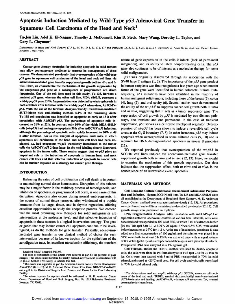
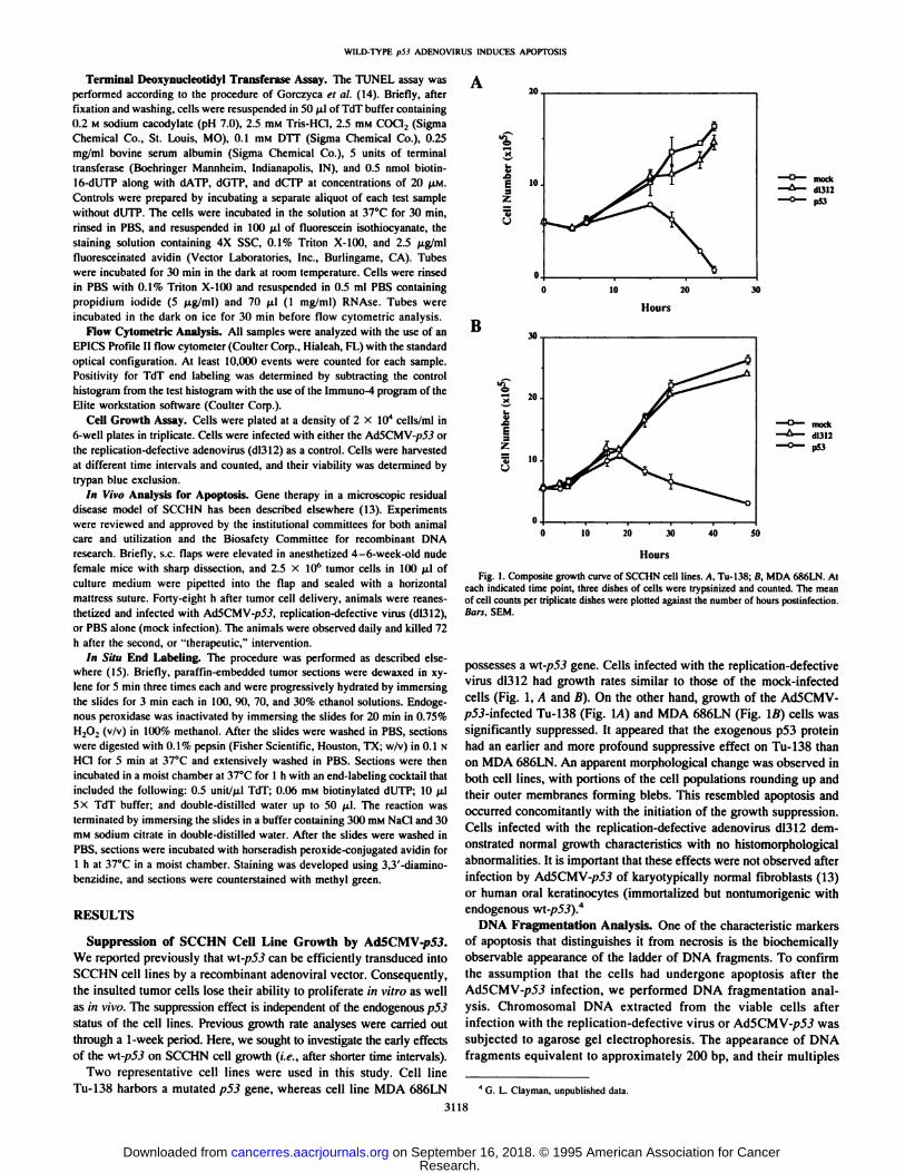
Fig. 1. Composite growth curve of SC'CHN cell lines. A. Tu-138; B, MDA 686LN. Al
each indicated time point, three dishes of cells were Irypsinized and counted. The meanof cell counts per triplicate dishes were plotted against the number of hours postinfection.Bars. SEM.
possesses a wi-p53 gene. Cells infected with the replication-defectivevirus dl312 had growth rates similar to those of the mock-infectedcells (Fig. 1, A and B). On the other hand, growth of the Ad5CMV-p5J-infected Tu-138 (Fig. IA) and MDA 686LN (Fig. \B) cells was
significantly suppressed. It appeared that the exogenous p53 proteinhad an earlier and more profound suppressive effect on Tu-138 than
on MDA 686LN. An apparent morphological change was observed inboth cell lines, with portions of the cell populations rounding up andtheir outer membranes forming blebs. This resembled apoptosis andoccurred concomitantly with the initiation of the growth suppression.Cells infected with the replication-defective adenovirus dl312 dem
onstrated normal growth characteristics with no histomorphologicalabnormalities. It is important that these effects were not observed afterinfection by Ad5CMV-/;>5.? of karyotypically normal fibroblasts (13)
or human oral keratinocytes (immortalized but nontumorigenic withendogenous wt-/)5J).4
DNA Fragmentation Analysis. One of the characteristic markersof apoptosis that distinguishes it from necrosis is the biochemicallyobservable appearance of the ladder of DNA fragments. To confirmthe assumption that the cells had undergone apoptosis after theAd5CMV-p5J infection, we performed DNA fragmentation anal
ysis. Chromosomal DNA extracted from the viable cells afterinfection with the replication-defective virus or Ad5CMV-/;5J was
subjected to agarose gel electrophoresis. The appearance of DNAfragments equivalent to approximately 200 bp, and their multiples
* G. L. dayman, unpublished data.
3118
Research. on September 16, 2018. © 1995 American Association for Cancercancerres.aacrjournals.org Downloaded from
WILD-TYPE p53 ADENOVIRUS INDUCES APOPTOSIS
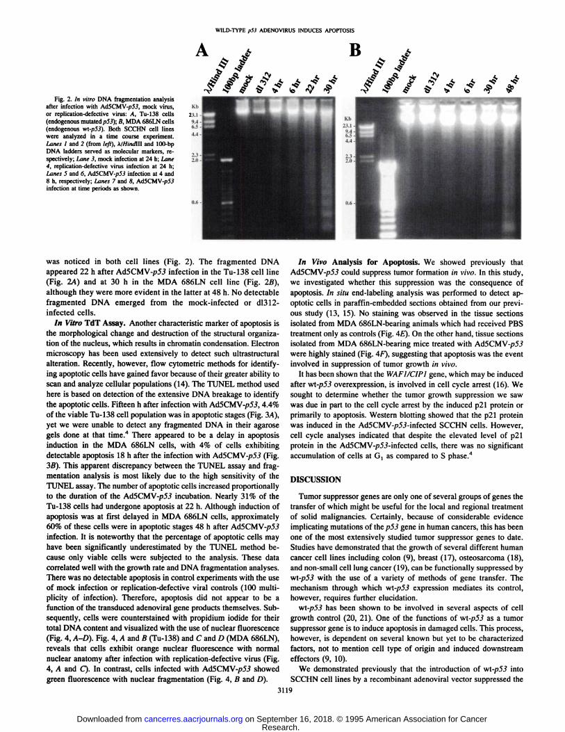
Fig. 2. In vitro DNA fragmentation analysisafter infection wiih Ad5CMV-/;5J, mock virus,or replication-defective virus: A, Tu-138 cells
(endogenous mutated p53); B, MDA 686LN cells(endogenous wt-p53). Both SCCHN cell lineswere analyzed in a time course experiment.Lanes I and 2 (from ¡eft),KIHtndm and HK)-bp
DNA ladders served as molecular markers, respectively; Lane 3, mock infection at 24 h; Lane4, replication-defective virus infection at 24 h;Lanes 5 and 6, Au5CMV-p53 infection at 4 and8 h, respectively; Lanes 7 and 8, Ad5CMV-p53infection at time periods as shown.
was noticed in both cell lines (Fig. 2). The fragmented DNAappeared 22 h after Ad5CMV-p53 infection in the Tu-138 cell line
(Fig. 2A) and at 30 h in the MDA 686LN cell line (Fig. 2B),although they were more evident in the latter at 48 h. No detectablefragmented DNA emerged from the mock-infected or dl312-
infected cells.In Vitro TdT Assay. Another characteristic marker of apoptosis is
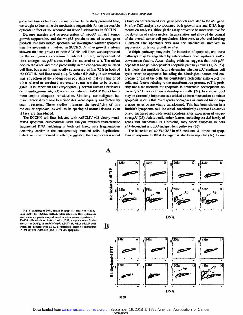
the morphological change and destruction of the structural organization of the nucleus, which results in chromatin condensation. Electronmicroscopy has been used extensively to detect such ultrastructuralalteration. Recently, however, flow cytometric methods for identifying apoptotic cells have gained favor because of their greater ability toscan and analyze cellular populations (14). The TÚNEL method usedhere is based on detection of the extensive DNA breakage to identifythe apoptotic cells. Fifteen h after infection with Ad5CMV-p5J, 4.4%of the viable Tu-138 cell population was in apoptotic stages (Fig. 3A),
yet we were unable to detect any fragmented DNA in their agarosegels done at that time.4 There appeared to be a delay in apoptosis
induction in the MDA 686LN cells, with 4% of cells exhibitingdetectable apoptosis 18 h after the infection with Ad5CMV-/)55 (Fig.
3ß).This apparent discrepancy between the TÚNEL assay and fragmentation analysis is most likely due to the high sensitivity of theTÚNEL assay. The number of apoptotic cells increased proportionallyto the duration of the A.d5CMV-p53 incubation. Nearly 31% of theTu-138 cells had undergone apoptosis at 22 h. Although induction of
apoptosis was at first delayed in MDA 686LN cells, approximately60% of these cells were in apoptotic stages 48 h after Ad5CMV-p5J
infection. It is noteworthy that the percentage of apoptotic cells mayhave been significantly underestimated by the TÚNEL method because only viable cells were subjected to the analysis. These datacorrelated well with the growth rate and DNA fragmentation analyses.There was no detectable apoptosis in control experiments with the useof mock infection or replication-defective viral controls (100 multi
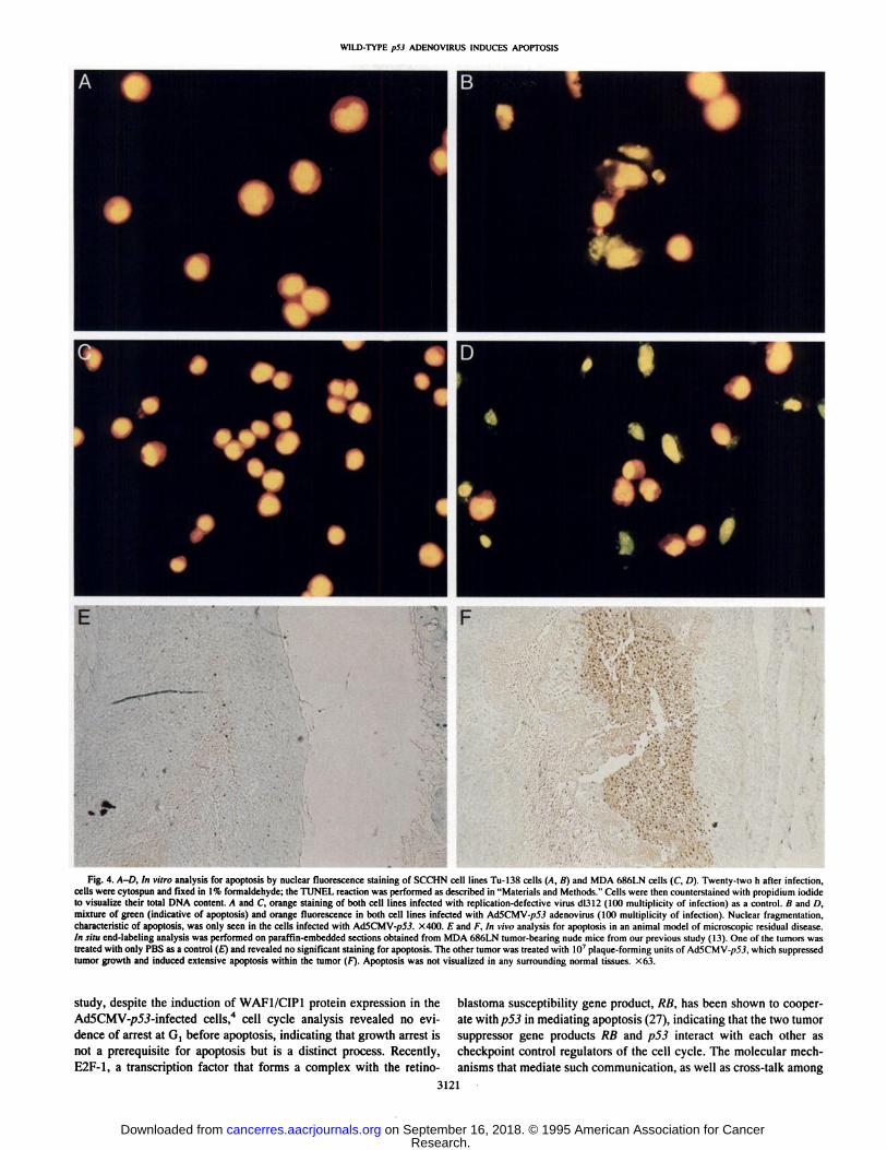
plicity of infection). Therefore, apoptosis did not appear to be afunction of the transduced adenoviral gene products themselves. Subsequently, cells were counterstained with propidium iodide for theirtotal DNA content and visualized with the use of nuclear fluorescence(Fig. 4, A-D). Fig. 4, A and B (Tu-138) and C and D (MDA 686LN),
reveals that cells exhibit orange nuclear fluorescence with normalnuclear anatomy after infection with replication-defective virus (Fig.4, A and C). In contrast, cells infected with Ad5CMV-/>5J showed
green fluorescence with nuclear fragmentation (Fig. 4, B and D).
In Vivo Analysis for Apoptosis. We showed previously thatAd5CMV-p5^ could suppress tumor formation in vivo. In this study,
we investigated whether this suppression was the consequence ofapoptosis. In situ end-labeling analysis was performed to detect apoptotic cells in paraffin-embedded sections obtained from our previ
ous study (13, 15). No staining was observed in the tissue sectionsisolated from MDA 686LN-bearing animals which had received PBS
treatment only as controls (Fig. 4E). On the other hand, tissue sectionsisolated from MDA 686LN-bearing mice treated with Ad5CMV-p5J
were highly stained (Fig. 4F), suggesting that apoptosis was the eventinvolved in suppression of tumor growth /';; vivo.
It has been shown that the WAFI/CIP1 gene, which may be inducedafter wl-p53 overexpression, is involved in cell cycle arrest ( 16). We
sought to determine whether the tumor growth suppression we sawwas due in part to the cell cycle arrest by the induced p21 protein orprimarily to apoptosis. Western blotting showed that the p21 proteinwas induced in the Ad5CMV-p5_?-infected SCCHN cells. However,
cell cycle analyses indicated that despite the elevated level of p21protein in the Ad5CMV-/?5.?-infected cells, there was no significantaccumulation of cells at G, as compared to S phase.4
DISCUSSION
Tumor suppressor genes are only one of several groups of genes thetransfer of which might be useful for the local and regional treatmentof solid malignancies. Certainly, because of considerable evidenceimplicating mutations of the p53 gene in human cancers, this has beenone of the most extensively studied tumor suppressor genes to date.Studies have demonstrated that the growth of several different humancancer cell lines including colon (9), breast (17), osteosarcoma (18),and non-small cell lung cancer (19), can be functionally suppressed bywt-p53 with the use of a variety of methods of gene transfer. Themechanism through which v/\-p53 expression mediates its control,
however, requires further elucidation.wl-p53 has been shown to be involved in several aspects of cell
growth control (20, 21). One of the functions of wl-p53 as a tumor
suppressor gene is to induce apoptosis in damaged cells. This process,however, is dependent on several known but yet to be characterizedfactors, not to mention cell type of origin and induced downstreameffectors (9, 10).
We demonstrated previously that the introduction of wl-p53 into
SCCHN cell lines by a recombinant adenoviral vector suppressed the
3119
Research. on September 16, 2018. © 1995 American Association for Cancercancerres.aacrjournals.org Downloaded from
WILD-TYPE ¡>53ADENOVIRUS INDUCES APOPTOSIS
growth of tumors both in vitro and HIvivo. In the study presented here,we sought to determine the mechanism responsible for the irreversiblecytocidal effect of the recombinant wt-p53 adenovirus in SCCHN.
Because transfer and overexpression of wt-/?5J initiated tumorgrowth suppression, and the wt-p53 protein is one of several gene
products that may induce apoptosis, we investigated whether apoptosiswas the mechanism involved in SCCHN. In vitro growth analysisshowed that the growth of both SCCHN cell lines was suppressedby the exogenous expression of wt-p53 protein, independent of
their endogenous p53 status (whether mutated or wt). The effectoccurred earlier and more profoundly in the endogenously mutatedcell line, but growth was totally suppressed within 72 h in both ofthe SCCHN cell lines used (13). Whether this delay in suppressionwas a function of the endogenous p53 status of that cell line or ofother related or unrelated molecular events remains to be investigated. It is important that karyotypically normal human fibroblasts(with endogenous wl-p53) were insensitive to Ad5CMV-/?5J treat
ment despite adequate transduction. Similarly, nonmalignant human immortalized oral keratinocytes were equally unaffected bysuch treatment. These studies illustrate the specificity of thismolecular approach, as well as its sparing of normal tissues, evenif they are transduced.
The SCCHN cell lines infected with Ad5CMV-/;5J clearly mani
fested apoptosis. Nucleosomal DNA analysis revealed characteristicfragmented DNA laddering in both cell lines, with fragmentationoccurring earlier in the endogenously mutated cells. Replication-
defective virus produced no effect, suggesting that the process was not
a function of transduced viral gene products unrelated to the p53 gene.In vitro TdT analysis corroborated both growth rate and DNA fragmentation analyses, although the assay proved to be more sensitive forthe detection of earlier nuclear fragmentation and allowed the perusalof the overall tumor cell population. Moreover, HI situ end labelingillustrated that apoptosis was also the mechanism involved insuppression of tumor growth in vivo.
Multiple pathways may exist for induction of apoptosis, and thesepathways may be regulated by interventions from upstream and/ordownstream factors. Accumulating evidence suggests that both p53-dependent and/>5J-independent apoptotic pathways exist (11, 22, 23).
It is likely that multiple factors determine whether p53 mediates cellcycle arrest or apoptosis, including the histológica! source and embryonic origin of the cells, the constitutive molecular make-up of the
cells, and factors relating to the transformation process. p53 is probably not a requirement for apoptosis in embryonic development because "p53 knock-out" mice develop normally (24). In contrast, p53
may be extremely important as a critical defense mechanism to induceapoptosis in cells that overexpress oncogenes or mutated tumor suppressor genes or are virally transformed. This has been shown in aBurkitt's lymphoma cell line which constitutively expressed an active
c-myc oncogene and underwent apoptosis after expression of exoge
nous p53 (25). Additionally, other factors, including the Bel family ofgenes and adenoviral E1B proteins, may block apoptosis in both/j5J-dependent and /^-independent pathways (26).
The induction of WAFI/CIPÃŒin />5.?-mediated G, arrest and apop
tosis in response to DNA damage has also been reported (16). In our
Fig. 3. Labeling of DNA breaks in apoptotic cells with biotiny-latcd dUTP by TÚNEL method. After infection, flow cytomclricanalysis for apoptosis was performed in a time course experiment. A.Tu-l3S cells which are infected with dl3l2. a replication-defectiveadenovirus (A-D). or MSCMV-p53 (£-//). B. MDA 6X6LN cellswhich are infected with dl3l2, a replication-defective adenovirus(A-D). or with AdSCMV-pSJ (E-H). Ap. apoplosis.
B
DNA
30hr 48hr D
30hr
DNA
3120
Research. on September 16, 2018. © 1995 American Association for Cancercancerres.aacrjournals.org Downloaded from
WILD-TYPE p53 ADENOVIRUS INDUCES APOPTOSIS
Fig. 4. A-D, In vitro analysis for apoptosis by nuclear fluorescence staining of SCCHN cell lines Tu-138 cells (A, B) and MDA 686LN cells (C, D). Twenty-two h after infection,cells were cylospun and fixed in \% formaldehyde; the TÚNEL reaction was performed as described in "Materials and Methods." Cells were then counterstained with propidium iodide
to visualize their total DNA content. A and C orange staining of both cell lines infected with replication-defective virus dl312 (KM) multiplicity of infection) as a control. B and /),mixture of green (indicative of apoptosis) and orange fluorescence in both cell lines infected with Ad5CMV-/>5.i adenovirus (KM) multiplicity of infection). Nuclear fragmentation,characteristic of apoptosis. was only seen in the cells infected with Ad5CMV-/;5.f. X400. E and F, In vim analysis for apoptosis in an animal model of microscopic residual disease.in situ end-labeling analysis was performed on paraffin-embedded sections obtained from MDA 686LN tumor-bearing nude mice from our previous study (13). One of the tumors wastreated with only PBS as a control (£")and revealed no significant staining for apoptosis. The other tumor was treated with K)7plaque-forming units of Ad5CMV-/>5J, which suppressed
tumor growth and induced extensive apoptosis within the tumor (F). Apoptosis was not visualized in any surrounding normal tissues. X63.
study, despite the induction of WAF1/CIP1 protein expression in theAd5CMV-/?5.?-infected cells,4 cell cycle analysis revealed no evi
dence of arrest at G, before apoptosis, indicating that growth arrest isnot a prerequisite for apoptosis but is a distinct process. Recently,E2F-1, a transcription factor that forms a complex with the retino-
blastoma susceptibility gene product, RB, has been shown to cooperate with p53 in mediating apoptosis (27), indicating that the two tumorsuppressor gene products RB and p53 interact with each other ascheckpoint control regulators of the cell cycle. The molecular mechanisms that mediate such communication, as well as cross-talk among
3121
Research. on September 16, 2018. © 1995 American Association for Cancercancerres.aacrjournals.org Downloaded from
WILD.TYPE p53 ADENOV1RUS INDUCES APOPTOSIS
other important cell cycle-regulating components such as pió, p21,cyclins, and cyclin-dependent kinases remain to be elucidated.
In summary, the studies presented here demonstrated thatAd5CMV-/>5.? induced apoptosis in SCCHN but spared normal cells.
In contrast, Roth et al. (19) found that the same intervention suppressed the growth of malignant non-small cell lung cancer cells
(H358) but did not induce apoptosis. This supports the concept that thereare inherent constitutive differences between these neoplasms that mayregulate the apoptotic process. We conclude that selective induction ofapoptosis of solid malignancies that spares normal cells is an attractivestrategy for molecular therapy and requires further investigation.
ACKNOWLEDGMENTS
The authors express their sincere gratitude to Drs. Jack A. Roth andWei-Wei Zhang for generously providing Ad5CMV-p5.i. to Dr. No Hee Park
for the immortalized human oral kcratinocyte cell lines: to Dr. Peter Sacks forthe MDA 686LN cell line; to Kathryn Hale for manuscript editing; and to JoseJuarez for computer medical graphic assistance.
REFERENCES
1. Linzcr. D. I., and Levine. A. J. Characterization of a 54K dalton cellular SV40 tumorantigen present in SV40-transformed cells and uninfected embryonal carcinoma cells.Cell, 17: 43-52, 1979.
2. Lane, D. P.. and Crawford. L. V. T antigen is bound to a host protein in SV40-transformcd cells. Nature (Lond.). 278: 261-263, 1979.
3. Rodrigues, N. R., Rowan, A., Smith, M. E. F., Kerr, I. B., Bodmer, W. F., Gannon,J. V., and Lane, D. P. p53 mutations in colorectal cancer. Proc. Nati. Acad. Sci. USA,87: 7555-7559, 1990.
4. Bartek, J., Iggo. R.. Gannon. J., and Lane, D. P. Genetic and immunochemical analysis ofmutant p53 in human breast cancer cell lines. Oncogene, 5: 893-899, 1990.
5. Takahashi. T.. Takahashi. T., Suzuki, H.. Hida. T., Sekido, Y., Ariyoshi. Y., andUeda, R. The p53 gene is very frequently mutated in small-cell lung cancer with adistinct nuclcotide substitution pattern. Cancer Res.. 52: 734-736, 1992.
6. Maestro, R., Dolcetti, R., Gasparotto, D., Doglioni. C., Pelucchi, S., Barzan, L.,Grandi. E., and Boiocchi, M. High frequency of p53 gene alterations associated withprotein overexpression in human squamous cell carcinoma of the larynx. Oncogene,7: 1159-1166, 1992.
7. Martinez. J., Georgoff, I.. Martinez. J.. and Levine, A. J. Cellular localization and cellcycle regulation by a temperature-sensitive p53 protein. Genes Dev., 5: 151-159,1991.
8. Diller. L., Kassel, J., Nelson, C. E., Gryka, M. A., Litwak, G., Gebhardt. M., Bressac,B.. Ozturk. M., Baker, S. J., Vogclstein. B. and Friend. S. p53 function as a cell cyclecontrol protein in osteosarcomas. Mol. Cell. BioL. 10: 5772-5781, 1990.
9. Shaw, P., Bovey, R.. Tardy. S., Sahli, R., Sorda!, B., and Costa, J. Induction ofapoptosis by wild-type p53 in a human colon tumor-derived celt line. Proc. Nati.Acad. Sci. USA, 8V: 4495-4499, 1992.
10. Yonish-Rouach. E.. Resnitzky. D., Lotem, J., Sachs, L.. Kimchi, A., and Oren, M.Wild-type p53 induces apoptosis of myeloid leukaemic cells that is inhibited byinterleukin-6. Nature (Lond.), 352: 345-347, 1991.
11. Lowe, S. W., Schmitt, E. M.. Smith, S. W., Osborne, B. A., and Jacks, T. p53 isrequired for radiation-induced apoptosis in mouse thymocytes. Nature (Lond.). 362:847-849, 1993.
12. Liu, T.-J., Zhang, W.-W., Taylor, D. L., Roth. J. A., Goepfert, H., and dayman,
G. L. Growth suppression of human head and neck cancer cells by the introduction of a wild-type p53 gene via a recombinant adenovirus. Cancer Res., 54:3662-3667, 1994.
13. dayman, G. L., EI-Naggar. A. K.. Zhang, W.-W., Taylor, D. L.. Roth, J. A.,Goepfert, H.. and Liu, T-J. In vivo molecular therapy with p53 adenovirus formicroscopic residual head and neck squamous carcinoma. Cancer Res., 55: 1-6,
1995.14. Gorczyca, W., Gong, J., and Darzynkiewicz, Z. Detection of DNA strand breaks in
individual apoptotic cells by the m .sii«terminal deoxynucleotidyl transferase and nicktranslation assays. Cancer Res.. 53: 1945-1951, 1993.
15. Wijsman, J. H., Jonker, R. R.. Keijzer. R., Van De Velde, C. J. H.. Cornelisse,C. J., and Van Dierendonck, J. H. A new method to detect apoptosis in paraffinsection: in .sii«end-labeling of fragmented DNA. J. Histochem. Cytochem., 41:7-12, 1993.
16. El-Deiry, W. S., Harper, J. W., O'Connor, P. M., Velculescu, V. E., Canman, C. E.,
Jackman. J., Pietenpol, J. A.. Burrell, M., Hill. D. E., Wang, Y., Wiman, K. G.,Mercer, W. E., Kastan, M. B., Kohn, K. W.. Elledge, S. J., Kinzler, K. W., andVogelstein B. WAFl/CIPI is induced in /j5J-mediated G, arrest and apoptosis.Cancer Res., 54: 1169-1174, 1994.
17. Casey, G., Lo-Hsueh. M., Lopez, M. E.. Vogelstein, B., and Stanbridge, E. J. Growthsuppression of human breast cancer cells by the introduction of a wild-type p53 gene.Oncogene. 6: 1791-1797, 1991.
18. Chen, P. L., Chen. Y. M., Bookstein. R., and Lee, W. H. Genetic mechanisms oftumor suppression by the human p53 gene. Science (Washington DC), 250:1576-1580, 1990.
19. Zhang, W-W., Fang, X., Mazur, W.. French, B. A., Georges, R., and Roth, J. A.High-efficiency gene transfer and high-level expression of wild-type p53 in humanlung cancer cells mediated by recombinant adenovirus. Cancer Gene Ther., /: 5-13,
1994.20. Mietz, J. A., Unger, T., Huibregtse, J. M., and Howley, P. M. The transcriptional
transactivation function of wild-type p53 is inhibited by SV40 large T-antigen and byHPV-16 E6 oncoprotein. EMBO J.. //: 5013-5020, 1992.
21. Wilcock. D.. and Lane. D. P. Localization of p53, retinoblastoma, and host replicationproteins at sites of viral replication in herpes-infected cells. Nature (Lond.), 349:429-431. 1991.
22. Clarke, A. R., Purdie, C. A., Harrison. D. J., Morris, R. G.. Bird, C. C., Hooper, M. L.,and Wyllie, A. H. Thymocyte apoptosis induced by p53-dependent and independentpathways. Nature (Lond.), 362: 849-852, 1993.
23. Berges, R. R., Furuya. Y., Remington, L., English, H. F., Jacks, T., and Isaacs, J. T.Cell proliferation, DNA repair, and p53 function are not required for programmed celldeath of prostatic glandular cells induced by androgcn ablation. Proc. Nati. Acad. Sci.USA, 90: 8910-8914. 1993.
24. Donehower. L. A.. Harvey. M., Slagle, B. L., McArthur, M. J., Montgomery, C. A.,Jr., Butel, J. S., and Bradley, A. Mice deficient for p53 arc developmentally normalbut susceptible to spontaneous tumors. Nature (Lond.). 356: 215-221. 1992.
25. Ramqvist, T., Magnusson. K. P.. Wang, Y.. Szekely. L., Klein. G.. and Wiman, K. G.Wild-type p53 induces apoptosis in a Burkitt lymphoma (BL) line that carries mutantp53. Oncogene, 8: 1495-1500. 1993.
26. Rao, L., Debbas. M., Sabbatini. P., Hockenbery. D., Korsmeyer, S., and White, E. Theadenovirus EIA proteins induce apoptosis which is inhibited by the E1B 19K andBcl-2 proteins. Proc. Nati. Acad. Sci. USA, 89: 7742-7746. 1992.
27. Wu, X.. and Levine. A. J. p53 and E2F-I cooperate to mediate apoptosis. Proc. Nail.Acad. Sci. USA, 91: 3602-3606, 1994.
3122
Research. on September 16, 2018. © 1995 American Association for Cancercancerres.aacrjournals.org Downloaded from
1995;55:3117-3122. Cancer Res Ta-Jen Liu, Adel K. El-Naggar, Timothy J. McDonnell, et al. NeckGene Transfer in Squamous Cell Carcinoma of the Head and
Adenoviralp53Apoptosis Induction Mediated by Wild-Type
Updated version
http://cancerres.aacrjournals.org/content/55/14/3117
Access the most recent version of this article at:
E-mail alerts related to this article or journal.Sign up to receive free email-alerts
Subscriptions
Reprints and
To order reprints of this article or to subscribe to the journal, contact the AACR Publications
Permissions
Rightslink site. Click on "Request Permissions" which will take you to the Copyright Clearance Center's (CCC)
.http://cancerres.aacrjournals.org/content/55/14/3117To request permission to re-use all or part of this article, use this link
Research. on September 16, 2018. © 1995 American Association for Cancercancerres.aacrjournals.org Downloaded from