Embed Size (px)
Citation preview
BioMed CentralC
TIONALINTERNACANCER CELLCancer Cell International
ss
Open AccePrimary researchCytogenetic characterization and H-ras associated transformation of immortalized human mammary epithelial cellsKrishna Rao*1, Özge Alper2, Kent E Opheim3, George Bonnet4, Kristine Wolfe5, Eileen Bryant6,7, Siobhan O'Hara Larivee7, Peggy Porter8,9 and James K McDougall8,9Address: 1Southern Illinois University School of Medicine Cancer Institute, P.O. Box 19678, Springfield, IL 62794-9678, USA, 2National Institutes of Health, National Institute of Neurological Disorders, Surgical Neurology, Bldg 10, 5D37, Bethesda, MD 20892-1414, USA, 3Children's Hospital and Regional Medical Center, Dept. of Laboratories, A-6901 4800 Sand Point Way, NE, Seattle, WA 98105, USA, 4Cytogenetics Studio, Inc., 41 Myrtle Ave., Cambridge, MA 02138, USA, 52511 Boundary St., San Diego, CA 92104-5313, USA, 6Fred Hutchinson Cancer Research Center, Clinical Research Division, P.O. Box 19024, 1100 Fairview Avenue North, D2-190, Seattle, WA 98109-1024, USA, 7Seattle Cancer Care Alliance, 825 Eastlake Avenue East, Mailstop G7500, Seattle, WA 98109, USA, 8Fred Hutchinson Cancer Research Center, Cancer Biology Program, P.O. Box 19024, 1100 Fairview Avenue North, C1-015, Seattle, WA, 98109-1024, USA and 9Department of Pathology, University of Washington School of Medicine, Seattle, WA 98195, USA
Email: Krishna Rao* - [email protected]; Özge Alper - [email protected]; Kent E Opheim - [email protected]; George Bonnet - [email protected]; Kristine Wolfe - [email protected]; Eileen Bryant - [email protected]; Siobhan O'Hara Larivee - [email protected]; Peggy Porter - [email protected]; James K McDougall - [email protected]
* Corresponding author
AbstractIntroduction: Immortalization is a key step in malignant transformation, but immortalization alone isinsufficient for transformation. Human mammary epithelial cell (HMEC) transformation is a complexprocess that requires additional genetic changes beyond immortalization and can be accomplished in vitroby accumulation of genetic changes and expression of H-ras.
Methods: HMEC were immortalized by serial passaging and transduction with the catalytic subunit of thehuman telomerase gene (hTERT). The immortalized cells were passaged in vitro and studied by acombination of G- banding and Spectral Karyotyping (SKY). H-ras transduced, hTERT immortalized cellswere cloned in soft agar and injected into nude mice. Extensive analysis was performed on the tumors thatdeveloped in nude mice, including immunohistochemistry and western blotting.
Results: Immortal HMEC alone were not tumorigenic in γ-irradiated nude mice and could not grow insoft agar. Late passage hTERT immortalized HMEC from a donor transduced with a retroviral vectorcontaining the mutant, autoactive, human H-ras61L gene acquired anchorage independent growthproperties and the capacity for tumorigenic growth in vivo. The tumors that developed in the nude micewere poorly differentiated epithelial carcinomas that continued to overexpress ras. These cells wereresistant to doxorubicin mediated G1/S phase arrest but were sensitive to treatment with afarnesyltransferase inhibitor.
Conclusion: Some of the cytogenetic changes are similar to what is observed in premalignant andmalignant breast lesions. Despite these changes, late passage immortal HMEC are not tumorigenic andcould only be transformed with overexpression of a mutant H-ras oncogene.
Published: 26 May 2006
Cancer Cell International 2006, 6:15 doi:10.1186/1475-2867-6-15
Received: 10 August 2005Accepted: 26 May 2006
This article is available from: http://www.cancerci.com/content/6/1/15
© 2006 Rao et al; licensee BioMed Central Ltd.This is an Open Access article distributed under the terms of the Creative Commons Attribution License (http://creativecommons.org/licenses/by/2.0), which permits unrestricted use, distribution, and reproduction in any medium, provided the original work is properly cited.
Page 1 of 11(page number not for citation purposes)
Cancer Cell International 2006, 6:15 http://www.cancerci.com/content/6/1/15
IntroductionImmortalization is a key step in malignant transforma-tion, as it allows a cell to indefinitely proliferate whileaccumulating genetic abnormalities. Normal humanmammary epithelial cells (HMEC) initially proliferaterobustly in tissue culture before encountering a prolifera-tion block termed stasis [1,2]. Cells in stasis are less pro-liferative, appear large and flattened, stain positively forbeta galactosidase, but are non-apoptotic since the cellsare negative by terminal deoxynucleotidyl transferasemediated dUTP nick end labeling (TUNEL) staining.HMEC can escape stasis spontaneously, as evidenced bythe fact that a small percentage of HMEC in culture maypass through stasis and continue to proliferate [1]. Theexact number of passages and percentage of cells thatevade this initial growth arrest are highly dependent uponindividual specimens and culture conditions [1]. It hasbeen demonstrated that the p16 INK4A tumor suppressor isinactivated in HMEC escaping stasis [3], primarily by pro-gressive promoter methylation [4-6]. Cells undergo a sec-ond mortality stage, termed agonescence [7]. Cells inagonescence appear small and vacuolated and have criti-cally shortened telomeres. Activation of telomeraseexpression at this stage stabilizes telomere length, allow-ing cells to pass through agonescence and proliferateindefinitely.
DNA tumor viruses immortalize cells and provide a usefulmodel for studying neoplasia. For example, the combinedexpression of the catalytic subunit of human telomerase(hTERT), together with SV40 large T antigen can immor-talize HMEC. The E6/E7 gene products from "high risk"human papillomavirus types such as HPV16 can uniquelyimmortalize HMEC at any stage without the aid of hTERT[8].
In vitro models of mammary cell transformation eitherrequire the introduction of viral oncogenes [9-11] ormake use of chemical carcinogens [12], such as benzo[a]pyrene. Stampfer and Bartley [12] exposed rapidlygrowing primary cultures of human mammary epithelialcells to benzo [a]pyrene for 2–3 days and produced twoimmortalized cell lines- 184 A1 and 184 B5. The introduc-tion of Ras or erbB2 into subline 184A1N4 or 184B5respectively results in production of a malignant pheno-type [12,13]. Both 184 A1 and 184 B5 have acquired mul-tiple cytogenetic abnormalities [14]. HBL 100, a cell linederived from an immortalized mammary cell culturedfrom mother's milk, also transforms with the introductionof a single oncogene [15], but carries an integrated copy ofthe SV40 genome and demonstrates a near triploid karyo-type.
More recently, Elenbaas et al. [9] transformed HMEC byserial transduction of SV40 large T antigen, SV40 small t
antigen, hTERT, and a mutant, constitutively active,human V12G H-ras. Substantial overexpression of H-ras(12 fold) was required in order to transform the HMEC.The tumors that were obtained were epithelial, stainingpositively for cytokeratin, and negative for the estrogenand progesterone receptor, consistent with the poorly dif-ferentiated histology seen on hematoxylin and eosinstaining. Cytogenetic data presented in the paper revealedfrequent increased copy number of the locus containingthe c-Myc oncogene, either through trisomy 8, transloca-tions involving 8q24, or by creation of isochromosomefor the long arm of chromosome 8 [i(8)(q10)]. Interest-ingly, cells in the tumor, in which i(8)(q10) was fre-quently seen, displayed extra copies of chromosomes 5.One tumor had 22% metaphases with del(12p). One ofthe tumors was near tetraploid while the other two werenear diploid (90% and 62% of total cells analyzed).
Toouli et al. [11] transformed HMEC from a single donorpurely through the use of SV40 large T antigen and SV40small t antigen. Fifty percent of the late passage cell linesexpressing both large T antigen and small t antigen weretumorigenic. One of the lines showed a 30% take rate,with a latency of twenty-one days, while the other lineshowed a 20% take rate with a latency of sixty-eight days.No histologic, cytogenetic, or immunohistochemical datawere presented on the tumors. The authors also noted thatnone of the hTERT immortalized HMEC lines were tum-origenic.
In this paper, we describe the cytogenetic characteristics ofa hTERT immortalized human mammary epithelial cellline, and of a tumorigenic human mammary epithelialcell line produced through hTERT immortalization, pas-saging, transduction of H-ras61L, and soft agar cloning.
Materials and methodsIsolation and culture of HMECThe mammary epithelial cells (HMEC) were all derivedfrom reduction mammoplasties of individuals with noknown breast pathology, as reconfirmed by histopathol-ogy of the post-surgical specimens. HMEC were preparedby the method of Smith et al. [16] and grown at 37°C and5% CO2 in DFCI-1 media [17]. Cells were allowed to passthrough stasis before infection with the hTERT construct,as described by Foster et al.[18], Kiyono et al.[3], andRothenberg and Nolan [19].
Cell proliferation and stainingHMEC (human normal mammary epithelial cells) andour tumorigenic cells were seeded in glass 8-well chamberslides at a concentration of 104 cells per well and treatedwith FTI-277 and DMSO at 2.5, 5.0, 10.0, 15.0 and 20.0μM concentrations for 5 days in duplicate. Cells werecounted using Hemocytometer (blood cell counter) on
Page 2 of 11(page number not for citation purposes)
Cancer Cell International 2006, 6:15 http://www.cancerci.com/content/6/1/15
1st, 2nd and 5th days as well as stained with Phalloidin-TXRfor cytoskeletal staining and DAPI for nuclear staining.
CytogeneticsMetaphase chromosomes from HMEC were prepared andG-banded (Wright's stain) using standard cytogenetictechniques [20]. Briefly, after the cells were incubatedwith 0.06 ug/ul of colcemid at 37°C for 2.5 hours, theywere trypsinized with trypsin/EDTA (0.5%/0.1%) andincubated in 2 ml of a 1:1 (by vol) mixture of 0.8%sodium citrate and 0.075 M potassium chloride for thirtyminutes at 37 degrees C. This was followed by addition of1 ml of Carnoy's fixative (3:1 by vol; methanol:aceticacid) and incubation at room temperature. The fixed cellswere then spun down and resuspended in 1 ml of Car-noy's fixative. Metaphase chromosomes were prepared forG-banding and Spectral Karyotyping (SKY). Cells wereanalyzed based on clonality criteria and karyotypes weredescribed by the recommendations of the InternationalSystem for Human Cytogenetic Nomenclature (ISCN2005) [21].
Spectral karyotypingSKY [22] was used to identify or confirm the G-band kary-otype assignments. Briefly, SKY is a form of fluorescencein situ hybridization (FISH) which utilizes a mixture of"whole chromosome paint" DNA probes to uniquelylabel each of the 24 different human chromosomes,allowing simultaneous visualization and identification ofeach chromosome or chromosome derivative. The limit ofresolution of SKY for structural abnormalities is currentlyunknown, and the technique will likely not detect subtle,intra-chromosomal rearrangements, such as small dele-tions, duplications or inversions. We also cannot rule outthe possibility of mosaicism for numerical or structuralabnormalities given the limited number of cells (usuallyfive), which are evaluated in this study. Unbanded or de-stained G-banded metaphase chromosome preparationswere hybridized with the SKY probe mixture and wereanalyzed using SKYView imaging software, following themanufacturer's instructions (Applied Spectral Imaging,Inc.; Carlsbad, CA). The hybridized metaphase cells weresimultaneously stained with the nuclear stain, DAPI.When the bright-dark fluorescent bands from chromo-somes stained with DAPI are reversed by the SKYViewimaging software, a low-resolution G-band-like pattern isproduced that is used to confirm the chromosome iden-tity and to assign cytogenetic breakpoints on rearrangedchromosomes.
For both G-band and SKY analyses, structural chromo-some abnormalities and chromosome gains were consid-ered clonal if the aberration was exhibited in at least twocells from a culture. Whole chromosome losses were con-
sidered clonal if observed in at least three cells from a cul-ture, per ISCN convention [21].
Flow cytometryThe protocol as previously described was followed [23].Cells were lifted from the plate with trypsin/EDTA (0.5%/0.1%) and fixed overnight in 80% ethanol at 4°C. Cellswere washed with PBS the following day, and this was fol-lowed by staining in PBS with 10 ug/ml of propidiumiodide for twenty minutes at room temperature.
Transduction of H-ras 61L into HMECpCTV3H H-ras61L (a generous gift of Dr. Channing Der)or pCTV3H (empty control vector) was precipitated andincubated with Phoenix cells to produce infectious super-natant [19]. Cells were transduced with either construct byincubation with 1 ml of infectious supernatant with 4 pg/ml of polybrene for four hours. Two days later, continu-ous selection with hygromycin B (20 ug/ml) was initiatedover a several week time span until sustained growth froma robust population was maintained.
Soft agar assaySix-well plates were filled with two ml of 0.66% nobleagar in DFCI-1 media as a bottom layer and allowed tosolidify at 4°C overnight. 105 cells were suspended in oneml of 0.33% noble agar in DFCI-1 media and plated ontowells. Each well was fed once a week with one ml of0.33% agar in DFCI-1 media. Wells were scored for colonygrowth over the next four weeks. Cell clumps greater than60 um were scored as colonies.
Nude mice injection and tumor harvestingFour-week-old female nude mice were γ-irradiated to 300rads using a linear accelerator and injected subcutane-ously over the abdomen with 107 cells derived from thesoft agar colony that was regrown in monolayer. The micewere observed for tumor formation over the following 30days. Tumors were harvested and cut into two parts. Onepart was sent for paraffin embedding, while the other halfwas minced and disaggregated with trypsin/EDTA (0.5%/0.1%) overnight at 4°C, followed by trypsinization for anadditional thirty minutes at 37°C. Single cells were platedand allowed to grow in DFCI-1 media.
ImmunohistochemistryImmunohistochemistry on paraffin embedded tumor sec-tions was performed as previously described by Gown andVogel [24] at Phenopath Laboratories (Seattle, WA) or thelaboratory of Dr. Peggy Porter (Program in Cancer Biol-ogy, Fred Hutchinson Cancer Research Center, Seattle,WA) using 4 um thick sections baked onto positivelycharged slides. Immunohistochemical studies were per-formed on formalin-fixed, paraffin-embedded, tissue sec-tions using the avidin-biotin-peroxidase complex (ABC)
Page 3 of 11(page number not for citation purposes)
Cancer Cell International 2006, 6:15 http://www.cancerci.com/content/6/1/15
method. Sections were incubated with a cocktail of twoanti- cytokeratin mouse monoclonal antibodies (AE1 andAE3, Roche, Indianapolis, IN, 1:200 dilution) that recog-nizes a wide range of high and low molecular weightcytokeratins; anti-cytokeratin-7 (OV-TL 12/30, DAKO,Carpinteria, CA, 1:1000), anti-smooth muscle myosinheavy chain (SMMS-1, DAKO, 1:30); anti-estrogen recep-tor (ID5, Zymed, South San Francisco, CA, 1:200); anti-progesterone receptor (IA6, Lab Vision, 1:50); anti-mam-moglobin(304-1A5 1;2000, 31A5 1:400, Corixa, Seattle,WA); anti-cytokeratin 5/6(5/16 B4, Chemicon, Temecula,CA, 1:1000); anti-cytokeratin 19 (BA 17, DAKO, 1:100),anti-calponin (Calp, DAKO, 1;1000); anti-cytokeratin 20(KS 20.8, DAKO, 1:250); anti-cytokeratin 8 (35βH11,DAKO, 1:50); anti-E-cadherin (HECD-1, Zymed, SanFrancisco, CA,1:300); brst-2 (D6, Signet, Dedham, MA,1:200). After overnight incubation, slides were incubatedwith secondary antibody. For polyclonal antibodies raisedin rabbit, biotinylated goat anti-rabbit immunoglobulin(1:200) was used. For murine monoclonal antibodies, weused biotinylated goat anti-mouse immunoglobulin(1:200). Sections were incubated in secondary antibodyfor 30 minutes. The peroxidase staining procedure wasdone using the ABC Elite kit (Vector laboratories, Burlin-game, CA). The immunostaining reaction was visualizedusing 0.05 % 3,3'- diaminobenzidine in Tris- HCl buffercontaining 0.01 % hydrogen peroxide, pH 7.6.
Western blottingTotal cell protein was extracted from transduced cells bysonication in We 16 buffer (25 mM Tris HCl- pH 7.5, 125mM NaCl, 2.5 mM EDTA, 0.05% SDS, 0.5% NP40, 0.5%DOC, 10% glycerol, 1 mM DTT, 0.5 mg/ml Pefablock,0.025 mg/ml leupeptin, 0.01 mg/ml pepstatin, 0.01 mg/ml aprotinin, 0.08 M β glycerophosphate, 0.005 Na3VO4,0.05 M NaF). Twenty micrograms of protein from eachcell population was electrophoresed and transferred to apolyvinylidene difluoride membrane (NEN). The mem-branes were blotted with the following antibodies: H-ras(LA-069, Quality Biotech, Camden, NJ, 1:10,000); c-Myc(9E10, BD Biosciences, San Diego, CA, 1:250), Mdm2(IF2, Oncogene Research Products, Boston, MA, 1:50),p53 (DO-1, Oncogene Research Products, 1:100), andactin (c-11, Santa Cruz Biotechnology, Santa Cruz, CA,1:100). Samples destined for H-ras western blotting wererun on a 15% SDS polyacrylamide gel, while all othersamples were run on 8% polyacrylamide gel. After thesamples were transferred onto the membrane, the mem-brane was blocked in 10% milk in TBST buffer (10 mMTris-HCl, pH 7.5, 150 mM NaCl, 0.05% Tween-20) forone hour at room temperature or overnight at 4°C. Mem-branes were then incubated overnight at 4°C with 10 mlof 5% milk in TBST/0.5% Tween 20 with the primary anti-body. The following day, membranes were briefly washedand incubated for one hour with secondary antibody in
5% milk in TBST/0.5% Tween 20 with the appropriate sec-ondary antibody. Anti- mouse IgG- HRP from JacksonImmunoresearch Labs (Bar Harbor, ME) was used againstthe H-ras (1:35,000), c-Myc (1:10,000), and p53(1:10,000) primaries. Donkey anti-goat antibody (SantaCruz Biotechnology) was used against the polyclonalactin primary (1:5,000). Blots were developed using theRenaissance chemiluminescence system (NEN) andimaged on Kodak Xomat blue film.
ResultshTERT immortalized HMEC from donor # 4 (hereinreferred to as HMEC 4-hTERT or RAO-1) at late passagedemonstrated a derivative chromosome 1,[der(1)t(1;8)(p13;q13)], in addition to two normal cop-ies of chromosome 1 and two normal copies of chromo-some 8, in four of five metaphase cells analyzed by SKY.HMEC 4-hTERT demonstrated trisomy of 1q and 8q dueto the presence of the derivative chromosome, andshowed complete trisomy of chromosomes 5 and 20.Flow cytometry of HMEC 4-hTERT confirmed a near dip-loid population. These immortal cell lines analyzed werenot tumorigenic in nude mice and did not demonstrateanchorage independent growth in soft agar.
Transduction of pCTV3H H-ras61L into hTERT-immortal-ized late passage (passage 32) HMEC from donor 4 (RAO-2 cell population) resulted in high levels (at least six fold)of H-ras expression in these cells as compared to HMEC(passage 43). At passage 43, 105 cells were cast into softagar. H-ras-transduced HMEC demonstrated growth insoft agar, with at least fifty colonies generated. Of the threecolonies picked at random, the second colony wasexpanded in monolayer and injected, at passage 47, intosix nude mice, producing mammary epithelial carcinomas(RAO-4 cell line). The other two colonies, when injectedinto nude mice, produced spindle cell carcinomas (RAO-3 cell line) and have been described separately [25].
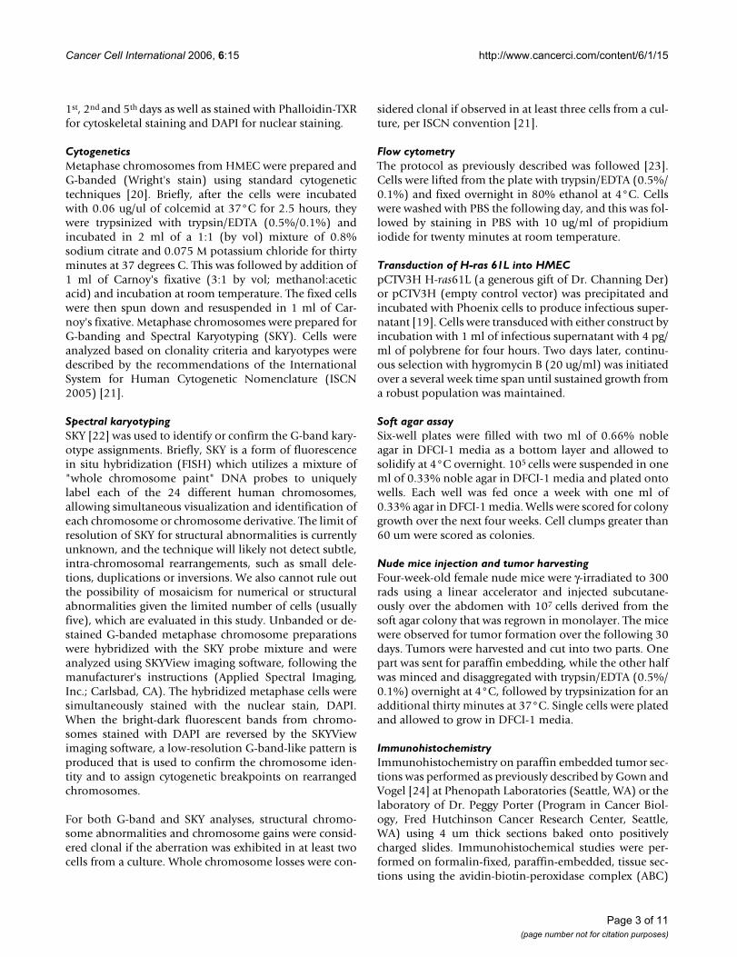
All six mice injected with cells from colony 2 of H-ras61Ltransduced HMEC developed tumors subcutaneously atthe site of injection within 21 days. Figures 1a and 1billustrate the hematoxylin/eosin staining of a paraffin-embedded section of the epithelial tumor. The cells areround and circumscribed. The cells form a sheet with nodifferentiated structure and display areas of necrosis, allconsistent with a poorly differentiated carcinoma. Thepan-cytokeratin staining (Figure 1c) is strongly positive,consistent with the tumor's epithelial origin. The tumor iscytokeratin 7 positive and negative for cytokeratins 8, 18,and 20. It is negative for basal/myoepithelial markerssuch as cytokeratin 5, smooth muscle myosin heavychain, and calponin. The tumor is negative for the estro-gen receptor and the progesterone receptor. It is negative
Page 4 of 11(page number not for citation purposes)
Cancer Cell International 2006, 6:15 http://www.cancerci.com/content/6/1/15
for mammoglobin and the brst-2 antigen. The tumor hasalso lost E-cadherin expression (data not shown).
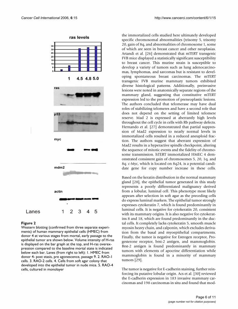
Western blot analysis of HMEC from donor 4 revealssome interesting patterns, shown in Figure 2. H-ras levelsare very low in early passage HMEC. With immortaliza-tion and subsequent passaging, H-ras levels increase. Aftertransduction of pCTV3 H-ras61L, H-ras levels increasesubstantially and are maintained at a five-fold level ofoverexpression through to the tumor. Expression of p53was at low levels in mortal cells. In late passage immortalcells, it is expressed at high levels. Ras transduction appar-ently reduces the level of p53 expression, and a reducedlevel of p53 expression is observed in the tumor. c-Myctakes a very interesting pattern of expression. c-Myc is atlow levels in our post-selection HMEC (lane 1, Figure 2).It is strongly expressed in RAO-1 (lane 2, Figure 2), butdiminished in RAO-2 cells (lane 3, Figure 2), suggestingthat H-ras61L transduction reduces c-Myc expression.Finally, c-Myc expression is detected in the soft agar col-ony (lane 4, Figure 2) that gave rise to the mammary epi-thelial tumor, and in the RAO-4 tumor cells cultured inmonolayer (lane 5, Figure 2). Mdm 2 levels are stablethroughout this time course.
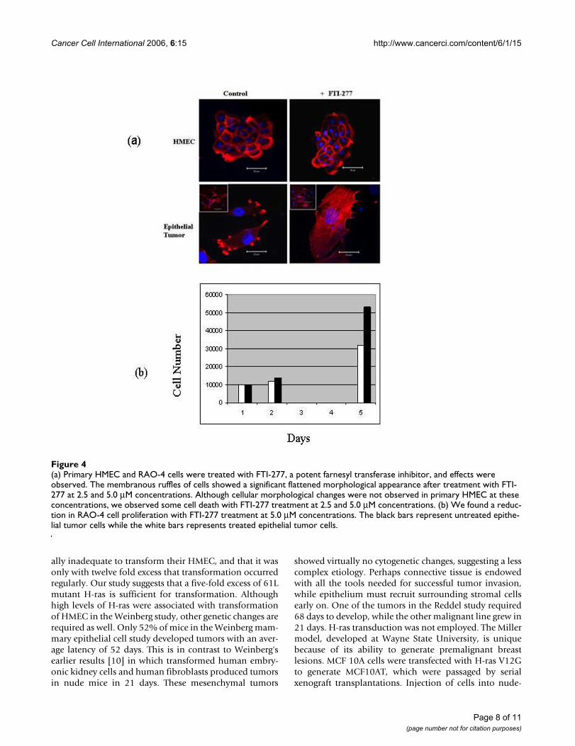
Single color (DAPI) flow cytometry (Figure 3) of mam-mary epithelial tumor cells revealed a near diploid popu-lation with no significant tetraploid population,confirming the results of cytogenetic analysis. The treat-ment of H-ras transduced cells with FTI-277, a farnesyltransferase inhibitor, resulted in a significant change incell phenotype observed by a flattened membraneousmorphology in (Figure 4a) and a decrease in cell number
over a period of five days (figure 4b), suggesting that thecell line is quite sensitive to ras inhibition. No cellularmorphological changes were observed in HMEC at theseconcentrations.
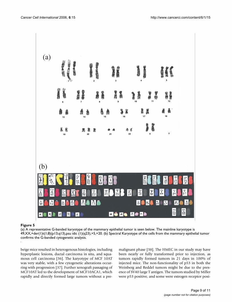
Cytogenetic analysis of the tumor revealed at least tworelated cell populations: 49, XX, +der(1)t(1;8)(p13;q13),psu idic(1)(q23),+5,+20 (Figure 5a; Figure 6) and 49, XX,add(1)(q21),+der(1)t(1;8)(p13;q13),+5,+20 (Figure 6).Among the cells analyzed, there were chromosome losses,and analysis could not determine whether they wereclonal or non-clonal.
Spectral Karyotyping was performed on late passagehTERT immortalized cells (prior to Hras61L transduction)from donor 4, confirming the presence of many of thecytogenetic abnormalities seen in the epithelial tumor.The mainline karyotype in these cells was 49, XX,+der(1)t(1;8)(p13;q13), +5, +20. Despite the presence ofthese extensive cytogenetic abnormalities, these cells werenon-tumorigenic in gamma-irradiated nude mice. Spec-tral Karyotyping of the epithelial tumor (Figure 5b)revealed the mainline karyotype to be 49,XX,+der(1)t(1;8)(p13;q13), psu idic(1)(q23), +5, +20.
DiscussionWe hypothesize that the HMEC studied here escaped a tel-omere length associated crisis through early passage trans-duction with hTERT resulting in stabilization of telomeresand transient maintenance of a diploid state, as well asimmortalization. However, the constitutive expression ofthe catalytic subunit of telomerase through agonescenceand the disruption of the Rb pathway may carry a price, as
a. Photomicrograph displays a hematoxylin/eosin section (of a section of the mammary epithelial tumor at 40×)Figure 1a. Photomicrograph displays a hematoxylin/eosin section (of a section of the mammary epithelial tumor at 40×). b. This same section at 200×. c. The pan-cytokeratin stain (AE1/AE3) is strongly positive, confirming the epithelial nature of the tumor. Mag-nification is 200×.
Page 5 of 11(page number not for citation purposes)
Cancer Cell International 2006, 6:15 http://www.cancerci.com/content/6/1/15
the immortalized cells studied here ultimately developedspecific chromosomal abnormalities (trisomy 5, trisomy20, gain of 8q, and abnormalities of chromosome 1, someof which are seen in breast cancer and other neoplasias.Artandi et al. [26] demonstrated that mTERT transgenicFVB mice displayed a statistically significant susceptibilityto breast cancer. This murine strain is susceptible todevelop a variety of tumors such as lung adenocarcino-mas, lymphomas, and sarcomas but is resistant to devel-oping spontaneous breast carcinomas. The mTERTtransgenic FVB murine mammary tumors exhibiteddiverse histological patterns. Additionally, preinvasivelesions were noted in anatomically separate regions of themammary gland, suggesting that constitutive mTERTexpression led to the promotion of preneoplastic lesions.The authors concluded that telomerase may have dualroles of stabilizing telomeres and have a second role thatdoes not depend on the setting of limited telomerereserve. Mad 2 is expressed at aberrantly high levelsthroughout the cell cycle in cells with Rb pathway defects.Hernando et al. [27] demonstrated that partial suppres-sion of Mad2 expression to nearly normal levels inimmortalized cells resulted in a reduced aneuploid frac-tion. The authors suggest that aberrant expression ofMad2 results in a hyperactive spindle checkpoint, alteringthe sequence of mitotic events and the fidelity of chromo-some transmission. hTERT immortalized HMEC 4 dem-onstrated consistent gain of chromosomes 5, 20, 1q, and8q. c-Myc, which is located on 8q24, is a potential candi-date gene for copy number increase in these cells.
Based on the keratin distribution in the normal mammarygland [28], the epithelial tumor generated in this studyrepresents a poorly differentiated malignancy derivedfrom a lobular, luminal cell. This phenotype most likelyappears after selection in soft agar as the preceding cellsdo express luminal markers. The epithelial tumor stronglyexpresses cytokeratin 7, which is found predominantly inluminal cells. It is negative for cytokeratin 20, consistentwith its mammary origins. It is also negative for cytokerat-ins 8 and 18, which are found predominantly in the duc-tal cells. It completely lacks cytokeratin 5, smooth musclemyosin heavy chain, and calponin, which excludes deriva-tion from the basal and myoepithelial compartments.Finally, the tumor is negative for Estrogen receptor, Pro-gesterone receptor, brst-2 antigen, and mammoglobin.Brst-2 antigen is found predominantly in mammarytumors with elements of apocrine differentiation whilemammoglobin is found in a minority of mammarytumors [29].
The tumor is negative for E-cadherin staining, further rein-forcing its putative lobular origin. Acs et al. [30] reviewedthe E-cadherin expression in 183 invasive mammary car-cinomas and 198 carcinomas in situ and found that mod-
Western blotting (confirmed from three separate experi-ments) of human mammary epithelial cells (HMEC) from donor 4 at various stages from mortal, early passage to the epithelial tumor are shown belowFigure 2Western blotting (confirmed from three separate experi-ments) of human mammary epithelial cells (HMEC) from donor 4 at various stages from mortal, early passage to the epithelial tumor are shown below. Volume intensity of H-ras is displayed on the bar graph at the top, and H-ras overex-pression compared to the baseline mortal state is indicated below each bar. Lanes (from right to left): 1. HMEC from donor 4- post stasis, pre agonescence, passage 9. 2. RAO-1 cells. 3. RAO-2 cells. 4. Cells from soft agar colony that developed into the epithelial tumor in nude mice. 5. RAO-4 cells, cultured in monolayer
Page 6 of 11(page number not for citation purposes)

Cancer Cell International 2006, 6:15 http://www.cancerci.com/content/6/1/15
erate to strong staining was present in all invasive and insitu ductal carcinomas while virtually all in situ and inva-sive lobular carcinomas lost expression of E-cadherin.Droufakou et al. [31] analyzed 22 invasive lobular carci-nomas and demonstrated several different mechanismsfor loss of E-cadherin expression including promotermethylation, mutation, or loss of heterozygosity in anycombination. Methylation may also play a key role insilencing E-cadherin expression in the tumor generated inthis study. As noted previously, the HMEC in this studywere transduced with mutant H-ras, cloned in soft agar,and injected into nude mice. During this process, c-Mycexpression was reduced after passaging and transduction,but apparently regained during soft agar cloning. The re-expression of myc may be critical to the transformation ofHMEC. The oncogenic potency of the myc/ras combina-tion has been known for quite some time [32]. The levelsof c-Myc and p53 rise with repeated passaging [11,33] inhTERT immortalized HMEC. However, they are reducedquite dramatically following H-ras61L transduction.Because c-myc expression is detected in the soft agar col-ony that gave rise to the epithelial tumor, as well as in thetumor itself, we hypothesize that c-myc expression playsan important role in mammary cell transformation. Thelevels of p53 are reduced after Hras61L transduction, andremain reduced which suggested that p53 is non-func-tional in the epithelial tumors, as demonstrated byabsence of doxorubicin-induced G1 arrest.
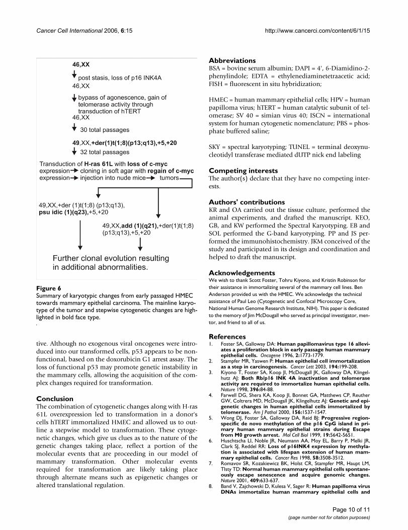
Our summary of karyotypic changes involved in mam-mary cell transformation is outlined in Figure 6. NormalHMEC were allowed to passage through stasis, during
which time the p16 promoter was likely to have been epi-genetically silenced. They were then immortalized withhTERT. Cells first acquire the +der(1)t(1;8)(p13;q13), tri-somy 5, trisomy 20. Following this, a fraction of the cellsundergo further rearrangement involving chromosome 1,resulting in two related cell populations, one with translo-cation of unidentified material to the long arm of chro-mosome 1 at band 1q21 and the other with apseudoisodicentric chromosome 1 (Figure 6). We hypoth-esize that these cell lines demonstrating the add(1) andpsu idic(1) represent the primary karyotypes of the mam-mary carcinoma. Further complex karyotypic evolutionoccurs beyond these cell lines populations.
Other in vitro transformation models include the Wein-berg model [9], the Reddel model [11], and the Millermodel [34]. The tumors generated in the Weinberg studywere epithelial based on the pan-cytokeratin staining.Toouli et al. [11] did not discuss the morphology of thetumor generated in their study; however, it was presuma-bly epithelial. To achieve mammary cell transformation,the Weinberg laboratory [9,35] used a combination offour genes (H-rasV12G (12×), hTERT, small t antigen, andlarge T antigen), and the Reddel lab, the two viral onco-genes (small t antigen, large T antigen) alone. In the Red-del study, prolonged passaging of HMEC in vitroproduced malignancy, in contrast to a series of transduc-tions. In our study, passaged immortal HMEC accumu-lated some cytogenetic abnormalities but did not acquiretumorigenicity. Transformation of immortal HMEC wasonly associated with late-passage transduction of H-ras.Elenbaas et al. [9] state a seven-fold excess of Ras was usu-
Flow cytometry data is represented belowFigure 3Flow cytometry data is represented below. There is no tetraploid population noted.
Page 7 of 11(page number not for citation purposes)
Cancer Cell International 2006, 6:15 http://www.cancerci.com/content/6/1/15
ally inadequate to transform their HMEC, and that it wasonly with twelve fold excess that transformation occurredregularly. Our study suggests that a five-fold excess of 61Lmutant H-ras is sufficient for transformation. Althoughhigh levels of H-ras were associated with transformationof HMEC in the Weinberg study, other genetic changes arerequired as well. Only 52% of mice in the Weinberg mam-mary epithelial cell study developed tumors with an aver-age latency of 52 days. This is in contrast to Weinberg'searlier results [10] in which transformed human embry-onic kidney cells and human fibroblasts produced tumorsin nude mice in 21 days. These mesenchymal tumors
showed virtually no cytogenetic changes, suggesting a lesscomplex etiology. Perhaps connective tissue is endowedwith all the tools needed for successful tumor invasion,while epithelium must recruit surrounding stromal cellsearly on. One of the tumors in the Reddel study required68 days to develop, while the other malignant line grew in21 days. H-ras transduction was not employed. The Millermodel, developed at Wayne State University, is uniquebecause of its ability to generate premalignant breastlesions. MCF 10A cells were transfected with H-ras V12Gto generate MCF10AT, which were passaged by serialxenograft transplantations. Injection of cells into nude-
(a) Primary HMEC and RAO-4 cells were treated with FTI-277, a potent farnesyl transferase inhibitor, and effects were observedFigure 4(a) Primary HMEC and RAO-4 cells were treated with FTI-277, a potent farnesyl transferase inhibitor, and effects were observed. The membranous ruffles of cells showed a significant flattened morphological appearance after treatment with FTI-277 at 2.5 and 5.0 μM concentrations. Although cellular morphological changes were not observed in primary HMEC at these concentrations, we observed some cell death with FTI-277 treatment at 2.5 and 5.0 μM concentrations. (b) We found a reduc-tion in RAO-4 cell proliferation with FTI-277 treatment at 5.0 μM concentrations. The black bars represent untreated epithe-lial tumor cells while the white bars represents treated epithelial tumor cells.
Page 8 of 11(page number not for citation purposes)
Cancer Cell International 2006, 6:15 http://www.cancerci.com/content/6/1/15
beige mice resulted in heterogenous histologies, includinghyperplastic lesions, ductal carcinoma in situ, and squa-mous cell carcinoma [36]. The karyotype of MCF 10ATwas very stable, with a few cytogenetic alterations occur-ring with progression [37]. Further xenograft passaging ofMCF10AT led to the development of MCF10ACA1, whichrapidly and directly formed large tumors without a pre-
malignant phase [38]. The HMEC in our study may havebeen nearly or fully transformed prior to injection, astumors rapidly formed tumors in 21 days in 100% ofinjected mice. The non-functionality of p53 in both theWeinberg and Reddel tumors might be due to the pres-ence of SV40 large T antigen. The tumors studied by Millerwere p53 positive, and some were estrogen receptor posi-
(a) A representative G-banded karyotype of the mammary epithelial tumor is seen belowFigure 5(a) A representative G-banded karyotype of the mammary epithelial tumor is seen below. The mainline karyotype is 49,XX,+der(1)t(1;8)(p13;q13),psu idic (1)(q23),+5,+20. (b) Spectral Karyotype of the cells from the mammary epithelial tumor confirms the G-banded cytogenetic analysis.
Page 9 of 11(page number not for citation purposes)
Cancer Cell International 2006, 6:15 http://www.cancerci.com/content/6/1/15
tive. Although no exogenous viral oncogenes were intro-duced into our transformed cells, p53 appears to be non-functional, based on the doxorubicin G1 arrest assay. Theloss of functional p53 may promote genetic instability inthe mammary cells, allowing the acquisition of the com-plex changes required for transformation.
ConclusionThe combination of cytogenetic changes along with H-ras61L overexpression led to transformation in a donor'scells hTERT immortalized HMEC and allowed us to out-line a stepwise model to transformation. These cytoge-netic changes, which give us clues as to the nature of thegenetic changes taking place, reflect a portion of themolecular events that are proceeding in our model ofmammary transformation. Other molecular eventsrequired for transformation are likely taking placethrough alternate means such as epigenetic changes oraltered translational regulation.
AbbreviationsBSA = bovine serum albumin; DAPI = 4', 6-Diamidino-2-phenylindole; EDTA = ethylenediaminetetraacetic acid;FISH = fluorescent in situ hybridization;
HMEC = human mammary epithelial cells; HPV = humanpapilloma virus; hTERT = human catalytic subunit of tel-omerase; SV 40 = simian virus 40; ISCN = internationalsystem for human cytogenetic nomenclature; PBS = phos-phate buffered saline;
SKY = spectral karyotyping; TUNEL = terminal deoxynu-cleotidyl transferase mediated dUTP nick end labeling
Competing interestsThe author(s) declare that they have no competing inter-ests.
Authors' contributionsKR and OA carried out the tissue culture, performed theanimal experiments, and drafted the manuscript. KEO,GB, and KW performed the Spectral Karyotyping. EB andSOL performed the G-band karyotyping. PP and JS per-formed the immunohistochemistry. JKM conceived of thestudy and participated in its design and coordination andhelped to draft the manuscript.
AcknowledgementsWe wish to thank Scott Foster, Tohru Kiyono, and Kristin Robinson for their assistance in immortalizing several of the mammary cell lines. Ben Anderson provided us with the HMEC. We acknowledge the technical assistance of Paul Leo (Cytogenetic and Confocal Microscopy Core, National Human Genome Research Institute, NIH). This paper is dedicated to the memory of Jim McDougall who served as principal investigator, men-tor, and friend to all of us.
References1. Foster SA, Galloway DA: Human papillomavirus type 16 allevi-
ates a proliferation block in early passage human mammaryepithelial cells. Oncogene 1996, 2:1773-1779.
2. Stampfer MR, Yaswen P: Human epithelial cell immortalizationas a step in carcinogenesis. Cancer Lett 2003, 194:199-208.
3. Kiyono T, Foster SA, Koop JI, McDougall JK, Galloway DA, Klingel-hutz AJ: Both Rb/p16 INK 4A inactivation and telomeraseactivity are required to immortalize human epithelial cells.Nature 1998, 396:84-88.
4. Farwell DG, Shera KA, Koop JI, Bonnet GA, Matthews CP, ReutherGW, Coltrera MD, McDougall JK, Klingelhutz AJ: Genetic and epi-genetic changes in human epithelial cells immortalized bytelomerase. Am J Pathol 2000, 156:1537-1547.
5. Wong DJ, Foster SA, Galloway DA, Reid BJ: Progressive region-specific de novo methylation of the p16 CpG island in pri-mary human mammary epithelial strains during Escapefrom M0 growth arrest. Mol Cell Biol 1999, 19:5642-5651.
6. Huschtscha LI, Noble JR, Neumann AA, Moy EL, Barry P, Melki JR,Clark SJ, Reddel RR: Loss of p16INK4 expression by methyla-tion is associated with lifespan extension of human mam-mary epithelial cells. Cancer Res 1998, 58:3508-3512.
7. Romavov SR, Kozakiewicz BK, Holst CR, Stampfer MR, Haupt LM,Tlsty TD: Normal human mammary epithelial cells spontane-ously escape senescence and acquire genomic changes.Nature 2001, 409:633-637.
8. Band V, Zajchowski D, Kulesa V, Sager R: Human papilloma virusDNAs immortalize human mammary epithelial cells and
Summary of karyotypic changes from early passaged HMEC towards mammary epithelial carcinomaFigure 6Summary of karyotypic changes from early passaged HMEC towards mammary epithelial carcinoma. The mainline karyo-type of the tumor and stepwise cytogenetic changes are high-lighted in bold face type.
Page 10 of 11(page number not for citation purposes)
Cancer Cell International 2006, 6:15 http://www.cancerci.com/content/6/1/15
Publish with BioMed Central and every scientist can read your work free of charge
"BioMed Central will be the most significant development for disseminating the results of biomedical research in our lifetime."
Sir Paul Nurse, Cancer Research UK
Your research papers will be:
available free of charge to the entire biomedical community
peer reviewed and published immediately upon acceptance
cited in PubMed and archived on PubMed Central
yours — you keep the copyright
Submit your manuscript here:http://www.biomedcentral.com/info/publishing_adv.asp
BioMedcentral
reduce their growth factor requirements. Proc Natl Acad SciUSA 1990, 87:463-467.
9. Elenbaas B, Spirio L, Koerner F, Fleming MD, Zimonjic DB, DonaherJL, Popescu NC, Hahn WC, Weinberg RA: Human cancer cellsgenerated by oncogenic transformation of primary mam-mary epithelial cells. Genes Dev 2001, 15:50-65.
10. Hahn WC, Counter CM, Lundberg AS, Beijersbergen RL, BrooksMW, Weinberg RA: Creation of human tumour cells withdefined genetic elements. Nature 1999, 400:464-468.
11. Toouli CD, Huschtscha LI, Neumann AA, Noble JR, Colgin LM,Hukku B, Reddel RR: Comparison of human mammary epithe-lial cells immortalized by simian virus 40 T-antigen or by thetelomerase catalytic subunit. Oncogene 2002, 21:128-139.
12. Stampfer MR, Bartley JC: Induction of transformation and con-tinuous cell lines from normal human mammary epithelialcells after exposure to benzo[a]pyrene. Proc Natl Acad Sci USA1985, 82:2394-2398.
13. Pierce JH, Arnstein P, DiMarco E, Artrip J, Kraus MH, Lonardo F,DiFiore PP, Aaronson SA: Oncogenic potential of erbB2 inhuman mammary epithelial cells. Oncogene 1991, 6:1189-1194.
14. Walen KH, Stampfer MR: Chromosome Analyses of HumanMammary Epithelial Cells at Stages of Chemical-InducedTransformation Progression to Immortality. Cancer GenetCytogenet 1989, 37:249-261.
15. Lebeau J, Le Chalony C, Prosperi M-T, Goubin G: Constitutiveoverexpression of a 89 kDa heat shock protein gene in theHBL100 human mammary cell line converted to a tumori-genic phenotype by the EJ/T24 Harvey-ras oncogene. Onco-gene 1991, 6:1125-1132.
16. Smith HS, Hackett AJ, Lan S, Stampfer MR: Use of an efficientmethod for culturing human mammary epithelial cells tostudy adriamycin sensitivity. Cancer Chemother Pharmacol 1981,6:237-244.
17. Band V, Sager R: Distinctive traits of normal and tumor-derived epithelial cells expressed in a medium that supportslong term growth of both cell types. Proc Natl Acad Sci USA 1989,86:1249-1252.
18. Foster SA, Wong DJ, Barrett MT, Galloway DA: Inactivation of p16in human mammary epithelial cells by CpG island methyla-tion. Mol Cell Biol 1996, 18:1793-1801.
19. Rothenberg M, Nolan GP: Preparing retroviruses using phoenixlines. [http://www.stanford.edu/group/nolan/retroviral_systems/phx.html].
20. Barch MJ, Knutsen T, Spurbeck JL: The AGT Cytogenetics LaboratoryManual 3rd edition. Philadelphia: Lipponcott-Raven Press; 1997.
21. Shaffer LG, Mitelman F: An International System for Human CytogeneticNomenclature Basel: Karger Press; 2005.
22. Schrock E, du Manoir S, Veldman T, Schoell B, Wienberg J, Ferguson-Smith MA, Ning Y, Ledbetter DH, Bar-Am I, Soenksen D, Garini Y,Reid T: Multicolor spectral karyotyping of human chromo-somes. Science 1996, 273:494-497.
23. Darzynkiewicz Z, Gong J, Traganos F: Analysis of DNA contentand cyclin protein expression in studies of DNA ploidy,growth fraction, lymphocyte stimulation, and the cell cycle.In Methods in Cell Biology Volume 41. Edited by: Darzynkiewicz Z, Rob-inson JP, Crissman HA. San Diego: Academic Press; 1994:421-424.
24. Gown AM, Vogel AM: Monoclonal antibodies to human inter-mediate filament proteins. III. Analysis of tumors. Am J ClinPathol 1985, 84:413-424.
25. Rao K, Bryant E, O'Hara Larivee S, McDougall JK: Production ofspindle cell carcinoma by transduction of H-Ras 61L intoimmortalized human mammary epithelial cells. Cancer Lett2003, 201:79-88.
26. Artandi SE, Alson S, Tietze MK, Sharpless NE, Ye S, Greenberg RA,Castrillon DH, Horner JW, Weler SR, Carrasco RD, DePinho RA:Constitutive telomerase expression promotes mammarycarcinomas in aging mice. Proc Natl Acad Sci USA 2002,99:8191-8196.
27. Hernando E, Nahlé , Juan G, Diaz-Rodriguez E, Alaminos M, HemannM, Michel L, Mittal V, Gerald W, Benezra R, Lowe SW, Cordon-Cardo C: Rb inactivation promotes genomic instability byuncoupling cell cycle progression from mitotic control.Nature 2004, 430:797-802.
28. Taylor-Papadimitriou J, Stampfer M, Bartek J, Lewis A, Boshell M, LaneEB, Leigh IM: Keratin expression in human mammary epithe-lial cells cultured from normal and malignant tissue: relation
to in vivo phenotypes and influence of medium. J Cell Sci 1989,94:403-413.
29. Watson MA, Fleming TP: Mammoglobin, a mammary-specificmember of the uteroglobin gene family, is overexpressed inhuman breast cancer. Cancer Res 1996, 56:860-865.
30. Acs G, Lawton TJ, Rebbeck TR, LiVolsi VA, Zhang PJ: Differentialexpression of E-cadherin in lobular and ductal neoplasms ofthe breast and its biologic and diagnostic implications. Am JClin Pathol 2001, 115:85-98.
31. Droufakou S, Deshmane V, Roylance R, Hanby A, Tomlinson I, HartIR: Multiple ways of silencing E-cadherin gene expression inlobular carcinoma of the breast. Int J Cancer 2001, 92:404-408.
32. Land H, Parada LF, Weinberg RA: Tumorigenic conversion of pri-mary embryo fibroblasts requires at least two cooperatingoncogenes. Nature 1983, 304:596-602.
33. Wang J, Hannon GJ, Beach DH: Cell biology: risky immortaliza-tion by telomerase. Nature 2000, 405:755-756.
34. Strickland LB, Dawson PJ, Santner SJ, Miller FR: Progression of pre-malignant MCF10AT generates heterogenous malignantvariants with characteristic histologic types and immunohis-tochemical markers. Breast Cancer Res Treat 2000, 64:235-240.
35. Hahn WC, Dessain SK, Brooks MW, King JE, Elenbaas B, Sabatini DM,DeCaprio JA, Weinberg RA: Enumeration of the simian virus 40early region elements necessary for human cell transforma-tion. Mol Cell Biol 2002, 22:2111-2123.
36. Dawson PJ, Wolman SR, Tait L, Heppner GH, Miller FR: MCF10AT:A model for the evolution of cancer from proliferative breastdisease. Am J Pathol 1996, 148:313-319.
37. Miller FR: Xenograft models of premalignant breast disease. JMammary Gland Biol Neoplasia 2000, 5:379-391.
38. Santner SJ, Dawson PJ, Tait L, Soule HD, Eliason J, Mohamed AN,Wolman SR, Heppner GH, Miller FR: Malignant MCF10CA1 celllines derived from premalignant human breast epithelialMCF10AT cells. Breast Cancer Res Treat 2001, 65:101-110.
Page 11 of 11(page number not for citation purposes)