Embed Size (px)
Citation preview
REVIEWbph_1729 1288..1305
Cyclic nucleotidephosphodiesterase (PDE)isozymes as targets of theintracellular signallingnetwork: benefits of PDEinhibitors in various diseasesand perspectives for futuretherapeutic developmentsThérèse Keravis and Claire Lugnier
CNRS UMR 7213, Laboratoire de Biophotonique et Pharmacologie, Université de Strasbourg,
Faculté de Pharmacie, Illkirch, France
CorrespondenceDr Claire Lugnier, Université deStrasbourg, CNRS UMR 7213,Laboratoire de Biophotonique etPharmacologie, Faculté dePharmacie, 74 route du Rhin, B.P.60024, 67401 Illkirch, France.E-mail: claire.lugnier@unistra.fr----------------------------------------------------------------
KeywordscAMP; cGMP; cyclic nucleotidephosphodiesterases; regulation;expression; distribution; PDEinhibitors; pathologies;therapeutic strategies;signalosome----------------------------------------------------------------
Received21 June 2011Revised14 September 2011Accepted14 October 2011
Cyclic nucleotide phosphodiesterases (PDEs) that specifically inactivate the intracellular messengers cAMP and cGMP in acompartmentalized manner represent an important enzyme class constituted by 11 gene-related families of isozymes (PDE1 toPDE11). Downstream receptors, PDEs play a major role in controlling the signalosome at various levels of phosphorylationsand protein/protein interactions. Due to the multiplicity of isozymes, their various intracellular regulations and their differentcellular and subcellular distributions, PDEs represent interesting targets in intracellular pathways. Therefore, the investigationof PDE isozyme alterations related to various pathologies and the design of specific PDE inhibitors might lead to thedevelopment of new specific therapeutic strategies in numerous pathologies.
This manuscript (i) overviews the different PDEs including their endogenous regulations and their specific inhibitors; (ii)analyses the intracellular implications of PDEs in regulating signalling cascades in pathogenesis, exemplified by two diseasesaffecting cell cycle and proliferation; and (iii) discusses perspectives for future therapeutic developments.
AbbreviationsAKAP, A-kinase anchoring protein; ALL, acute lymphoblastic leukaemia; ANP, atrial natriuretic peptide; BRAF, B-RAF;CaM-PDE, calmodulin-stimulated PDE; CaM, calmodulin; CAM, chorioallantoic membrane; cAMP-GEFS, cAMP-regulatedguanine nucleotide exchange factors; cGI-PDE, cGMP-inhibited PDE; cGS-PDE, cGMP-stimulated PDE; CLL, chroniclymphocytic leukaemia; CNGC, cyclic nucleotide gated channel; COPD, chronic obstructive pulmonary disease; CREB,cAMP response element-binding; CREM, cAMP response element modulator; EPAC, exchange protein activated bycAMP; FDA, Food and Drug Administration; GAF domain, cGMP-activated PDE, adenylyl cyclase- and Fh1A-bindingdomain; HUVEC, human umbilical vein endothelial cell; ICER, inducible cAMP early repressor; IKK, IkB kinase; IPAH,idiopathic pulmonary arterial hypertension; i.t., intratracheal; KO, knock out; NHRs, possess N-terminal hydrophobicmembrane association regions; PAS, Per–Arnt–Sim; PASMC, pulmonary arterial smooth muscle cell; PBMC, peripheralblood mononuclear cell; PDE, cyclic nucleotide phosphodiesterase; PHT, pulmonary hypertension; PI3K, PI3 kinase;Rap1, Ras-related protein 1; REC, Response Regulator Receiver; RT-PCR, real-time PCR; SMC, smooth muscle cell; SPH,secondary pulmonary hypertension; TAPAS, tryptophan anchoring phosphatidic acid selective-binding domain; UCR,upstream conserved region; UHRF1, ubiquitin-like, containing PHD and RING finger domains 1
BJP British Journal ofPharmacology
DOI:10.1111/j.1476-5381.2011.01729.xwww.brjpharmacol.org
1288 British Journal of Pharmacology (2012) 165 1288–1305 © 2011 The AuthorsBritish Journal of Pharmacology © 2011 The British Pharmacological Society
Cyclic nucleotide phosphodiesterases(PDEs)
Cyclic nucleotide phosphodiesterases play a critical role inintracellular signalling by selectively hydrolysing the secondmessengers cAMP and cGMP that control cAMP- and cGMP-regulated proteins and transcription factors. In the 1970s,‘the cyclic nucleotide system’ concept was developed asproviding a powerful tool to enhance the opportunity fordrug discovery (Amer, 1977). This system encompassedadenylyl and guanylyl cyclases, PDEs and cAMP-dependentand cGMP-dependent kinases. Very early, the cAMP- andcGMP-PDEs have been considered as targets for drugdevelopment (Pang, 1988). At that time, only three typesof PDE (PDEI, PDEII and PDEIII) were described in cardio-vascular tissues on the basis of their chromatographicelution order (Lugnier et al., 1986; Weishaar et al., 1987;Pang, 1988). Nowadays, advances in PDE methodology andtechnology (see book: Lugnier, 2005) have favoured thedevelopment of knowledge concerning PDE families (Beavoand Brunton, 2002; and see book: Beavo et al., 2006). Pres-ently, PDEs represent an important enzyme class constitutedby 11 gene-related families of isozymes (PDE1 to PDE11),giving rise to a multiplicity of isozymes. Each PDE familyencompasses one to four distinct genes, giving rise to morethan 20 genes in mammals that encode more than 50 dif-ferent PDE proteins that are all probably expressed in mam-malian cells (Lugnier, 2006; Conti and Beavo, 2007; Franciset al., 2011). PDE isozymes generally exist as dimers. Theirmonomeric structure has common features with three dis-tinct domains. The N-terminal regulatory domain character-izes each family and their variants. The catalytic domain isquite conserved, sharing 25% to 52% homology with othermammalian PDE catalytic domains, and contains a Zn2+
binding site. The C-terminal domain can be prenylated(Anant et al., 1992) or phosphorylated by MAPK (Baillieet al., 2000). New mechanisms for cAMP action have beenhighlighted with the findings of GAF domains (cGMP-activated PDE, adenylyl cyclase- and Fh1A-binding domain)(Heikaus et al., 2009; Schultz, 2009), exchange protein acti-vated by cAMP (EPAC), cAMP-regulated guanine nucleotideexchange factors (cAMP-GEFS), and cyclic nucleotide gatedchannels (CNGC; Cukkemane et al., 2011). This has resultedin expanding and diversifying cyclic-nucleotide-relatedpathways (for review, Borland et al., 2009; Gloerich and Bos,2010).
In many pathologies including inflammation, cardiovas-cular diseases, neurodegenerescence and cancer, alterationsof intracellular signalling related to PDE isozyme deregula-tion might contribute in explaining the difficulties observedin the prevention and treatment of these pathologies. Themultiplicity of biochemical and structural properties ofPDEs, their specific subcellular compartments, their tran-scriptional and posttranscriptional regulation make possibleto envision to target only the altered PDE isozymes, thusavoiding and/or decreasing the adverse effects induced bynon-specific treatments.
Below are described the 11 PDE families, their contribu-tion in physiological functions and the therapeutical use ofsome specific PDE inhibitors (Table 1).
PDE1Previously named calmodulin-stimulated PDE (CaM-PDE),PDE1 is the unique PDE family that is Ca2+-dependently regu-lated via calmodulin (CaM, a 16 kDa Ca2+-binding protein)complexed with four Ca2+ (for review, Stoclet et al., 1988;Kakkar et al., 1999; Bender, 2006). Thus, this family repre-sents an interesting regulatory link between cyclic nucle-otides and intracellular Ca2+, as shown in olfactory mucosa inwhich Ca2+ stimulation of PDE1 might be necessary for ter-mination of olfactory signals (Borisy et al., 1992). The PDE1family is encoded by three genes: PDE1A (mapped on humanchromosome 2q32), PDE1B (human chromosome location,hcl: 12q13) and PDE1C (hcl: 7p14.3). They have alternativepromoters and give rise to a multitude of proteins by alter-native splicing. More than 10 human isoforms are identified(Bolger, 2006). Their molecular weights vary from 58 to86 kDa per monomer. The N-terminal regulatory domain thatcontains two Ca2+/CaM binding domains and two phospho-rylation sites differentiate their corresponding proteins andmodulate their biochemical functions. PDE1A and PDE1Bpreferentially hydrolyse cGMP, whereas PDE1C hydrolysescAMP and cGMP with similar Km values (see Table 1). Theassociation of Ca2+/CaM complex changes PDE1 isozyme con-formation, increasing their Vmax for cyclic nucleotide hydroly-sis without modifying their Km. The sensitivity of PDE1 toCa2+ activation, expressed in EC50, varies from 0.27 mM forPDE1A1 to 3.01 mM for PDE1C1 (Yan et al., 1996). Phospho-rylation of PDE1A1 (59 kDa) and PDE1A2 (61 kDa) by PKAand phosphorylation of PDE1B1 by CaM kinase II decreasetheir calmodulin and Ca2+ sensitivities, resulting in a decreaseof PDE1 activity. Interestingly, PDE1A2 isozyme is sensitive toendogenous proteolysis by calpain that activates PDE1. Thiscould be an alternative mechanism for the permanent acti-vation of PDE1A2 (for review, see Sharma et al., 2006). PDE1is mainly present in brain, cardiac tissues and smoothmuscles. Its presence in isolated cardiomyocytes was recentlyreported (Keravis et al., 2007).
Initially, PDE1 activity has been described as cytosolic. Itnow appears that PDE1A is not restricted to the cytosol but isalso present in the nucleus where it contributes to the regu-lation of transcription factors (Nagel et al., 2006). This opensa new field of research in transcriptional regulation. Forexample, we recently showed that PDE1A controls the expres-sion of the epigenetic integrator UHRF1 (Abusnina et al.,2011). Changes in PDE1 location associated with cell differ-entiation might contribute to compartmental signalling(Nagel et al., 2006).
Until now, very few inhibitors of PDE1 were available forevaluating the contribution of PDE1 in tissue and cell func-tion. Vinpocetine (Ahn et al., 1989) and 8-methoxymethyl-IBMX (Ahn et al., 1997) are common PDE1 inhibitors.However, these compounds are not specific for PDE1; theyalso inhibit PDE5 at lower concentrations and vinpocetineinhibits PDE7B with an IC50 of 59 mM (Sasaki et al., 2004).Furthermore, vinpocetine directly inhibits BKCa channels (Wuet al., 2001), and recently vinpocetine was shown to inhibitNF-kB-dependent inflammation via an IKK-dependent butPDE-independent mechanism (Jeon et al., 2010). Previously,we showed that nimodipine, a dihydropyridine Ca2+ antago-nist, selectively inhibits PDE1 in basal- and calmodulin-
BJPPDEs: intracellular network targets for diseases
British Journal of Pharmacology (2012) 165 1288–1305 1289
activated states (Lugnier et al., 1984), indicating that thiscompound is a helpful tool to assess the contribution of PDE1in tissue or cell homogenates. Indeed, the use of nimodipinein cardiac tissue showed that PDE1 is up-regulated in earlycardiac hypertrophy induced by angiotensin II (Mokni et al.,2010). Additionally, this compound, despite its Ca2+ antago-nist effect, has been used to assess the participation of PDE1in relaxation of noradrenaline-contracted smooth muscle(Noguera et al., 2001). Use of two potent and specific PDE1inhibitors, namely IC86340 (IC50 from 0.06 to 0.44 mMdepending on PDE1 variants; Nagel et al., 2006) and IC295(Vandeput et al., 2007) synthesized by ICOS Corporationclearly established the contribution of PDE1 in cardiovascularfunction. Notably, PDE1 inhibition by IC86340 or down-regulation of PDE1A using siRNA allowed demonstrating thatPDE1A may regulate specific cGMP pools with elevations of[Ca2+]i during hypertrophic stimulation (Miller et al., 2009).Unfortunately, these compounds are no longer available tothe scientific community. We recently showed that diocleinselectively inhibits PDE1 (IC50 of 0.62 � 0.14 mM for basalPDE1 and of 0.55 � 0.07 mM for activated PDE1) and inducesPKG-dependent vasodilatation (Gonçalves et al., 2009), indi-cating that this compound is helpful for studying basal- andstimulated-PDE1 contributions in cellular signalling.
PDE1 was reported to offer a new target for therapeuticintervention in pulmonary hypertension (PHT). Indeed, astrong up-regulation of PDE1C and PDE1A in pulmonary
arterial smooth muscle cells (PASMCs) was noted, respec-tively, in idiopathic pulmonary arterial hypertension (IPAH)lungs and in lungs from animal models of PHT, and inhibi-tion of PDE1 was reversing structural lung vascular remodel-ling and right heart hypertrophy in two animal models(Schermuly et al., 2007). The expression of both PDE1A andPDE1C was enhanced in PASMCs from IPAH and secondarypulmonary hypertension (SPH) patients compared withcontrol PASMCs (Murray et al., 2007). PDE1C has beenreported to be expressed in proliferating human SMCs but tobe absent in quiescent human aorta and to be expressed invivo in human foetal aorta containing proliferating SMCs,making PDE1C a marker of human SMC proliferation ex vivoand in vivo (Rybalkin et al., 2002). Furthermore, inhibition ofPDE1C in SMCs isolated from normal aorta or from lesions ofatherosclerosis was resulting in suppression of SMC prolifera-tion, making PDE1C a good target to inhibit proliferatingSMCs in lesions of atherosclerosis or restenosis (Rybalkinet al., 2002).
PDE2PDE2 hydrolyses both cAMP and cGMP. It is encoded by asingle gene PDE2A (hcl: 11q 13.4) and expressed as PDE2A1,PDE2A2 and PDE2A3. These isozymes differ by theirN-terminal residues allowing different subcellular locations:PDE2A1 is cytosolic, whereas PDE2A2 and PDE2A3 aremembrane-associated. We showed that PDE2 is associated
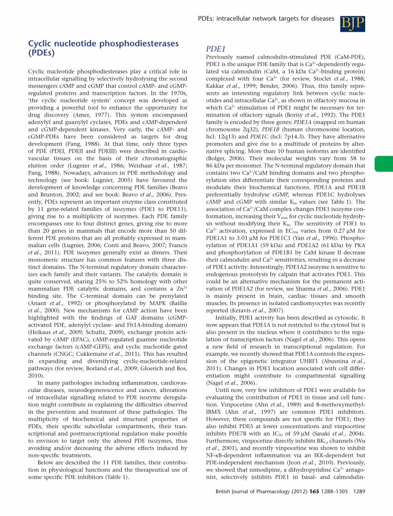
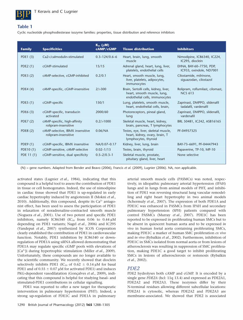
Table 1Cyclic nucleotide phosphodiesterase isozyme families: properties, tissue distribution and reference inhibitors
Family SpecificitiesKm (mM)cAMP/cGMP Tissue distribution Inhibitors
PDE1 (3) Ca2+/calmodulin-stimulated 0.3–124/0.6–6 Heart, brain, lung, smoothmuscle
Nimodipine, IC86340, IC224,IC295, dioclein
PDE2 (1) cGMP-stimulated 15/15 Adrenal gland, heart, lung, liver,platelets, endothelial cells
EHNA, BAY-60–7750, PDP,IC933, oxindole, ND7001
PDE3 (2) cAMP-selective, cGMP-inhibited 0.2/0.1 Heart, smooth muscle, lung,liver, platelets, adipocytes,immunocytes
Cilostamide, milrinone,siguazodan, cilostazol
PDE4 (4) cAMP-specific, cGMP-insensitive 2/>300 Brain, Sertolli cells, kidney, liver,heart, smooth muscle, lung,endothelial cells, immunocytes
Rolipram, roflumilast, cilomast,NCS 613
PDE5 (1) cGMP-specific 150/1 Lung, platelets, smooth muscle,heart, endothelial cells, brain,
Zaprinast, DMPPO, sildenafiltadalafil, vardenafil
PDE6 (3) cGMP-specific, transducinactivated
2000/60 Photoreceptors, pineal gland,lung
Zaprinast, DMPPO, sildenafil,vardenafil
PDE7 (2) cAMP-specific, high-affinityrolipram-insensitive
0.2/>1000 Skeletal muscle, heart, kidney,brain, pancreas, T lymphocytes
BRL 50481, IC242, ASB16165
PDE8 (2) cAMP-selective, IBMX insensitiverolipram-insensitive
0.06/NA Testes, eye, liver, skeletal muscle,heart, kidney, ovary, brain, Tlymphocytes, thyroid
PF-04957325
PDE9 (1) cGMP-specific, IBMX insensitive NA/0.07–0.17 Kidney, liver, lung, brain BAY-73–6691, PF-04447943
PDE10 (1) cGMP-sensitive, cAMP-selective 0.02–1/13 Testes, brain, thyroid Papaverine, TP-10, MP-10
PDE 11 (1) cGMP-sensitive, dual specificity 0.5–2/0.3–1 Skeletal muscle, prostate,pituitary gland, liver, heart
None selective
(N) = gene numbers. Adapted from Bender and Beavo (2006), Francis et al. (2009), Lugnier (2006). NA, non applicable.
BJP T Keravis and C Lugnier
1290 British Journal of Pharmacology (2012) 165 1288–1305
with the nuclear envelope (Lugnier et al., 1999) and to puri-fied Golgi endosomal fraction, and its phosphorylation byPKC increases its activity (Geoffroy et al., 1999). TheN-terminal domain of the monomer has two cGMP-bindingdomains, GAF-A and GAF-B. The GAF-A domain mediatesPDE2 dimerization (Martinez et al., 2002). Recently, crystalstructure studies extended this function to GAF-A and GAF-Bdomains (Heikaus et al., 2009).
The binding of cGMP (1–5 mM) to GAF-B domain allos-terically and positively stimulates up to a 30-fold increase incAMP hydrolysis, inducing Michaelian behaviour with Km
values from 10–30 mM (for review, Martinez, 2006). For thisreason, PDE2 was first named cGMP-stimulated PDE (cGS-PDE). This specific regulation gives a major feedback role forPDE2 by restoring the basal cyclic nucleotide level inresponse to elevated cGMP (Bender and Beavo, 2006),notably following NO, ANP or BNP production. PDE2 is thusable to induce a crosstalk between cAMP and cGMP pathways(Lugnier, 2006).
When activated by cGMP, PDE2 is selectively inhibited byEHNA in comparison with other PDEs (Duncan et al., 1982;Podzuweit et al., 1995) with an IC50 value of 2 mM for acti-vated PDE2 and 38 mM for basal PDE2 (Keravis et al., 2006).However, EHNA is also a potent adenosine deaminase inhibi-tor (Ki = 10 nM; Schaeffer and Schwender, 1974). Bay 60–7550(Boess et al., 2004), IC933 (Snyder et al., 2005), PDP (Seyboldet al., 2005) and oxindole (Chambers et al., 2006) are newpotent and specific PDE2 inhibitors; however, only Bay60–7550 is available. Interestingly, it was reported in a cell-based assay that EHNA, Bay 60–7550 and PDP increase cGMPlevels only when combined with ANP, indicating that theseinhibitors work only on cGMP-stimulated PDE2 (Wunderet al., 2009). Recently, our group designed and patentedND7001 as a PDE2 inhibitor that inhibits with the samepotency basal and cGMP-stimulated PDE2 (Bourguignonet al., 2004). This compound antagonizes the anxiogeniceffects of restraint stress on behaviour in mouse, similarly toBay 60–7550, and in primary cultures of rat cerebral corticalneurons, ND7001 increases cGMP levels similarly to Bay60–7550 (Masood et al., 2009). These data confirm thatND7001 is efficacious both in vivo and in vitro. Interestingly,in a mouse model of acute lung injury, LPS by intratracheal(i.t.) administration increases lung PDE2A mRNA and proteinexpression. PDE2A knockdown with an adenovirus adminis-trated i.t. 3 days before LPS decreases PDE2A protein expres-sion and attenuates alveolar inflammation and protein leak(Rentsendorj et al., 2011).
For complementary information, an extensive and up-to-date review on PDE2A has recently appeared online (Gesell-chen and Zaccolo, 2011).
PDE3PDE3 hydrolyses both cAMP and cGMP. The Vmax for cAMPhydrolysis is 10-fold higher than for cGMP hydrolysis and thesubstrate affinity for cGMP being higher than for cAMP,cGMP behaves as a competitive inhibitor for cAMP hydrolysisand therefore participates in cAMP/cGMP crosstalk (in plate-lets: Maurice and Haslam, 1990; in vascular smooth muscle:Komas et al., 1991; Eckly and Lugnier, 1994). Accordingly,PDE3 was ‘cGMP-inhibited PDE’ (cGI-PDE; Beavo, 1995).PDE3 is encoded by two genes: PDE3A (hcl: 12p12) and
PDE3B (hcl: 11p15.1). Three variants are expressed for PDE3A(Wechsler et al., 2002): PDE3A1 (136 kDa), PDE3A2 (118 kDa)and PDE3A3 (94 kDa), whereas only a single PDE3B1 varianthas been identified (Bolger, 2006) even if PDE3B of varioussizes have been reported. These variants possess N-terminalhydrophobic membrane association regions (NHRs) and a44-amino acid insert in the catalytic domain. A PKA phos-phorylation site is present on PDE3A1 and PDE3A2, whereasan Akt/protein kinase B (PKB) phosphorylation site is onlypresent on PDE3A1. PDE3A3 lacks PKA and PKB phosphory-lation sites (Wechsler et al., 2002). PKA phosphorylation acti-vates PDE3A, inducing a negative-feedback regulation incAMP signalling. PKA phosphorylation of human PDE3B pro-motes 14-3-3-protein binding and inhibits phosphatase-catalysed inactivation (Palmer et al., 2007). PKB-dependentphosphorylation activates PDE3A and PDE3B. PKC phospho-rylates and activates PDE3A (Pozuelo Rubio et al., 2005).PDE3A1 contains two membrane-associated domains.PDE3A2 contains only one membrane-associated domain,whereas PDE3A3 lacks membrane-associated domains. PDE3is associated with cardiac sarcoplasmic reticulum (Lugnieret al., 1993), cardiac nuclear envelope near nucleopore com-plexes (Lugnier et al., 1999) as well as with liver Golgi endo-somal fraction (Geoffroy et al., 2001). In primary adipocytes,PDE3B is associated with caveolae (Nilsson et al., 2006).PDE3B is present in hepatocyte caveolae and smooth endo-plasmic reticulum (Berger et al., 2009) and adipocyte caveo-lae, endoplasmic reticulum and Golgi (Ahmad et al., 2009).
PDE3A is mainly present in heart, platelet, vascularsmooth muscle and oocytes, whereas PDE3B is mainly asso-ciated with adipocytes, hepatocytes and spermatocytes.Reviews have appeared on the implications of PDE3 isoformsin heart failure and hypertension (Movsesian and Smith,2006) and the role of PDE3B in energy homeostasis(Degerman and Manganiello, 2006). In adipocytes,insulin-mediated phosphorylation/activation of membrane-associated PDE3B leads to a reduction in PKA activity andinhibition of lipolysis. Inhibition of adipocyte PDE3B blocksthe antilipolytic action of insulin and reduces insulin-stimulated lipogenesis and glucose uptake (Zmuda-Trzebiatowska et al., 2006).
PDE3B has the ability to act as an important scaffoldprotein with PI3K. A PDE3B-based signalling complex inte-grates EPAC1 and PI3K signals in human arterial endothelialcells, and this signalosome may be of importance in woundrepair and angiogenesis (Wilson et al., 2011). Also, a recentstudy (Perino et al., 2011) shows that p110g-anchored PKAactivates PDE3B thus enhancing cAMP degradation and phos-phorylating p110g to inhibit PIP3 production, and this pro-vides a local feedback control of PIP3 and cAMP signallingevents. The authors show that pharmacological inhibition ofp110g normalizes b-adrenergic receptor density and improvescontractility in failing hearts.
The first potent and selective inhibitor for PDE3 was cil-ostamide, shown as a platelet anti-aggregant (Hidaka et al.,1979). PDE3 was considered as a cardiotonic target. There-fore, medicinal chemistry has been focusing on PDE3 inhibi-tors, giving rise to amrinone, milrinone and enoximone.Milrinone was the first PDE3 inhibitor given in heart failure.However, due to some mortality in chronic treatment relatedto tachyarrhythmia and tachycardia, its prescription in
BJPPDEs: intracellular network targets for diseases
British Journal of Pharmacology (2012) 165 1288–1305 1291
humans is now only for a short period. In these conditions,milrinone does not induce exacerbation of myocardial injuryin patients with severe heart failure and low cardiac outputbut instead is associated with a reduced inflammatory andapoptotic signalling (Lanfear et al., 2009). Nevertheless, con-tinuous milrinone therapy use is shown to be safe as bridge totransplant in patients with short waiting time (<100 days)(Assad-Kottner et al., 2008). Recently, the PDE3 inhibitor cil-ostazol was marketed as Pletal® for treatment of intermittentclaudication, the most common symptom of peripheralartery disease in which the narrowing and hardening of thearteries that supply legs leads to decreased blood flow. Whena patient with peripheral artery disease exercises, the insuffi-cient blood flow can produce muscle ischemia, and walkingbecomes problematic.
Pharmacological therapy for intermittent claudication inpatients with peripheral artery disease is limited. Along withpentoxyfilline, cilostazol is the only medication approved bythe Food and Drug Administration (FDA) for intermittentclaudication. Cilostazol inhibits platelet aggregation and is adirect arterial vasodilator. Its main effects are dilation of thearteries supplying blood to the legs and decreasing plateletcoagulation. Interestingly, cilostazol might increase O2 deliv-ery by prediabetes erythrocytes whose PDE3B is up-regulatedby insulin (Hanson et al., 2010). Long-term safety of cilosta-zol has been questioned because other PDE3 inhibitors,notably milrinone, have been previously associated withexcess mortality in patients with heart failure (Movsesian andKukreja, 2011). However, a recent study has shown that cil-ostazol demonstrated no increased risk of all-cause mortality(Pande et al., 2010).
PDE4The PDE4 family that selectively hydrolyses cAMP is the mostextended family, encoded by four genes, namely PDE4A (hcl:19p13.2), PDE4B (hcl: 1p31), PDE4C (hcl: 19p13.1) and PDE4D(hcl: 5p12) that have different promoters and give rise to amultitude of proteins by alternative splicing. More than 25human isoforms (from 50 to 125 kDa) are identified (Bolger,2006). These proteins contain a unique amino acid signatureregion called upstream conserved regions 1 and 2 (UCR1 andUCR2; Bolger et al., 1993). Long PDE4 isozymes exhibit bothUCR1 and UCR2, short PDE4 isozymes lack UCR1 and super-short isozymes contain only half of UCR2. UCR1 contains aPKA phosphorylation site that upon phosphorylation attenu-ates the ability of UCR1 to interact with UCR2 and therebyactivates PDE4 activity (Beard et al., 2000). The carboxyl-terminal end of the catalytic region contains an ERK phospho-rylation site; its phosphorylation induces the activation ofPDE4 short forms and the inhibition of PDE4 long forms(Baillie et al., 2000). PDE4, like other PDEs, may exist as adimer. Since the UCR modules mediate dimerization, onlylong-form PDE4 splice variants containing UCR1 and UCR2are dimerized (Richter and Conti, 2004). PDE4A1 is a super-short isoform that possesses a specific N-terminal regionentirely associated with membranes (Scotland and Houslay,1995) and formed from two helices separated by a mobilehinge. The helix-2 contains a TAPAS-1 microdomain thatallows rapid Ca2+-triggered membrane association with phos-phatidic acid (Baillie et al., 2002). The helix-1 is important forintracellular targeting of PDE4A1 in living cells, facilitating
membrane association, targeting to the trans-Golgi stack andconferring Ca2+-stimulated intracellular redistribution in amanner that is dependent on the phospholipase-D-mediatedgeneration of phosphatidic acid (Huston et al., 2006). PDE4variants are specifically localized by A-kinase anchoringprotein (AKAPs; Skroblin et al., 2010; Welch et al., 2010) insubcellular compartments nearby PKA, which regulates func-tional responses (Dodge et al., 2001; for review see Houslay,2010). We have shown that PDE4 is associated with cardiacsarcoplasmic reticulum (Lugnier et al., 1993), and that PDE4Band PDE4D are associated with the nuclear envelope (Lugnieret al., 1999). There is a predominant co-localisation of PDE4D3with centrosome-associated AKAP450 (McCahill et al., 2005).Furthermore, PDE4s interact directly with many other intrac-ellular proteins, thereby defining the PDE4 interactome (for adetailed recent review, see Houslay, 2010). PDE4D tetheringEPAC1 regulates both the activity and subcellular localizationof EPAC1 and controls cAMP-mediated vascular permeability(Rampersad et al., 2010). Furthermore, a relationship betweenPDE4A and PDE4B expression and the regulation of [Ca2+]i wasshown in human endothelial cells (Campos-Toimil et al.,2008). PDE4s are present in brain, smooth muscle, heart,kidney, endothelial cells and immunocytes.
Rolipram, an antidepressant compound (Schwabe et al.,1976), specifically binds with high affinity to membranebrain fractions (Schneider et al., 1986). Rolipram was shownto be a potent inhibitor of cAMP hydrolysis in brain homo-genates. Rolipram and Ro 20-1724 are highly selective forPDE4 (Lugnier et al., 1983, 1986; Komas et al., 1989). Accord-ingly, PDE4 became an archetype for the synthesis of newpotent and selective PDE4 inhibitors (Marivet et al., 1989).PDE4 subtypes are expressed in a number of cell types that areconsidered suitable drug targets for the treatment of respira-tory diseases such as asthma and COPD (for review, see:Spina, 2008). Suppression of human inflammatory cell func-tion by subtype-selective PDE4 inhibitors correlates withinhibition of PDE4A and PDE4B (Manning et al., 1999). Inter-estingly, PDE4A4 is significantly upregulated in lung mac-rophages from smokers with COPD when compared withcontrol smokers (Barber et al., 2004). Pharmaceutical compa-nies synthesized many PDE4 inhibitors as anti-inflammatoryagents for asthma and COPD, since they decreased produc-tion of TNF-a and cytokines (for review, Houslay et al., 2005;Boswell-Smith et al., 2006; Lugnier, 2006; Pagès et al., 2009).The PDE4 inhibitor NCS 613, a potent adenine analoguesynthesized by our group, inhibits TNFa production inhuman lymphocytes; interestingly, this compound does notstimulate gastric acid secretion in rats (Boichot et al., 2000).Today, roflumilast is the first PDE4 inhibitor on the market (asDaxas®) for COPD treatment (Hatzelmann et al., 2010).Recently, a very potent and subtype-specific PDE4D inhibitorwas reported (Aspiotis et al., 2010) and PDE4D allostericmodulators for enhancing cognition with improved safetyhave been designed (Burgin et al., 2010).
PDE5PDE5 specifically hydrolyses cGMP and is encoded by onegene PDE5A (hcl: 4q27) with three variants being expressed:PDE5A1 (ª100 kDa), PDE5A2 (ª95 kDa) and PDE5A3(ª95 kDa). Their N-terminal domain contains GAF-A andGAF-B domains. The GAF-A domain is responsible for allos-
BJP T Keravis and C Lugnier
1292 British Journal of Pharmacology (2012) 165 1288–1305
teric binding of cGMP that promotes phosphorylation ofPDE5 (Turko et al., 1998). Such phosphorylation activatescatalytic activity and it increases cGMP-binding affinity andconverts PDE5 into a stably activated form. The GAF-Bdomain modulates cGMP binding by GAF-A, thereby contrib-uting to PDE5 dimerization (Francis et al., 2006). Despite asimilar overall folding of the two GAF domains, only one(GAF-A) binds cGMP. A recent study suggests a complicatedand cooperative process of cGMP signalling, which is com-posed of conformation changes in the GAF-A pocket uponcGMP binding, relay of the cGMP signal from GAF-A toGAF-B, and involvement of N-terminal segment 101–127 inthe cGMP signalling (Wang et al., 2010). The first three-dimensional structure resolved by electron microscopicanalysis of a native PDE revealed a dimeric arrangement forPDE5 having GAF-A, GAF-B and catalytic domains (Kameni-Tcheudji et al., 2001). PDE5 is cytosolic in vascular smoothmuscle cells (Lugnier et al., 1986) and is mainly present inlung, smooth muscles, platelets and corpus cavernosum. Ini-tially, no detectable levels of PDE5 in cardiac ventricles werereported (Komas et al., 1989; Wallis et al., 1999). PDE5 isassociated with Z-bands in cardiac ventricle tissues (Takimotoet al., 2005) and with membrane fractions in isolated cardi-omyocytes (Keravis et al., 2007). PDE5 is localized in endot-helial caveolae, where it modulates NOS3 activity (Gebskaet al., 2011).
Zaprinast (M&B 22948) was the first selective PDE5inhibitor described (Lugnier et al., 1986), and it was used toinvestigate various functional implications of PDE5. Sildena-fil was the first potent and selective PDE5 inhibitor marketed(ViagraTM, Pfizer Inc.) for male erectile dysfunction (Franciset al., 2008). Thereafter, compounds more potent were devel-oped. Tadalafil (CialisTM, Lilly-ICOS) was synthetized withgreater selectivity for PDE5 versus PDE1-4 than sildenafil andwith the highest long-lasting effect (>7h) (Daugan et al.,2003). Vardenafil (Levitra TM, Bayer-GSK) was synthetizedwith the lowest IC50. Both sildenafil and vardenafil are alsopotent PDE6 inhibitor and induce visual perturbation (Cote,2006). Furthermore, tadalafil although inhibiting PDE11might not induce cross-reaction in patients (Weeks et al.,2005). Pulmonary hypertension (Wilkens et al., 2001; Ghof-rani et al., 2002; Michelakis et al., 2002; for review, Croomand Curran, 2008), high altitude mountain sickness (Bateset al., 2007), memory dysfunctions (Puzzo et al., 2008) andcardiovascular diseases (Kass et al., 2007; for reviews, Kumaret al., 2009 and Tsai and Kass, 2009) are new therapeutic fieldsfor PDE5 inhibitors. Recently, it has been reported that,human heart PDE5 being present at much lower levels thanthose seen in animal models, the applicability of PDE5 inhibi-tors in treating heart failure could be questioned (Movsesianand Kukreja, 2011). However, PDE5 inhibition with sildenafilimproves left ventricular diastolic function, cardiac geometry,and clinical status in patients with stable systolic heart failure(Guazzi et al., 2011).
Moreover, PDE5 plays a role in cancer. In melanoma cells,oncogenic BRAF induces invasion through down-regulationof PDE5A (Arozarena et al., 2011; Houslay, 2011). Neverthe-less, PDE5 inhibition is responsible for the breast tumour cellgrowth inhibitory activity and apoptosis inducing activity ofsulindac sulphite and may contribute to the chemopreven-tive properties of sulindac (Tinsley et al., 2009).
PDE6In contrast to other PDE families, the PDE6 family isrestricted to retinal rod and cone cells (Miki et al., 1975; Ridgeet al., 2003) and to pineal gland (Morin et al., 2001). PDE6family, that hydrolyses cGMP, is a key component in thevisual transduction cascade and is known as the photorecep-tor phosphodiesterase (Cote, 2006). PDE6 is composed of twolarge catalytic subunits (a and b in rod, 100 and 98 kDa,respectively, and two a′ in cones, 99 kDa) associated with twocopies of small inhibitory subunits (g in rods, 9.5 kDa and g ′in cones, 8.9 kDa; Cote, 2006). The catalytic subunits areencoded by PDE6A for rod a subunit, PDE6B for rod b subunitand PDE6C for cone a′ subunit. The inhibitory subunits areencoded by PDE6G (hcl: 17q25) and PDE6H (hcl: 12p13), forg and g ′ respectively. The PDE6D (hcl: 2q35-q36) gene thatencodes a d subunit (17 kDa) co-purified with PDE6 wasrecently excluded from PDE6 family and revised as a prenylbinding protein (PrBP/d). Contrary to PDE6, PrBP/d is presentin numerous tissues and may interact with other proteins(Cote, 2006). The structure for the membrane-bound rodPDE6 holoenzyme is abg2, whereas it is a′2g2 for cone PDE6(Artemyev et al., 1996). Each catalytic subunit contains thecGMP-binding GAF-A and GAF-B domains. The GAF-Adomain may allow dimerization and binding of the inhibi-tory subunit g ′. The binding of cGMP is required for highaffinity binding of g subunit. PDE6 activation, mediated byphototransduction, decreases cGMP levels, and it induces theclosure of cyclic nucleotide-gated channels (CNGC), therebyconverting a light signal into an electrical signal (Wensel,2008). For now, there is no specific PDE6 inhibitor. However,some PDE5 inhibitors also inhibit PDE6 (for review, Franciset al., 2009).
Subtypes of PDE6 have been found outside the eye, espe-cially PDEg in human embryonic kidney 293 cells (Wan et al.,2001) and in mouse lung (Tate et al., 2002), which seems tohave a critical role in regulating p42/p44 MAPK signalling.PDEg is also in pulmonary vessels from rats as well as incultured human pulmonary SMCs, both maintained underchronic hypoxic conditions (Murray et al., 2003). Recently,PDE6D and PDE6G/H subtypes have been shown to bepresent in alveolar epithelial cells and to be altered in lungfibrosis (Nikolova et al., 2010). These studies are encouragingto investigate whether some PDE6 subtypes could be thera-peutic targets in pulmonary hypertension.
PDE7The PDE7 family specifically hydrolyses cAMP and is insen-sitive to rolipram, the specific inhibitor of the PDE4 family,but is sensitive to isobutyl-methylxanthine (IBMX). There isno known regulatory domain on the N-terminal region. Thisfamily includes two genes PDE7A (hcl: 8q13) and PDE7B (hcl:6q23-q24), with alternative splicing for PDE7A (Michaeli,2006) giving rise to PDE7A1 (57 kDa), PDE7A2 (50 kDa) andPDE7A3 (50 kDa). PDE7A1, expressed in lymphocytes andproinflammatory cells, is a bifunctional protein. In additionto its cAMP hydrolytic activity, its N-terminal domain, byinteracting directly with the catalytic subunit of PKA, inhibitskinase activity with high affinity. PDE7A2, present in skeletaland cardiac muscles, has a hydrophobic N-terminus. PDE7A3lacks a part of the catalytic domain structure and retains thecapacity of PDE7A1 to interact and inhibit the catalytic
BJPPDEs: intracellular network targets for diseases
British Journal of Pharmacology (2012) 165 1288–1305 1293
subunit of PKA. Although no endogenous PDE7B proteinshave been yet detected, four alternative splice PDE7B tran-scripts have been identified: PDE7B1, PDE7B2, PDE7B3 andan additional variant found in GenBank (Michaeli, 2006).Two putative phosphorylation sites for PKA would be onPDE7B1 and PDE7B3. PDE7B1 is the only variant to be acti-vated by D1 agonist through the cAMP/PKA/CREB pathwayin striatal neuron, arguing for a role for PDE7B1 in memoryfunction and promoting PDE7B1 as a possible target for Par-kinson and Huntington diseases (Sasaki et al., 2004). Thereare few PDE7 inhibitors. IC242 was the first reported selectivePDE7A inhibitor with an IC50 value of 0.37 mM (Lee et al.,2002). BRL 50481was discovered as a PDE7 inhibitor (Ki =180 nM) and has an acceptable in vitro selectivity (Smithet al., 2004). The thiadiazoles is a structural class of potentand selective PDE7 inhibitors, synthetized by Pfizer andacting in the nanomolar range (Vergne et al., 2004). Lastly,ASB16165, a novel inhibitor for PDE7A (IC50 = 15 nM) wasreported to suppress IL-12-induced IFN-g production bymouse activated T lymphocytes (Kadoshima-Yamaoka et al.,2009).
PDE7 family is a pharmacological target in chronic lym-phocytic leukaemia (CLL). Indeed, PDE7A is present inhuman splenic B lymphocytes from either healthy donors orB-lineage malignancy CLL patients as well as in WSU-CLL, aCLL-derived cell line (Lee et al., 2002). Amongst the differentcAMP-PDE isozymes present in WSU-CLL, only PDE7A levelsincrease after a treatment with methylxanthines; theophyl-line, IBMX and IC242 inhibit PDE7A activity with respectiveIC50 values of 343.5, 8.6 and 0.84 mM. These data mightexplain the observation that theophylline, a non-specificmethylxanthine PDE inhibitor, induces apoptosis in CLLB-lymphocytes in vitro (Mentz et al., 1995). Another study hasshown that PDE7B of peripheral blood mononuclear cells(PBMC) was increased at both mRNA (23-fold increase) andprotein (10-fold increase) levels in patients with CLL com-pared with healthy donors and that the PDE7 inhibitors BRL-50481 and IR-202 selectively increased apoptosis in CLL cellscompared with normal PBMC or B cells (Zhang et al., 2008).
PDE8The PDE8 family specifically hydrolyses cAMP with thehighest affinity amongst all PDEs and is insensitive to rolip-ram, IBMX and cGMP (Vasta, 2006). It is encoded by PDE8A(hcl: 15q25.3) and PDE8B (hcl: 5q13.3). PDE8A mRNA hasthe highest expression in testis, followed be eye, liver, skeletalmuscle, heart, kidney ovary and brain, in decreasing order.The primary structure of PDE8 includes N-terminal REC andPAS domains whose regulatory functions for PDE8 areunknown. Three putative PKA and PKG phosphorylationsites are located between the PAS domain and the catalyticdomain. One to five splice variants of PDE8 were cloned fromtestis and T cells. PDE8A1, the longest (93 kDa) and mostfrequently expressed variant, contains REC and PAS domains.PDE8A2 lacks the PAS domain, whereas PDE8A3 and thetruncated PDE8A4 and PDE8A5 lack both REC and PASdomains (Wang et al., 2001). Five variants of PDE8B havebeen identified (Gamanuma et al., 2003). PDE8B1 andPDE8B4 contain both REC and PAS domains, whereasPDE8B2 and PDE8B3 have a deletion in PAS domain. RT-PCRstudies showed that PDE8B1 is mainly present in thyroid
gland, PDE8B1 and PDE8B3 are equally expressed in placentaand PDE8B3 is the most abundant form in brain (Hayashiet al., 2002). Interestingly, PDE8 regulates excitation–contraction coupling in ventricular myocytes, revealing itsparticipation in cardiac function (Patrucco et al., 2010).PF-04957325, the recently produced PDE8 inhibitor (Vanget al., 2010) will be helpful for studying the role of PDE8 incardiovascular pathologies.
PDE9The PDE9 family specifically hydrolyses cGMP with thehighest affinity amongst all PDE families and is insensitive upto 100 mM to most reference PDE inhibitors, notably toIBMX. However, zaprinast inhibits PDE9 with an IC50 value of35 mM (Fisher et al., 1998). Twenty-one N-terminal mRNAvariants have been identified for the PDE9A gene (hcl:21q22.3) (Rentero et al., 2003). There are no regulatorysequences identified on the N-terminal domain of PDE9(Bender and Beavo, 2006). There is no characterization ofPDE9A phosphorylation (Omori and Kotera, 2007). OnlyPDE9A1 and PDE9A6 (originally named PDE9A5 in the refer-enced paper) proteins have been expressed and characterized.They are predominant in some immune tissues. PDE9A6 iscytosolic, whereas PDE9A1 is associated to the nucleus (Wanget al., 2003). BAY 73-6691 inhibits PDE9A with a 25-foldgreater selectivity compared with all other PDEs (Wunderet al., 2005). The use of BAY 73-6691 in rodents has demon-strated that PDE9 inhibition increases learning and memory(van der Staay et al., 2008). An orally bioavailable PDE9inhibitor was synthetized for use as a potential hypoglycemicagent (DeNinno et al., 2009). Lastly, PF-04447943, a brainpenetrant PDE9A inhibitor that enhances synaptic plasticityand cognitive function in rodents has been published(Hutson et al., 2011).
PDE10The PDE10 family is a dual-substrate gene family encoded bythe unique gene PDE10A. Human PDE10A maps to chromo-some 6q26-27 (Fujishige et al., 1999; 2000). The deducedamino acid sequence contains 779 amino acids (88 kDa),including two GAF domains in the N-terminal region. Incontrast to other PDEs, the GAF-A domain apparently bindsonly cAMP. Due to its kinetic properties for cAMP and cGMPhydrolysis, cGMP hydrolysis by PDE10 is potently inhibitedby cAMP, which is notably the opposite compared with thePDE3 family (Bender and Beavo, 2006). The first knownpotent and specific PDE10A inhibitor is papaverine, with anEC50 value of 36 nM (Siuciak et al., 2006). There are 18 splicevariants for PDE10A (PDE10A1–PDE10A18; for details, seeStrick et al., 2006). PDE10A is expressed mainly in brain,particularly in striatal medium spiny neurons, in pineal glandand, to a lower extent, in testis. TP-10, a new potent and veryspecific PDE10 inhibitor, synthesized by Pfizer, was underinvestigation in preclinical study as a new therapeuticapproach for the treatment of schizophrenia (Schmidt et al.,2008). Furthermore, papaverine and MP-10 treatments act onthe negative symptoms of schizophrenia (Grauer et al., 2009).Recently, it was shown that this PDE10 inhibitor improvesstriatal and cortical pathology in the R6/2 mouse model ofHuntington’s disease (Giampà et al., 2010), and that PDE10A
BJP T Keravis and C Lugnier
1294 British Journal of Pharmacology (2012) 165 1288–1305
plays a key role in the pathophysiology of Parkinson’s disease(Giorgi et al., 2011). A recent study demonstrates for the firsttime that PDE10A has a central role in progressive pulmonaryvascular remodelling and suggests a novel therapeuticapproach for the treatment of pulmonary hypertension (Tianet al., 2011).
PDE11The PDE11 family represents a dual-substrate PDE familyhaving a catalytic site more similar to PDE5 than to PDE10A.PDE11A1 (491 amino acids; predicted molecular mass 56 kDa)was cloned from human skeletal muscle and was shown tocontain only one GAF domain. PDE11 is mainly present inprostate and to a lower amount in pituitary gland, liver andheart. Four N-terminal variants are encoded from the PDE11Agene mapped on human chromosome 2 (2q31.2): PDE11A1 toPDE11A4. PDE11A2 (65.8 kDa) and PDE11A3 (78 kDa)contain one complete and one incomplete GAF domain in theN-terminal region (Omori and Kotera, 2006). PDE11A4(100 kDa) is the longest PDE11 protein, including two GAFdomains and two phosphorylation sites for PKA and PKG(Yuasa et al., 2000). The role of the GAF domain in PDE11 isstill unknown. PDE11A is inhibited by dipyridamole (IC50 =0.9–1.8 mM), zaprinast (IC50 = 5–33 mM), IBMX (IC50 =25–81 mM) and is insensitive to EHNA, rolipram and mil-rinone. There are no specific inhibitors for PDE11A. However,amongst the newly launched PDE5 inhibitors, tadalafil wasthe most potent (IC50 = 37–73 nM), inhibiting both cAMP andcGMP hydrolysis (Saenz de Tejada et al., 2002; for review, seeMakhlouf et al., 2006). Although PDE11A is expressed in veryrestricted brain areas, such as ventral hippocampus, its dele-tion in a KO mouse model causes psychiatric disease-relatedphenotypes (Kelly et al., 2010). Carney complex patientspossess PDE11A variants with a high frequency, suggestingthat PDE11A is a genetic factor for the development of tes-ticular and adrenal tumours (Libé et al., 2011).
The search for specific PDE family inhibitors might takeadvantage of yeast-based assays, which have been reported tobe useful for detecting and characterising inhibitors of eithercAMP- or cGMP-metabolizing PDEs (Demirbas et al., 2011).
Intracellular implication of PDEs inregulating signalling cascades
Intracellular cyclic nucleotide targets andPDE regulationThe multiplicity of intracellular cyclic nucleotide targets giverise to an expanding role for PDEs in controlling cellularevents. For example, in addition to their very well knownstimulating effect on PKA and PKG, binding of cyclic nucle-otides to CNGCs results in activation of cation-permeablechannels that have key cellular signalling roles (for review,see Kaupp and Seifert, 2002; Matulef and Zagotta, 2003; Bieland Michalakis, 2009). Amongst the direct cAMP targets,EPAC was newly discovered as a Rap1 guanine nucleotideexchange factor that is activated directly by cAMP (de Rooijet al., 1998; Gloerich and Bos, 2010). As described above,cyclic nucleotides regulate PDEs either by their binding toGAF domains or by competitive inhibition. The discovery of
EPAC enriches the complexity of the network of cyclicnucleotide-mediated pathways in which PDEs play a criticalfunction. EPAC proteins transduce diverse cellular actions ofcAMP (for review, see: Borland et al., 2009). Studies of themolecular mechanisms of EPAC-related signalling have dem-onstrated that these novel cAMP sensors regulate manyphysiological processes either alone and/or in concert withPKA (Grandoch et al., 2010). Mechanisms for the anti-apoptotic response to cAMP likely involve EPAC. Therapeuticapproaches that activate PKA-mediated pro-apoptosis orblock EPAC-mediated anti-apoptosis may provide a means toenhance cell killing, such as in certain cancers. In contrast,efforts to block PKA or stimulate EPAC have the potential tobe useful in disease settings (such as heart failure) associatedwith cAMP-promoted apoptosis (Insel et al., 2011).
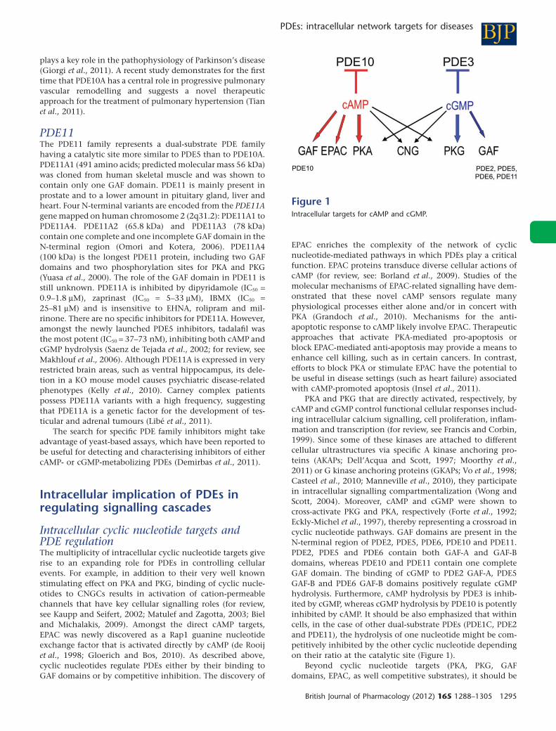
PKA and PKG that are directly activated, respectively, bycAMP and cGMP control functional cellular responses includ-ing intracellular calcium signalling, cell proliferation, inflam-mation and transcription (for review, see Francis and Corbin,1999). Since some of these kinases are attached to differentcellular ultrastructures via specific A kinase anchoring pro-teins (AKAPs; Dell’Acqua and Scott, 1997; Moorthy et al.,2011) or G kinase anchoring proteins (GKAPs; Vo et al., 1998;Casteel et al., 2010; Manneville et al., 2010), they participatein intracellular signalling compartmentalization (Wong andScott, 2004). Moreover, cAMP and cGMP were shown tocross-activate PKG and PKA, respectively (Forte et al., 1992;Eckly-Michel et al., 1997), thereby representing a crossroad incyclic nucleotide pathways. GAF domains are present in theN-terminal region of PDE2, PDE5, PDE6, PDE10 and PDE11.PDE2, PDE5 and PDE6 contain both GAF-A and GAF-Bdomains, whereas PDE10 and PDE11 contain one completeGAF domain. The binding of cGMP to PDE2 GAF-A, PDE5GAF-B and PDE6 GAF-B domains positively regulate cGMPhydrolysis. Furthermore, cAMP hydrolysis by PDE3 is inhib-ited by cGMP, whereas cGMP hydrolysis by PDE10 is potentlyinhibited by cAMP. It should be also emphasized that withincells, in the case of other dual-substrate PDEs (PDE1C, PDE2and PDE11), the hydrolysis of one nucleotide might be com-petitively inhibited by the other cyclic nucleotide dependingon their ratio at the catalytic site (Figure 1).
Beyond cyclic nucleotide targets (PKA, PKG, GAFdomains, EPAC, as well competitive substrates), it should be
Figure 1Intracellular targets for cAMP and cGMP.
BJPPDEs: intracellular network targets for diseases
British Journal of Pharmacology (2012) 165 1288–1305 1295
noticed that CaM, CaM-Kinase, ERK, PKC and phosphatidicacid also participate in short-term regulation of PDEs (Keravisand Lugnier, 2010).
Long-term regulation of PDEs is mediated by PKA-dependent phosphorylation of transcription factors such asCREB and CREM (Lamas and Sassone-Corsi, 1997) and ICERexpression (Cho et al., 2005). Therefore, these molecularmechanisms will contribute to regulate physiological cellularevents as well as dysfunctions associated with variouspathologies.
Two examples of PDE regulation inaltered signallingSince the 1980s, the use of various PDE inhibitors in normaland diseased cells and tissues as well as in animal models hasallowed to increase and more precisely define the possibleimplication of specific PDE families in various physiologicalsystems and thereafter in associated pathologies, pointingout their roles in the CNS, in the cardiovascular system and ininflammation. Currently, biochemical and molecular biologi-cal tools as well as fluorescent imaging, allow specifyingwhich PDE family and how a variant contribute to thestudied dysfunction. In that way, hereafter, we present twoexamples where we studied the implications of different PDEsin diseases with altered cell cycle: angiogenesis and acutelymphoid leukaemia (ALL).
PDEs in angiogenesis. Angiogenesis, the development of newvessels from pre-existing ones, contributes mainly in tumourgrowth and metastasis that are dependent on VEGF secretion,which induces endothelial cell proliferation. EndothelialPDEs were first described as PDE2 and PDE4 in pig aorticendothelial cell (Souness et al., 1990) and in bovine aorticendothelial cells (Lugnier and Schini, 1990). On one hand,
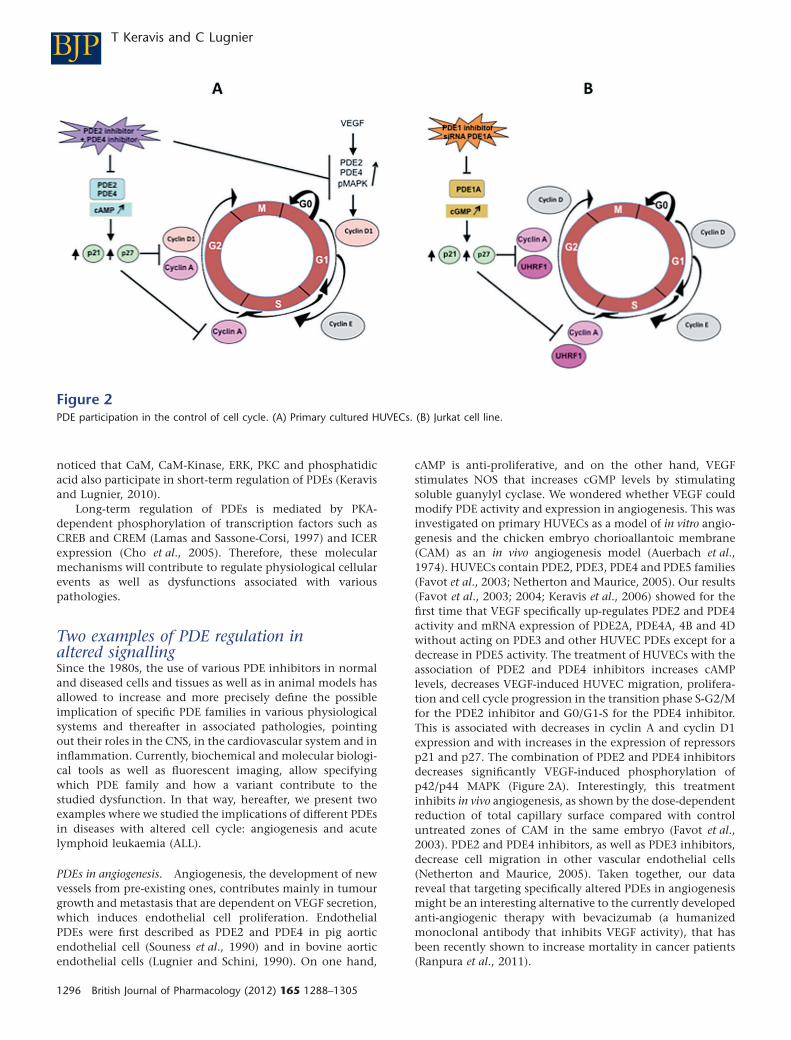
cAMP is anti-proliferative, and on the other hand, VEGFstimulates NOS that increases cGMP levels by stimulatingsoluble guanylyl cyclase. We wondered whether VEGF couldmodify PDE activity and expression in angiogenesis. This wasinvestigated on primary HUVECs as a model of in vitro angio-genesis and the chicken embryo chorioallantoic membrane(CAM) as an in vivo angiogenesis model (Auerbach et al.,1974). HUVECs contain PDE2, PDE3, PDE4 and PDE5 families(Favot et al., 2003; Netherton and Maurice, 2005). Our results(Favot et al., 2003; 2004; Keravis et al., 2006) showed for thefirst time that VEGF specifically up-regulates PDE2 and PDE4activity and mRNA expression of PDE2A, PDE4A, 4B and 4Dwithout acting on PDE3 and other HUVEC PDEs except for adecrease in PDE5 activity. The treatment of HUVECs with theassociation of PDE2 and PDE4 inhibitors increases cAMPlevels, decreases VEGF-induced HUVEC migration, prolifera-tion and cell cycle progression in the transition phase S-G2/Mfor the PDE2 inhibitor and G0/G1-S for the PDE4 inhibitor.This is associated with decreases in cyclin A and cyclin D1expression and with increases in the expression of repressorsp21 and p27. The combination of PDE2 and PDE4 inhibitorsdecreases significantly VEGF-induced phosphorylation ofp42/p44 MAPK (Figure 2A). Interestingly, this treatmentinhibits in vivo angiogenesis, as shown by the dose-dependentreduction of total capillary surface compared with controluntreated zones of CAM in the same embryo (Favot et al.,2003). PDE2 and PDE4 inhibitors, as well as PDE3 inhibitors,decrease cell migration in other vascular endothelial cells(Netherton and Maurice, 2005). Taken together, our datareveal that targeting specifically altered PDEs in angiogenesismight be an interesting alternative to the currently developedanti-angiogenic therapy with bevacizumab (a humanizedmonoclonal antibody that inhibits VEGF activity), that hasbeen recently shown to increase mortality in cancer patients(Ranpura et al., 2011).
Figure 2PDE participation in the control of cell cycle. (A) Primary cultured HUVECs. (B) Jurkat cell line.
BJP T Keravis and C Lugnier
1296 British Journal of Pharmacology (2012) 165 1288–1305

PDEs in ALL. It has been reported that PDE4 inhibitors,which activate cAMP signalling by reducing cAMP catabo-lism, are known to induce apoptosis in B-lineage CLL cells(Lerner et al., 2000), but not in normal human T cells (Meyerset al., 2009) and T leukaemic cell lines (Tiwari et al., 2005). Werecently showed in Jurkat cells (a human T-cell line, model ofALL) that the apoptotic effect of the natural compound thy-moquinone is mediated by p73 up-regulation that inducesdown-regulation of the epigenetic integrator UHRF1 andG1 cell cycle arrest in a dose-dependent manner (Alhosinet al., 2010). Since thymoquinone has anti-oxidant andanti-inflammatory properties similarly to PDE4 inhibitors(Houslay et al., 2005), since some natural compounds are ableto inhibit PDEs and since PDE4 inhibitors decrease cell pro-liferation in leukaemia (Kim and Lerner, 1998), we wonderedwhether a specific PDE isoform controlling apoptosis wouldbe responsible for the thymoquinone effects. The determina-tion of the PDE family profile contributing to total cAMP- andcGMP-hydrolysing activities reveals that PDE4 contributesmainly to cAMP hydrolysis, whereas cGMP-hydrolysingactivity is mainly due to PDE5, PDE1 contributing only for1%. Assessed as a PDE inhibitor, thymoquinone only inhibitscAMP hydrolysis by PDE4 with an IC50 value of 60 mM, inaccordance with its previous described anti-oxidant and anti-inflammatory properties. However, and intriguingly, 24 hthymoquinone treatment dose-dependently decreases cAMPlevels but increases cGMP levels, despite that thymoquinonedoes not act on cGMP hydrolysis by PDE1, PDE2, PDE3 andPDE5. This treatment inhibits Jurkat cell growth in a dose-dependent manner and induces apoptosis. In contrast toPDE3, PDE4 and PDE5 inhibitors, nimodipine, a PDE1 inhibi-tor, is able, at a concentration selectively active on PDE1, toinhibit the growth and apoptosis of Jurkat cells, similarly tothymoquinone. The effects of nimodipine and thymo-quinone induce concomitant decreased expression of PDE1Aand UHRF1, whereas p73 expression is upregulated. Knockingdown PDE1A by RNA-interference leads to the same results.Conversely, re-expression of PDE1A induces UHRF1 expres-sion and p73 down-regulation. Taken together, these dataclearly and newly demonstrate that inhibition of PDE1Aexpression induces apoptosis of Jurkat cells by up-regulatingp73 repressor and down-regulating the expression of the epi-genetic integrator UHRF1 (Figure 2B). This also indicates forthe first time that a PDE family, such as PDE1A, apparentlyparticipates in epigenetic regulation (Abusnina et al., 2011).
These two examples of alterations of PDEs participating incell cycle regulation illustrate one aspect of how PDEs areimplicated in cellular functions via critical partners (Figure 2).They point out that, although thymoquinone inhibits onlyPDE4 activity, its effect in Jurkat cells is not mediated by PDE4but dependent on PDE1A regulation, illustrating the com-plexity of cyclic nucleotide pathways. This complexity couldbe linked to subcellular compartmentalization orchestratedby PDEs, AKAP and protein kinases (Houslay and Adams,2003), together with subcellular changes in location of PDEsinduced by some PDE inhibitors as recently shown for PDE4inhibitors (Day et al., 2011). Furthermore, PDE/protein inter-actions as largely reviewed for the PDE4 interactome (Houslay,2010) might well be extended to other ‘PDE interactomes’, assuggested for PDE1 by our work on PDE1A/UHRF1in Jurkatcells (Abusnina et al., 2011), for PDE3B in endothelial cells
(Wilson et al., 2011). Other ways to disrupt this interactomeinclude the design of specific peptides that would modifycritical peptidic interaction during PDE/protein interaction assuggested recently (Keravis and Lugnier, 2010) and the stimu-lation of changes in microdomain location of a critical PDEvariant for a particular cell function.
Perspectives for future therapeuticdevelopments
After numerous disappointments in the development ofPDE3 and PDE4 inhibitors due to their adverse effects (mor-tality and emesis, respectively), the success in PDE5 inhibitordevelopment with Viagra®, associated with increasing knowl-edge in the PDE field, renews interest in developing selectivePDE inhibitors as specific therapeutic agents with few adverseeffects.
Since PDEs participate in the interactome that regulatescellular signalling as recently exemplified by PDE3B (Perinoet al., 2011; Wilson et al., 2011), PDEs might be implicated inmultiple dysfunctions and pathologies associated withderegulated signals. Recent studies demonstrate the interestof PDE inhibitors to treat and to reverse heart hypertrophy(Takimoto et al., 2005), to enhance cognition (Reneerkenset al., 2009) and to treat sleep deprivation (Vecsey et al.,2009). Concerning alterations of PDE expression, increase inPDE5 expression in human failing left ventricle heart tissuerelates to oxidative stress and sildenafil treatment counteractsthis alteration (Lu et al., 2010). Similarly, expression of PDE5and PDE9 increases in aging and Alzheimer’s disease (Domek-Łopacinska and Strosznajder, 2010). Lastly and interestingly,by using a global PDE8B KO mouse model and a new PDE8-selective inhibitor (PF-04957325), Beavo and colleaguesreported that PDE8B controls steroidogenesis in mouseadrenal gland (Tsai et al., 2011).
To improve therapeutic index, dual PDE inhibitors induc-ing synergic potencies with lesser adverse effect have beenenvisionned. Indeed, dual PDE3/4 inhibitors are nowdesigned as therapeutic agents for chronic obstructive pulmo-nary disease. By combining both inhibitions, these com-pounds have additive and synergistic bronchodilatory andanti-inflammatory effects (Banner and Press, 2009). Further-more, PDE7/PDE4 dual inhibitors would represent a novelclass of drugs that could regulate pro-inflammatory andimmune T-cell function and be especially useful in treating awide variety of immune and inflammatory disorders with lessundesirable side effects (Castaño et al., 2009).
Another way to obtain an efficient therapeutic effect,without inducing adverse effects, may be to specifically targetthe altered PDE family and in particular the altered variant sothat the other PDEs present are not modified. To do so, it isfirst of all necessary to (i) characterize the various PDEisozymes present in the studied model, (ii) identify thealtered PDE isozyme(s) related to the given pathology (thera-peutic target) and (iii) validate the therapeutic treatment bythe induction of concomitant restoration of the alteredPDE(s) and associated cellular function without changing theother non-altered PDEs. This approach might be useful in thecase of diseases for which no specific therapy is available. It
BJPPDEs: intracellular network targets for diseases
British Journal of Pharmacology (2012) 165 1288–1305 1297
could also be worth considering in established treatmentsthat induce too many adverse effects.
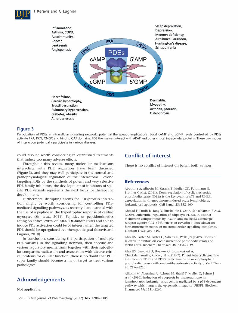
Throughout this review, many molecular mechanismsinteracting with PDE regulation have been discussed(Figure 3), and they may well participate in the normal andpathophysiological regulation of the interactome. Beyondtargeting PDEs by the synthesis of potent and very selectivePDE family inhibitors, the development of inhibitors of spe-cific PDE variants represents the next focus for therapeuticdevelopment.
Furthermore, disrupting agents for PDE/protein interac-tion might be worth considering for controlling PDE-mediated signalling pathways, as recently demonstrated withthe use of a peptide in the hypertrophic response of cardiacmyocytes (Sin et al., 2011). Peptides or peptidomimeticsacting on critical extra- or intra-PDE-binding sites and able toinduce PDE activation could be of interest when the targetedPDE should be upregulated as a therapeutic goal (Keravis andLugnier, 2010).
In conclusion, considering the participation of multiplePDE variants in the signalling network, their specific andvarious regulatory mechanisms together with their subcellu-lar compartmentalization and association with diverse criti-cal proteins for cellular function, there is no doubt that PDEsuper family should become a major target to treat variouspathologies.
Acknowledgements
Not applicable.
Conflict of interest
There is no conflict of interest on behalf both authors.
ReferencesAbusnina A, Alhosin M, Keravis T, Muller CD, Fuhrmann G,Bronner C et al. (2011). Down-regulation of cyclic nucleotidephosphodiesterase PDE1A is the key event of p73 and UHRF1deregulation in thymoquinone-induced acute lymphoblasticleukemia cell apoptosis. Cell Signal 23: 152–160.
Ahmad F, Lindh R, Tang Y, Ruishalme I, Ost A, Sahachartsiri B et al.(2009). Differential regulation of adipocyte PDE3B in distinctmembrane compartments by insulin and the beta3-adrenergicreceptor agonist CL316243: effects of caveolin-1 knockdown onformation/maintenance of macromolecular signalling complexes.Biochem J 424: 399–410.
Ahn HS, Foster M, Foster C, Sybertz E, Wells JN (1989). Effects ofselective inhibitors on cyclic nucleotide phosphodiesterases ofrabbit aorta. Biochem Pharmacol 38: 3331–3339.
Ahn HS, Bercovici A, Boykow G, Bronnenkant A,Chackalamannil S, Chow J et al. (1997). Potent tetracyclic guanineinhibitors of PDE1 and PDE5 cyclic guanosine monophosphatephosphodiesterases with oral antihypertensive activity. J Med Chem40: 2196–2210.
Alhosin M, Abusnina A, Achour M, Sharif T, Muller C, Peluso Jet al. (2010). Induction of apoptosis by thymoquinone inlymphoblastic leukemia Jurkat cells is mediated by a p73-dependentpathway which targets the epigenetic integrator UHRF1. BiochemPharmacol 79: 1251–1260.
Figure 3Participation of PDEs in intracellular signalling network: potential therapeutic implications. Local cAMP and cGMP levels controlled by PDEsactivate PKA, PKG, CNGC and bind to GAF domains. PDE themselves interact with AKAP and other critical intracellular proteins. These two modesof interaction potentially participate in various diseases.
BJP T Keravis and C Lugnier
1298 British Journal of Pharmacology (2012) 165 1288–1305
Amer MS (1977). Cyclic nucleotides as targets for drug design. AdvDrug Res 12: 1–38.
Anant JS, Ong OC, Xie HY, Clarke S, O’Brien PJ, Fung BK (1992).In vivo differential prenylation of retinal cyclic GMPphosphodiesterase catalytic subunits. J Biol Chem 267: 687–690.
Arozarena I, Sanchez-Laorden B, Packer L, Hidalgo-Carcedo C,Hayward R, Viros A et al. (2011). Oncogenic BRAF inducesmelanoma cell invasion by downregulating the cGMP-specificphosphodiesterase PDE5A. Cancer Cell 19: 45–57.
Artemyev NO, Surendran R, Lee JC, Hamm HE (1996). Subunitstructure of rod cGMP-phosphodiesterase. J Biol Chem 271:25382–25388.
Aspiotis R, Deschênes D, Dubé D, Girard Y, Huang Z, Laliberté Fet al. (2010). The discovery and synthesis of highly potent subtypeselective phosphodiesterase 4D inhibitors. Bioorg Med Chem Lett20: 5502–5505.
Assad-Kottner C, Chen D, Jahanyar J, Cordova F, Summers N,Loebe M et al. (2008). The use of continuous milrinone therapy asbridge to transplant is safe in patients with short waiting times.J Card Fail 14: 839–843.
Auerbach R, Kubai L, Knighton D, Folkman J (1974). A simpleprocedure for the long-term cultivation of chicken embryos. DevBiol 41: 391–394.
Baillie GS, MacKenzie SJ, McPhee I, Houslay MD (2000). Sub-familyselective actions in the ability of Erk2 MAP kinase to phosphorylateand regulate the activity of PDE4 cyclic AMP-specificphosphodiesterases. Br J Pharmacol 131: 811–819.
Baillie GS, Huston E, Scotland G, Hodgkin M, Gall I, Peden AHet al. (2002). TAPAS-1, a novel microdomain within the uniqueN-terminal region of the PDE4A1 cAMP-specific phosphodiesterasethat allows rapid, Ca2+-triggered membrane association withselectivity for interaction with phosphatidic acid. J Biol Chem 277:28298–28309.
Banner KH, Press NJ (2009). Dual PDE3/4 inhibitors as therapeuticagents for chronic obstructive pulmonary disease. Br J Pharmacol157: 892–906.
Barber R, Baillie GS, Bergmann R, Shepherd MC, Sepper R,Houslay MD et al. (2004). Differential expression of PDE4 cAMPphosphodiesterase isoforms in inflammatory cells of smokers withCOPD, smokers without COPD, and nonsmokers. Am J PhysiolLung Cell Mol Physiol 287: L332–L343.
Bates MG, Thompson AA, Baillie JK (2007). Phosphodiesterase type5 inhibitors in the treatment and prevention of high altitudepulmonary edema. Curr Opin Investig Drugs 8: 226–231.
Beard MB, Olsen AE, Jones RE, Erdogan S, Houslay MD, Bolger GB(2000). UCR1 and UCR2 domains unique to the cAMP-specificphosphodiesterase family form a discrete module via electrostaticinteractions. J Biol Chem 275: 10349–10358.
Beavo JA (1995). Cyclic nucleotide phosphodiesterases: functionalimplications of multiple isoforms. Physiol Rev 75: 725–748.
Beavo JA, Brunton LL (2002). Cyclic nucleotide research – stillexpanding after half a century. Nat Rev Mol Cell Biol 3: 710–718.
Beavo JA, Francis SH, Houslay MD (eds) (2006). Cyclic NucleotidePhosphodiesterases in Health and Disease. CRC Press: Boca Raton,USA.
Bender AT (2006). Calmodulin-stimulated cyclic nucleotidephosphodiesterases. In: Beavo JA, Francis SH, Houslay MD (eds).Cyclic Nucleotide Phosphodiesterases in Health and Disease. CRCPress: Boca Raton, pp. 35–54.
Bender AT, Beavo JA (2006). Cyclic nucleotide phosphodiesterases:molecular regulation to clinical use. Pharmacol Rev 58: 488–520.
Berger K, Lindh R, Wierup N, Zmuda-Trzebiatowska E, Lindqvist A,Manganiello VC et al. (2009). Phosphodiesterase 3B is localized incaveolae and smooth ER in mouse hepatocytes and is important inthe regulation of glucose and lipid metabolism. PLoS ONE 4: e4671.
Biel M, Michalakis S (2009). Cyclic nucleotide-gated channels.Handb Exp Pharmacol 191: 111–136.
Boess FG, Hendrix M, van der Staay FJ, Erb C, Schreiber R,van Staveren W et al. (2004). Inhibition of phosphodiesterase 2increases neuronal cGMP, synaptic plasticity and memoryperformance. Neuropharmacology 47: 1081–1092.
Boichot E, Wallace JL, Germain N, Corbel M, Lugnier C, Lagente Vet al. (2000). Anti-inflammatory activities of a new series of selectivephosphodiesterase 4 inhibitors derived from 9-benzyladenine.J Pharmacol Exp Ther 292: 647–653.
Bolger GB (2006). Phosphodiesterase isoforms – an annoted list. In:Beavo JA, Francis SH, Houslay MD (eds). Cyclic NucleotidePhosphodiesterases in Health and Disease. CRC Press: Boca Raton,pp. 19–31.
Bolger G, Michaeli T, Martins T, St John T, Steiner B, Rodgers Let al. (1993). A family of human phosphodiesterases homologous tothe dunce learning and memory gene product of Drosophilamelanogaster are potential targets for antidepressant drugs. MolCell Biol 13: 6558–6571.
Borisy FF, Ronnett GV, Cunningham AM, Juilfs D, Beavo J,Synder SH (1992). Calcium/calmodulin-activated phosphodiesteraseexpressed in olfactory receptor neurons. J Neurosci 12: 915–923.
Borland G, Smith BO, Yarwood SJ (2009). EPAC proteins transducediverse cellular actions of cAMP. Br J Pharmacol 158: 70–86.
Boswell-Smith V, Spina D, Page CP (2006). Phosphodiesteraseinhibitors. Br J Pharmacol 147: S252–S257.
Bourguignon JJ, Lugnier C, Abarghaz M, Lagouge Y, Wagner P,Mondadori C et al. (2004). Cyclic nucleotide phosphodiesteraseinhibitors, preparation and uses. Patent WO2004041258, published21.05.2004. International Application N°: PCT/FR2003/003247(30.10.2003).
Burgin AB, Olafur T, Magnusson OT, Singh J, Witte P, Bart L et al.(2010). Design of phosphodiesterase 4D (PDE4D) allostericmodulators for enhancing cognition with improved safety. NatBiotechnol 28: 63–70.
Campos-Toimil M, Keravis T, Orallo F, Takeda K, Lugnier C (2008).Short-term or long-term treatments with a phosphodiesterase-4(PDE4) inhibitor result in opposing agonist-induced Ca(2+)responses in endothelial cells. Br J Pharmacol 154: 82–92.
Castaño T, Wang H, Campillo NE, Ballester S, González-García C,Hernández J et al. (2009). Synthesis, structural analysis, andbiological evaluation of thioxoquinazoline derivatives asphosphodiesterase 7 inhibitors. ChemMedChem 4: 866–876.
Casteel DE, Smith-Nguyen EV, Sankaran B, Roh SH, Pilz RB, Kim C(2010). A crystal structure of the cyclic GMP-dependent proteinkinase Ib dimerization/docking domain reveals molecular details ofisoform-specific anchoring. J Biol Chem 285: 32684–32688.
Chambers RJ, Abrams K, Garceau NY, Kamath AV, Manley CM,Lilley SC et al. (2006). A new chemical tool for exploring thephysiological function of the PDE2 isozyme. Bioorg Med Chem Lett16: 307–310.
BJPPDEs: intracellular network targets for diseases
British Journal of Pharmacology (2012) 165 1288–1305 1299
Cho ES, Yu JH, Kim MS, Yim M (2005). Rolipram, aphosphodiesterase 4 inhibitor, stimulates inducible cAMP earlyrepressor expression in osteoblasts. Yonsei Med J 46: 149–154.
Conti M, Beavo J (2007). Biochemistry and physiology of cyclicnucleotide phosphodiesterases: essential components in cyclicnucleotide signaling. Annu Rev Biochem 76: 481–511.
Cote RH (2006). Photoreceptor phosphodiesterase (PDE6): aG-protein-activated PDE regulating visual excitation in rod andcone photoreceptor cells. In: Beavo JA, Francis SH, Houslay MD(eds). Cyclic Nucleotide Phosphodiesterases in Health and Disease.CRC Press: Boca Raton, pp. 165–193.
Croom KF, Curran MP (2008). Sildenafil: a review of its use inpulmonary arterial hypertension. Drugs 68: 383–397.
Cukkemane A, Seifert R, Kaupp UB (2011). Cooperative anduncooperative cyclic-nucleotide-gated ion channels. TrendsBiochem Sci 36: 55–64.
Daugan A, Grondin P, Ruault C, Le Monnier de Gouville AC,Coste H, Kirilovsky J et al. (2003). The discovery of tadalafil: a noveland highly selective PDE5 inhibitor. 1: 5,6,11,11a-tetrahydro-1H-imidazo[1′,5′:1,6]pyrido[3,4-b]indole-1,3(2H)-dione analogues.J Med Chem 46: 4525–4532.
Day JP, Lindsay B, Riddell T, Jiang Z, Allcock RW, Abraham A et al.(2011). Elucidation of a structural basis for the inhibitor-driven,p62 (SQSTM1)-dependent intracellular redistribution of cAMPphosphodiesterase-4A4 (PDE4A4). J Med Chem 54: 3331–3347.
Degerman E, Manganiello V (2006). Phosphodiesterase 3B: animportant regulator of energy homeostasis. In: Beavo JA,Francis SH, Houslay MD (eds). Cyclic NucleotidePhosphodiesterases in Health and Disease. CRC Press: Boca Raton,pp. 79–97.
Dell’Acqua ML, Scott JD (1997). Protein kinase A anchoring. J BiolChem 272: 12881–12884.
Demirbas D, Ceyhan O, Wyman AR, Ivey FD, Allain C, Wang Let al. (2011). Use of a Schizosaccharomyces pombe PKA-repressiblereporter to study cGMP metabolising phosphodiesterases. CellSignal 23: 594–601.
DeNinno MP, Andrews M, Bell AS, Chen Y, Eller-Zarbo C,Eshelby N (2009). The discovery of potent, selective and orallybioavailable PDE9 inhibitors as potential hypoglycemic agents.Bioorg Med Chem Lett 19: 2537–2541.
Dodge KL, Khouangsathiene S, Kapiloff MS, Mouton R, Hill EV,Houslay MD et al. (2001). mAKAP assembles a protein kinaseA/PDE4 phosphodiesterase cAMP signaling module. EMBO J 20:1921–1930.
Domek-Łopacinska KU, Strosznajder JB (2010). Cyclic GMP andnitric oxide synthase in aging and Alzheimer’s disease. MolNeurobiol 41: 129–137.
Duncan GS, Wolberg G, Schmitges CJ, Deeprose RD,Zimmerman TP (1982). Inhibition of lymphocyte-mediatedcytolysis and cyclic AMP phosphodiesterase by erythro-9-(2-hydroxy-3-nonyl)adenine. J Immunopharmacol 4: 79–100.
Eckly AE, Lugnier C (1994). role of phosphodiesterase-III andphosphodiesterase-IV in the modulation of vascular cyclic-AMPcontent by the NO/cyclic-GMP pathway. Br J Pharmacol 113:445–450.
Eckly-Michel A, Martin V, Lugnier C (1997). Involvement of cyclicnucleotide-dependent protein kinases in cyclic AMP-mediatedvasorelaxation. Br J Pharmacol 122: 158–164.
Favot L, Keravis T, Holl V, Le Bec A, Lugnier C (2003).VEGF-induced HUVEC migration and proliferation are decreased byPDE2 and PDE4 inhibitors. Thromb Haemost 90: 334–343.
Favot L, Keravis T, Lugnier C (2004). Modulation of VEGF-inducedendothelial cell cycle protein expression through cyclic AMPhydrolysis by PDE2 and PDE4. Thromb Haemost 92: 634–645.
Fisher DA, Smith JF, Pillar JS, St. Denis SH, Cheng JB (1998).Isolation and characterization of PDE9A, a novel humancGMP-specific phosphodiesterase. J Biol Chem 273: 15559–15564.
Forte LR, Thorne PK, Eber SL, Krause WJ, Freeman RH, Francis SHet al. (1992). Stimulation of intestinal Cl-transport by heat-stableenterotoxin: activation of cAMP-dependent protein kinase bycGMP. Am J Physiol 263: C607–C615.
Francis SH, Corbin JD (1999). Cyclic nucleotide-dependent proteinkinases: intracellular receptors for cAMP and cGMP action. Crit RevClin Lab Sci 36: 275–328.
Francis SH, Zoraghi R, Kotera J, Ke H, Bessay EP, Blount MA et al.(2006). Phosphodiesterase 5: molecular characteristics relating tostructure function and regulation. In: Beavo JA, Francis SH,Houslay MD (eds). Cyclic Nucleotide Phosphodiesterases in Healthand Disease. CRC Press: Boca Raton, pp. 131–164.
Francis SH, Morris GZ, Corbin JD (2008). Molecular mechanismsthat could contribute to prolonged effectiveness of PDE5 inhibitorsto improve erectile function. Int J Impot Res 20: 333–342.
Francis SH, Corbin JD, Bischoff E (2009). Cyclic GMP-hydrolyzingphosphodiesterases. Handb Exp Pharmacol 191: 367–408.
Francis SH, Blount MA, Corbin JD (2011). Mammalian cyclicnucleotide phosphodiesterases: molecular mechanisms andphysiological functions. Physiol Rev 91: 651–690.
Fujishige K, Kotera J, Michibata H, Yuasa K, Takebayashi S,Okumura K et al. (1999). Cloning and characterization of a novelhuman phosphodiesterase that hydrolyzes both cAMP and cGMP(PDE10A). J Biol Chem 274: 18438–18445.
Fujishige K, Kotera J, Yuasa K, Omori K (2000). The humanphosphodiesterase PDE10A gene genomic organization andevolutionary relatedness with other PDEs containing GAF domains.Eur J Biochem 267: 5943–5951.
Gamanuma M, Yuasa K, Sasaki T, Sakurai N, Kotera J, Omori K(2003). Comparison of enzymatic characterization and geneorganization of cyclic nucleotide phosphodiesterase 8 family inhumans. Cell Signal 15: 565–574.
Gebska MA, Stevenson BK, Hemnes AR, Bivalacqua TJ, Haile A,Hesketh GG et al. (2011). Phosphodiesterase-5A (PDE5A) is localizedto the endothelial caveolae and modulates NOS3 activity.Cardiovasc Res 90: 353–363.
Geoffroy V, Fouque F, Nivet V, Clot JP, Lugnier C, Desbuquois Bet al. (1999). Activation of a cGMP-stimulated cAMPphosphodiesterase by protein kinase C in a liver Golgi-endosomalfraction. Eur J Biochem 259: 892–900.
Geoffroy V, Fouque F, Lugnier C, Desbuquois B, Benelli C (2001).Characterization of an in vivo hormonally regulatedphosphodiesterase 3 (PDE3) associated with a liver Golgi-endosomalfraction. Arch Biochem Biophys 387: 154–162.
Gesellchen F, Zaccolo M (2011). Phosphodiesterase 2A, cGMPstimulated. UCSD Nat Mol. DOI: 10.1038/mp.a001750.01.
Ghofrani HA, Wiedemann R, Rose F, Schermuly RT, Olschewski H,Weissmann N et al. (2002). Sildenafil for treatment of lung fibrosisand pulmonary hypertension: a randomised controlled trial. Lancet360: 895–900.
BJP T Keravis and C Lugnier
1300 British Journal of Pharmacology (2012) 165 1288–1305
Giampà C, Laurenti D, Anzilotti S, Bernardi G, Menniti FS,Fusco FR (2010). Inhibition of the striatal specificphosphodiesterase PDE10A ameliorates striatal and corticalpathology in R6/2 mouse model of Huntington’s disease. PLoS ONE5: e13417.
Giorgi M, Melchiorri G, Nuccetelli V, D’Angelo V, Martorana A,Sorge R et al. (2011). PDE10A and PDE10A-dependent cAMPcatabolism are dysregulated oppositely in striatum and nucleusaccumbens after lesion of midbrain dopamine neurons in rat: a keystep in Parkinsonism physiopathology. Neurobiol Dis 43: 293–303.
Gloerich M, Bos JL (2010). Epac: defining a new mechanism forcAMP action. Annu Rev Pharmacol Toxicol 50: 355–375.
Gonçalves RL, Lugnier C, Keravis T, Lopes MJ, Fantini FA,Schmitt M et al. (2009). The flavonoid dioclein is a selectiveinhibitor of cyclic nucleotide phosphodiesterase type 1 (PDE1) anda cGMP-dependent protein kinase (PKG) vasorelaxant in humanvascular tissue. Eur J Pharmacol 620: 78–83.
Grandoch M, Roscioni SS, Schmidt M (2010). The role of Epacproteins, novel cAMP mediators, in the regulation of immune, lungand neuronal function. Br J Pharmacol 159: 265–284.
Grauer SM, Pulito VL, Navarra RL, Kelly MP, Kelley C, Graf R et al.(2009). Phosphodiesterase 10A inhibitor activity in preclinicalmodels of the positive, cognitive, and negative symptoms ofschizophrenia. J Pharmacol Exp Ther 331: 574–590.
Guazzi M, Vicenzi M, Arena R, Guazzi MD (2011). PDE5 inhibitionwith sildenafil improves left ventricular diastolic function, cardiacgeometry, and clinical status in patients with stable systolic heartfailure: results of a 1-year, prospective, randomized,placebo-controlled study. Circ Heart Fail 4: 8–17.
Hanson MS, Stephenson AH, Bowles EA, Sprague RS (2010). Insulininhibits human erythrocyte cAMP accumulation and ATP release:role of phosphodiesterase 3 and phosphoinositide 3-kinase. ExpBiol Med 235: 256–262.
Hatzelmann A, Morcillo EJ, Lungarella G, Adnot S, Sanjar S,Beume R et al. (2010). The preclinical pharmacology of roflumilast –a selective, oral phosphodiesterase 4 inhibitor in development forchronic obstructive pulmonary disease. Pulm Pharmacol Ther 23:235–256.
Hayashi M, Shimada Y, Nishimura Y, Hama T, Tanaka T (2002).Genomic organization, chromosomal localization, and alternativesplicing of the human phosphodiesterase 8B gene. BiochemBiophys Res Commun 297: 1253–1258.
Heikaus CC, Pandit J, Klevit RE (2009). Cyclic nucleotide bindingGAF domains from phosphodiesterases: structural and mechanisticinsights. Structure 17: 1551–1557.
Hidaka H, Hayashi H, Kohri H, Kimura Y, Hosokawa T, Igawa Tet al. (1979). Selective inhibitor of platelet cyclic adenosine mono-phosphate phosphodiesterase, cilostamide, inhibits plateletaggregation. J Pharmacol Exp Ther 211: 26–30.
Houslay MD (2010). Underpinning compartmentalised cAMPsignalling through targeted cAMP breakdown. Trends Biochem Sci35: 91–100.
Houslay MD (2011). Hard times for oncogenic BRAF-expressingmelanoma cells. Cancer Cell 19: 3–4.
Houslay MD, Adams DR (2003). PDE4 cAMP phosphodiesterases:modular enzymes that orchestrate signalling cross-talk,desensitization and compartmentalization. Biochem J 370: 1–18.
Houslay MD, Schafer P, Zhang KY (2005). Keynote review:phosphodiesterase-4 as a therapeutic target. Drug Discov Today 10:1503–1519.
Huston E, Gall I, Houslay TM, Houslay MD (2006). Helix-1 of thecAMP-specific phosphodiesterase PDE4A1 regulates itsphospholipase-D-dependent redistribution in response to release ofCa 2+. J Cell Sci 119: 3799–3810.
Hutson PH, Finger EN, Magliaro BC, Smith SM, Converso A,Sanderson PE et al. (2011). The selective phosphodiesterase 9(PDE9) inhibitor PF-04447943 (6-[(3S,4S)-4-methyl-1-(pyrimidin-2-ylmethyl)pyrrolidin-3-yl]-1-(tetrahydro-2H-pyran-4-yl)-1,5-dihydro-4H-pyrazolo[3,4-d]pyrimidin-4-one) enhances synaptic plasticityand cognitive function in rodents. Neuropharmacology 61:665–676.
Insel PA, Zhang L, Murray F, Yokouchi H, Zambon AC (2011).Cyclic AMP is both a pro-apoptotic and anti-apoptotic secondmessenger. Acta Physiol (Oxf). DOI: 10.1111/j.1748-1716.2011.02273 [Epub ahead of print].
Jeon KI, Xu X, Aizawa T, Lim JH, Jono H, Kwon DS et al. (2010).Vinpocetine inhibits NF-kB-dependent inflammation via anIKK-dependent but PDE-independent mechanism. Proc Natl AcadSci U S A 107: 9795–9800.
Kadoshima-Yamaoka K, Murakawa M, Goto M, Tanaka Y, Inoue H,Murafuji H et al. (2009). ASB16165, a novel inhibitor forphosphodiesterase 7A (PDE7A), suppresses IL-12-inducedIFN-gamma production by mouse activated T lymphocytes.Immunol Lett 122: 193–197.
Kakkar R, Raju RV, Sharma RK (1999). Calmodulin-dependent cyclicnucleotide phosphodiesterase (PDE1). Cell Mol Life Sci 55:1164–1186.
Kameni-Tcheudji JF, Lebeau L, Virmaux N, Maftei CG, Cote RH,Lugnier C et al. (2001). Molecular organization of bovine rodcGMP- phosphodiesterase 6. J Mol Biol 310: 781–791.
Kass DA, Champion HC, Beavo JA (2007). Phosphodiesterase type5: expanding roles in cardiovascular regulation. Circ Res 101:1084–1095.
Kaupp UB, Seifert R (2002). Cyclic nucleotide-gated ion channels.Physiol Rev 82: 769–824.
Kelly MP, Logue SF, Brennan J, Day JP, Lakkaraju S, Jiang L et al.(2010). Phosphodiesterase 11A in brain is enriched in ventralhippocampus and deletion causes psychiatric disease relatedphenotypes. Proc Natl Acad Sci USA 107: 8457–8462.
Keravis T, Lugnier C (2010). Cyclic nucleotide phosphodiesterases(PDE) and peptide motifs. Curr Pharm Des 16: 1114–1125.
Keravis T, Silva AP, Favot L, Lugnier C (2006). Role of PDEs invascular health and disease: endothelial PDEs and angiogenesis. In:Beavo JA, Francis SH, Houslay MD (eds). Cyclic NucleotidePhosphodiesterases in Health and Disease. CRC Press: Boca Raton,pp. 417–439.
Keravis T, Vandecasteele G, Mokni W, Fischmeister R, Lugnier C(2007). Identification of PDE1 and PDE5 in adult rat left ventriclescardiomyocytes. J Mol Cell Cardiol 42: S49–S50.
Kim DH, Lerner A (1998). Type 4 cyclic adenosine monophosphatephosphodiesterase as a therapeutic target in chronic lymphocyticleukemia. Blood 92: 2484–2494.
Komas N, Lugnier C, Le Bec A, Serradeil-Le Gal C, Barthelemy G,Stoclet JC (1989). Differential sensitivity to cardiotonic drugs ofcyclic AMP phosphodiesterases isolated from canine ventricular andsinoatrial-enriched tissues. J Cardiovasc Pharmacol 14: 213–220.
Komas N, Lugnier C, Stoclet JC (1991). Endothelium-dependentand independent relaxation of the rat aorta by cyclic-nucleotidephosphodiesterase inhibitors. Br J Pharmacol 104: 495–503.
BJPPDEs: intracellular network targets for diseases
British Journal of Pharmacology (2012) 165 1288–1305 1301
Kumar P, Francis GS, Tang WH (2009). Phosphodiesterase 5inhibition in heart failure: mechanisms and clinical implications.Nat Rev Cardiol 6: 349–355.
Lamas M, Sassone-Corsi P (1997). The dynamics of thetranscriptional response to cyclic adenosine 3,5-monophosphate:recurrent inducibility and refractory phase. Mol Endocrinol 11:1415–1424.
Lanfear DE, Hasan R, Gupta RC, Williams C, Czerska B, Tita C et al.(2009). Short term effects of milrinone on biomarkers of necrosis,apoptosis, and inflammation in patients with severe heart failure.J Transl Med 7: 67. DOI: 10.1186/1479-5876-7-67.
Lee R, Wolda S, Moon E, Esselstyn J, Hertel C, Lerner A (2002).PDE7A is expressed in human B-lymphocytes and is up-regulatedby elevation of intracellular cAMP. Cell Signal 14: 277–284.
Lerner A, Kim DH, Lee R (2000). The cAMP signaling pathway as atherapeutic target in lymphoid malignancies. Leuk Lymphoma 37:39–51.
Libé R, Horvath A, Vezzosi D, Fratticci A, Coste J, Perlemoine Ket al. (2011). Frequent phosphodiesterase 11A gene (PDE11A)defects in patients with Carney complex (CNC) caused by PRKAR1Amutations: PDE11A may contribute to adrenal and testiculartumors in CNC as a modifier of the phenotype. J Clin EndocrinolMetab 96: E208–E214.
Lu Z, Xu X, Hu X, Lee S, Traverse JH, Zhu G et al. (2010). Oxidativestress regulates left ventricular PDE5 expression in the failing heart.Circulation 121: 1474–1483.
Lugnier C (ed.) (2005). Phosphodiesterase Methods and Protocols,Methods in Molecular Biology 307. Humana Press: Totowa.
Lugnier C (2006). Cyclic nucleotide phosphodiesterase (PDE)superfamily: a new target for the development of specifictherapeutic agents. Pharmacol Ther 109: 366–398.
Lugnier C, Schini VB (1990). Characterization of cyclic nucleotidephosphodiesterases from cultured bovine aortic endothelial cells.Biochem Pharmacol 39: 75–84.
Lugnier C, Stierlé A, Beretz A, Schoeffter P, Le Bec A, Wermuth CGet al. (1983). Tissue and substrate specificity of inhibition byalkoxy-aryl-lactams of platelet and arterial smooth muscle cyclicnucleotide phosphodiesterases: relationship to pharmacologicalactivity. Biochem Biophys Res Commun 113: 954–959.
Lugnier C, Follénius A, Gérard D, Stoclet JC (1984). Bepridil andflunarizine as calmodulin inhibitors. Eur J Pharmacol 98: 157–158.
Lugnier C, Schoeffter P, Le Bec A, Strouthou E, Stoclet JC (1986).Selective inhibition of cyclic nucleotide phosphodiesterases ofhuman, bovine and rat aorta. Biochem Pharmacol 35: 1743–1751.
Lugnier C, Muller B, Le Bec A, Beaudry C, Rousseau E (1993).Characterization of indolidan- and rolipram-sensitive cyclicnucleotide phosphodiesterases in canine and human cardiacmicrosomal fractions. J Pharmacol Exp Ther 265: 1142–1151.
Lugnier C, Keravis T, Le Bec A, Pauvert O, Proteau S, Rousseau E(1999). Characterization of cyclic nucleotide phosphodiesteraseisoforms associated to isolated cardiac nuclei. Biochim Biophys Acta1472: 431–446.
Makhlouf A, Kshirsagar A, Niederberger C (2006).Phosphodiesterase 11: a brief review of structure, expression andfunction. Int J Impot Res 18: 501–509.
Manneville JB, Jehanno M, Etienne-Manneville S (2010). Dlg1 bindsGKAP to control dynein association with microtubules, centrosomepositioning, and cell polarity. J Cell Biol 191: 585–598.
Manning CD, Burman M, Christensen SB, Cieslinski LB,Essayan DM, Grous M et al. (1999). Suppression of humaninflammatory cell function by subtype-selective PDE4 inhibitorscorrelates with inhibition of PDE4A and PDE4B. Br J Pharmacol128: 1393–1398.
Marivet MC, Bourguignon JJ, Lugnier C, Mann A, Stoclet JC,Wermuth CG (1989). Inhibition of cyclic adenosine-3′,5′-monophosphate phosphodiesterase from vascular smooth muscleby rolipram analogues. J Med Chem 32: 1450–1457.
Martinez SE (2006). PDE2 structure and functions. In: Beavo JA,Francis SH, Houslay MD (eds). Cyclic NucleotidePhosphodiesterases in Health and Disease. CRC Press:Boca Raton, pp. 55–77.
Martinez SE, Wu AY, Glavas NA, Tang XB, Turley S, Hol WG et al.(2002). The two GAF domains in phosphodiesterase 2A havedistinct roles in dimerization and in cGMP binding. Proc Natl AcadSci USA 99: 13260–13265.
Masood A, Huang Y, Hajjhussein H, Xiao L, Li H, Wang W et al.(2009). Anxiolytic effects of phosphodiesterase-2 inhibitorsassociated with increased cGMP signaling. J Pharmacol Exp Ther331: 690–699.
Matulef K, Zagotta WN (2003). Cyclic nucleotide-gated ionchannels. Annu Rev Cell Dev Biol 19: 23–44.
Maurice DH, Haslam RJ (1990). Molecular basis of the synergisticinhibition of platelet function by nitrovasodilators and activators ofadenylate cyclase: inhibition of cyclic AMP breakdown by cyclicGMP. Mol Pharmacol 37: 671–681.
McCahill A, McSorley T, Huston E, Hill EV, Lynch MJ, Gall I et al.(2005). In resting COS1 cells a dominant negative approach showsthat specific, anchored PDE4 cAMP phosphodiesterase isoforms gatethe activation, by basal cyclic AMP production, of AKAP-tetheredprotein kinase A type II located in the centrosomal region. CellSignal 17: 1158–1173.
Mentz F, Merle-Beral H, Ouaaz F, Binet JL (1995). Theophylline, anew inducer of apoptosis in B-CLL: role of cyclic nucleotides. Br JHaematol 90: 957–959.
Meyers JA, Su DW, Lerner A (2009). Chronic lymphocytic leukemiaand B and T cells differ in their response to cyclic nucleotidephosphodiesterase inhibitors. J Immunol 182: 5400–5411.
Michaeli T (2006). PDE7. In: Beavo JA, Francis SH, Houslay MD(eds). Cyclic Nucleotide Phosphodiesterases in Health and Disease.CRC Press: Boca Raton, pp. 195–203.
Michelakis E, Tymchak W, Lien D, Webster L, Hashimoto K,Archer S (2002). Oral sildenafil is an effective and specificpulmonary vasodilator in patients with pulmonary arterialhypertension: comparison with inhaled nitric oxide. Circulation105: 2398–2403.
Miki N, Baraban JM, Keirns JJ, Boyce JJ, Bitensky MW (1975).Purification and properties of the light-activated cyclic nucleotidephosphodiesterase of rod outer segments. J Biol Chem 250:6320–6327.
Miller CL, Oikawa M, Cai Y, Wojtovich AP, Nagel DJ, Xu X et al.(2009). Role of Ca2+/calmodulin-stimulated cyclic nucleotidephosphodiesterase 1 in mediating cardiomyocyte hypertrophy. CircRes 105: 956–964.
Mokni W, Keravis T, Etienne-Selloum N, Walter A, Kane MO,Schini-Kerth VB et al. (2010). Concerted regulation of cGMP andcAMP phosphodiesterases in early cardiac hypertrophy induced byangiotensin II. PLoS ONE 5: e14227.
BJP T Keravis and C Lugnier
1302 British Journal of Pharmacology (2012) 165 1288–1305
Moorthy BS, Gao Y, Anand GS (2011). Phosphodiesterases catalyzehydrolysis of cAMP bound to regulatory subunit of protein kinase Aand mediate signal termination. Mol Cell Proteomics 10:M110.002295.
Morin F, Lugnier C, Kameni J, Voisin P (2001). Expression and roleof phosphodiesterase 6 in the chicken pineal gland. J Neurochem78: 88–99.
Movsesian MA, Kukreja RC (2011). Phosphodiesterase inhibition inheart failure. Handb Exp Pharmacol 204: 237–249.
Movsesian MA, Smith CJ (2006). Role of cyclic nucleotidesphosphodiesterases in heart failure and hypertension. In:Beavo JA, Francis SH, Houslay MD (eds). Cyclic NucleotidePhosphodiesterases in Health and Disease. CRC Press: Boca Raton,pp. 485–500.
Murray F, MacLean MR, Pyne NJ (2003). An assessment of the roleof the inhibitory gamma subunit of the retinal cyclic GMPphosphodiesterase and its effect on the p42/p44 mitogen-activatedprotein kinase pathway in animal and cellular models ofpulmonary hypertension. Br J Pharmacol 138: 1313–1319.
Murray F, Patel HH, Suda RY, Zhang S, Thistlethwaite PA, Yuan JXet al. (2007). Expression and activity of cAMP phosphodiesteraseisoforms in pulmonary artery smooth muscle cells from patientswith pulmonary hypertension: role for PDE1. Am J Physiol LungCell Mol Physiol 292: L294–L303.
Nagel DJ, Aizawa T, Jeon KI, Liu W, Mohan A, Wei H et al. (2006).Role of nuclear Ca2+/calmodulin-stimulated phosphodiesterase 1Ain vascular smooth muscle cell growth and survival. Circ Res 98:777–784.
Netherton SJ, Maurice DH (2005). Vascular endothelial cell cyclicnucleotide phosphodiesterases and regulated cell migration:implications in angiogenesis. Mol Pharmacol 67: 263–272.
Nikolova S, Guenther A, Savai R, Weissmann N, Ghofrani HA,Konigshoff M et al. (2010). Phosphodiesterase 6 subunits areexpressed and altered in idiopathic pulmonary fibrosis. Respir Res11: 146.
Nilsson R, Ahmad F, Swärd K, Andersson U, Weston M,Manganiello V et al. (2006). Plasma membrane cyclic nucleotidephosphodiesterase 3B (PDE3B) is associated with caveolae inprimary adipocytes. Cell Signal 18: 1713–1721.
Noguera MA, Ivorra MD, Lugnier C, D’Ocon P (2001). Role of cyclicnucleotide phosphodiesterase isoenzymes in contractile responsesof denuded rat aorta related to various Ca2+ sources. NaunynSchmiedebergs Arch Pharmacol 363: 612–619.
Omori K, Kotera J (2006). PDE11. In: Beavo JA, Francis SH,Houslay MD (eds). Cyclic Nucleotide Phosphodiesterases in HealthDisease. CRC Press: Boca Raton, pp. 255–274.
Omori K, Kotera J (2007). Overview of PDEs and their regulation.Circ Res 100: 309–327.
Pagès L, Gavaldà A, Lehner MD (2009). PDE4 inhibitors: a review ofcurrent developments (2005-2009). Expert Opin Ther Pat 19:1501–1519.
Palmer D, Jimmo SL, Raymond DR, Wilson LS, Carter RL,Maurice DH (2007). Protein kinase A phosphorylation of humanphosphodiesterase 3B promotes 14-3-3 protein binding and inhibitsphosphatase-catalyzed inactivation. J Biol Chem 282: 9411–9419.
Pande RL, Hiatt WR, Zhang P, Hittel N, Creager MA, McDermott M(2010). A pooled analysis of the durability and predictors oftreatment response of cilostazol in patients with intermittentclaudication. Vasc Med 15: 181–188.
Pang DC (1988). Cyclic AMP and cyclic GMP phosphodiesterases:target for drug development. Drug Dev Res 12: 85–92.
Patrucco E, Albergine MS, Santana LF, Beavo JA (2010).Phosphodiesterase 8A (PDE8A) regulates excitation-contractioncoupling in ventricular myocytes. J Mol Cell Cardiol 49: 330–333.
Perino A, Ghigo A, Ferrero E, Morello F, Santulli G, Baillie GS et al.(2011). Integrating cardiac PIP(3) and cAMP signaling through aPKA anchoring function of p110g. Mol Cell 42: 84–95.
Podzuweit T, Nennstiel P, Müller A (1995). Isozyme selectiveinhibition of cGMP-stimulated cyclic nucleotide phosphodiesterasesby erythro-9-(2-hydroxy-3-nonyl) adenine. Cell Signal 7: 733–738.
Pozuelo Rubio M, Campbell DG, Morrice NA, Mackintosh C (2005).Phosphodiesterase 3A binds to 14-3-3 proteins in response toPMA-induced phosphorylation of Ser428. Biochem J 392: 163–172.
Puzzo D, Sapienza S, Arancio O, Palmeri A (2008). Role ofphosphodiesterase 5 in synaptic plasticity and memory.Neuropsychiatr Dis Treat 4: 371–387.
Rampersad SN, Ovens JD, Huston E, Umana MB, Wilson LS,Netherton SJ et al. (2010). Cyclic AMP phosphodiesterase 4D(PDE4D) Tethers EPAC1 in a vascular endothelial cadherin(VE-Cad)-based signaling complex and controls cAMP-mediatedvascular permeability. J Biol Chem 285: 33614–33622.
Ranpura V, Hapani S, Wu S (2011). Treatment-related mortalitywith bevacizumab in cancer patients: a meta-analysis. JAMA 305:487–494.
Reneerkens OA, Rutten K, Steinbusch HW, Blokland A, Prickaerts J(2009). Selective phosphodiesterase inhibitors: a promising targetfor cognition enhancement. Psychopharmacology 202: 419–443.
Rentero C, Monfort A, Puigdomènech P (2003). Identification anddistribution of different mRNA variants produced by differentialsplicing in the human phosphodiesterase 9A gene. BiochemBiophys Res Commun 301: 686–692.
Rentsendorj O, Damarla M, Aggarwal NR, Choi JY, Johnston L,D’Alessio FR et al. (2011). Knockdown of lung phosphodiesterase 2Aattenuates alveolar inflammation and protein leak in a two-hitmouse model of acute lung injury. Am J Physiol Lung Cell MolPhysiol 301: L161–L170.
Richter W, Conti M (2004). The oligomerization state determinesregulatory properties and inhibitor sensitivity of type 4cAMP-specific phosphodiesterases. J Biol Chem 279: 30338–30348.
Ridge KD, Abdulaev NG, Sousa M, Palczewski K (2003).Phototransduction: crystal clear. Trends Biochem Sci 28: 479–487.
de Rooij J, Zwartkruis FJ, Verheijen MH, Cool RH, Nijman SM,Wittinghofer A et al. (1998). Epac is a Rap1 guanine-nucleotide-exchange factor directly activated by cyclic AMP. Nature396: 474–477.
Rybalkin SD, Rybalkina I, Beavo JA, Bornfeldt KE (2002). Cyclicnucleotide phosphodiesterase 1C promotes human arterial smoothmuscle cell proliferation. Circ Res 90: 151–157.
Saenz de Tejada I, Argulo Frutos J, Gadau M, Florio V (2002).Comparative selectivity profiles of tadalafil, sildenafil and vardenafilusing an in vitro phosphodiesterase activity assay. Int J Impot Res14: S20.
Sasaki T, Kotera J, Omori K (2004). Transcriptional activation ofphosphodiesterase 7B1 by dopamine D1 receptor stimulationthrough the cyclic AMP/cyclic AMP-dependent protein kinase/cyclic AMP-response element binding protein pathway in primarystriatal neurons. J Neurochem 89: 474–483.
BJPPDEs: intracellular network targets for diseases
British Journal of Pharmacology (2012) 165 1288–1305 1303
Schaeffer HJ, Schwender CF (1974). Enzyme inhibitors. 26. Bridginghydrophobic and hydrophilic regions on adenosine deaminase withsome 9-(2-hydroxy-3-alkyl)adenines. J Med Chem 17: 6–8.
Schermuly RT, Pullamsetti SS, Kwapiszewska G, Dumitrascu R,Tian X, Weissmann N et al. (2007). Phosphodiesterase 1upregulation in pulmonary arterial hypertension: target forreverse-remodeling therapy. Circulation 115: 2331–2339.
Schmidt CJ, Chapin DS, Cianfrogna J, Corman ML, Hajos M,Harms JF et al. (2008). Preclinical characterization of selectivephosphodiesterase 10A inhibitors: a new therapeutic approach tothe treatment of schizophrenia. J Pharmacol Exp Ther 325:681–690.
Schneider HH, Schmiechen R, Brezinski M, Seidler J (1986).Stereospecific binding of the antidepressant rolipram to brainprotein structures. Eur J Pharmacol 127: 105–115.
Schultz JE (2009). Structural and biochemical aspects of tandemGAF domains. Handb Exp Pharmacol 191: 93–109.
Schwabe U, Miyake M, Ohga Y, Daly JW (1976).4-(3-Cyclopentyloxy-4-methoxyphenyl)-2-pyrrolidone (ZK 62711):a potent inhibitor of adenosine cyclic 3′,5′-monophosphatephosphodiesterases in homogenates and tissue slices from rat brain.Mol Pharmacol 12: 900–910.
Scotland G, Houslay MD (1995). Chimeric constructs show that theunique N-terminal domain of the cyclic AMP phosphodiesteraseRD1 (RNPDE4A1A; rPDE-IVA1) can confer membrane associationupon the normally cytosolic protein chloramphenicolacetyltransferase. Biochem J 308: 673–681.
Seybold J, Thomas D, Witzenrath M, Boral S, Hocke AC, Bürger Aet al. (2005). Tumor necrosis factor-alpha-dependent expression ofphosphodiesterase 2: role in endothelial hyperpermeability. Blood105: 3569–3576.
Sharma RK, Das SB, Lakshmikuttyamma A, Selvakumar P,Shrivastav A (2006). Regulation of calmodulin-stimulated cyclicnucleotide phosphodiesterase (PDE1): review. Int J Mol Med 18:95–105.
Sin YY, Edwards HV, Li X, Day JP, Christian F, Dunlop AJ et al.(2011). Disruption of the cyclic AMP phosphodiesterase-4(PDE4)-HSP20 complex attenuates the b-agonist inducedhypertrophic response in cardiac myocytes. J Mol Cell Cardiol 50:872–883.
Siuciak JA, Chapin DS, Harms JF, Lebel LA, McCarthy SA,Chambers L et al. (2006). Inhibition of the striatum-enrichedphosphodiesterase PDE10A: a novel approach to the treatment ofpsychosis. Neuropharmacology 51: 386–396.
Skroblin P, Grossmann S, Schäfer G, Rosenthal W, Klussmann E(2010). Mechanisms of protein kinase A anchoring. Int Rev. CellMol Biol 283: 235–330.
Smith SJ, Cieslinski LB, Newton R, Donnelly LE, Fenwick PS,Nicholson AG et al. (2004). Discovery of BRL 50481[3-(N,N-dimethylsulfonamido)-4-methyl-nitrobenzene], a selectiveinhibitor of phosphodiesterase 7: in vitro studies in humanmonocytes, lung macrophages, and CD8+ T-lymphocytes. MolPharmacol 66: 1679–1689.
Snyder PB, Esselstyn JM, Loughney K, Wolda SL, Florio VA (2005).The role of cyclic nucleotide phosphodiesterases in the regulationof adipocyte lipolysis. J Lipid Res 46: 494–503.
Souness JE, Diocee BK, Martin W, Moodie SA (1990). Pig aorticendothelial-cell cyclic nucleotide phosphodiesterases. Use ofphosphodiesterase inhibitors to evaluate their roles in regulatingcyclic nucleotide levels in intact cells. Biochem J 266: 127–132.
Spina D (2008). PDE4 inhibitors: current status. Br J Pharmacol 155:308–315.
van der Staay FJ, Rutten K, Bärfacker L, Devry J, Erb C, Heckroth Het al. (2008). The novel selective PDE9 inhibitor BAY 73-6691improves learning and memory in rodents. Neuropharmacology 55:908–918.
Stoclet JC, Boulanger-Saunier C, Lassegue B, Lugnier C (1988).Cyclic nucleotides and calcium regulation in heart and smoothmuscle cells. Ann NY Acad Sci 522: 106–115.
Strick CA, Schmidt CJ, Menneti FS (2006). PDE10A: astriatum-enriched, dual-substrate phosphodiesterase. In:Beavo JA, Francis SH, Houslay MD (eds). Cyclic NucleotidePhosphodiesterases in Health and Disease. CRC Press: Boca Raton,pp. 237–254.
Takimoto E, Champion HC, Belardi D, Moslehi J, Mongillo M,Mergia E et al. (2005). cGMP catabolism by phosphodiesterase 5Aregulates cardiac adrenergic stimulation by NOS3-dependentmechanism. Circ Res 96: 100–109.
Tate RJ, Arshavsky VY, Pyne NJ (2002). The identification of theinhibitory gamma-subunits of the type 6 retinal cyclic guanosinemonophosphate phosphodiesterase in non-retinal tissues:differential processing of mRNA transcripts. Genomics 79: 582–586.
Tian X, Vroom C, Ghofrani HA, Weissmann N, Bieniek E,Grimminger F et al. (2011). Phosphodiesterase 10A upregulationcontributes to pulmonary vascular remodeling. PLoS ONE 6:e18136. DOI: 10.1371/journal.pone.0018136.
Tinsley HN, Gary BD, Keeton AB, Zhang W, Abadi AH, Reynolds RCet al. (2009). Sulindac sulfide selectively inhibits growth andinduces apoptosis of human breast tumor cells byphosphodiesterase 5 inhibition, elevation of cyclic GMP, andactivation of protein kinase G. Mol Cancer Ther 2009: 3331–3340.
Tiwari S, Dong H, Kim EJ, Weintraub L, Epstein PM, Lerner A(2005). Type 4 cAMP phosphodiesterase (PDE4) inhibitors augmentglucocorticoid-mediated apoptosis in B cell chronic lymphocyticleukemia (B-CLL) in the absence of exogenous adenylyl cyclasestimulation. Biochem Pharmacol 69: 473–483.
Tsai EJ, Kass DA (2009). Cyclic GMP signaling in cardiovascularpathophysiology and therapeutics. Pharmacol Ther 122: 216–238.
Tsai LC, Shimizu-Albergine M, Beavo JA (2011). The high-affinitycAMP-specific phosphodiesterase 8B controls steroidogenesis in themouse adrenal gland. Mol Pharmacol 79: 639–648.
Turko IV, Francis SH, Corbin JD (1998). Binding of cGMP to bothallosteric sites of cGMP-binding cGMP-specific phosphodiesterase(PDE5) is required for its phosphorylation. Biochem J 329: 505–510.
Vandeput F, Wolda SL, Krall J, Hambleton R, Uher L, McCaw KNet al. (2007). Cyclic nucleotide phosphodiesterase PDE1C1 inhuman cardiac myocytes. J Biol Chem 282: 32749–32757.
Vang AG, Ben-Sasson SZ, Dong H, Kream B, DeNinno MP,Claffey MM et al. (2010). PDE8 regulates rapid Teff cell adhesionand proliferation independent of ICER. PLoS ONE 5: e12011.
Vasta V (2006). cAMP-phosphodiesterase 8 family. In: Beavo JA,Francis SH, Houslay MD (eds). Cyclic NucleotidePhosphodiesterases in Health and Disease. CRC Press: Boca Raton,pp. 205–219.
Vecsey CG, Baillie GS, Jaganath D, Havekes R, Daniels A,Wimmer M et al. (2009). Sleep deprivation impairs cAMP signalingin the hippocampus. Nature 461: 1122–1125.
BJP T Keravis and C Lugnier
1304 British Journal of Pharmacology (2012) 165 1288–1305
Vergne F, Bernardelli P, Lorthiois E, Pham N, Proust E, Oliveira Cet al. (2004). Discovery of thiadiazoles as a novel structural class ofpotent and selective PDE7 inhibitors: Part 2. Metabolism-directedoptimization studies towards orally bioavailable derivatives. BioorgMed Chem Lett 14: 4615–4621.
Vo NK, Gettemy JM, Coghlan VM (1998). Identification ofcGMP-dependent protein kinase anchoring proteins (GKAPs).Biochem Biophys Res Commun 246: 831–835.
Wallis RM, Corbin JD, Francis SH, Ellis P (1999). Tissue distributionof phosphodiesterase families and the effects of sildenafil on tissuecyclic nucleotides, platelet function, and the contractile responsesof trabeculae carneae and aortic rings in vitro. Am J Cardiol 83:3C–12C.
Wan KF, Sambi BS, Frame M, Tate R, Pyne NJ (2001). The inhibitorygamma subunit of the type 6 retinal cyclic guanosinemonophosphate phosphodiesterase is a novel intermediateregulating p42/p44 mitogen-activated protein kinase signaling inhuman embryonic kidney 293 cells. J Biol Chem 276:37802–37808.
Wang H, Robinson H, Ke H (2010). Conformation Changes,N-terminal Involvement, and cGMP Signal Relay in thePhosphodiesterase-5 GAF Domain. J Biol Chem 285: 38149–38156.
Wang P, Wu P, Egan RW, Billah MM (2001). Humanphosphodiesterase 8A splice variants: cloning, gene organization,and tissue distribution. Gene 280: 183–194.
Wang P, Wu P, Egan RW, Billah MM (2003). Identification andcharacterization of a new human type 9 cGMP-specificphosphodiesterase splice variant (PDE9A5). Differential tissuedistribution and subcellular localization of PDE9A variants. Gene314: 15–27.
Wechsler J, Choi YH, Krall J, Ahmad F, Manganiello VC,Movsesian MA (2002). Isoforms of cyclic nucleotidephosphodiesterase PDE3A in cardiac myocytes. J Biol Chem 277:38072–38078.
Weeks JL, Zoraghi R, Beasley A, Sekhar KR, Francis SH, Corbin JD(2005). High biochemical selectivity of tadalafil, sildenafil andvardenafil for human phosphodiesterase 5A1 (PDE5) over PDE11A4suggests the absence of PDE11A4 cross-reaction in patients. Int JImpot Res 17: 5–9.
Weishaar RE, Kobylarz-Singer DC, Steffen RP, Kaplan HR (1987).Subclasses of cyclic AMP-specific phosphodiesterase in leftventricular muscle and their involvement in regulating myocardialcontractility. Circ Res 61: 539–547.
Welch EJ, Jones BW, Scott JD (2010). Networking with AKAPs:context-dependent regulation of anchored enzymes. Mol Interv 10:86–97.
Wensel TG (2008). Signal transducing membrane complexes ofphotoreceptor outer segments. Vision Res 48: 2052–2061.
Wilkens H, Guth A, König J, Forestier N, Cremers B, Hennen B et al.(2001). Effect of inhaled iloprost plus oral sildenafil in patients withprimary pulmonary hypertension. Circulation 104: 1218–1222.
Wilson LS, Baillie GS, Pritchard LM, Umana B, Terrin A, Zaccolo Met al. (2011). A phosphodiesterase 3B-based signaling complexintegrates exchange protein activated by cAMP andphosphatidylinositol 3-kinase signals in human arterial endothelialcells. J Biol Chem 286: 16285–16296.
Wong W, Scott JD (2004). AKAP signalling complexes: focal pointsin space and time. Nat Rev Mol Cell Biol 5: 959–970.
Wu SN, Li HF, Chiang HT (2001). Vinpocetine-induced stimulationof calcium-activated potassium currents in rat pituitary GH3 cells.Biochem Pharmacol 61: 877–892.
Wunder F, Tersteegen A, Rebmann A, Erb C, Fahrig T, Hendrix M(2005). Characterization of the first potent and selective PDE9inhibitor using a cGMP reporter cell line. Mol Pharmacol 68:1775–1781.
Wunder F, Gnoth MJ, Geerts A, Barufe D (2009). A novel PDE2Areporter cell line: characterization of the cellular activity of PDEinhibitors. Mol Pharmacol 6: 326–336.
Yan C, Zhao AZ, Bentley JK, Beavo JA (1996). Thecalmodulin-dependent phosphodiesterase gene PDE1C encodesseveral functionally different splice variants in a tissue-specificmanner. J Biol Chem 271: 25699–25706.
Yuasa K, Kotera J, Fujishige K, Michibata H, Sasaki T, Omori K(2000). Isolation and characterization of two novelphosphodiesterase PDE11A variants showing unique structure andtissue-specific expression. J Biol Chem 275: 31469–31479.
Zhang L, Murray F, Zahno A, Kanter JR, Chou D, Suda R et al.(2008). Cyclic nucleotide phosphodiesterase profiling revealsincreased expression of phosphodiesterase 7B in chroniclymphocytic leukemia. Proc Natl Acad Sci U S A 105: 19532–19537.
Zmuda-Trzebiatowska E, Oknianska A, Manganiello V, Degerman E(2006). Role of PDE3B in insulin-induced glucose uptake, GLUT-4translocation and lipogenesis in primary rat adipocytes. Cell Signal18: 382–390.
BJPPDEs: intracellular network targets for diseases
British Journal of Pharmacology (2012) 165 1288–1305 1305