Embed Size (px)
Citation preview

of February 13, 2018.This information is current as
of Its Recognition DomainHeparan Sulfate through Neighboring Sites Cutting Edge: C1q Binds Deoxyribose and
Thielens, Gérard J. Arlaud and Christine GaboriaudVivès, Helena Païdassi, Hugues Lortat-Jacob, Nicole M. Virginie Garlatti, Anne Chouquet, Thomas Lunardi, Romain
http://www.jimmunol.org/content/185/2/808doi: 10.4049/jimmunol.10001842010;
2010; 185:808-812; Prepublished online 14 JuneJ Immunol
MaterialSupplementary
4.DC1http://www.jimmunol.org/content/suppl/2010/06/14/jimmunol.100018
average*
4 weeks from acceptance to publicationSpeedy Publication! •
Every submission reviewed by practicing scientistsNo Triage! •
from submission to initial decisionRapid Reviews! 30 days* •
?The JIWhy
Referenceshttp://www.jimmunol.org/content/185/2/808.full#ref-list-1
, 14 of which you can access for free at: cites 35 articlesThis article
Subscriptionhttp://jimmunol.org/subscription
is online at: The Journal of ImmunologyInformation about subscribing to
Permissionshttp://www.aai.org/About/Publications/JI/copyright.htmlSubmit copyright permission requests at:
Email Alertshttp://jimmunol.org/alertsReceive free email-alerts when new articles cite this article. Sign up at:
Print ISSN: 0022-1767 Online ISSN: 1550-6606. Immunologists, Inc. All rights reserved.Copyright © 2010 by The American Association of1451 Rockville Pike, Suite 650, Rockville, MD 20852The American Association of Immunologists, Inc.,
is published twice each month byThe Journal of Immunology
by guest on February 13, 2018http://w
ww
.jimm
unol.org/D
ownloaded from
by guest on February 13, 2018
http://ww
w.jim
munol.org/
Dow
nloaded from

Cutting Edge: C1q Binds Deoxyribose and Heparan Sulfatethrough Neighboring Sites of Its Recognition DomainVirginie Garlatti,* Anne Chouquet,* Thomas Lunardi,† Romain Vives,† Helena Paıdassi,†
Hugues Lortat-Jacob,† Nicole M. Thielens,† Gerard J. Arlaud,† and Christine Gaboriaud*
C1q, the recognition subunit of the C1 complex of com-plement, is an archetypal pattern recognition moleculewith the striking ability to sense a wide variety of targets,including a number of altered self-motifs. The recogni-tion properties of its globular domain were further deci-phered bymeans of x-ray crystallography using deoxy-D-ribose and heparan sulfate as ligands. Highly specificrecognition of deoxy-D-ribose, involving interactionswith Arg C98, Arg C111, and Asn C113, was observedat 1.2 A resolution. Heparin-derived tetrasaccharide in-teracted more loosely through Lys C129, Tyr C155, andTrp C190. These data together with previous findingsdefine a unique binding area exhibiting both polyanionand deoxy-D-ribose recognition properties, located onthe inner face of C1q. DNA and heparin compete forC1q binding but are poor C1 activators compared withimmune complexes. How the location of this bindingarea in C1q may regulate the level of C1 activation isdiscussed. The Journal of Immunology, 2010, 185:808–812.
Well characterized as the innate immune recognitionprotein responsible for triggering the classical Cpathway (1), C1q is a 460-kDa hexameric protein
with the overall shape of a bouquet of flowers. Its three chains,A, B, and C, associate to form six heterotrimeric collagen-likefibers prolonged byC-terminal globular regions (GRs).Whereasthe collagen domains hold the hexameric architecture and inter-act with effector molecules, the GRs support the recognitionproperties of C1q (2–4).C1qbindsIgM,IgG,orC-reactiveproteindepositedonpatho-
gen surfaces (5, 6), but also recognizes various repeating molec-ular patterns on pathogens (bacterial porins, LPSs) (7, 8). C1q-triggered C activation on these non–self-surfaces leads to theirenhanced phagocytosis and lysis and to inflammatory signals (1).Moreover, C1q also senses various altered self-structures, such asabnormal proteins [prion (9) and b-amyloid fibrils (10)] and
apoptotic cells (11). In this latter case, C1q binding elicits clear-ance, but inflammation and cytolysis are inhibited (12, 13),which is essential for immune tolerance (14). It is not knownwhether, in addition to the control exerted by C regulators (15,16), this differential effect may also result from a regulation atthe level of C1 activation.Twomolecules known to provide eat-me signals on apoptotic
cells,phosphatidylserineandDNA,were showntoberecognizedby C1q (17, 18). The C1q–DNA interaction at the apoptoticcell surface involves recognition of the deoxy-D-ribose moietyof DNA (18). To decipher the structural determinants for thespecific C1q binding to deoxy-D-ribose compared with ribose,x-ray analyses of C1q–GR crystals were performed in the pres-ence of these two molecules. In contrast, sulfated molecules,such as triterpenoid sulfates, heparin (Hp) sulfates, or chondroi-tin sulfates, have been shown to interact with C1q and inhibitactivation of the classical C pathway (19–21). This promptedus to investigate C1q binding to heparan sulfates throughthe combined use of surface plasmon resonance (SPR) andx-ray analyses. Interestingly, these studies reveal that deoxy-D-ribose and hp-derived tetrasaccharide (HS-4) bind to an area ofthe C1q-GR close to a site previously identified for phospho-serine (PS) (17). The recognition specificity of this regionand its possible implication in the regulation of C1 activationwill be discussed.
Materials and MethodsC1q was purified from human serum (22), and its GRs were produced bycollagenase treatment as described previously (4, 10).
SPR analysis
Six-kiloDalton heparin (Hp6) was biotinylated and immobilized on aBiacor CM4 sensorchip (Biacore, GE Healthcare, Uppsala, Sweden), asdescribed previously (23). Briefly, two flow cells were prepared by sequentialinjections of 1-ethyl-3-(3-dimethylaminopropyl)carbodiimide hydrochloride/N-hydroxysuccinimide, streptavidin, and ethanolamine. One cell was used asa negative control surface, whereas biotinylated Hp6 was injected on the othercell to reach an immobilization level of 40–50 response units. All analyses wereperformed using 10 mMHEPES, 150 mMNaCl, and 0.005% surfactant P20(pH 7.4) at a flow rate of 20 ml/min. A range of C1q-GR concentrations (0–80mg/ml) was injected for 8 min over both surfaces, and the complexes formed
*Laboratoire de Cristallogenese et Cristallographie des Proteines and †Laboratoired’Enzymologie Moleculaire, Institut de Biologie Structurale Jean-Pierre Ebel, Commis-sariat a l’Energie Atomique, Centre National de la Recherche Scientifique, UniversiteJoseph Fourier, Grenoble, France
Received for publication January 22, 2010. Accepted for publication May 17, 2010.
This work was supported by the Commissariat a l’Energie Atomique, the CentreNational de la Recherche Scientifique, the Universite Joseph Fourier, Grenoble, France,and Grant ANR-05-MIIM-023-01 from the Agence Nationale de la Recherche.
Coordinates and structure factors have been submitted to ProteinData Bank (www.pdb.org) under accession numbers 2wnu and 2wnv.
Address correspondence and reprint requests to Dr. Christine Gaboriaud, Institut deBiologie Structurale, 41 Rue Jules Horowitz, 38027 Grenoble Cedex 1, France. E-mailaddress: [email protected]
The online version of this article contains supplemental material.
Abbreviations used in this paper: gC1q, C1q globular domain; GR, globular region; Hp,heparin; HS-4, heparin-derived tetrasaccharide; IC, IgG–OVA complexes; PS, phospho-serine; SPR, surface plasmon resonance.
Copyright� 2010 by TheAmerican Association of Immunologists, Inc. 0022-1767/10/$16.00
www.jimmunol.org/cgi/doi/10.4049/jimmunol.1000184
by guest on February 13, 2018http://w
ww
.jimm
unol.org/D
ownloaded from

were allowed to dissociate by washing the surfaces with the buffer for 6 min.Regeneration of the Hp6-bound surface was achieved by injection of 10 mMHEPES, 2 M NaCl, and 0.005% surfactant P20 (pH 7.4). The sensorgramsshown correspond to the online subtraction of the negative control to the Hp6-bound surface signal. Data analysis was performed using the BIAeval 3.1 soft-ware (Biacore, GE Healthcare).
C1 activation assay
C1 was reconstituted from purified C1q and the proenzyme C1s-C1r-C1r-C1stetramer (10). The complex (0.25 mM) was incubated for 90 min at 37˚C in50 mM triethanolamine-HCl, 145 mM NaCl, and 1 mM CaCl2 (pH 7.4) inthe presence of 1 mM C1 inhibitor and varying amounts of IgG–OVAimmune complexes, Hp15 (Sigma-Aldrich, Saint Quentin, France), or calfthymus DNA (Invitrogen, Cergy-Pontoise, France). The C1 activation extentwas measured by SDS-PAGE followed by Western blot analysis using an anti-C1s Ab (10).
X-ray analysis
C1q-GR crystals suitable for diffraction were obtained as described previously(4). Microseeding was used to obtain more reproducible native crystals. Crys-tals were soaked in highly concentrated ligand solutions (200 mM ribose ordeoxy-D-ribose for 5 h or 10 mM HS-4 for 8 d). HS-4 is an Hp-derivedtetrasaccharide prepared as previously described (24). X-ray diffraction datawere collected at the European Synchrotron Radiation Facility (Grenoble,France) beamlines ID14-eh2 and ID14-eh1 and processed using XDS (MaxPlanck Institute for Medical Research, Heidelberg, Germany) (25). The struc-ture was solved by molecular replacement with PHASER (University of Cam-bridge, Cambridge, U.K.) (26) using native C1q-GR as a search model.Refinement was performed using Refmac5 (27), and model corrections usingCoot (28). Crystal solvent content and crystal packing were analyzed using theCCP4 Matthews and Contact procedures, respectively (29).
Results and DiscussionSPR analysis reveals binding of C1q-GR to Hp
The binding ofC1q and itsGR to anHp6-functionalized sensorchip was monitored by SPR. C1q was found to bind avidly toHp6, but no model could fit the data, likely because of thepresence of different kinds of binding sites (19). The isolatedC1q-GR also bound toHp6 (Fig. 1), with aKD of 1546 6 nM,comparable to the value of 500 nM previously determined forthe interaction of this domain with PS (17). This representshigh binding affinity, considering that this value is expectedto increase through avidity at the level of the hexameric C1qmolecule (17).
Deoxyribose and HS-4 bind neighboring sites on C1q-GR
X-ray structural analyses were carried out to determine preciseinteractions between C1q-GR and its ligands deoxy-D-riboseand HS-4. Despite our efforts, no crystal of a C1q-GR/ligand
complex could be generated by cocrystallization experiments.We therefore used the soaking technique to introduce ligandsinto native C1q-GR crystals, as this approach was usedsuccessfully to investigate interaction with PS (17). C1q-GRcrystals were found to collapse in highly concentrated HS-4–containing solutions; therefore, the ligand concentration wasreduced to 10 mM, and the soaking time was increased. X-raydiffraction data were collected, and ligands could be observedin the electron density maps after soaking the crystals intodeoxy-D-ribose– and HS-4–containing solutions. In contrast,the density maps remained unmodified when using ribose, inagreement with previous data showing that deoxy-D-ribose,but not ribose, prevents C1q binding to DNA, galactose, orN-acetyl glucosamine (18).The complete data collection and refinement statistics cor-
responding to the crystallographic analyses performed withdeoxy-D-ribose and HS-4 are provided in Supplemental TableI. The C1q-GR/HS crystals diffracted only to 2.3 A resolu-tion compared with native data sets previously obtained at 1.9A resolution (4). The final Rwork and Rfree factors were 0.198and 0.256, respectively. The additional electron density wasmuch smaller and less elongated than the ligand itself, indicat-ing that only part of the HS-4 molecule was stabilized throughidentical binding interactions in the crystal. The observedbinding site is located in the outer part of the C chain, betweenthe surface loops 98–113 and 125–130 (Fig. 2A). The ligandmoiety corresponding to the extra electron density was inter-preted as a sulfate group and part of its associated carbohydratering, including the link to the next carbohydrate. As depictedin Fig. 2B, this part of the molecule is accommodated into asite lined by Tyr155, Trp190, and Lys129. This is reminiscentof the binding of Hp to trombospondin-1, where a funnellined by several arginines, a lysine, and a tryptophan was de-scribed (30). In this latter case, discrete electron density patcheswere observed and interpreted as a signature pattern corre-sponding to the binding of sulfate groups (30). In our case,the main electron density patch also likely corresponds to asulfate group, stabilized by a polar interaction with the side-chain nitrogen of Trp190. The hydroxyl group of Tyr155 in-teracts with the carbohydrate ring. In addition, an electrostaticinteraction with Lys129 likely stabilizes the sulfate group of theneighboring carbohydrate unit (Fig. 2B).Soaking C1q-GR crystals into deoxy-D-ribose-containing
solutions significantly improved the diffraction resolution to1.25 A. The final Rwork and Rfree factors were 0.175 and 0.202,respectively. A clear density patch corresponding to deoxy-D-ribose was observed, also located in the vicinity of the 98–113surface loop of the C1q globular domain (gC1q) C subunit(Fig. 2A). As depicted in Fig. 2C, the deoxy-D-ribose moleculeis inserted between Arg98 and Arg111. Polar interactionswith Asn113 and Arg111 stabilize its 3-OH group. Additionalwater-mediated interactions with Arg98 and Arg111 stabilizeits 5-OH group. This ligand orientation provides a structuralbasis for the C1q-binding specificity toward deoxy-D-ribose(18), because grafting the additional OH group of ribose ontothe bound deoxy-D-ribose would induce steric hindrance withAsn113.As illustrated in Fig. 2D, these analyses thus provide evidence
that both HS and deoxy-D-ribose are attracted to the same areawithin subunit C, close to the PS-binding site previously iden-tified, involving Arg111, Ser126, and Thr127 (17). However,
FIGURE 1. SPR analysis of C1q-GR binding to Hp. Sensorgrams were
obtained by injections of C1q-GR at 0, 50, 100, 200, 400, 800, and 1600
nM (from bottom to top) onto a Hp6-bound sensor chip. Data were fitted
using a 1:1 Langmuir model (chi2: 0.44).
The Journal of Immunology 809
by guest on February 13, 2018http://w
ww
.jimm
unol.org/D
ownloaded from

binding is significantly improved for the smaller deoxy-D-ribose ligand compared with larger ones. This observation pro-mpted us to investigate whether crystal packing could result inrestricted access to large ligands. Two solvent channels, C1and C2 (Fig. 2D), represent ∼41% of the crystal content.The binding area identified in this study (shown in magentain Fig. 2A, 2D) is located on the edge of the smaller C1channel. More precisely, Gln99, Thr100, Gln102, Arg111,Asn113, and Ser126 of gC1qC mediate polar and Van derWaals crystal packing interactions with the 162–165 loop ofgC1qA. Thus, whereas the relatively small deoxy-D-ribose mol-ecule has clearly reached its specific binding pocket, largerphysiological ligands have reduced access to their binding sitein this crystal context. In particular, access to arginines C98and C111, which can attract anionic molecules, is clearly ham-pered by crystal packing, preventing interaction with largersulfate or phosphate groups, as discussed previously in the caseof PS (17).
C1q-GR binds the polyanionic Hp and DNA molecules throughneighboring sites
Because deoxy-D-ribose is likely a molecular determinant rec-ognized by C1q in DNA (18), we have checked whether itspositioning in the x-ray structure is actually compatible with
its recognition in the context of a nucleotide. The model de-picted in Fig. 2E suggests that a DNA 39 end can indeed beaccommodated in this binding site, the 59 phosphate groupbeing stabilized by Arg C98, and the nitrogenous base beingstacked between Asn C113 and Thr C125.We observe binding of a sulfate group to Tyr C155 and Trp
C190 and previously showed binding of a phosphate group toArg C111 and Ser C126 (17). In addition, it is likely thatanother HS sulfate group could bind to Lys C129 and that a
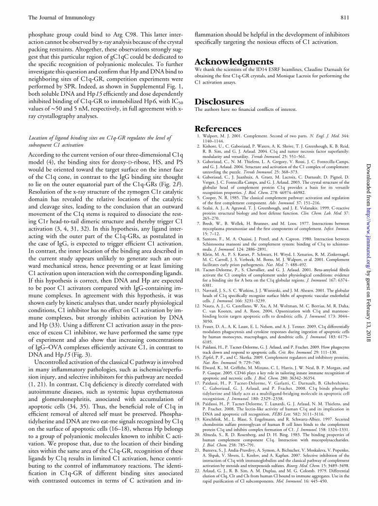
FIGURE 2. Structural characterization of the gC1qC binding area. A, Overall structure of C1q-GR, highlighting the location of the binding area observed in
gC1qC (magenta). Labels indicate the color code of the C1q subunits. The yellow sphere represents the Ca2+ ion. A deoxy-D-ribose molecule is shown in sticks in
its binding orientation. B, Zoom on the HS binding site. Sticks correspond to the HS moiety fitted into the additional electron density map observed after
soaking, whereas thin lines represent the adjacent mobile parts (black, yellow, and red). The C1q binding site residues Y155, W190, and K129 are shown in
sticks. Polar interactions and their corresponding distance are shown with dotted red lines. The position of the nearby mobile Arg111 is also shown. C, Zoom on
the deoxy-D-ribose binding site, using the same color code as in B. The 2Fo-Fc electron density map corresponding to the deoxy-D-ribose molecule is shown in
blue. D, Crystal packing environment in the C1q–HS complex. Solvent channels are labeled C1 and C2. Stars indicate the approximate position of the HS, PS,
and deoxyribose binding sites. The color code for gC1q is the same as in A, except for the A162–A165 loop (orange) and the B160–B169 loop (dark blue). E, Athymidine nucleotide was modeled from the bound deoxy-D-ribose, and its additional interactions are shown. F, Model of the whole C1q molecule highlighting
the position of the known C1q-GR binding sites.
FIGURE 3. C1 activation by IC, Hp, and DNA. C1 (0.25 mM) was
incubated for 90 min at 37˚C in the presence of C1 inhibitor (1 mM) and
increasing concentrations of IC, Hp, or DNA. C1 activation was monitored
as described under Materials and Methods. DNA, calf thymus DNA; Hp,
Hp15; and IC, IgG–OVA complexes.
810 CUTTING EDGE: C1q BINDS DEOXYRIBOSE AND HEPARAN SULFATE
by guest on February 13, 2018http://w
ww
.jimm
unol.org/D
ownloaded from
phosphate group could bind to Arg C98. This latter inter-action cannot be observed by x-ray analysis because of the crystalpacking restraints. Altogether, these observations strongly sug-gest that this particular region of gC1qC could be dedicated tothe specific recognition of polyanionic molecules. To furtherinvestigate this question and confirm thatHp andDNAbind toneighboring sites of C1q-GR, competition experiments wereperformed by SPR. Indeed, as shown in Supplemental Fig. 1,both soluble DNA and Hp15 efficiently and dose dependentlyinhibited binding of C1q-GR to immobilized Hp6, with IC50
values of ∼50 and 5 nM, respectively, in full agreement with x-ray crystallography analyses.
Location of ligand binding sites on C1q-GR regulates the level ofsubsequent C1 activation
According to the current version of our three-dimensional C1qmodel (4), the binding sites for deoxy-D-ribose, HS, and PSwould be oriented toward the target surface on the inner faceof the C1q cone, in contrast to the IgG binding site thoughtto lie on the outer equatorial part of the C1q-GRs (Fig. 2F).Resolution of the x-ray structure of the zymogen C1r catalyticdomain has revealed the relative locations of the catalyticand cleavage sites, leading to the conclusion that an outwardmovement of the C1q stems is required to dissociate the rest-ing C1r head-to-tail dimeric structure and thereby trigger C1activation (3, 4, 31, 32). In this hypothesis, any ligand inter-acting with the outer part of the C1q-GRs, as postulated inthe case of IgG, is expected to trigger efficient C1 activation.In contrast, the inner location of the binding area described inthe current study appears unlikely to generate such an out-ward mechanical stress, hence preventing or at least limitingC1 activation upon interaction with the corresponding ligands.If this hypothesis is correct, then DNA and Hp are expectedto be poor C1 activators compared with IgG-containing im-mune complexes. In agreement with this hypothesis, it wasshown early by kinetic analyses that, under nearly physiologicalconditions, C1 inhibitor has no effect on C1 activation by im-mune complexes, but strongly inhibits activation by DNAand Hp (33). Using a different C1 activation assay in the pres-ence of excess C1 inhibitor, we have performed the same typeof experiment and also show that increasing concentrationsof IgG–OVA complexes efficiently activate C1, in contrast toDNA and Hp15 (Fig. 3).Uncontrolled activation of the classical C pathway is involved
in many inflammatory pathologies, such as ischemia/reperfu-sion injury, and selective inhibitors for this pathway are needed(1, 21). In contrast, C1q deficiency is directly correlated withautoimmune diseases, such as systemic lupus erythematosusand glomerulonephritis, associated with accumulation ofapoptotic cells (34, 35). Thus, the beneficial role of C1q inefficient removal of altered self must be preserved. Phospha-tidylserine and DNA are two eat-me signals recognized by C1qon the surface of apoptotic cells (16–18), whereas Hp belongsto a group of polyanionic molecules known to inhibit C acti-vation. We propose that, due to the location of their bindingsites within the same area of the C1q-GR, recognition of theseligands by C1q results in limited C1 activation, hence contri-buting to the control of inflammatory reactions. The identi-fication in C1q-GR of different binding sites associatedwith contrasted outcomes in terms of C activation and in-
flammation should be helpful in the development of inhibitorsspecifically targeting the noxious effects of C1 activation.
AcknowledgmentsWe thank the scientists of the ID14 ESRF beamlines, Claudine Darnault for
obtaining the first C1q-GR crystals, and Monique Lacroix for performing the
C1 activation assays.
DisclosuresThe authors have no financial conflicts of interest.
References1. Walport, M. J. 2001. Complement. Second of two parts. N. Engl. J. Med. 344:
1140–1144.2. Kishore, U., C. Gaboriaud, P. Waters, A. K. Shrive, T. J. Greenhough, K. B. Reid,
R. B. Sim, and G. J. Arlaud. 2004. C1q and tumor necrosis factor superfamily:modularity and versatility. Trends Immunol. 25: 551–561.
3. Gaboriaud, C., N. M. Thielens, L. A. Gregory, V. Rossi, J. C. Fontecilla-Camps,and G. J. Arlaud. 2004. Structure and activation of the C1 complex of complement:unraveling the puzzle. Trends Immunol. 25: 368–373.
4. Gaboriaud, C., J. Juanhuix, A. Gruez, M. Lacroix, C. Darnault, D. Pignol, D.Verger, J. C. Fontecilla-Camps, and G. J. Arlaud. 2003. The crystal structure of theglobular head of complement protein C1q provides a basis for its versatilerecognition properties. J. Biol. Chem. 278: 46974–46982.
5. Cooper, N. R. 1985. The classical complement pathway: activation and regulationof the first complement component. Adv. Immunol. 37: 151–216.
6. Szalai, A. J., A. Agrawal, T. J. Greenhough, and J. E. Volanakis. 1999. C-reactiveprotein: structural biology and host defense function. Clin. Chem. Lab. Med. 37:265–270.
7. Bredt, W., B. Wellek, H. Brunner, and M. Loos. 1977. Interactions betweenmycoplasma pneumoniae and the first components of complement. Infect. Immun.15: 7–12.
8. Santoro, F., M. A. Ouaissi, J. Pestel, and A. Capron. 1980. Interaction betweenSchistosoma mansoni and the complement system: binding of C1q to schistoso-mula. J. Immunol. 124: 2886–2891.
9. Klein, M. A., P. S. Kaeser, P. Schwarz, H. Weyd, I. Xenarios, R. M. Zinkernagel,M. C. Carroll, J. S. Verbeek, M. Botto, M. J. Walport, et al. 2001. Complementfacilitates early prion pathogenesis. Nat. Med. 7: 488–492.
10. Tacnet-Delorme, P., S. Chevallier, and G. J. Arlaud. 2001. Beta-amyloid fibrilsactivate the C1 complex of complement under physiological conditions: evidencefor a binding site for A beta on the C1q globular regions. J. Immunol. 167: 6374–6381.
11. Navratil, J. S., S. C. Watkins, J. J. Wisnieski, and J. M. Ahearn. 2001. The globularheads of C1q specifically recognize surface blebs of apoptotic vascular endothelialcells. J. Immunol. 166: 3231–3239.
12. Nauta, A. J., G. Castellano, W. Xu, A. M. Woltman, M. C. Borrias, M. R. Daha,C. van Kooten, and A. Roos. 2004. Opsonization with C1q and mannose-binding lectin targets apoptotic cells to dendritic cells. J. Immunol. 173: 3044–3050.
13. Fraser, D. A., A. K. Laust, E. L. Nelson, and A. J. Tenner. 2009. C1q differentiallymodulates phagocytosis and cytokine responses during ingestion of apoptotic cellsby human monocytes, macrophages, and dendritic cells. J. Immunol. 183: 6175–6185.
14. Paidassi, H., P. Tacnet-Delorme, G. J. Arlaud, and P. Frachet. 2009. How phagocytestrack down and respond to apoptotic cells. Crit. Rev. Immunol. 29: 111–130.
15. Zipfel, P. F., and C. Skerka. 2009. Complement regulators and inhibitory proteins.Nat. Rev. Immunol. 9: 729–740.
16. Elward, K., M. Griffiths, M. Mizuno, C. L. Harris, J. W. Neal, B. P. Morgan, andP. Gasque. 2005. CD46 plays a key role in tailoring innate immune recognition ofapoptotic and necrotic cells. J. Biol. Chem. 280: 36342–36354.
17. Paıdassi, H., P. Tacnet-Delorme, V. Garlatti, C. Darnault, B. Ghebrehiwet,C. Gaboriaud, G. J. Arlaud, and P. Frachet. 2008. C1q binds phospha-tidylserine and likely acts as a multiligand-bridging molecule in apoptotic cellrecognition. J. Immunol. 180: 2329–2338.
18. Paıdassi, H., P. Tacnet-Delorme, T. Lunardi, G. J. Arlaud, N. M. Thielens, andP. Frachet. 2008. The lectin-like activity of human C1q and its implication inDNA and apoptotic cell recognition. FEBS Lett. 582: 3111–3116.
19. Kirschfink, M., L. Blase, S. Engelmann, and R. Schwartz-Albiez. 1997. Secretedchondroitin sulfate proteoglycan of human B cell lines binds to the complementprotein C1q and inhibits complex formation of C1. J. Immunol. 158: 1324–1331.
20. Almeda, S., R. D. Rosenberg, and D. H. Bing. 1983. The binding properties ofhuman complement component C1q. Interaction with mucopolysaccharides.J. Biol. Chem. 258: 785–791.
21. Bureeva, S., J. Andia-Pravdivy, A. Symon, A. Bichucher, V. Moskaleva, V. Popenko,A. Shpak, V. Shvets, L. Kozlov, and A. Kaplun. 2007. Selective inhibition of theinteraction of C1q with immunoglobulins and the classical pathway of complementactivation by steroids and triterpenoids sulfates. Bioorg. Med. Chem. 15: 3489–3498.
22. Arlaud, G. J., R. B. Sim, A. M. Duplaa, and M. G. Colomb. 1979. Differentialelution of Clq, Clr and Cls from human Cl bound to immune aggregates. Use in therapid purification of Cl subcomponents. Mol. Immunol. 16: 445–450.
The Journal of Immunology 811
by guest on February 13, 2018http://w
ww
.jimm
unol.org/D
ownloaded from
23. Vives, R. R., R. Sadir, A. Imberty, A. Rencurosi, and H. Lortat-Jacob. 2002. A
kinetics and modeling study of RANTES(9-68) binding to heparin reveals a mech-
anism of cooperative oligomerization. Biochemistry 41: 14779–14789.24. Vanhaverbeke, C., J. P. Simorre, R. Sadir, P. Gans, and H. Lortat-Jacob. 2004. NMR
characterization of the interaction between the C-terminal domain of interferon-g and
heparin-derived oligosaccharides. Biochem. J. 384: 93–99.25. Kabsch, W. 1993. Automatic processing of rotation diffraction data from crystals of
initially unknown symmetry and cell constants. J. Appl. Crystallogr. 26: 795–800.26. McCoy, A. J., R. W. Grosse-Kunstleve, P. D. Adams, M. D. Winn, L. C. Storoni,
and R. J. Read. 2007. Phaser crystallographic software. J. Appl. Crystallogr. 40: 658–674.
27. Murshudov, G. N., A. A. Vagin, and E. J. Dodson. 1997. Refinement of macro-
molecular structures by the maximum-likehood method. Acta Crystallogr. D. Biol.Crystallogr. 53: 240–255.
28. Emsley, P., and K. Cowtan. 2004. Coot: model-building tools for molecular
graphics. Acta Crystallogr. D. Biol. Crystallogr. 60: 2126–2132.29. Collaborative Computational Project, Number 4. 1994. The CCP4 suite: programs
for protein crystallography. Acta Crystallogr. D. Biol. Crystallogr. 50: 760–763.
30. Tan, K., M. Duquette, J. H. Liu, K. Shanmugasundaram, A. Joachimiak, J. T.Gallagher, A. C. Rigby, J. H. Wang, and J. Lawler. 2008. Heparin-induced cis- andtrans-dimerization modes of the thrombospondin-1 N-terminal domain. J. Biol.Chem. 283: 3932–3941.
31. Budayova-Spano, M., M. Lacroix, N. M. Thielens, G. J. Arlaud, J. C. Fontecilla-Camps, and C. Gaboriaud. 2002. The crystal structure of the zymogen catalyticdomain of complement protease C1r reveals that a disruptive mechanical stress isrequired to trigger activation of the C1 complex. EMBO J. 21: 231–239.
32. Bally, I., V. Rossi, T. Lunardi, N. M. Thielens, C. Gaboriaud, and G. J. Arlaud.2009. Identification of the C1q-binding Sites of Human C1r and C1s: a refinedthree-dimensional model of the C1 complex of complement. J. Biol. Chem. 284:19340–19348.
33. Ziccardi, R. J. 1982. A new role for C-1-inhibitor in homeostasis: control of activationof the first component of human complement. J. Immunol. 128: 2505–2508.
34. Bowness, P., K. A. Davies, P. J. Norsworthy, P. Athanassiou, J. Taylor-Wiedeman,L. K. Borysiewicz, P. A. R. Meyer, and M. J. Walport. 1994. Hereditary C1qdeficiency and systemic lupus erythematosus. QJM 87: 455–464.
35. Botto, M. 1998. C1q knock-out mice for the study of complement deficiency inautoimmune disease. Exp. Clin. Immunogenet. 15: 231–234.
812 CUTTING EDGE: C1q BINDS DEOXYRIBOSE AND HEPARAN SULFATE
by guest on February 13, 2018http://w
ww
.jimm
unol.org/D
ownloaded from