Embed Size (px)
Citation preview
69
Chapter 2.4
Testicular microlithiasis in boys and young men with congenital or acquired undescended (ascending) testis
J GoedeWWM HackLM van der Voort-DoedensFH PierikLHJ LooijengaK Sijstermans
J Urol 2010; 183:1539-1543

70
Chapter 2.4 Testicular microlithiasis in boys and young men with undescended testis
Abstract
Purpose
We assessed the prevalence of testicular microlithiasis by ultrasound in boys and young men with congenital or acquired undescended (ascending) testis.
Materials and methods
During follow-up for testicular growth patients with congenital or acquired undescended (ascending) testis were also screened by ultrasound for testicular microlithiasis, which was defined as echogenic foci without shadowing within the testis parenchyma. Classical microlithiasis was defined as 5 or more echogenic foci in either or both testes and limited microlithiasis as fewer than 5 foci.
Results
We performed 181 ultrasounds in 181 patients (199 congenital undescended testes) with a mean age of 12.6 years (range 2.6 to 28.6) and 636 ultrasounds in 320 patients (350 acquired undescended/ascending testes) with a mean age of 12.4 years (4.1 to 24.1). Age in both groups was equivalent. Median followup was 1.34 years (range 0 to 3.2). Testicular microlithiasis was found in 14 patients (2.8%), of whom 11 (2.2%) displayed classical testicular microlithiasis and 3 (0.6%) exhibited limited testicular microlithiasis. Among this 14 patients 5 had congenital undescended testis, which demonstrated classical microlithiasis. Of these 5 patients 4 had chromosomal deformities. The remaining 9 patients had acquired undescended (ascending) testis, which exhibited classical microlithiasis in 6 instances and limited microlithiasis in 3.
Conclusion
The prevalence of testicular microlithiasis in patients with undescended testis is 2.8%. There is no difference in the prevalence of testicular microlithiasis between congenital and acquired undescended (ascending) testes.

71
Testicular microlithiasis in boys and young men with undescended testis
Introduction
Undescended testis is categorised as either congenital or acquired (ascending).1 Patients with undescended testis have a 4 to 7-fold increased risk of malignant testicular germ cell tumor.2 No definite factor triggering this tumor in later life has been identified thus far. Testicular germ cell tumor is seen more frequently in boys with abnormal external genitalia, an intra-abdominal testis or an abnormal karyotype.3 Furthermore, boys treated for undescended testis at 13 years or older are at increased risk for this tumor.4
Testicular microlithiasis is considered to be an additional predisposing factor for TGCT. Testicular microlithiasis is characterised by multiple echogenic foci less than 3 mm without shadowing within the testis parenchyma. There are some indications that testicular microlithiasis is more frequently seen in UDT.5 Ultrasound following orchiopexy revealed a 10% microlithiasis rate in the operated testis. Moreover, approximately 10% of these patients had TGCT during followup.6 Hence, testicular microlithiasis may be an additional risk factor for TGCT in patients with UDT, necessitating tighter surveillance of these patients. The aim of this prospective study was to determine by ultrasound the prevalence of testicular microlithiasis in boys and young men with congenital or acquired undescended (ascending) testis.
Materials and methods
Patients with congenital undescended testis
We recently analysed 181 boys and young men with 199 congenital UDTs for long-term testicular growth after orchiopexy. US assessment of both testes was performed for testicular growth. During this examination the presence of TM was also considered. All patients underwent 1 scrotal US.
Patients with acquired undescended (ascending) testis
Since the mid-1990s we have annually assessed patients with acquired undescended (ascending) testis. In accordance with the Dutch Consensus spontaneous descent at puberty is awaited and orchiopexy is performed during puberty only in cases of non-descent.7 Preliminary results of these assessments have already been published.8 To date, 636 ultrasounds have been performed in 320 patients with 350 acquired undescended (ascending) testes.
Definitions
A retractile testis was defined as a normally developed testis that could be manipulated into a low scrotal position, where it remained until the cremasteric reflex was elicited.
72
Chapter 2.4 Testicular microlithiasis in boys and young men with undescended testis
There was no shortening of cord structures. UDT was defined as a testis that could not be manipulated into a stable scrotal position and further tension on cord structures was not possible due to shortening. UDT can be high scrotal, inguinal or nonpalpable. Congenital UDT was defined as a testis that had not been documented in a scrotal position since birth, whereas an acquired undescended (ascending) testis had previously been descended. TM involves multiple hyperechogenic, nonshadowing small foci within the testis parenchyma. Classical TM was considered present if 5 or more echogenic foci 1 to 3 mm in diameter were present in either or both testes, while limited TM was defined as fewer than 5 foci. TM was differentiated as diffusely scattered throughout the parenchyma or segmented.
Study protocol
A questionnaire was used that included the items medical problems, medication, major surgery, previous groin surgery (other than orchiopexy), gestational age, birth weight. Because it is possible that the rate of TM varies in different ethnical groups, we also assessed patients ethnicity. Adenotonsillectomy and/or middle ear drainage was not considered. Physical examination of the left testis was performed first, followed by the right testis with the patient in the supine and crossed-legged position. Testes were categorised as descended, retractile or undescended. Scrotal US was performed by 1 physician (KS) in the 181 patients with 199 congenital UDTs and by 1 physician (WH) in the 320 patients with 350 acquired undescended (ascending) testis. All ultrasounds were performed with the same equipment (12 MHz linear array transducer and Falco Auto Image, Falco Software Co, Tomsk, Russia). To assess the presence or absence of TM, we placed the scanner on the scrotum while recording transverse and longitudinal images of both testes. We performed US on both testes (undescended and normally descended), documenting the testicle involved and the number of microliths detected. Color Doppler ultrasound of the testis was not performed.
Followup after TM diagnosis
If TM was diagnosed, a full physical examination was performed. Additionally US was repeated to confirm the diagnosis. Brothers of the boys diagnosed with TM were also requested for scrotal ultrasound because TM has been reported in relatives of patients with TGCT.9
Statistics
Chi-square tests were conducted to compare birth weight, gestational age, groin surgery, chromosomal abnormalities, medication use and ethnicity between patients
73
Testicular microlithiasis in boys and young men with undescended testis
with and without TM. Chi-square tests were also used to compare the frequency of TM in congenital and acquired undescended (ascending) testes. Fisher’s exact test was used to compare the frequency in different age groups. A p value of less than 0.05 was considered significant.
Results
Numbers of patients and ultrasounds
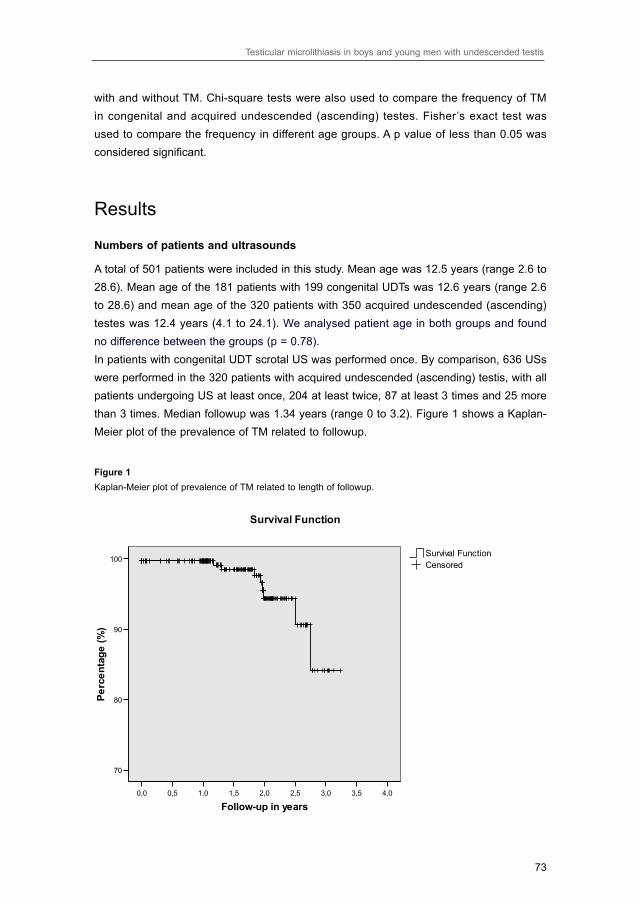
A total of 501 patients were included in this study. Mean age was 12.5 years (range 2.6 to 28.6). Mean age of the 181 patients with 199 congenital UDTs was 12.6 years (range 2.6 to 28.6) and mean age of the 320 patients with 350 acquired undescended (ascending) testes was 12.4 years (4.1 to 24.1). We analysed patient age in both groups and found no difference between the groups (p = 0.78). In patients with congenital UDT scrotal US was performed once. By comparison, 636 USs were performed in the 320 patients with acquired undescended (ascending) testis, with all patients undergoing US at least once, 204 at least twice, 87 at least 3 times and 25 more than 3 times. Median followup was 1.34 years (range 0 to 3.2). Figure 1 shows a Kaplan-Meier plot of the prevalence of TM related to followup.
Figure 1Kaplan-Meier plot of prevalence of TM related to length of followup.
74
Chapter 2.4 Testicular microlithiasis in boys and young men with undescended testis
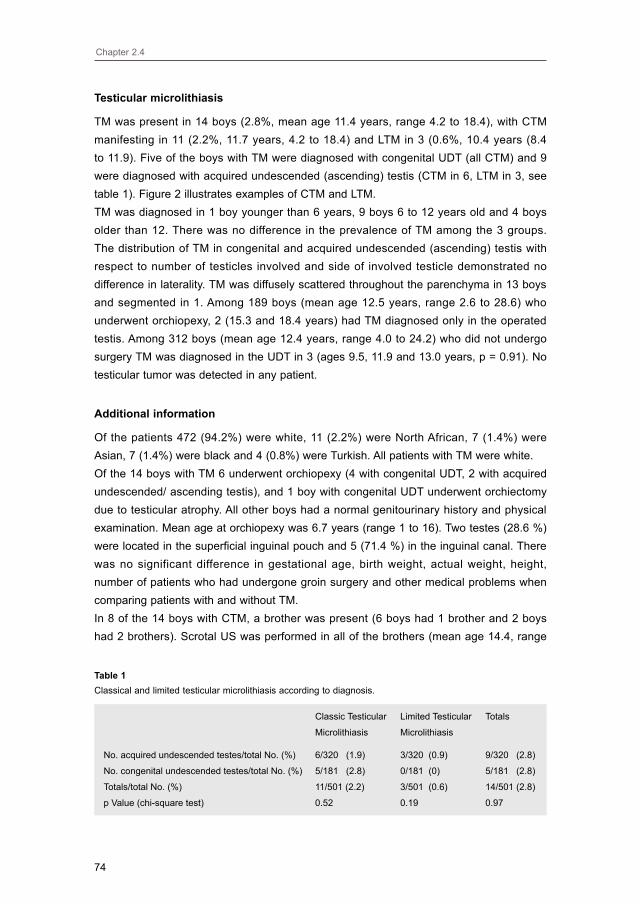
Classic Testicular Limited Testicular Totals
Microlithiasis Microlithiasis
No. acquired undescended testes/total No. (%) 6/320 (1.9) 3/320 (0.9) 9/320 (2.8)
No. congenital undescended testes/total No. (%) 5/181 (2.8) 0/181 (0) 5/181 (2.8)
Totals/total No. (%) 11/501 (2.2) 3/501 (0.6) 14/501 (2.8)
p Value (chi-square test) 0.52 0.19 0.97
Testicular microlithiasis
TM was present in 14 boys (2.8%, mean age 11.4 years, range 4.2 to 18.4), with CTM manifesting in 11 (2.2%, 11.7 years, 4.2 to 18.4) and LTM in 3 (0.6%, 10.4 years (8.4 to 11.9). Five of the boys with TM were diagnosed with congenital UDT (all CTM) and 9 were diagnosed with acquired undescended (ascending) testis (CTM in 6, LTM in 3, see table 1). Figure 2 illustrates examples of CTM and LTM.TM was diagnosed in 1 boy younger than 6 years, 9 boys 6 to 12 years old and 4 boys older than 12. There was no difference in the prevalence of TM among the 3 groups. The distribution of TM in congenital and acquired undescended (ascending) testis with respect to number of testicles involved and side of involved testicle demonstrated no difference in laterality. TM was diffusely scattered throughout the parenchyma in 13 boys and segmented in 1. Among 189 boys (mean age 12.5 years, range 2.6 to 28.6) who underwent orchiopexy, 2 (15.3 and 18.4 years) had TM diagnosed only in the operated testis. Among 312 boys (mean age 12.4 years, range 4.0 to 24.2) who did not undergo surgery TM was diagnosed in the UDT in 3 (ages 9.5, 11.9 and 13.0 years, p = 0.91). No testicular tumor was detected in any patient.
Additional information
Of the patients 472 (94.2%) were white, 11 (2.2%) were North African, 7 (1.4%) were Asian, 7 (1.4%) were black and 4 (0.8%) were Turkish. All patients with TM were white.Of the 14 boys with TM 6 underwent orchiopexy (4 with congenital UDT, 2 with acquired undescended/ ascending testis), and 1 boy with congenital UDT underwent orchiectomy due to testicular atrophy. All other boys had a normal genitourinary history and physical examination. Mean age at orchiopexy was 6.7 years (range 1 to 16). Two testes (28.6 %) were located in the superficial inguinal pouch and 5 (71.4 %) in the inguinal canal. There was no significant difference in gestational age, birth weight, actual weight, height, number of patients who had undergone groin surgery and other medical problems when comparing patients with and without TM.In 8 of the 14 boys with CTM, a brother was present (6 boys had 1 brother and 2 boys had 2 brothers). Scrotal US was performed in all of the brothers (mean age 14.4, range
Table 1Classical and limited testicular microlithiasis according to diagnosis.
75
Testicular microlithiasis in boys and young men with undescended testis
10.3 to 17.9). In 1 brother (age 11.8 years) TM was also diagnosed, the third brother in this family had no TM.Of the boys with TM 1 with congenital UDT was diagnosed with Klinefelter syndrome. Three other boys with congenital UDT were diagnosed with Down syndrome, 18q deletion syndrome or pseudoxanthoma elasticum. Boys with TM had no medical history or use of medications, except 1 who was taking methylphenidate for attention deficit hyperactivity disorder with hyperactivity.
Discussion
This study represents one of the first large prospective ultrasound series examining the prevalence of TM in boys and young men with congenital or acquired undescended (ascending) testis. The overall prevalence of TM was 2.8%, CTM 2.2% and of LTM 0.6%. In this series no difference was found in TM rates between congenital (2.8%) and acquired undescended (ascending) testis (2.8%). These rates are slightly lower than the rate of 4.2% in asymptomatic boys.10 The exact pathogenesis of TM remains unclear, although the formation of microliths is thought to be the result of degeneration of cells in the seminiferious tubules in which hydroxyapatite deposits develop surrounded by concentric layers.11 TM can be segmented or scattered diffusely throughout the testicular parenchyma. Segmented TM might be more prevalent in patients with TGCT and, therefore, must be differentiated from diffuse TM.12 The age of onset is largely unknown but TM seems to originate at a later age.10 In this study we found no difference in prevalence between different age groups. Median age in our series (11.8 years) was not lower than in the study of asymptomatic boys (8.6). If TM manifests later in life, a lower median age in our series might have caused the lower TM rate.Recently TM has been reported in relatives of TGCT patients.9 In this study, TM was identified in 1 younger brother of a boy with TM. Figure 3 illustrates examples of TM as found in these brothers. Moreover, TM has recently been reported in siblings of patients with fragile X syndrome and pseudoxanthoma elasticum.13,14 The TM rate in asymptomatic boys is 4.2%10, compared to 2.0% in symptomatic boys.15 In symptomatic adults the rate varies from 0.6% to 9%, while 2 studies in asymptomatic populations revealed rates of 2.4 to 5.6%.16,17 Patients were considered symptomatic when US was performed for scrotal complaints and asymptomatic in the absence of scrotal complaints.There are several studies on the prevalence of TM in patients with UDT. All of these series except 1 suggest higher TM rates than our study. This discrepancy may be due to the fact that the TM rate in asymptomatic boys has only recently been reported and
76
Chapter 2.4 Testicular microlithiasis in boys and young men with undescended testis
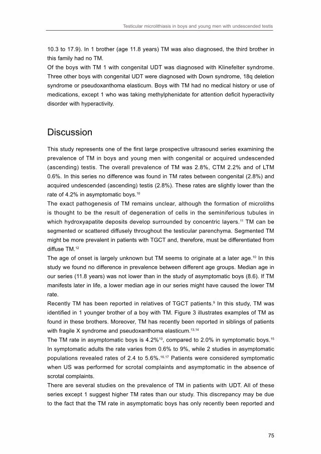
Figure 2A: Longitudinal plane of classical testicular microlithiasis (arrows) of the left testicle in 11-year-old boy with bilateral acquired undescended (ascending) testis and bilateral testicular microlithiasis.B: Longitudinal plane of limited testicular microlithiasis (arrows) of the right testicle in 9-year-old boy with right acquired undescended (ascending) testis and right testicular microlithiasis.
that different methods were used to determine TM. Furthermore, prior series were mostly retrospective and were performed in small groups. Also, TM could still develop at a later age in some of the patients in this study, although TM was not more prevalent in older patients.The lifetime risk of TGCT in the general population is approximately 0.3 to 0.7%, whereas in men with a history of UDT this risk is 3 to 5%.4 Orchiopexy before age 13 seems to decrease the risk of TGCT. In 2 of our 6 patients (33.3%) with TM who underwent orchiopexy TM was present in the operated testis and not in the controlateral testis. In these boys TM may be the result of testicular infarction due to trauma caused by orchiopexy. It remains uncertain whether the risk of TGCT in boys with acquired undescended
A B
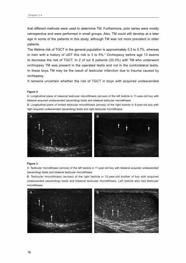
Figure 3A: Testicular microlithiasis (arrows) of the left testicle in 11-year-old boy with bilateral acquired undescended (ascending) testis and bilateral testicular microlithiasis.B: Testicular microlithiasis (arrows) of the right testicle in 12-year-old brother of boy with acquired undescended (ascending) testis and bilateral testicular microlithiasis. Left testicle also had testicular microlithiasis.
A B
77
Testicular microlithiasis in boys and young men with undescended testis
(ascending) testis is the same as for those with congenital UDT. TM has been associated with TGCT and with the precursor carcinoma in situ. Recently testis biopsy has been suggested in cases where there is a combination of UDT and TM, since both are risk factors for developing TGCT.18 TM has also been suggested to be an indication of manifestation of a TGCT susceptibility allele.19 Based on the literature, patients with UDT and TM are at increased risk for TGCT. This risk may be further increased if TM occurs bilaterally. However, this series do not support this hypothesis. There is some controversy regarding exact followup procedure and duration in patients with TM. At the least some degree of followup seems to be indicated.Hopefully it will soon be possible to identify carcinoma in situ with immunohistochemical analysis using OCT3/4 in semen. Eventually this method will enable identification of adolescents with a history of UDT who are prone to TGCT. It will then become clear whether TM is indeed an additional risk factor, as some studies suggest. There are limitations to this study. Unlike patients with acquired undescended (ascending) testis, patients with congenital UDT were only examined once. In 204 (63.8 %) of the 320 patients with acquired undescended (ascending) testis 2 or more USs were performed, and in 3 (21.4 %) of the 14 patients with TM, the condition was not diagnosed during the first US. Therefore, the rate of TM in patients with congenital UDT may have been greater if US had been performed more than once during a longer followup. The prevalence of TM would have been 2.2% (congenital 2.8%, acquired/ ascending 1.9%) if only the first US had been used to determine the TM rate. Overall length of followup in patients with acquired undescended (ascending) testis is too short (maximum 3.2 years) to determine whether the prevalence of TM increases with time. Another limitation is that 4 of the 14 patients (28.6%, all with congenital UDT) suffered from chromosomal deformities. There was a statistically significant difference (p < 0.001) in the number of patients with chromosomal deformities between the groups with and without TM. It may be speculated that TM is due to the chromosomal abnormality itself, which further decreases the TM rate in congenital UDT. Especially in the patient with Down syndrome the chromosomal deformity might be more important than the TM in the development of TGCT, since patients with Down syndrome have a higher risk of TGCT.20
Conclusion
In patients with congenital or acquired undescended (ascending) testis the overall TM rate is 2.8% (CTM 2.2%, LTM 0.6%). There is no significant difference between TM rates in congenital and acquired undescended (ascending) testis. The TM rate is comparable with the prevalence of TM in asymptomatic patients.
78
Chapter 2.4 Testicular microlithiasis in boys and young men with undescended testis
References
1. Barthold, J. S. and Gonzalez, R.: The epidemiology of congenital cryptorchidism, testicular ascent and orchiopexy. J Urol, 170: 2396, 2003
2. Moller, H., Cortes, D., Engholm, G. et al.: Risk of testicular cancer with cryptorchidism and with testicular biopsy: cohort study. BMJ, 317: 729, 1998
3. Cortes, D., Thorup, J. M., and Visfeldt, J.: Cryptorchidism: aspects of fertility and neoplasms. A study including data of 1,335 consecutive boys who underwent testicular biopsy simultaneously with surgery for cryptorchidism. Horm Res, 55: 21, 2001
4. Pettersson, A., Richiardi, L., Nordenskjold, A. et al.: Age at surgery for undescended testis and risk of testicular cancer. N Engl J Med, 356: 1835, 2007
5. Patel, R. P., Kolon, T. F., Huff, D. S. et al.: Testicular microlithiasis and antisperm antibodies following testicular biopsy in boys with cryptorchidism. J Urol, 174: 2008, 2005
6. Husmann, D. A.: Cryptorchidism and its relationship to testicular neoplasia and microlithiasis. Urology, 66: 424, 2005
7. de Muinck Keizer-Schrama SM: [Consensus on management of the undescended testis]. Ned Tijdschr Geneeskd, 131: 1817, 1987
8. Sijstermans, K., Hack, W. W., van der Voort-Doedens LM et al.: Puberty stage and spontaneous descent of acquired undescended testis: implications for therapy? Int J Androl, 29: 597, 2006
9. Chia, V. M., Li, Y., Goldin, L. R. et al.: Risk of cancer in first- and second-degree relatives of testicular germ cell tumor cases and controls. Int J Cancer, 124: 952, 2009
10. Goede, J., Hack, W. W., van der Voort-Doedens LM et al.: Prevalence of testicular microlithiasis in asymptomatic males 0 to 19 years old. J Urol, 182: 1516, 2009
11. Vegni-Talluri, M., Bigliardi, E., Vanni, M. G. et al.: Testicular microliths: their origin and structure. J Urol, 124: 105, 1980
12. Backus, M. L., Mack, L. A., Middleton, W. D. et al.: Testicular microlithiasis: imaging appearances and pathologic correlation. Radiology, 192: 781, 1994
13. Pourbagher, M. A., Pourbagher, A., and Erol, I.: Fragile x syndrome associated with testicular microlithiasis in siblings. J Ultrasound Med, 24: 1727, 2005
14. Goede, J., Hack, W. W., Sijstermans, K. et al.: Testicular microlithiasis in a 2-year-old boy with pseudoxanthoma elasticum. J Ultrasound Med, 27: 1503, 2008
15. Miller, F. N., Rosairo, S., Clarke, J. L. et al.: Testicular calcification and microlithiasis: association with primary intra-testicular malignancy in 3,477 patients. Eur Radiol, 17: 363, 2007
16. Peterson, A. C., Bauman, J. M., Light, D. E. et al.: The prevalence of testicular microlithiasis in an asymptomatic population of men 18 to 35 years old. J Urol, 166: 2061, 2001
79
Testicular microlithiasis in boys and young men with undescended testis
17. Serter, S., Gumus, B., Unlu, M. et al.: Prevalence of testicular microlithiasis in an asymptomatic population. Scand J Urol Nephrol, 40: 212, 2006
18. van Casteren, N. J., Looijenga, L. H., and Dohle, G. R.: Testicular microlithiasis and carcinoma in situ overview and proposed clinical guideline. Int J Androl, 2008
19. Kanetsky, P. A., Mitra, N., Vardhanabhuti, S. et al.: Common variation in KITLG and at 5q31.3 predisposes to testicular germ cell cancer. Nat Genet, 41: 811, 2009
20. Hill, D. A., Gridley, G., Cnattingius, S. et al.: Mortality and cancer incidence among individuals with Down syndrome. Arch Intern Med, 163: 705, 2003

80