Embed Size (px)
Citation preview
Volume 30 Issue 2 Article 4
2020
Curve of Spee: Development and Orthodontic Leveling Curve of Spee: Development and Orthodontic Leveling
Yu-Hsin Lee School of Dentistry, College of Dental Medicine, Kaohsiung Medical University; Department of Orthodontics, Dental Clinics, Kaohsiung Medical University Memorial Hospital
Yu-Chuan Tseng School of Dentistry, College of Dental Medicine, Kaohsiung Medical University; Department of Orthodontics, Dental Clinics, Kaohsiung Medical University Memorial Hospital, [email protected]
Follow this and additional works at: https://www.tjo.org.tw/tjo
Part of the Orthodontics and Orthodontology Commons
Recommended Citation Recommended Citation Lee, Yu-Hsin and Tseng, Yu-Chuan (2020) "Curve of Spee: Development and Orthodontic Leveling," Taiwanese Journal of Orthodontics: Vol. 30 : Iss. 2 , Article 4. DOI: 10.30036/TJO.201806_30(2).0004 Available at: https://www.tjo.org.tw/tjo/vol30/iss2/4
This Review Article is brought to you for free and open access by Taiwanese Journal of Orthodontics. It has been accepted for inclusion in Taiwanese Journal of Orthodontics by an authorized editor of Taiwanese Journal of Orthodontics.
98 Taiwanese Journal of Orthodontics. 2018, Vol. 30. No. 2
Review Article
Curve of Spee (CoS) is characterized as human occlusal curvature viewed in the sagittal plane. This
naturally occurring phenomenon has clinical importance in orthodontics and prosthodontic dentistry. The
purpose of this article is to examine the formation of the CoS regarding of when, how, or why the CoS develops.
The mechanism of orthodontic leveling the CoS will be discussed. (Taiwanese Journal of Orthodontics. 30(2): 98-103, 2018)
Keywords: Curve of Spee; craniofacial morphology; orthodontic treatment.
curve of spee: developmenT and orThodonTic levelinG
Yu-Hsin Lee,1,2
Yu-Chuan Tseng1,2
1School of Dentistry, College of Dental Medicine, Kaohsiung Medical University
2 Department of Orthodontics, Dental Clinics, Kaohsiung Medical University Memorial Hospital
Received: March 14, 2018 Revised: May 18, 2018 Accepted: May 22, 2018Reprints and correspondence to: Dr. Yu-Chuan Tseng, 100 Shih-Chuan 1st Road, Kaohsiung 80708, Taiwan Tel: +886-7-3121101 ext 7009 Fax: +886-7-3221510 E-mail: [email protected]
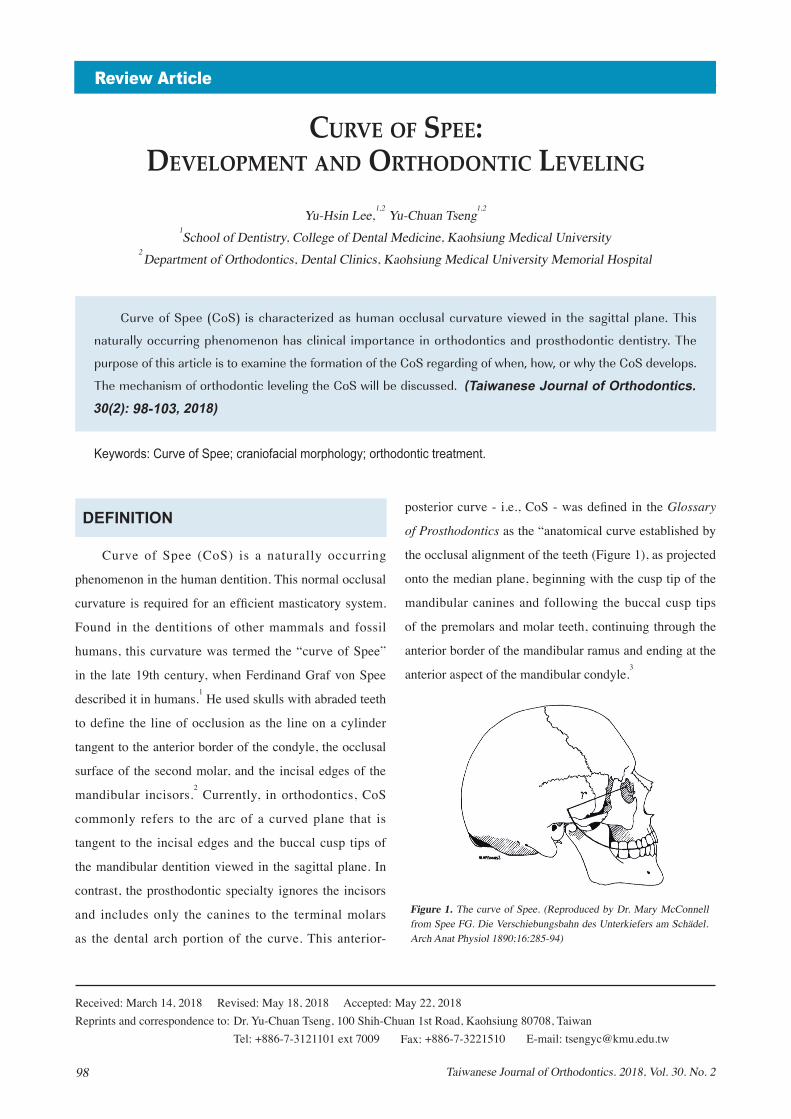
posterior curve - i.e., CoS - was defined in the Glossary
of Prosthodontics as the “anatomical curve established by
the occlusal alignment of the teeth (Figure 1), as projected
onto the median plane, beginning with the cusp tip of the
mandibular canines and following the buccal cusp tips
of the premolars and molar teeth, continuing through the
anterior border of the mandibular ramus and ending at the
anterior aspect of the mandibular condyle.3
DEFINITION
Curve of Spee (CoS) is a naturally occurring
phenomenon in the human dentition. This normal occlusal
curvature is required for an efficient masticatory system.
Found in the dentitions of other mammals and fossil
humans, this curvature was termed the “curve of Spee”
in the late 19th century, when Ferdinand Graf von Spee
described it in humans.1 He used skulls with abraded teeth
to define the line of occlusion as the line on a cylinder
tangent to the anterior border of the condyle, the occlusal
surface of the second molar, and the incisal edges of the
mandibular incisors.2 Currently, in orthodontics, CoS
commonly refers to the arc of a curved plane that is
tangent to the incisal edges and the buccal cusp tips of
the mandibular dentition viewed in the sagittal plane. In
contrast, the prosthodontic specialty ignores the incisors
and includes only the canines to the terminal molars
as the dental arch portion of the curve. This anterior-
Figure 1. The curve of Spee. (Reproduced by Dr. Mary McConnell from Spee FG. Die Verschiebungsbahn des Unterkiefers am Schädel. Arch Anat Physiol 1890;16:285-94)
99Taiwanese Journal of Orthodontics. 2018, Vol. 30. No. 2
Curve of Spee
DEVELOPMENT
The description of how the CoS develops is limited
in the literature. The developmental and functional
significance of the CoS had been investigated by several
researchers. Some have suggested that its development
probably results from a combination of factors including
growth of orofacial structures, eruption of teeth, and
development of the neuromuscular system.4 It has been
suggested that the mandibular sagittal and vertical position
relative to the cranium is related to the CoS, which is
presented in various forms in mammals. In humans, an
increased CoS is often observed in brachycephalic facial
pattern and is associated with short mandibular bodies.5,6
In a mechanical sense, the presence of a CoS may make
it possible for a dentition to resist the forces of occlusion
during mastication. Although several theories have been
proposed to explain the presence of a CoS in natural
dentitions, its role during normal mandibular function has
been questioned. It has been proposed that an imbalance
between the anterior and the posterior components of
occlusal force can cause the lower incisors to over-erupt,
the premolars to infra-erupt, and the lower molars to be
mesially inclined.7,8
In describing the 6 characteristics
of normal occlusion, Andrews9 found that the CoS in
subjects with good occlusion ranged from flat to mild,
noting that optimal static intercuspation occurred when
the occlusal plane was relatively flat. The author proposed
that flattening the occlusal plane should be a treatment
goal in orthodontics. This concept, especially applied
to the correction of deep overbite, has been supported
by others and produces variable results with regard to
maintaining a level curve after treatment.
DECIDUOUS TEETH VERSUS PERMANENT TEETH
It has been suggested that the deciduous dentition has
a CoS ranging from flat to mild, whereas the adult CoS is
more pronounced. On average, eruption of the mandibular
permanent first molars precedes that of the maxillary
permanent first molars by 1 to 2 months, and eruption of
the mandibular permanent central incisors precedes that of
the maxillary permanent central incisors by 12 months.10
Moreover, the mean age for the eruption of the mandibular
second molars is 6 months before the maxillary second
molars. This difference in tooth eruption timing could
permit unopposed mandibular permanent first molar and
incisor eruptions beyond the established mandibular
occlusal plane.10 The greatest increase in CoS occurs in
the early mixed dentition as a result of permanent first
molar and central incisor eruption. It maintains this depth
until it increases to maximum depth with eruption of the
permanent second molars, and then remains relatively
stable into late adolescence and early adulthood.11-13
These
findings also support those of Carter and McNamara11
and
Bishara et al,12
that once established in adolescence, the
CoS appears to be relatively stable.
GENDER-RELATED VARIATION OF COS
Marshall et al reported that there are no significant
differences in maximum depth of CoS between either the
right and left sides of the mandibular arch, or according
to gender.10
Carter and McNamara reported that no
difference in depth of the CoS between male and female
subjects when it was measured from dental casts taken
before treatment.11
AGE-RELATED CHANGES IN COS
The present review found that no evidence supported
the inter-relationship between CoS and age. It was
reported that the depth of the CoS was stable throughout
adolescence and into adulthood.14,15,16
The effect of age
on CoS has been investigated in growing subjects up to
young adulthood. However, to our knowledge, there are
no available data regarding variation of CoS with age
100 Taiwanese Journal of Orthodontics. 2018, Vol. 30. No. 2
Lee YH, Tseng YC
in adults. With increasing of age, it can be expected that
homogeneous dental wear may occur as a consequence
of masticatory function. It is interesting to note that the
CoS could be observed in most primates and mammals,
regardless of the extent of tooth wearing.17,18
Tooth wear
does not appear to affect the forward tilt of the posterior
teeth in the sagittal plane. Although the cusps become flat
gradually, the CoS is maintained throughout the years.19
DENTAL RELATIONS RELATED TO COS
In the study of Veli et al, depth of the CoS was
greatest in the Class II division 1 malocclusion group,
followed by Class II division 2, Class I, and Class III
malocclusion.20
Shannon and Nanda reported that patients
with a Class II malocclusion had a significantly deeper
CoS than those with a Class I malocclusion.21
A similar
study examined 100 untreated patients and also reported
that the CoS was the most severe in the Class II division
2 patients, followed by Class II Division 1, Class I, and
Class III patients.22
The CoS is deeper in individuals with Class II
malocclusions with consensus. Some Class III patients
may have deep Cos, it could be assumed that Class III
patients with a negative increased overjet exhibit greater
eruption of the anterior teeth because of uncovering.20
Thus, CoS has a direct relationship with the anterio-
posterior position of maxilla and mandible.
INFLUENCE OF CRANIOFACIAL MORPHOLOGY ON THE COS
According to a previous study, the influence
of craniofacial morphology on the CoS has been
systematically investigated in very few studies and with
conflicting findings. Kumar and Tamizharasi reported
that the CoS was influenced only to a minor extent by
craniofacial morphology.1 On the other hand, Farella et
al reported that condylar height (relative to the occlusal
plane) and anteroposterior position of the mandible
(relative to the cranial base) are associated with CoS
depth.19
Based on the results reported by Southard10
and Farella et al,19
in patients with small mandibles, the
mandibular permanent incisors would keep erupting (i.e.,
CoS increasing) until they contact the palate.
Farella et al also reported that the CoS was also
influenced by the position of the mandible with respect to
the anterior cranial base (i.e., SNB angle), regardless of
the reciprocal position of the lower and upper jaw in the
sagittal plane (ANB angle).19
The more the mandible was
forward positioning, the less marked the Cos. Orthlieb
found that the radius of the CoS was shorter in Class III
than in Class II malocclusions.23
LEVELING OF THE COS
Correction of exaggerated CoS can achieve by
extrusion of molars, intrusion of incisors, or combination
of the above tooth movement. Before starting this
orthodontic treatment, a precise diagnosis must be
decided which mechanics to be used. If patients have
short lower facial height (orthodivergent to hypodivergent
facial pattern), excessive CoS, moderate-to-minimal
incisor display and deep overbite, it is suitable for molar
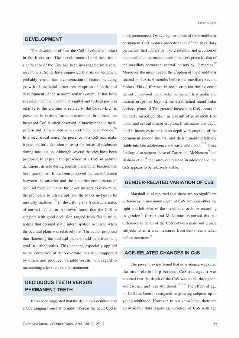
extrusion. One millimeter of molar extrusion could
reduce the incisor overlap by 1.5 to 2.5 mm. Use the
continuous archwires to level the Cos is common and
simple,24
such as the use of 0.016 × 0.022" reverse curve
NiTi wire (Figure 2). However, flaring of the incisors is
the disadvantage of continuous archwires. Second, wire
bending with increasing step bends in an archwire, can
also level the CoS. A third method was the use of a lever
arm (Figure 3) and intrusive arch, which could not only
extrude the posterior teeth but also intrude the anterior
teeth. The CoS can also be levelled by using a bite plate,
which enables the posterior teeth to erupt. Although all
of these methods can help to correct an exaggerated CoS,
the stability of molar extrusion is questionable in non-
growing patients.
101Taiwanese Journal of Orthodontics. 2018, Vol. 30. No. 2
Curve of Spee
Intrusion of the upper and/or lower incisors is a
desirable method to level CoS in many adolescent and
adult patients. It is particularly indicated for patients with
an excessive mandibular plane angle, hyperdivergent
facial pattern, excessive incision-stomion distance, and
a large interlabial gap. The methods for intruding the
anterior teeth include modification of bracket bonding
position, a utility arch without tie back, intrusion bend,
lever arm, and 0.016 × 0.022" reverse NiTi wire. If more
intrusion is required, temporary anchorage devices could
be provided as an absolute anchorage. A major risk factor
associated with intrusion of anterior teeth is external
apical root resorption. In some cases, intruding anterior
teeth may even worsen in patients with periodontal
compromised condition.
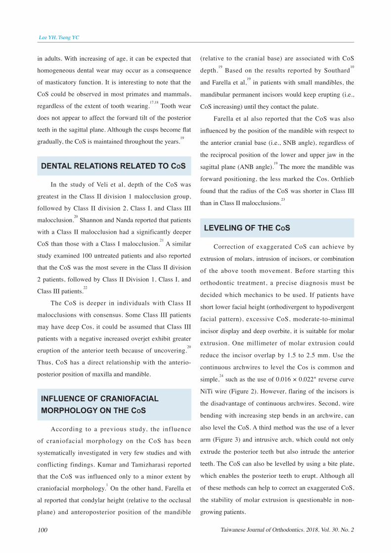
Figure 2. The lower anterior teeth were intruded by 0.016X0.022" reverse NiTi wire for leveling the Cos.
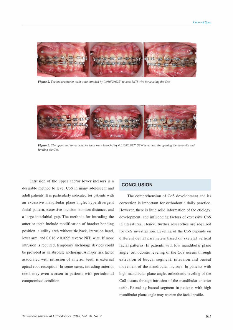
Figure 3. The upper and lower anterior teeth were intruded by 0.016X0.022" SSW lever arm for opening the deep bite and leveling the Cos.
CONCLUSION
The comprehension of CoS development and its
correction is important for orthodontic daily practice.
However, there is little solid information of the etiology,
development, and influencing factors of excessive CoS
in literatures. Hence, further researches are required
for CoS investigation. Leveling of the CoS depends on
different dental parameters based on skeletal vertical
facial patterns. In patients with low mandibular plane
angle, orthodontic leveling of the CoS occurs through
extrusion of buccal segment, intrusion and buccal
movement of the mandibular incisors. In patients with
high mandibular plane angle, orthodontic leveling of the
CoS occurs through intrusion of the mandibular anterior
teeth. Extruding buccal segment in patients with high
mandibular plane angle may worsen the facial profile.
102 Taiwanese Journal of Orthodontics. 2018, Vol. 30. No. 2
Lee YH, Tseng YC
13. Veli I, Ozturk MA, Uysal T. Development of the
curve of Spee in Class II subdivision malocclusion: a
longitudinal study. Eur J Orthod. 2015;37(4):412–7.
14. Ferrario VF, Sforza C, Poggio CE, Serrao G,
Colombo A. Three-dimensional dental arch curvature
in human adolescents and adults. Am J Orthod.
1999;115(4):401–5.
15. Enlow DH. Normal variations in facial form and the
anatomic basis for malocclusion. In: Enlow, DH, ed.
Facial growth, 3rd edn. Philadelphia: WB Saunders,
1990:193–221.
16. Osborn JW, Francis LJ. The position of the dentition
in the mandible and its possible relation to orthodontic
abnormalities. Am J Orthod. 1989;96(4):327–32.
17. Osborn JW. Relationship between the mandibular
condyle and the occlusal plane during hominid
evolution: some of its effects on jaw mechanics. Am J
Phys Anthropol. 1987;73(2):193–207.
18. Baragar FA, Osborn JW. Efficiency as a predictor of
human jaw design in the sagittal plane. J Biomech.
1987;20(5):447–57.
19. Farella M, Michelotti A, van Eijden TM, Martina
R. The curve of Spee and craniofacial morphology:
a multiple regression analysis. Eur J Oral Sci.
2002;110(4):277–81.
20. Veli I, Ozturk MA, Uysal T. Curve of Spee and
its relationship to vertical eruption of teeth among
different malocclusion groups. Am J Orthod Dentofacial
Orthop. 2015;147(3):305–12.
21. Shannon KR, Nanda RS. Changes in the curve of
Spee with treatment and at 2 years posttreatment. Am
J Orthod Dentofacial Orthop. 2004;125(5):589–96.
22. Ahmed I, Nazir R, Gul-e-Erum, Ahsan T. Influence
of malocclusion on the depth of curve of Spee. J Pak
Med Assoc. 2011;61(11):1056–9.
23. Orthlieb JD. The curve of Spee: understanding the
sagittal organization of mandibular teeth. Cranio.
1997;15(4):333-340.
REFERENCES
1. Kumar KP and Tamizharasi S. Significance of curve
of Spee: An orthodontic review. J Pharm Bioallied
Sci. 2012;4(Suppl 2):323–8
2. Spee FG, Biedenbach MA, Hotz M, Hitchcock HP.
The gliding path of the mandible along the skull. J Am
Dent Assoc. 1980;100:670–5.
3. Van Blarcom CW. Glossary of Prosthodontics terms,
8th ed. St. Louis: Mosby 2005.
4. Osborn JW. Orientation of the masseter muscle and
the curve of Spee in relation to crushing forces on
the molar teeth of primates. Am J Phys Anthropol.
1993;92:99–106.
5. Wylie WL. Overbite and vertical facial dimensions in
terms of muscle balance. Angle Orthod. 1994;19:13–7.
6. Björk A. Variability and age changes in overjet and
overbite. Am J Orthod. 1953;39:779–801.
7. Strang RHM and Thompson WM. A Textbook of
Orthodontia. Philadelphia: Lea and Fiebiger; 1958.
p.335-61
8. Gresham H. A manual of orthodontics. Christ Church:
N.M. Peryer; 1957.
9. Andrews LF. The six keys to normal occlusion. Am J
Orthod. 1972;62:296–309.
10. Marshall SD, Caspersen M, Hardinger RR, Franciscus
RG, Aquilino SA, Southard TE. Development of the
curve of Spee. Am J Orthod Dentofacial Orthop.
2008;134:344–52.
11. Carter GA, McNamara JA Jr. Longitudinal dental arch
changes in adults. Am J Orthod Dentofacial Orthop.
1998;114(1):88–99.
12. Bishara SE, Jakobsen JR, Treder JE, Stasi MJ.
Changes in the maxillary and mandibular tooth size-
arch length relationship from early adolescence to
early adulthood. A longitudinal study. Am J Orthod
Dentofacial Orthop. 1989;95(1):46–59.

103Taiwanese Journal of Orthodontics. 2018, Vol. 30. No. 2
Curve of Spee
24. Weiland FJ, Bantleon HP, Droschl H. Evaluation
of continuous arch and segmented arch leveling
techniques in adult patients – a clinical study. Am J
Orthod Dentofacial Orthop. 1996;110(6):647–52.