Embed Size (px)
Citation preview

1988
FYSI
KAALISEN FARMASIAN YHDISTY
S
SOCIETY OF PHYSICAL PHARM
ACY
**
Current trends in pharmaceutical preformulation and
manufacturing process design
POLYMORFI
2018

Sess
ion
ISe
ssio
n II
Ready for
21 CFR part 11X-RAY DIFFRACTION
SOLUTIONS FOR THE PHARMACEUTICAL INDUSTRY
Easily swith between a wide range of pharmaceutical applications in a matter of minutes:
• Structural investigation of NCE’s • High througput, high resolution polymorph,
salts, hydrates and solvates screening• In situ crystallization and scale-up studies• Investigation of amorphous and nano-
crystalline compounds• Stability and compatibility studies • Detectionandquantificationoflowamounts
of polymorphic impurities • Microstructure analysis of tablets by CT
Microstructure analysis of counterfeit and original tablet
Detection of polymorphic impurities
For more information, please contact:
Malvern Panalytical B.V., Branch FinlandLinnoitustie 4 BFIN-02600 ESPOOT +358 9 2212 580
8.25 8.35 8.45 8.55 8.65 8.75
372100
384400
396900
409600
0 %
0.5 %
0.25 %
0.15 %
422500
435600
Inte
nsi
ty [
a.u
.]
2theta [deg]
PN11452_1_col.indd 1 11/01/18 15:08
3
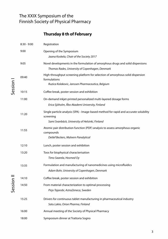
Thursday 8 th of February
8:30 - 9:00 Registration
9:00 Opening of the Symposium
Jaana Koskela, Chair of the Society 2017
9:05 Novel developments in the formulation of amorphous drugs and solid dispersions
Thomas Rades, University of Copenhagen, Denmark
09:40High-throughput screening platform for selection of amorphous solid dispersion
formulationsRuzica Kolakovic, Janssen Pharmaceutica, Belgium
10:15 Coffee break, poster session and exhibition
11:00 On-demand inkjet-printed personalized multi-layered dosage forms
Erica Sjöholm, Åbo Akademi University, Finland
11:20Single particle analysis (SPA) - Image-based method for rapid and accurate solubility
screening
Sami Svanbäck, University of Helsinki, Finland
11:55Atomic pair distribution function (PDF) analysis to assess amorphous organic
compounds
Detlef Beckers, Malvern Panalytical
12:10 Lunch, poster session and exhibition
13:20 Toos for biophysical characterisation
Timo Saarela, Hosmed Oy
13:35 Formulation and manufacturing of nanomedicines using microfluidics
Adam Bohr, University of Copenhagen, Denmark
14:10 Coffee break, poster session and exhibition
14:50 From material characterization to optimal processing
Pirjo Tajarobi, AstraZeneca, Sweden
15:25 Drivers for continuous tablet manufacturing in pharmaceutical industry
Satu Lakio, Orion Pharma, Finland
16:00 Annual meeting of the Society of Physical Pharmacy
18:00 Symposium dinner at Trattoria Sogno
The XXIX Symposium of the Finnish Society of Physical Pharmacy
Sess
ion
ISe
ssio
n II
Ready for X-RAY DIFFRACTION
SOLUTIONS FOR THE PHARMACEUTICAL INDUSTRY
Easily swith between a wide range of pharmaceutical applications in a matter of minutes:
• Structural investigation of NCE’s • High througput, high resolution polymorph,
salts, hydrates and solvates screening• In situ crystallization and scale-up studies• Investigation of amorphous and nano-
crystalline compounds• Stability and compatibility studies • Detectionandquantificationoflowamounts
of polymorphic impurities • Microstructure analysis of tablets by CT
Microstructure analysis of counterfeit and original tablet
Detection of polymorphic impurities
For more information, please contact:
Malvern Panalytical B.V., Branch FinlandLinnoitustie 4 BFIN-02600 ESPOOT +358 9 2212 580
8.25 8.35 8.45 8.55 8.65 8.75
372100
384400
396900
409600
0 %
0.5 %
0.25 %
0.15 %
422500
435600
Inte
nsi
ty [
a.u
.]
2theta [deg]
PN11452_1_col.indd 1 11/01/18 15:08

4
The cellZscope from nanoAnalytics is a device for measuring TEER; the transepithelial / -endothelial impedance of cell layers under physiological conditions. It is computer-controlled and allows automated, long-term monitoring experiments in the incubator with up to 24 different cell cultures simultaneously or 24 different exposures. It also measures CCL: Cell Layer Capacity.
Baker Ruskinn provide solutions for cell biology, stem cell and regenerative medicine, including accurate and stable anaerobic chambers, hypoxia workstations, and precise oxygen regulation in culture media. Sci-tive is the flexible Hypoxia system with many sizes and shapes. InvivO2 is single or dual chamber Hypoxia Workbench - latest technology.
Svanholm.com - Your Biomedical expert in Denmark, Sweden, Norway and Finland Phone: +45-7026 5811 - Mail: [email protected] - Web: www.svanholm.com
Nordic Biotech, Pharma and Biomedical Center Visit the Svanholm.com booth at IVBM2018

Friday 9 th of February
9:30 Opening of the second day
09:35 Implementation and examples of pharma OSD continuous manufacturing
Cait Boyd, GEA Group, Belgium
10:10 Significance and measurement of residence time distributions in
continuous manufacturing
Ossi Korhonen, University of Eastern Finland, Finland
11:45The influence of co-monomers applied in the SFPP course on the electrokinetic
potential of thermosensitive microparticles for controlled drug delivery
Witold Musial, Wroclaw Medical University, Poland
11:05 Coffee break, poster session and exhibition
11:50 A continuous and controlled pharmaceutical freeze-drying technology for unit doses
Thomas De Beer, Ghent University, Belgium
12:25 Biophysical tools for characterizing protein self-association
Petteri Heljo, Novo Nordisk, Denmark
13:00 Closing of the symposium
The cellZscope from nanoAnalytics is a device for measuring TEER; the transepithelial / -endothelial impedance of cell layers under physiological conditions. It is computer-controlled and allows automated, long-term monitoring experiments in the incubator with up to 24 different cell cultures simultaneously or 24 different exposures. It also measures CCL: Cell Layer Capacity.
Baker Ruskinn provide solutions for cell biology, stem cell and regenerative medicine, including accurate and stable anaerobic chambers, hypoxia workstations, and precise oxygen regulation in culture media. Sci-tive is the flexible Hypoxia system with many sizes and shapes. InvivO2 is single or dual chamber Hypoxia Workbench - latest technology.
Svanholm.com - Your Biomedical expert in Denmark, Sweden, Norway and Finland Phone: +45-7026 5811 - Mail: [email protected] - Web: www.svanholm.com
Nordic Biotech, Pharma and Biomedical Center Visit the Svanholm.com booth at IVBM2018

6
Table of Contents
Editorial 8
From the chairman 9
Lecture abstracts 11
Novel developments in the formulation of amorphous drugs and solid dispersions 12
High-throughput screening platform for selection of amorphous solid dispersion formulations 13
Single Particle Analysis (SPA) - Image-based Method for Rapid andAccurate Solubility Screening 15
Formulation and manufacturing of nanomedicines using microfluidics 16
From material characterization to optimal processing 17
Drivers for Continuous Tablet Manufacturing in Pharmaceutical Industry 18
Implementation and Examples of Pharma OSD Continuous Manufacturing 19
Significance and measurement of residence time distribution in continuous manufacturing 20
A Continuous and Controlled Pharmaceutical Freeze-Drying Technology for Unit Doses 22
Biophysical tools for characterizing protein self-association 23
On-demand Inkjet-printed Personalized Multilayered Dosage Forms 24
The influence of co-monomers applied in the SFPP course on the electrokinetic potential of thermosensitive microparticles for controlled drug delivery 26
Poster presentations 29
PCL-Gelatin nanofibers incorporating two antibiotics loaded MSNs: A potential wound dressing 30
Engineering of MSN-based structures to enhance anti-biofilm activity 31
Manufacturing Films for Individualized Dosing 32
Application of ultrasound-enhanced electrospinning for fabricating drug-loaded polymer nanocomposites 34
Bioadhesive nanofibrillated cellulose films for drug release 36
Influence of surface chemistry on adsorption and confinement of drug in porous silicon 38
Multimodal imaging of surface solid-state transformations 40
Preparation of ibuprofen-arginine salt by spray drying from water 41
Electrospinning of nanofibrillar cellulose reinforced nanofibers forpharmaceutical applications 42
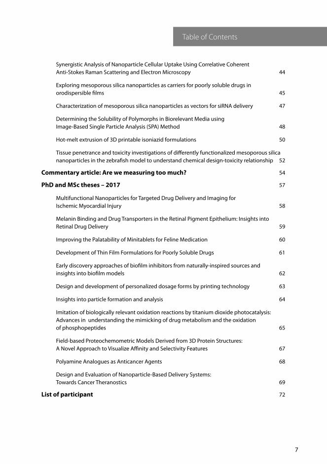
7
Table of Contents
Synergistic Analysis of Nanoparticle Cellular Uptake Using Correlative Coherent Anti-Stokes Raman Scattering and Electron Microscopy 44
Exploring mesoporous silica nanoparticles as carriers for poorly soluble drugs in orodispersible films 45
Characterization of mesoporous silica nanoparticles as vectors for siRNA delivery 47
Determining the Solubility of Polymorphs in Biorelevant Media using Image-Based Single Particle Analysis (SPA) Method 48
Hot-melt extrusion of 3D printable isoniazid formulations 50
Tissue penetrance and toxicity investigations of differently functionalized mesoporous silica nanoparticles in the zebrafish model to understand chemical design-toxicity relationship 52
Commentary article: Are we measuring too much? 54
PhD and MSc theses – 2017 57
Multifunctional Nanoparticles for Targeted Drug Delivery and Imaging for Ischemic Myocardial Injury 58
Melanin Binding and Drug Transporters in the Retinal Pigment Epithelium: Insights into Retinal Drug Delivery 59
Improving the Palatability of Minitablets for Feline Medication 60
Development of Thin Film Formulations for Poorly Soluble Drugs 61
Early discovery approaches of biofilm inhibitors from naturally-inspired sources and insights into biofilm models 62
Design and development of personalized dosage forms by printing technology 63
Insights into particle formation and analysis 64
Imitation of biologically relevant oxidation reactions by titanium dioxide photocatalysis: Advances in understanding the mimicking of drug metabolism and the oxidation of phosphopeptides 65
Field-based Proteochemometric Models Derived from 3D Protein Structures: A Novel Approach to Visualize Affinity and Selectivity Features 67
Polyamine Analogues as Anticancer Agents 68
Design and Evaluation of Nanoparticle-Based Delivery Systems: Towards Cancer Theranostics 69
List of participant 72

8
Editorial
For the 29th time the Finnish Society of Physical Pharmacy brings together professionals from academia, industry and regulatory authorities to interact and discuss about ongoing research and trends in physical pharmacy. Our aim was to arrange a valuable symposium with topics reflecting the current trends in the field. Thus, we are happy and honored to have gathered experts with knowledge about the recent advancements in preformulation and manufacturing process design.
This year’s commentary article “Are we measuring too much?” is written by Anne Juppo, Professor in Industrial Pharmacy at the University of Helsinki. In the article she shares the history about the development of the analytical technologies during the past decades. Furthermore, she also advises on to reflect on why we are doing certain measurements – are we doing the tests just because it is nice to know?
Wishing you interesting reading moments and a nice symposium!
Henrika Wickström
Editor in Chief - Polymorfi 2017
ISSN: 1236-4002
1458-5820 (pdf )
Painosalama Oy – Turku, Finland, 2018
Editor-in-chief: Henrika Wickström, Åbo Akademi University [email protected]
Proof readers: Jaana Koskela, University of Helsinki
Emma Hokkala, University of Helsinki
Publisher: Fysikaalisen farmasian yhdistys ry. www.fysikaalinenfarmasia.fi

9
From the chairman
The world is moving faster, and it has become ever more important to get new and better products to the market faster and more efficiently. Preformulation is the key for a new drug candidate to enter a successful drug development, and finding an appropriate formulation and process in a cost effective fashion is crucial. As these topics are both contemporary and interesting, the 29th Annual Symposium of the Finnish Society of Physical Pharmacy seeks to examine the latest trends in preformulation and manufacturing process design.
The purpose of the Society is to promote knowledge of physical pharmacy in Finland and internationally, and to enable broad sharing of knowledge and collaboration between aca-demic and industry professionals. Thus, the presentations and talks in the symposium aim to cover a broad range of inspiring topics from novel material sparing analytical methods in determining physiochemical properties of compounds to continuous manufacturing de-signs.
I would like to thank the participants, speakers, poster presenters and sponsors for your contributions. You and your active participation makes this symposium valuable. We hope that all participants will have inspiring and productive discussions during the symposium, and that together we make this a lively event.
Wishing you all a pleasant symposium!
Jaana Koskela
Chairman of the Finnish Society of Physical Pharmacy 2017
Jaana Koskela, Chair University of Helsinki
Emma Hokkala, Vice chair University of Helsinki
Flavia Fontana, Secretary University of Helsinki
Johan Nyman, Treasurer Åbo Akademi University
Henrika Wickström, Editor-in-Chief Åbo Akademi University
Anssi-Pekka Karttunen, Webmaster University of Eastern Finland
Rami Ojarinta University of Eastern Finland
Kirsi Salomäki Orion Pharma
Tiina Lipiäinen, Debuty member University of Helsinki
The Finnish Society of Physical Pharmacy
Members of the Board 2017–2018 and
the Organizing Committee of the XXIX Symposium
10

11
Lecture abstracts

12
Novel developments in the formulation of amorphous drugs and solid dispersions
Thomas Rades
Department of Pharmacy, University of Copenhagen, Denmark
Amorphous solid dosage forms are one of the most promising formulation strategies to overcome the limited bioavailability of many poorly water soluble drugs. However, the industrial application of amorphous sol-id dosage forms is still rather limited. This is likely to be due to an insufficient under-standing of the physico-chemical properties of amorphous solid dispersions including their physical stability, as well as due to the lack of predictive in vitro models. In this presentation, methods to predict amorphous drug stability and drug–polymer solubility will be discussed. We will then focus on alternatives to polymers in the formulation of amorphous solid dispersions and finally on the question how both the in vitro and in vivo performance of amorphous solid dis-persions is influenced by the use of amor-phous solid dispersions.
About the presenterSince March 2012 Professor Thomas Rades is the Re-search Chair in Pharmaceutical Design and Drug De-livery in the Department of Pharmacy, University of Copenhagen. Before that he has been the Chair in Phar-maceutical Sciences at the National School of Pharma-cy, University of Otago, New Zealand from 2003 – 2012.
In 1994 he received a PhD from the University of Braun-schweig, Germany for his work on thermotropic and lyotropic liquid crystalline drugs. After working as a Re-search Scientist in the Preclinical Development and For-mulation at F. Hoffmann-La Roche in Basel, Switzerland, he became a Senior Lecturer in Pharmaceutical Scienc-es at Otago in 1999 and since 2003 held the Chair in Pharmaceutical Sciences in Otago. Professor Rades has developed an international reputation for his research in the physical characterization of drugs and solid
dosage forms as well as in vaccine delivery using nanoparticulate systems (both polymeric and lipid based). He has published more than 350 papers in in-ternational peer reviewed journals as well as 18 book chapters, 11 patents and 3 books.
Professor Rades is an Editor of the Journal of Pharma-ceutical Sciences and the European Journal of Pharma-ceutics and Biopharmaceutics and an Associate Editor of the Journal of Pharmacy and Pharmacology.
He holds an honorary doctorate of Åbo Akademi Uni-versity, Finland, a visiting professorship at the Depart-ment of Medicine at the University of Adelaide, Aus-tralia and an honorary professorship at the University of Otago, New Zealand. He is an Eminent Fellow of the Academy of Pharmaceutical Sciences (UK), a Fellow of the New Zealand Institute of Chemistry and a mem-ber of the College of Fellows of the Controlled Release Society.
Professor Rades has successfully supervised more than 60 PhD students (completed) with currently supervising or co-supervising 12 PhD students. For his undergrad-uate and postgraduate teaching he was awarded the New Zealand Tertiary Teaching Excellence Award for Sustained Excellence (2005).
His research interests include The solid state of drugs and dosage forms, and Nanoparticles as delivery sys-tems for drugs and vaccines. Research in both areas aims to improve drug therapy through appropriate formulation and characterisation of medicines and to increase understanding of the physico-chemical prop-erties of drugs and medicines. It combines physical, chemical, and biological sciences and technology with analytics to optimally formulate drugs and vaccines for human and veterinary uses.
Current research projects include: Co-amorphous drug delivery systems, Solubility of drugs in solid polymers, lipid based drug delivery systems
Invited Lecture
13
High-throughput screening platform for selection of amorphous solid dispersion formulations
Ruzica Kolakovic
Janssen Pharmaceutica, Pharmaceutical Companies of Johnson & Johnson, Belgium
Vast majority of new active pharmaceutical ingredients (APIs) entering development pipeline from discovery possess insufficient water solubility and/or low dissolution rate. These cause challenges in achieving desired bioavailability.
Conversion of crystalline API to its amor-phous form is often taken as approach to achieve desired bioavailability based on higher solubility, faster dissolution rate, and enhanced oral bioavailability of amorphous APIs compared to their crystalline counter-parts. Over the last decade the production of stabilized amorphous drugs via amorphous solid dispersions ASDs has become an in-creasingly popular formulation strategy and the success of this strategy is reflected in the significant number of marketed amorphous products (2,4).
Variety of formulation variables can affect the development of ASDs with drug and polymer physicochemical properties, selec-tion of polymeric excipients, the definition of the drug polymer ratio, addition of sur-factants being some of them. A number of research laboratories have been developing their own screening methodologies, either based on experimental data or based on theoretical fundamentals with one common goal – choosing the right formulation com-position which will ensure both desired bio-availability and physical stability of ASD over intended shelf-life.
Selecting the optimal formulation can take a vast amount of time, can be extremely costly and importantly can require signif-icant amount of API which is not readily available in early phase of development. In order to reduce development time and cost, improve success rate and minimize API consumption, Janssen Pharmaceutica has developed automated high throughput screening platform for selection of ASD formulations. Such a screen is usually per-formed using 5-7 polymers and 2 additives and their combinations. 7 - 43 formulations can be tested simultaneously. The amor-phous solid dispersions are produced in 96 well-plate by low volume film casting meth-od resulting in amorphous films containing 25 - 75 μg of API. The produced films are evaluated for their dissolution rate by small scale two-stage dissolution method. Stabili-ty of the formulations is assessed as well by exposing films to stressing conditions (ele-vated temperature and/or relative humidity) and assessing crystallization propensity by cross-polarized imaging.
Use of automated high throughput screen-ing platform allows selection of 2-5 formu-lations for further scale-up with minimum time and API input (1 week and 1.5 g of API).
Invited Lecture

14
References
[1] Hywel D. Williams, Natalie L. Trev-askis, Susan A. Charman, Ravi M. Shanker, William N. Charman, Colin W. Pouton, and Christopher J. H. Porter. Pharmacol Rev 65:315–499, January 2013
[2] Smithey D, Gao P, Taylor L. Amor-phous solid dispersions: An enabling formulation technology for oral delivery of poorly water soluble drugs. AAPS Newsmagazine. 2013;16(1):11–4.
[3] R. Kolakovic, L. Wang, J. Herman. Annaual Formulation and Drug Deliv-ery Congress, London, UK, 17-18 May 2015.
About the presenterRuzica Kolakovic obtained her PhD in Pharmaceutical Technology from the University of Helsinki where she studied use of nanofibrillar cellulose as novel excipient in drug delivery. She worked as a postdoctoral scientist at Åbo Akdemi in Turku, Finland focusing on applica-tion of printing as a technique for production of drug delivery systems. In 2014 she joined Janssen Pharma-ceutica (Johnson & Johnson) in Belgium as scientist in preformulation group where she is responsible for developability assessment of compounds entering de-velopment pipeline. She is focused on defining the early drug development strategy through in depth un-derstanding of the compound properties and targeted product profile.
Bayer B5 Magazine 2017 dec add.indd 1 19/12/2017 10.20

15
One of the key physicochemical properties determining the developability of a drug is solubility. Currently in solubility measure-ment, there appears to exist a discrepancy between throughput, substance consump-tion and accuracy [1, 2].
While solubility is generally determined from bulk solutions after long incubation times, the diffusion layer dissolution rate model implies that solubility, as the rate limiting factor of dissolution, can be rapidly extracted from dissolution rate data of indi-vidual particles.
Automated image-based single-particle analysis (SPA) could substantially simplify data acquisition and processing, reducing sampling steps and operator contact with potentially hazardous substances. It could also allow for compounds of high value or scarce availability, such as in drug develop-ment, to be reliably analyzed. The non-spe-cific and non-interfering nature of image analysis further entails several potential ad-vantages.
We have applied the SPA method to measure the equilibrium solubility of drugs, excipi-ents, pesticides and sugars. The correlation with gold standard “shake-flask” equilibri-um solubility values is high. In addition, the SPA method has been used to measure the apparent solubility of salts, polymorphs and amorphous drug forms.
Examples form our work using the SPA method will be given in this presentation.
References
[1] Tetko, I. V., Poda, G.I., Ostermann, C., Mannhold, R., QSAR Comb. Sci. 28, (2009).
[2] Gardner, C.R., Walsh, C.T., Almarsson, O., Nat. Rev. Drug Discov. 3, (2004).
About the presenterSami Svanbäck received his PhD in 2016 from the University of Helsinki, for his work on developing im-age-based methods for drug analysis. Svanbäck has since continued the development as project manager of a Tekes (the Finnish Funding Agency for Innovation) funded project. The project has resulted in a new ana-lytical method currently being commercialized in col-laboration with the University of Helsinki innovation services.
Single Particle Analysis (SPA) - Image-based Method for Rapid and Accurate Solubility Screening
Sami Svanbäck
Division of Pharmaceutical Chemistry and Technology, Faculty of Pharmacy, University of Helsinki, Finland
Invited Lecture
Bayer B5 Magazine 2017 dec add.indd 1

16
Formulation and manufacturing of nanomedicines using microfluidics
Bohr Adam
Department of Pharmacy, University of Copenhagen, Denmark
Microfluidics has seen immense advances for analytical applications and is currently experiencing increasing popularity for the formulation and manufacturing of nano/micro systems intended for drug delivery. Nanopharmaceutical products have to some degree been limited by their production scalability and cost, and microfluidics-based systems may overcome these challenges and allow for the mass commercialization of nanopharmaceutical products.
This talk will focus on the application of microfluidic devices for preparing nanopar-ticulate pharmaceutical products, giving examples of different types of devices and nanoparticle systems, and the characteristics and performance of the resulting nanoparti-cles. Moreover, the application of 3D print-ing for fabrication of microfluidics devices and the application of in silico design and modelling for microfluidic systems will be will be discussed.
About the presenterAdam Bohr received his PhD degree in biomedical engineering in 2013 from University College London. From 2013-2015 he worked as a Postdoctoral research-er at the Institut Galien, University of Paris-Sud and from 2015-16 he worked as a Postdoctoral researcher at the Department of Pharmacy, University of Copen-hagen. In 2016 he was appointed as Assistant Professor at the Department of Pharmacy, University of Copenha-gen, Denmark. His research interests are in formulation and manufacturing of drug delivery systems, in par-ticular within the area of nanomedicine. This includes the formulation of polymeric drug-loaded nano- and microparticles for oral and pulmonary delivery for the treatment of infections and inflammation using novel polymers and drugs with low bioavailability. This also includes manufacturing of drug delivery systems and drug delivery devices using microfluidics and 3D print-ing. He has 31 publications in international, peer-re-viewed journals (h-index 12, determined from Google Scholar), as well as 3 patents.
Invited Lecture
17
From material characterization to optimal processing
Pirjo Tajarobi
Astra Zeneca, Gothenburg, Sweden
During processing of new drug compounds material related problems are quite com-mon. This has an immediate impact on the work within product development, as well as on manufacturability during scale-up and large scale production. A possible ear-ly identification of descriptors for material related issues would save both time and re-sources. This talk will focus on the materi-al characterization methods and predictive models typical for direct compression, roller compaction and wet granulation processes.
About the presenterPirjo Tajarobi (née Luukkonen) is a pharmacist and received her Ph.D. in the Pharmaceutical technology from the University of Helsinki (Finland) in 2001. She joined AstraZeneca Mölndal, Sweden 2001 and she has worked in the area of Product Development, PAT and Material Science. Currently Pirjo is working as an Asso-ciate Principal Scientist in the Drug Product Manufac-ture. She is an Associate Professor both at the Univer-sity of Helsinki and Åbo Akademi University in Finland. She is an expert in the areas of high-shear wet gran-ulation, continuous manufacturing, and powder flow characterization. She has published >20 peer-reviewed papers in these areas.
Invited Lecture

18
Invited Lecture
Continuous manufacturing (CM) can offer significant advantages over batch manufac-turing of solid dosage forms. These benefits include enhanced cost and quality aspects but CM also provides tools for accelerating product development which can decrease the time-to-market for new drug products. CM also offers more flexible manufacturing of tablets and thereby better enables per-sonalized medicine concept than traditional batch manufacturing does. Thus many phar-ma companies are taking steps towards CM. However, succeeding with CM requires not only significant investments on infrastruc-ture but also change in mindset, ways-of-working, new skillset and expertise.
About the presenterSatu Lakio is Senior Development Manager and Scien-tific Manager in Continuous Manufacturing at Orion Pharma, Finland. She got her PhD degree from Uni-versity of Helsinki, Finland and her PhD research cov-ered real time monitoring of pharmaceutical powder processing. Dr Lakio did her postdoctoral period at Monash University in Melbourne, Australia focusing on inhalation powder research. She has previously worked in several positions at academia and as an Associate Principal Scientist at AstraZeneca in Gothenburg, Swe-den. She also has long history of working in community pharmacies. Dr Lakio holds an adjunct professorship in University of Helsinki (Pharmaceutical technology) and she has supervised several under and post graduate as well as PhD students over the years. Her research cov-ers pharmaceutical powder characterization and pro-cessing all the way from inhalation powders to coated tablets including Process Analytical Technologies (PAT). Currently her main focus area is continuous tablet man-ufacturing.
Drivers for Continuous Tablet Manufacturing in Pharmaceutical Industry
Satu Lakio
Orion Pharma, Espoo, Finland

19
Implementation and Examples of Pharma OSD Continuous Manufacturing
Cait Boyd
GEA Group, Belgium
Traditionally the pharmaceutical industry has manufactured their drugs in batch pro-cesses. In recent years, there has been a shift to explore continuous manufacturing for oral solid dosage drugs. The drivers of this move to continuous manufacturing fall under two main areas: lean manufacturing to remove non-value added steps in a pro-cess and Six Sigma to use process under-standing to improve quality and efficiency.
Using the ConsiGma™ Continuous Tablet-ing Line, we explored how time to market was affected by moving to a continuous wet granulation process. It was found that com-mercial manufacturing could begin over 1 year earlier due to a reduction in scale up steps.
For a continuous direct compression pro-cess the ConsiGma™ CDC-50 was used to investigate the effect of lot to lot raw mate-rial variation on the system. After the raw material lot change the loss in weight feed-ers adjusted the feed factor to adapt to the change in raw material bulk density which in turn reduced variation in the final prod-uct.
About the presenterCait Boyd has a background in chemical engineering and started her career as a process engineer focusing on the scale up of emulsions. In 2012 she joined GEA and shortly after transitioned from engineering into sales of pharmaceutical powder processing equipment in the US market. In 2016 she moved to Belgium and is currently a sales support and business development manager focusing on pharma OSD continuous manu-facturing.
Invited Lecture

20
Significance and measurement of residence time distribution in continuous manufacturing
Ossi Korhonen
School of Pharmacy, University of Eastern Finland, Kuopio, Finland
The current manufacturing methods of drug products are very old-fashioned in compar-ison with to the other mass production in-dustries like food, chemical, pulp and paper, oil, mining, etc. Drug manufacturing based mostly on batch production. The main dis-advantage of batch production is that it is very time consuming due to the multiple quality checks between unit operations. Also the scale up of batch production is of-ten problematic from the R&D to produc-tion scale. Now Pharma industry has also started a transition from batch production to the continuous production. One of the driv-ing forces for the continuous production is many new guidelines (like QbD and PAT guidelines) from regulatory bodies which emphasizes that the quality of the drug product cannot be “tested in” but it has to be “build in” with the aid of PAT-tools. The main advantages of the continuous manu-facturing of drugs are a smaller footprint of the production site, smaller equipment, less material in process at the time, basically no scale up since the production scales up by time instead of the dimensions of production equipment as in the batch process, and pro-duction runs in the steady state most of the time whereas for example in batch mixing is a transient process. All of these enhance the efficiency and lower the costs of drug manu-facturing. Challenges are how to handle out of spec situations, how to feed accurately low content ingredients. Also some existing drug and excipient grades are very cohesive and thus difficult to feed accurately. Finally
how to synchronize the production rates of unit operation in series in order to avoid ma-terial overfill and/or run-out. For successful implementation of continuous manufactur-ing the key parameters are residence time distributions and mass hold-ups in each unit operations under different process and for-mulation conditions. Based on these param-eters, powder stream can be monitored and controlled along the serial unit operations. Also they enable the traceability of powder in out of spec situations.
The presentation will cover the introduc-tion of continuous PROMIS tableting line, the performance of unit operations, the res-idence time distributions and mass hold-ups of key unit operations and practical pros and cons observed during experiments.
Invited Lecture

21
About the presenterOssi Korhonen has graduated as a M.Sc. from the De-partment of Pharmaceutics, University of Kuopio, 1997. He received Ph.D. from the same University, 2004. Title of his thesis was ”Starch Acetate as a novel tablet ex-cipient for extended oral drug delivery”. He got the title of Docent in Pharmaceutical Technology, 2013. He has visited as a post-doc at the School of Pharmacy, Uni-versity of Connecticut, Storrs, USA in Mike Pikal’s Lab between 2004-2005. The topic of research conducted during the post-doc year was”The Stabilization of small molecule amorphous drugs”. Since then he has been in different teaching and project positions in the School of Pharmacy, University of Eastern Finland. He has su-pervised 4 Ph.D.s, 43 M.Sc., 22 Baccalautare thesis. Cur-rently he is supervising 5 Ph.D. students. He has pub-lished 43 papers and is the author of one book chapter. His main research topics are continuous manufacturing of tablets, Stabilization of small molecule, low soluble amorphous drug and development and optimization freeze-drying formulations and processes. He has an excellent knowledge of physical pharmacy, formulation and process optimization in solid dosage forms, Design of Experiment, multivariate analysis, spectroscopy, and thermal analysis.

22
A Continuous and Controlled Pharmaceutical Freeze-Drying Technology for Unit Doses
Thomas de Beer
Laboratory of pharmaceutical process analytical technology, Ghent University, Belgium
Driven by growing needs in the biophar-maceutical market and regulatory pressure, a continuous and controlled freeze-drying technology for unit doses to preserve bio-pharmaceuticals has been developed. Such continuous process allows a more efficient, cheaper, greener and controllable manufac-turing method compared to
traditional batch production systems, offer-ing competitive advantages and business opportunities.
Pharmaceutical freeze-drying (lyophiliza-tion) is a low-temperature drying process in which aqueous solutions of heat-labile bio-pharmaceuticals are converted into solids with sufficient stability for distribution and storage. Similar to all manufacturing pro-cesses of drug products (solids, semi-solids and liquids), conventional pharmaceutical freeze-drying is generally accomplished using batch processing that is considered time-consuming, costly, non-flexible and lacking robust quality control and real-time release.
Four major industrial drivers are demand-ing a more efficient and better controllable pharmaceutical freeze-drying technology for unit doses: cost-cutting, regulatory pres-sure, a fast growing biopharmaceutical mar-ket and an ageing population requiring more personalized medicines.
The continuous and controlled freeze-dry-ing technology, developed following the principle of model based design, offers clear advantages over current batch production such as cost reduction (up to 50%), track-and-trace product quality control, and a sig-nificant reduction of processing time (> 40 times faster, e.g. 1 hour instead of 5 days at a vial level), reduced need for clean room and a substantial sustainability gain.
About the presenterThomas De Beer graduated in pharmaceutical sciences in 2002 at the Ghent University in Belgium. He obtained his PhD at the same university in 2007. For his PhD research, he examined the suitability of Raman spec-troscopy as a Process Analytical Technology tool for pharmaceutical production processes. Within his PhD research period, he worked four months at University of Copenhagen in Denmark, Department of Pharma-ceutics and Analytical Chemistry (Prof. Jukka Rantanen). After his PhD, he was an FWO funded post-doctoral fellow at the Ghent University (2007-2010). Within his post-doc mandate, he worked 9 months at the Depart-ment of Pharmacy, Pharmaceutical Technology and Bio-pharmaceutics from the Ludwig-Maximilians-Universi-ty in Munich, Germany (Prof. Winter and Prof. Frieβ). In February 2010, he became professor in Process Analyt-ics & Technology at the Faculty of Pharmaceutical Sci-ences from the University of Ghent. His research goals include bringing innovation pharmaceutical production processes (freeze-drying, hot-melt extrusion, continu-ous from-powder-to-tablet processing etc.). More spe-cifically, Prof. De Beer contributes to the development of continuous manufacturing processes for drug prod-ucts such as solids, semi-solids, liquids and biologicals (continuous freeze-drying of unit doses).
Invited Lecture
23
Biophysical tools for characterizing protein self-association
Petteri Heljo
Structure, Biophysics & Formulation, Novo Nordisk A/S, Denmark
One possible form of instability for protein structured pharmaceuticals is aggregation, where protein molecules bind covalently or non-covalently together to form larger self-associated entities. The resulting ag-gregates tend to be biologically inactive, and they have also been suspected of being capable of eliciting the immune response, meaning that their formation is often con-sidered an undesidered instability event. However, controlled protein self-associa-tion can also be used to adjust the pharma-cokinetic profile of a protein product, when the availability of bioactive monomers in plasma is affected by the rate of dissociation of the self-associated state at the injection site. Furthermore, the physicochemical sta-bility of protein oligomers can be signifi-cantly better than that of monomers.
One of the aims of biophysical characteri-zation is to predict how protein monomers interact with one another. Protein self-asso-ciation tendency can be probed by trying to induce structural alterations within the mol-ecule, for example by heating or freezing the formulation, or subjecting it to mechan-ical stress. Individual amino acid residues can also be exchanged at key locations of the peptide backbone if these residues are expected to contribute to the oveall aggre-gation profile.
The complexity of the parameters affect-ing self-association makes it necessary to employ several complementary analytical techniques in order to understand the factors
which govern protein aggregation. Biophys-ical screening attempts to form an estimate of real-life shelf stability, and it is often used to weed out unstable molecules during lead candidate selection step of drug develop-ment.
This talk will give a brief introduction to protein self-association and unwanted ag-gregation, and describe some of the most common biophysical characterization tools used to study these. The aim is to provide an overview of the field rather than drilling deep into individual techniques and phe-nomena.
This is abstract template for abstract book Polymorfi in Symposium of Physical Phar-macy. Abstract can be 1-2 pages long.
Graphical work in abstract should fit to one column. If possible, send the image sepa-rately or at least make sure that word is not compressing images. Do not draw, edit or plot images in Word, images are not good enough for a print.
About the presenterPetteri Heljo finalized his PhD thesis with the title ”Comparison of disaccharides and polyalcohols as stabilizers in freeze-dried protein formulations” at the University of Helsinki in 2013 under the supervision of Prof. Anne Juppo. He spent the next two years as a postdoc at Roche (Switzerland) investigating peptide – excipient interactions, before moving to work as a Senior Research Scientist at Novo Nordisk (Denmark). His current research interests include high-throughput sterile formulation workflows and novel particle analy-sis techniques.
Invited Lecture

24
On-demand Inkjet-printed Personalized Multilayered Dosage Forms
Erica Sjöholm
Pharmaceutical Sciences Laboratory, Åbo Akademi University, Åbo, Finland
Producing flexible and tailored drug deliv-ery systems according to a patient’s need in-stead of manufacturing mass-oriented drug delivery systems in a ‘one size fits all’ man-ner, has gained interest in recent years [1]. Printing techniques have emerged as poten-tial technologies for on-demand manufac-turing. Inkjet printing with short manufac-turing time that enables precise deposition of an ink onto a given carrier matrix (sub-strate) is a potential new solution to produce tailored drug delivery systems at the point of care [2].
The main research focus of this work was to study the feasibility to deposit drug-loaded ink onto orodispersible polymer films by a 2D printing approach using inkjet printing to produce prednisolone containing multi-layered personalized dosage forms. An off-the-shelf office inkjet printer was selected as a quick and economical manufacturing method that potentially could be used in a hospital setting. A schematic illustration of the process steps can be seen in Figure 1.
In the 15% (w/w) HPC film solution, hy-droxypropyl cellulose (HPC) (Klucel–EXF PHARM, kindly provided by Ashland, Ger-many) served as film-forming agent and ethanol (Etax A, Altia, Finland) served as solvent.
Figure 1. A schematic illustration of the workflow for manufacturing printed multi-
layered dosage forms.
Figure 2. An Epson XP-760 desktop inkjet printer was used in this7 study for printing prednisolone containing multilayered dos-
age forms.
Oral Presentation

25
Dispersible films were cast to a wet thick-ness of 200 µm with a film-casting knife (Film Applicator MULTICATOR 411, Er-ichsen, Germany). Dried film sheets were used as printing substrates.
Prednisolone (≥ 99% Sigma-Aldrich, China) was used as a model drug. An ethanol-based ink solution consisting of 25 mg/ml pred-nisolone, 20% (v/v) distilled water and 1% (v/v) red food color was prepared. All water used was purified by Millipore SA-67120 from Millipore, Molsheim, France.
An Epson XP-760 desktop inkjet printer (Figure 2) was used to print the prepared drug ink onto the dried HPC orodispersible films according to the pattern set in the com-puter-aided design (CAD) (1x2 cm rectan-gle). A total of four different multilayered dosage forms were produced consisting of 5, 10, 15, and 20 printed drug layers with a fresh HPC layer cast after every fifth printed drug layer.
The multilayer dosage forms (n=3) were im-mersed in distilled water and drug content was measured using PerkinElmer Lambda 25 UV-Vis spectrophotometer (Singapore). As reference prednisolone ink was printed on copy paper.
The drug ink could successfully be printed on the casted HPC film and re-casted af-ter five printed drug layers. The reference drug amounts printed on copy paper were slightly higher than the obtained amounts for the multilayer dosage forms. For the 2 cm2 5, 10, 15, and 20 drug printed layers on copy paper the drug amounts achieved were 0.3, 0.7, 1.0, and 1.3 mg, whereas the drug amounts achieved for the printed 2 cm2 multilayered dosage forms were 0.3, 0.6, 0.9, and 1.2 mg respectively (Figure 3).
Figure 3. The Content of prednisolone printed on copy paper and in the multilay-
ered dosage forms (n=3).
Low standard deviation (± 0.0 mg for drug printed on copy paper and in the range of ± 0.00-0.05 mg for the multilayered dosage forms) and good linearity (R2 = 1 for drug ink printed on copy paper and R2 = 0.9984 for the multilayered dosage forms) indicates that aimed personalized doses can be pro-duced utilizing this method.
In conclusion, the desktop inkjet printer was successfully used to accurately imprint HPC films with prednisolone. Multiple printing layers could be used to increase the final drug content. In this study, one drug was investigated, however by printing various drugs between the casted film layers mul-tilayer multidrug formulations could be obtained. Overall, inkjet-printed orodispers-ible multi-layered dosage forms appear as a promising new approach to enable person-alized dosing.
This is an encore presentation which has been presented at AAPS 2017 Annual Meet-ing in San Diego, USA.
References
[1] R. Tutton, Soc Sci Med 75(10), 1721–1728 (2012).
[2] N. Sandler and M. Preis, Trends Phar-macol Sci. 37(12), 1070–1080 (2016).

26
Oral Presentation
The influence of co-monomers applied in the SFPP course on the electrokinetic potential of thermosensitive microparticles for controlled drug delivery
Witold Musial
Department of Physical Chemistry, Faculty of Pharmacy, Wroclaw Medical University, Poland
Various thermosensitive polymers, synthe-sized in the course of surfactant free precipi-tation polymerization, may be obtained, due to high number of possible co-monomers, cross-linkers, and solvents proposed for the process. One of the most interesting ther-mosensitive polymers with high applicative potential is poly-N-isopropylacrylamide, (pNIPA) and its variation may be applied as drug carriers and medical devices[1,2]. The microparticles may be characterized i.a. by electrokinetic potential, which is significant for the stability of colloidal dispersions.
The aim of the work was evaluation of the influence of variable co-monomers on the electrokinetic potential of polymeric micro-particles synthesized with the use of N-iso-propylacrylamide (NIPA).
The poly-NIPA particles were prepared in an aqueous solution using redox initiators, and in some cases accelerator. The compo-nents were dissolved in 900 mL of distilled water heated up to 70 °C in a four-necked round bottom flask equipped with magnetic paddle stirrer, reflux condenser, thermome-ter, and nitrogen gas inlet. The reaction ves-sel was kept at 70 °C for 6 h with respective mixing. After the reaction was terminated, the mixture was cooled to room tempera-ture. Obtained polymer solutions were pu-rified via dialysis for 15 days at room tem-
perature. The water was stirred and changed every 24 h. The purified products were fro-zen and freeze-dried by Chris Alpha 1–2 LD (Osterode am Harz, Germany) for 32 h.. Dynamic Light Scattering (DLS) Zeta Sizer Nano device of Malvern Instruments was used to measure values of zeta poten-tial in water dispersions of the synthesized polymers as a function of temperature. Zeta potential was measured in capillary cell type DTS-1070. Data of zeta potential were averaged from three replicates using Zeta-sizerNano Software, Version 7.11.
Figure 1. Electrokinetic potential of NI-PA-derivatives – polymeric microparticles synthesized with various co-monomers and
initiators at 18 °C.

27
The electrokinetic potential of synthesized microparticles was between -32,10 mV and 47,97 mV. The values depended on tempera-ture of the sample, and substrates used in the course of the synthesis (Figures 1 and 2). The values obtained for the samples at low-er temperature are smaller in absolute num-bers, comparing to the samples assessed at increased temperature. This indicates, that the differences in size, resulting from the temperature changes may influence the sta-bility of the colloid.
The further evaluation of the structures in comparison with the size changes may give pronounce information on the colloidal sta-bility of structures presumed for thermally triggered drug delivery.
References
[1] W. Musial, J. Pluta, J. Michalek, Acta Pol.Pharm. 409, (2015).
[2] M. Gasztych, A. Gola, J. Kobryń, W. Musiał, Molecules. 1473, (2016).

28

29
Poster presentations

30
PCL-Gelatin nanofibers incorporating two antibiotics loaded MSNs: A potential wound dressing
Z. Gounani1,2,3,4, M. A. Asadollahi*2, R. L. Meyer4,5 , J. M. Rosenholm3, A. Arpanaei*1
1Department of Industrial and Environmental Biotechnology, National Institute of Genetic Engineering and Biotechnology, Tehran, Iran.2Department of Biotechnology, Faculty of Advanced Sciences and Technologies, University of Isfahan, Isfahan, Iran.3Pharmaceutical Sciences Laboratory, Faculty of Science and Engineering, Åbo Akademi University, Turku 20520, Finland4Interdisciplinary Nanoscience Center (iNANO), Aarhus University, DK-8000 Aarhus C, Denmark.5Department of Bioscience, Aarhus University, DK-8000 Aarhus C, Denmark.
Wound healing is a challenging process for patients who suffer from severe burns or diabetic ulcers. The healing becomes more problematic when the wound becomes in-fected. Electrospun nanofibers are becom-ing highly considered for developing wound dressings due to high-surface area, mi-cro-porosity, and the ability of loading anti-microbial agents or required biomolecules. In this project, PCL (polycaprolactone) or PCL/Gelatin (70/30 or 50/50) were used in an acidic solvent system to develop electro-spun nanofiber mat. The carboxyl modified mesoporous silica nanoparticles (C-MSNs), unloaded C-MSNs or loaded with polymyx-in B and vancomycin (ABs^C-MSNs), were mixed with electrospinning solution in con-centrations of 1%, 2.5% and 5%. The size of the obtained nanofibers were between 122-138 nm. It was observed that the increasing concentration of gelatin or C-MSNs lead to
increased hydrophilicity and degradabili-ty of nanofibers. The antibacterial assays against P. aeruginosa and S. aureus showed high antibacterial efficiency of PCL/Gelatin nanofibers (70:30 and 50:50) incorporated with ABs^C-MSNs (2.5% and 5%). How-ever, PCL nanofibers incorporated with ABs^C-MSNs (2.5 % and 5%) showed mild antibacterial effects. Also low antibacterial effects were obtained for PCL or PCL/Gela-tin nanofibers incorporated with 1% ABs^C-MSNs. All types of the studied electrospun formulations showed high biocompatibility via MTT and hemolysis assays.
Figure 1. AAPTMS (N-(2-aminoeth-yl)-3-aminopropyltrimethoxy-silane) was used for amination of B-MSNs. Carboxyl modified mesoporous silica nanoparticles (C-MSNs) were obtained by adding sus-pension of N-MSNs in DMF to stirring
solution of succinic anhydride in DMF un-der N2 atmosphere. The Prepared C-MSNs were used to obtain dual antibiotics laoded C-MSNs (Abs^C-MSNs). Abs^C-MSNs
were then mixed with gelatin and PCl and used for electrospinning.

31
Engineering of MSN-based structures to enhance anti-biofilm activity
P. Govardhanam 1, A. Slita 1, D. Sen Karaman 1, S Manner 1, J. Rosenholm 1
1Pharmaceutical Science Laboratory, Faculty of Science and Engineering, Åbo Akademi University, Artillerigatan 6, Turku, Finland.
The study focuses on the development of tailored mesoporous silica nanoparticles (MSN)-based structures, namely, nano spheres (S-MSN), and nano rods (R-MSN) by using inorganic and organic constructs, for the in vitro treatment of Staphylococcus aureus (S. aureus) biofilm. These developed S-MSN and R-MSN, jointly named as an-ti-biofilm nanoparticles (AB-np), have been investigated for their antibacterial activity through surface modifications and natural antibacterial compound loading. In the fur-ther stages of the study the better penetrat-ing MSN-based structures are going to be employed to design species-specific AB-np with proper surface modification strategies.
Figure 1: Schematic of the step-by-step synthesis of S-MSN and R-MSN (Blue cir-cle indicates the present stage of the study) [* in the next stages of the study, PEI-AMP
conjugation is performed]
The AB-np are prepared with the incorpo-ration of fluorescein isothiocyanate (FITC) into MSN matrix. FITC helps in visualizing the penetration of the nanoparticles through biofilm by using confocal laser scanning microscopy (CLSM). The porous structure of the MSN enables it to be utilized as a drug carrier. The surface modification of the AB-np is performed using poly ethylene imine (PEI) to enhance the penetrative ca-pability of the AB-np through the extracel-lular polymeric substance (EPS) layer of the S. aureus biofilm and improve the loading capacity of MSN for the natural antibacte-rial compound, dehydroabietic acid (DHA), an abietane-type diterpene, to eliminate the biofilm from within upon penetration. The schematic of the AB-np developed are as shown in Figure 1.
The physicochemical characteristics of de-veloped AB-np were confirmed by checking the shape, size, surface modification and drug loading efficiency through, transmis-sion electron microscopy (TEM), dynamic light scattering (DLS), Zeta–potential and UV/Vis spectrophotometry measurements, respectively. The S. aureus biofilm were cultured in confocal dishes for the conve-nience of imaging. The in vitro tests were performed by incubating AB-np and its predecessors ((a), (b) and (d) in Figure 1) at 100 µg/ml in the confocal dishes for 24 hours. The penetrative capability of AB-np through S. aureus biofilm were investigated by resazurin cell viability assay, CLSM, and Image J.
The proposed AB-np were successfully syn-thesized and characterized. The AB-np will be enhanced and proceeded on to the next stage for the preparation of species-specific AB-np. According to the in vitro investiga-tions the penetration of S-MSN and R-MSN through S. aureus biofilm has been estab-lished through Z-stack images acquired from CLSM and upon particle analysis us-ing Image J.
32
The particles were visualized at different layers of the biofilm and abundantly at the bottom and the center of the biofilm. The cell viability assay established the eradica-tion of the biofilm with DHA-loaded S-MSN ((d) in Figure 1). Although, the DHA load-ed in S-MSN was ~11µg/ml, compared to the positive control (100µg/ml of DHA) the assay produced comparable results in these two samples.
The observed in vitro results reveal the successful multimodal nature of the MSN to accommodate dye (FITC), drug (DHA), and surface modification (PEI) to provide enhanced antibacterial activity against S. aureus biofilm in vitro. The obtained results have represented R-MSN and S-MSN rath-er uniquely. The results revealed the prom-inence of R-MSN at the bottom of the bio-film, whereas the S-MSN penetrates along all levels of the biofilm and more evenly compared to R-MSN. PEI functionaliza-tion improved the penetration of R-MSN through the biofilm, whereas PEI function-alization of S-MSN prevented the penetra-tion of S-MSN particles and accumulated at the upper level of the biofilm. The resazurin cell viability assay revealed the antibacterial efficiency of the DHA-loaded S-MSN and comparable behavior with respect to DHA alone. Hence, PEI functionalized R-MSN particles were successful in penetrating through biofilm, abundantly. PEI function-alized S-MSN particles were successful in accumulating at the upper levels of the bio-film, abundantly.
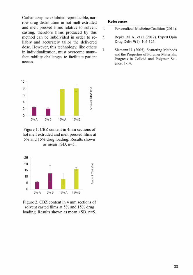
Manufacturing Films for Individualized Dosing
R. Govender1,2, S. Fennvik1,2, S. Folestad1, S. Abrahmsén-Alami1, A. Larsson2
1Global Product Development, AstraZeneca, Mölndal, Sweden2Chemistry and Chemical Engineering, Chalmers University of Technology, Gothenburg, SwedenBackground: Intra- and inter-individual variability in drug response is the premise for individualized therapy, which requires the design of the pharmaceutical product to be tailored to the characteristics of diverse patients to optimise health outcomes. One well studied product feature requiring indi-vidualization is the dose strength. Current pharmaceutical mass production can pro-duce some dose variants of solid oral dosage forms but lacks the flexibility required to in-dividualize dosing to the extent required to fully meet patient needs. Delivering flexible dosing via subdivision of a manufactured product to different sizes requires a uniform distribution of the drug prior to subdivision.
To investigate the suitability of different film manufacturing techniques to individu-alize dose strength in polymeric films.
Drug-loaded polymeric films containing 5% & 15% w/w carbamazepine (CBZ) in eth-yl cellulose were prepared by a) hot melt extrusion and melt pressing and b) solvent casting using 95% ethanol. 4mm sections of the film were evaluated for carbamaze-pine content by UV after dissolution in 95% ethanol.
33
Carbamazepine exhibited reproducible, nar-row drug distribution in hot melt extruded and melt pressed films relative to solvent casting, therefore films produced by this method can be subdivided in order to re-liably and accurately tailor the delivered dose. However, this technology, like others in individualization, must overcome manu-facturability challenges to facilitate patient access.
Figure 1. CBZ content in 4mm sections of hot melt extruded and melt pressed films at 5% and 15% drug loading. Results shown
as mean ±SD, n=5.
Figure 2. CBZ content in 4 mm sections of solvent casted films at 5% and 15% drug
loading. Results shown as mean ±SD, n=5.
References
1. Personalized Medicine Coalition (2014).
2. Repka, M. A., et al. (2012). Expert Opin Drug Deliv 9(1): 105-125.
3. Siemann U. (2005). Scattering Methods and the Properties of Polymer Materials. Progress in Colloid and Polymer Sci-ence: 1-14.

34
Application of ultrasound-enhanced electrospinning for fabricating drug-loaded polymer nanocompositesE. Hakkarainen1,2, I. Laidmäe2,3, A. Lust2, K. Semjonov2, K. Kogermann2, H. J. Nieminen4, A. Salmi4, O. Korhonen1, J. Heinämäki2 and E. Hæggström4
1School of Pharmacy, University of Eastern Finland, Kuopio, Finland 2Institute of Pharmacy, Faculty of Medicine, University of Tartu, Estonia 3Department of Immunology, Institute of Biomedicine and Translational Medicine, University of Tartu, Estonia 4Electronics Research Laboratory, Department of Physics, University of Helsinki, Finland
Electrospinning (ES) is an emerging nan-otechnology method for fabricating nano-composites for therapeutic agents. In tra-ditional ES (TES), a polymer solution is generated from a capillary toward a ground-ed metal collector plate by applying high voltage between the capillary and the plate [1,2]. The morphology and diameter of TES nanofibers depend on the intrinsic properties of the solution, type of polymer, conforma-tion of polymer chain, viscosity, elasticity, electric conductivity, as well as on the po-larity and surface tension of the solvent [3].
A novel ultrasound-enhanced ES (USES) provides an orifice-less ES technique that employs US for creating nanofibers [4]. In this technique, high-intensity focused US bursts are used to generate a liquid pro-trusion with a Taylor cone from the sur-face of a drug-polymer solution. When the drug-polymer solution is charged with a high negative voltage, a nanofibers jet from the tip of the protrusion is generated and
lead to an electrically grounded collector at a constant distance.
The objectives of the present study were (1) to compare the TES and USES techniques in fabricating drug-loaded polymer nano-fibers, and (2) to investigate the physico-chemical and pharmaceutical properties of nanofibers.
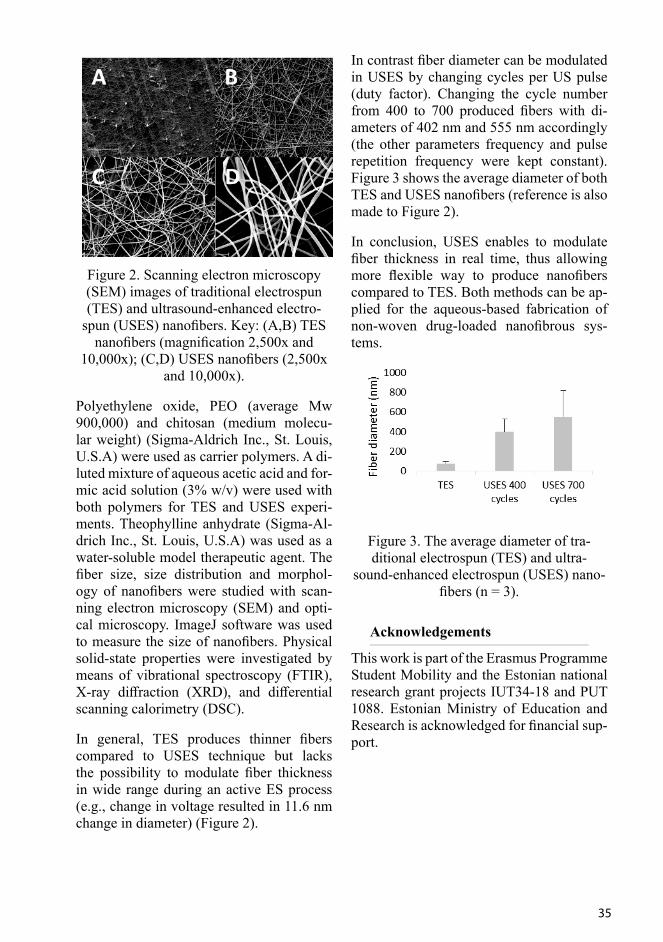
The nanofibers were fabricated with TES (ESR-200Rseries, eS-robot®, NanoNC, South Korea) and with an in-house USES method (Figure 1). The USES method is de-scribed in more detailed elsewhere [4]. To modulate fiber diameter, specific ultrasonic parameters (frequency, pulse repetition fre-quency and cycles per pulse) were applied during spinning.
Figure 1. A. Schematic of an ultrasound-en-hanced electrospinning (USES) setup. B.
Photograph of an USES device.
35
Figure 2. Scanning electron microscopy (SEM) images of traditional electrospun (TES) and ultrasound-enhanced electro-
spun (USES) nanofibers. Key: (A,B) TES nanofibers (magnification 2,500x and
10,000x); (C,D) USES nanofibers (2,500x and 10,000x).
Polyethylene oxide, PEO (average Mw 900,000) and chitosan (medium molecu-lar weight) (Sigma-Aldrich Inc., St. Louis, U.S.A) were used as carrier polymers. A di-luted mixture of aqueous acetic acid and for-mic acid solution (3% w/v) were used with both polymers for TES and USES experi-ments. Theophylline anhydrate (Sigma-Al-drich Inc., St. Louis, U.S.A) was used as a water-soluble model therapeutic agent. The fiber size, size distribution and morphol-ogy of nanofibers were studied with scan-ning electron microscopy (SEM) and opti-cal microscopy. ImageJ software was used to measure the size of nanofibers. Physical solid-state properties were investigated by means of vibrational spectroscopy (FTIR), X-ray diffraction (XRD), and differential scanning calorimetry (DSC).
In general, TES produces thinner fibers compared to USES technique but lacks the possibility to modulate fiber thickness in wide range during an active ES process (e.g., change in voltage resulted in 11.6 nm change in diameter) (Figure 2).
In contrast fiber diameter can be modulated in USES by changing cycles per US pulse (duty factor). Changing the cycle number from 400 to 700 produced fibers with di-ameters of 402 nm and 555 nm accordingly (the other parameters frequency and pulse repetition frequency were kept constant). Figure 3 shows the average diameter of both TES and USES nanofibers (reference is also made to Figure 2).
In conclusion, USES enables to modulate fiber thickness in real time, thus allowing more flexible way to produce nanofibers compared to TES. Both methods can be ap-plied for the aqueous-based fabrication of non-woven drug-loaded nanofibrous sys-tems.
Figure 3. The average diameter of tra-ditional electrospun (TES) and ultra-
sound-enhanced electrospun (USES) nano-fibers (n = 3).
Acknowledgements
This work is part of the Erasmus Programme Student Mobility and the Estonian national research grant projects IUT34-18 and PUT 1088. Estonian Ministry of Education and Research is acknowledged for financial sup-port.

36
References
[1] Z.-M. Huang, Y.-Z. Zhang, M. Kotakic, S. Ramakrishna, Comp. Sci Tech. 63, 2223 (2003).
[2] M. Naraghi, I. Chasiotis, Rev. Sci. In-strum. 78, 085108-1-7 (2007).
[3] S. Agarwal, J.H. Wendorff, A. Greiner, Polymer 49, 5603 (2008).
[4] I. Laidmäe, H. Nieminen, A. Salmi, T. Paulin, T. Rauhala, K. Falk, J. Yliruusi, J. Heinämäki, E. Hæggström, P. Veski, Int. Pat. Application WO/2016/151191,
PCT/FI2016/050170 (2016).
Bioadhesive nanofibrillated cellulose films for drug release
P. Laurén1, H. Paukkonen1, T. Lipiäinen1, T. Oksanen1, H. Räikkönen1, H. Ehlers1, P. Laaksonen2, M. Yliperttula1,3, T. Laaksonena,2
1Faculty of Pharmacy, University of Helsinki, Finland2Tampere University of Technology, Finland 3University of Padova, Italy
Bioadhesive materials have been gaining increasing interest due to inherently unsta-ble drug compounds [1]. Newly emerged peptide drugs are unable to cross biologi-cal barriers without being exposed to heavy enzymatic activity present in the GI-tract and liver, therefore resulting in poor bio-availability. Mucoadhesive formulations are generally designed to prolong GI-tract retention, however, local drug delivery sys-tems can enhance bioavailability by avoid-ing metabolic pathways, such as first-pass metabolism. Oral mucosa functions as a bi-ological barrier, which has been used in site
specific delivery of local oral diseases.
In addition to industrial applications, nano-fibrillar cellulose (NFC) has been inves-tigated in biomedical and pharmaceutical sciences, such as a scaffold that promotes three-dimensional cell culture or a drug-re-leasing matrix [2,3]. NFC fiber properties have several advantages to act as a func-tional biomaterial, e.g. inherent similarity to collagen fibers [2], great modification ca-pabilities, high water content, pseudoplas-tic and thixotropic properties. Additionally, NFC is considered as a safe, biocompatible and non-toxic biomaterial [4].In this study, we have fabricated bioadhesive films with the use of NFC and anionic type nanofibril-lar cellulose (ANFC). Mucin, pectin, and chitosan were investigated as mucoadhesive components to evaluate film mucoadhesive properties with texture analysis. Solid state characteristics and drug release properties of the films were examined with the use of metronidazole, an antibacterial drug com-pound used to treat periodontal diseases.
We observed that the bioadhesive properties of NFC could be enhanced by incorporat-ing mucoadhesive components into the film. This indicates potential local drug delivery systems for site specific medication of oral disease or to bypass metabolic routes to in-crease bioavailability.
This encore presentation has been presented at the 4th International Cellulose Confer-ence 2017, Fukuoka, Japan.
References
[1] Pettit DK & Gombotz WR. Trends Bio-technol. 16:343-9 (1998).
[2] Bhattacharya M, et al. J Control Re-lease. 164:291-8, 2012 (2012).
[3] Laurén P, et al. Eur J Pharm Sci. 18:79-88 (2014).
[4] Vartiainen J, et al. Cellulose. 18:775-86 (2011).
37
The society acknowledges the support from:
38
Influence of surface chemistry on adsorption and confinement of drug in porous silicon
E. Mäkilä1, H. Kivelä2, N. Shrestha3, A. Correia3, M. Kaasalainen1, E. Kukk1, J. Hirvonen3, H.A. Santos3, J. Salonen1
1Department of Physics and Astronomy, University of Turku, Finland2Department of Chemistry, University of Turku, Finland3Division of Pharmaceutical Chemistry and Technology, University of Helsinki, Finland
Drug delivery using PSi as dissolution en-hancing or payload protecting carrier ma-terial has been a widely studied application for the past 15 years [1,2]. By adsorbing small organic molecules into relatively small pores, where the average pore diame-ter is only 10–20x the size of the adsorbent molecule [3,4], porous materials become an effective tool for crystal engineering, which can be used to control e.g. specific polymorph nucleation within the porous matrix or even the complete suppression of the crystalline state. From drug delivery standpoint, the ability to prevent the forma-tion of crystalline structure through physical confinement leads to an effective method of keeping the adsorbed drug molecules in a disordered, amorphous-like state for pro-longed periods, while enhancing the aque-ous solubility and permeability, is a consid-erable advantage [4].
Here, the effect of hydrophobic and hydro-philic PSi surface chemistry (THCPSi and TOPSi, respectively) was studied with re-gard to the molecular dynamics of the ad-sorbed drug, ibuprofen, using thermal anal-ysis and variable temperature solid-state NMR because the selected mesopore size enabled the presence of both a nanocrystal-
line and an amorphous-like phase concur-rently inside of the mesopores [3,5]. Also, the effects of different parameters such as drug concentration and the loading solvent dielectric constant and (a)protic nature were studied for finding optimal loading param-eters.
The obtained results show that the drug load-ing appears most effective when adsorption occurs from solvent with low permittivity, such as CHF. However, after ca. 75% pore filling, corresponding 80% (w/w) drug pay-load, rapid accumulation of drug begins on the particle external surfaces, blocking fur-ther adsorption into the pores.
Interestingly, thermal analysis indicates that nucleation of ibuprofen nanocrystallites within the confinement of the pores begins already at low payloads of ca. 200 mg/cm3 (~20 % (w/w)). This separation of the nano-crystalline phase and an amorphous-like phase becomes more evident with larger drug payloads, with 40–50 % of the con-fined drug in the amorphous phase. Utiliz-ing the selectivity for molecular mobility between direct MAS and CP MAS 13C NMR measurements, this presence of two distinct populations of drug molecules with differing mobilities was also confirmed.
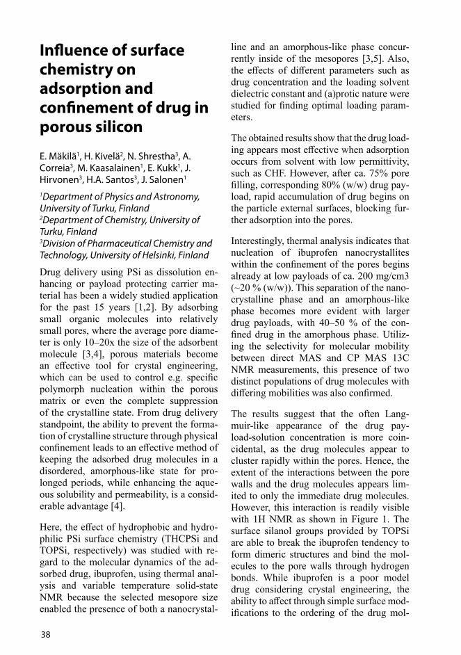
The results suggest that the often Lang-muir-like appearance of the drug pay-load-solution concentration is more coin-cidental, as the drug molecules appear to cluster rapidly within the pores. Hence, the extent of the interactions between the pore walls and the drug molecules appears lim-ited to only the immediate drug molecules. However, this interaction is readily visible with 1H NMR as shown in Figure 1. The surface silanol groups provided by TOPSi are able to break the ibuprofen tendency to form dimeric structures and bind the mol-ecules to the pore walls through hydrogen bonds. While ibuprofen is a poor model drug considering crystal engineering, the ability to affect through simple surface mod-ifications to the ordering of the drug mol-

39
ecules within the confined space prompts interesting possibilities. Combined with the complex fir-tree like wall structure inherent for mesoporous Si, these features could be exploited in e.g. polymorph selection and stabilization.
The adsorption of drug molecules within PSi can be optimized to yield high confined drug payloads. The interactions between the drug molecules and the pore walls character-ized with NMR spectroscopy indicate that while the extent of the interactions appears limited, the possibilities for using PSi as a crystal engineering platform for polymorph screening warrants further investigation.
References
1. H. A. Santos, E. Mäkilä, A. J. Airaksin-en, L. M. Bimbo, J. Hirvonen. Nano-medicine 2014, 9, 535–554.
2. J. Salonen, A. M. Kaukonen, J. Hir-vonen, V.-P.Lehto. J. Pharm. Sci. 2008, 97, 632–53.
3. E. Mäkilä, H. Kivelä, N. Shrestha, A. Correia, M. Kaasalainen, E. Kukk, J. Hirvonen, H. A. Santos, J. Salonen. Langmuir 2016, 32, 13020–13029.
4. E. Mäkilä, M. P. A. Ferreira, H. Kivelä, S.-M. Niemi, A. Correia, M. -A. Shah-bazi, J. Kauppila, J. Hirvonen, H. A. Santos, J. Salonen. Langmuir 2014, 30, 2196–2205.
5. J. Riikonen, E. Mäkilä, J. Salonen, V.-P. Lehto. Langmuir 2009, 25, 6137–42 Figure 1. Schematic representation of pos-
sible interactions near the pore walls with 1H MAS spectra of hydrophobic THCPSi
and hydrophilic TOPSi.
CH3
OOH
OHO CH2
CH3
CH3
16 12 8 4 0 -4 -8
Ibuprofen +TOPSi
Chemical shift (ppm)
Ibuprofen +THCPSi
1H MAS
OH O
HO
O
OHOH
OH
OH
40
Multimodal imaging of surface solid-state transformations
D. Novakovic1, J. Saarinen1, A. Isomäki2, S. J. Fraser-Miller3, T. Laaksonen4, L. Peltonen1, C. J. Strachan1 1 Division of Pharmaceutical Chemistry and Technology, University of Helsinki, Finland2 Biomedicum Imaging Unit, University of Helsinki, Finland3 Dodd-Walls Centre for Photonic and Quantum Technologies, Department of Chemistry, University of Otago, New Zealand4 Laboratory of Chemistry and Bioengineering, Tampere University of Technology, Finland
Surface and bulk regions of amorphous ma-terials differ in molecular mobility resulting in different behavior with regard to sus-ceptibility for solid-state transformations, for instance. Surface molecules with high-er mobility result in higher crystallization rates compared to molecules in the bulk. Crystallization on the free surfaces may af-fect critical properties such as dissolution. Nonlinear optical imaging is a relatively novel approach that provides both chemical and solid-state specificity. By simultaneous-ly combining second-order (sum frequency generation (SFG) and second harmonic gen-eration (SHG)) and third-order non-linear phenomena (coherent anti-Stokes Raman scattering (CARS)) differentiation between different solid-state forms of the drugs can be performed with greater confidence.1 The aim of this study was to combine SFG/SHG and CARS imaging to monitor surface sol-id-state transformations. Additionally, the effect of different levels of surface crystal-linity on the dissolution behavior was mon-itored.
The amorphous indomethacin was prepared by quench cooling the melted gamma form
of indomethacin (Orion, Finland). Pulver-ized amorphous indomethacin was com-pressed into tablets that were stored at 30°C at two different humidities: 23% RH and 75% RH. A Leica TCS SP8 CARS micro-scope was used for obtaining spectra and imaging. Tablet surfaces were analyzed on days 0, 1, 2, 5, 7 and 22. The intrinsic disso-lution rates of fresh and stored samples were measured using a channel flow system sim-ilar to the one described by Peltonen et al.2
The nature and level of surface crystallin-ity was dependent on storage conditions. Over the 22-day period, tablets stored at 30°C/23% RH crystalized mostly to the gamma indomethacin form whereas the tablets stored at 30°C/75% RH crystallized predominantly to the alpha indomethacin form. The crystallization was, however, not exclusive to only one of the polymorphs. Similarly, several small regions of tablets stored at 30°C/75% RH generated a CARS signal corresponding to the gamma form. Furthermore, storage induced surface sol-id-state changes were confirmed by having different intrinsic dissolution rate profiles.
The combination of two nonlinear imaging techniques was successfully used to study surface crystallization in amorphous indo-methacin tablets. Such surface crystalliza-tion can affect dissolution behavior.
References
[1] D. Novakovic, J. Saarinen, T. Rojalin, O. Antikainen, S. J. Fraser-Miller, T. Laaksonen, L. Peltonen+, Eur. J. Pharm. Sci. 19, (2003)
[2] L. Peltonen, P. Liljeroth, T. Heikkila, K. Kontturi, J. Hirvonen, Eur. J. Pharm. Sci. 19, (2003).
41
Preparation of ibuprofen-arginine salt by spray drying from water
R. Ojarinta1, L. Lerminiaux2 and R. Laitinen1
1 School of Pharmacy, University of Eastern Finland, Finland2 Laboratory for Pharmaceutical Process Analytical Technology, Department of Pharmaceutical Analysis, Faculty of Pharmaceutical Sciences, Ghent University, Belgium
Co-amorphous formulations are amorphous homogenous single-phase systems compris-ing of two or more low molecular weight compounds [1]. For example, co-amor-phous drug-amino acid systems have been shown to enhance both physical stability and dissolution properties of amorphous drugs. These effects have been especially significant with mixtures possessing strong interactions, such as salt formation, between the components.
In the present study, a co-amorphous mix-ture of IBU and ARG was prepared with an up-scalable method (spray drying). Our aim was to produce a stable co-amorphous mix-ture (1:1 molar ratio) with enhanced disso-lution properties and to investigate whether a salt would form between the components. Additionally, we aimed to conduct spray drying from aqueous solution without the use of organic solvents or solubilizers, such as surfactants.
Due to the solubilizing effect of ARG, dry IBU-ARG (1:1) powder (moisture content ~2.8%) could be obtained by spray drying from water, although the yield remained rather low (~34%). The resulting material was X-ray amorphous, and the temperature modulated DSC revealed a single Tg, which indicates the formation of a homogenous single-phase system. The Tg-value of the
spray dried IBU-ARG mixture (82.8 ± 1.93 °C) was also significantly higher than the theoretical Tg (-10.2 °C) calculated with the Gordon-Taylor equation, which suggests strong interactions between IBU and ARG. The FTIR spectrum analysis revealed the interaction to be salt formation.
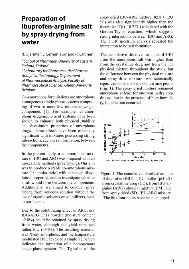
The cumulative dissolved amount of IBU from the amorphous salt was higher than from the crystalline drug and from the 1:1 physical mixture throughout the study, but the difference between the physical mixture and spray dried mixture was statistically significant only at 30 and 60 min time points (Fig. 1). The spray dried mixture remained amorphous at least for one year in dry con-ditions, but in the presence of high humidi-ty, liquefaction occurred.
Figure 1. The cumulative dissolved amount of ibuprofen (IBU) in HCl buffer (pH 1.2) from crystalline drug (CD), from IBU-ar-ginine (ARG) physical mixture (PM), and from spray dried (SD) IBU-ARG mixture. The first four hours have been enlarged.

42
Based on this study, spray drying from aqueous solutions seems to be a feasible up-scalable technique for the preparation of co-amorphous mixtures, if the co-former solubilizes the drug adequately. With this technique the amorphous form of very low Tg drugs may be stabilized at least in the presence of strong interactions between the components, and also the dissolution prop-erties may be enhanced.
References
[1] S. J. Dengale, H. Grohganz, T. Rades, K. Löbmann, Adv. Drug Deliver. Rev. 100, 116 (2016).
Electrospinning of nanofibrillar cellulose reinforced nanofibers for pharmaceutical applications
U. Paaver1, K. Kogermann1, L. Viidik1 and J. Heinämäki1
1Institute of Pharmacy, Faculty of Medicine, University of Tartu, Estonia
Nanofibrillar cellulose (NFC) is an interest-ing novel nanomaterial potential for a wide range of technical, biomedical and phar-maceutical applications. NFC has several unique material properties for an excipient use in both pharmaceutical immediate-re-lease and controlled-release dosage forms. NFC is non-toxic, has a high surface area, and possesses excellent mechanical strength that could be used to improve the mechan-ical properties of solid dosage forms [1,2]. NFC absorbs a tremendous amount of wa-ter and readily forms a gel which makes it a suitable candidate to be used also as a wound protective material [2].
Figure 1. Aqueous dispersion of 2.7% bio-fibrils form a continuous network of hy-
drated fibrils. The material is isolated from Pinus sylvestris and Picea abies or Betula
sp.
The main objectives of the present study were to investigate the effects of NFC on the formation and physicochemical properties of electrospun composite nanofibrous mats and the corresponding moulded thin films.
NFC was purchased as a 2.7-% aqueous dispersion from UPM Biofibrils AS 103, UPM-Kymmene Corporation, Finland (Figure 1). Polyethylene oxide, PEO (mw 900,000, Sigma-Aldrich) and polyvinyl al-cohol, PVA (mw 125,000, Mowiol®, Sig-ma-Aldrich) were used as a water-soluble polymer in nano-fibrous mats and thin films. Alginate (ALG) was studied as a native or-igin fibrous carrier material. Purified water was used as a main solvent.
For electrospinning (ES) and film mould-ing, aqueous solutions of both PEO (4% and 8% w/v) and PVA (Mowiol®) (4% and 12% w/v) were prepared. NFC was incor-porated in the polymeric solutions at dif-ferent concentrations ranging from 0.1 to 0.6% (w/v). The ALG solutions (1-3% w/v) were prepared by dissolving the material in purified water and used in different com-binations with PEO. The nanofibers were prepared using an ESR200RD robotized ES system (NanoNC, South-Korea) at an ambi-ent room temperature and relative humidity (RH). The flow rate of the solution was 0.1, 0.5 and 1.0 ml/h, and the voltage applied

43
9-16 kV. The distance between the spinneret and the fiber collector was 10-15 cm. The thin films were prepared by a solvent evap-oration method in polytetrafluoroethylene (Teflon®) molds. The molded solutions were allowed to dry for at least 24 hours at an ambient room temperature and RH.
The fiber size, size distribution, surface morphology and thickness (nanomats and thin films) were studied with a high-resolu-tion optical (CETI MagtexT) and scanning electron microscopy, SEM (Zeiss EVO® 15 MA, Germany). ImageJ software was used to measure the size of nanofibers. The thermal properties were studied by means of differential scanning calorimetry, DSC (DSC823e, Mettler Toledo AG, Switzer-land). The mechanical properties of nano-fibrous mats were tested with a Brookfield Texture Analyzer CT3 (U.S.A).
Figure 2 shows the SEM and optical micros-copy micrographs of the desiccated aqueous dispersion (2.7%) of NFC. After desicca-tion, a fragile NFC film was formed.
As seen in Figure 3, the 1:1 ratio of NFC (0.135% w/v aqueous dispersion) and PEO (8% w/v) solution in ES produced the nano-fibers with a uniform structure. The mean diameter of the fibers was less than 200 nm, and the fiber diameter was dependent on the voltage used.
Figure 2. SEM and optical microscopy im-ages of the desiccated aqueous dispersion
of nanofibrillar cellulose (NFC).
Figure 3. SEM images of nanofibrillar cel-lulose (NFC) reinforced polyethylene oxide (PEO), polyvinyl alcohol (PVA) or alginate (ALG) nanofibers. Magnification 10,000×.
When NFC was added to the PEO solution, the electrospun fibers decreased compared to that obtained with the pure PEO fibers. Increasing the NFC concentration resulted in the decrease in the diameter of the com-posite nanofibers. The higher concentration of NFC impaired the formation of nanofi-bers, and consequently, several beads were detected on the nanofibrous platforms. The corresponding composite thin films of NFC and PEO/PVA were transparent and uniform in structure (Figure 4).
Figure 4. Optical microscopy images of nanofibrillar cellulose (NFC) reinforced
polyethylene oxide (PEO) and polyvinyl al-cohol (PVA) thin films. Magnification 20×.
44
In conclusion, NFC can be successfully electrospun with PEO, PEO+ALG and PVA to obtain composite nanofibrous platforms for e.g., drug delivery applications. The corresponding composite free films can be fabricated with a simple moulding method. The concentration of NFC will greatly af-fect the formation and performance of the present nanofibrous mats and thin films. Further studies will reveal the storage sta-bility, swelling and dissolution behavior of the present composite platforms.
Acknowledgements
This work is part of the national research grant projects IUT34-18 and PUT 1088.
References
[1] A.J. Svagan, J.-W. Benjamins, Z. Al-An-sari, D.B. Shalom, A. Müllertz, L. Wåg-berg, K. Löbmann, J. Control. Release 244 (Part A), 74 (2016).
[2] R. Kolacović, L. Peltonen, A. Lauk-kanen, J. Hirvonen, Eur. J. Pharm. Bio-pharm. 82, 308 (2013)
Synergistic Analysis of Nanoparticle Cellular Uptake Using Correlative Coherent Anti-Stokes Raman Scattering and Electron Microscopy
J. Saarinen1, F. Gütter2, M. Lindman3, S. Fraser-Miller1,4, R. Scherließ2, A. Isomäki5 E. Jokitalo3,HA. Santos1, L. Peltonen1, CJ. Strachan1 1Drug Research Program, Division of Pharmaceutical Chemistry and Technology, Faculty of Pharmacy, University of Helsinki, Finland2Department of Pharmaceutics and Biopharmaceutics, Kiel University, Germany3Electron Microscopy Unit, Institute of Biotechnology, University of Helsinki, Finland4Dodd-Walls Center, Department of Chemistry, University of Otago, Dunedin, New Zealand5Biomedicum Imaging Unit, Haartmaninkatu 8, 00014 University of Helsinki, Finland
Nanoparticles are used routinely for exam-ple to achieve targeted drug delivery. It is important to have adequate analysis meth-ods for imaging these particles in biological matrices. Raman based chemically-specific label-free imaging holds potential for this purpose, while electron microscopy pro-vides very high nanometer-level spatial res-olution but lacks chemical specificity.
The aim of this study was to combine two imaging methods, chemically-specific la-bel-free (sub)micron-scale resolution CARS microscopy and (sub)nanometer-scale spa-tial resolution transmission electron micros-copy (TEM) to image the cellular uptake of nanoparticles. This synergistic correlative

45
coherent anti-Stokes Raman scattering and electron microscopy (C-CARS-EM) meth-od allows insights into nanoparticle uptake that would not be available with either of these techniques alone.
RAW 264.7 cells grown on glass bottomed gridded Petri dishes (Mattek) were incubat-ed with glibenclamidemide nanosuspen-sions (250 µg/ml) for 6 h. Cells were fixed (2% glutaraldehyde in sodium cacodylate buffer) and imaged with Leica TCS SP8 CARS microscope. 633 nm He/Ne laser was used for bright field (BF) imaging the grids on the dish. Z-stack CARS images were recorded and CARS spectrum from the in-side of the cells was measured. Cells were flat-embedded in plastic for microtome sectioning the area selected based on the grid markings and BF images [1]. 120 nm sections were imaged with Jeol JEM-1400 TEM (80 kV) using Gatan Orius SC1000B bottom mounted CCD-camera.
3080 cm-1 peak was used to image the uptake of crystals by cells in a chemical-ly-specific label-free manner. There was a good correlation in the observed particle localization inside the cells between CARS and TEM images. TEM could be used to visualize the subcellular localization of the nanocrystals, while the drug crystals were confirmed as such by CARS.
C-CARS-EM imaging of drug nanocrystal uptake in cells with CARS and TEM was successfully achieved. Both techniques had their inherent benefits. CARS is chemical-ly-specific with easy sample preparation, whereas TEM has better spatial resolution capable of showing fine structural details of the particles. TEM images revealed that most of the nanoparticles were located in membrane bound vesicles.
References
[1] Seemann, J.; Jokitalo, E. J.; Warren, G. The Role of the Tethering Proteins p115 and GM130 in Transport through the Golgi Apparatus in Vivo. Mol. Biol. Cell 2000, 11, 635–645.
Exploring mesoporous silica nanoparticles as carriers for poorly soluble drugs in orodispersible films
D. Sen Karaman1, G. Patrignani1, A. Orłowska1, R. Mustafa1, E. Rosqvist2, J-H Smått2, M. Preis1 JM.Rosenholm1
1Pharmaceutical Sciences Laboratory , Faculty of Science and Enginnering, Åbo Akademi University, Artillerigatan 6A, 20520 Turku, Finland2Laboratory of Physical Chemistry, Faculty of Science and Enginnering, Åbo Akademi University, Porthansgatan 3-5 , 20500 Turku Finland
Fast dissolving orodispersible films (ODF) are rapidly gaining interest especially to solve the problems encountered in the ad-ministration of drugs to pediatric and elder-ly patients. To be able to deliver poorly sol-uble drugs with ODFs, solubility enhancers are often required. Various substances have been incorporated as enhancers into ODFs to increase the absorption of drugs through the mucosa. However, they are associat-ed with irritation, membrane damage, and toxicity, which thereby limits their use.Biocompatible nanoparticles as carriers for poorly soluble drugs have been shown to be promising in improving the solubility of drugs. In the last two decades, mesoporous silica nanoparticles (MSNs) have been ex-tensively used in drug delivery applications. MSNs possess unique properties,

46
such as high specific surface area, high pore volume and appropriate pore sizes in the molecular range, ordered pore structures and silanol groups on their surfaces.2 These features make MSNs optimal drug carriers. Immediate and controlled release MSN-based drug delivery systems have been de-veloped for oral,3 transdermal,4 and intra-venous5 administration of compounds with poor stability or solubility.
In this study, we explored MSNs as carri-ers for the delivery of prednisolone in fast dissolving polyvinyl alcohol orodispersible films. In detail, two different methods (i.e. pre-incorporation and dip-coating) were used to accommodate MSNs into the ODF matrix. Furthermore, mechanical properties of the ODFs were investigated to evaluate the resulting changes of ODF due to the incorporation of the MSNs carrier. In addi-tion, the drug loading efficiency was inves-tigated. Lastly, the drug release properties of the optimized the ODF formulation was investigated in simulated saliva fluid (SSF).
The results revealed that the MSN incor-poration strategies have a significant im-pact on the mechanical properties of the ODF. The thickness of ODF significantly decreases when the number of immersions is increased (>5 times) in the dip-coat-ing process, whereas pre-incorporation of MSN into thr polymeric matrix increased the thickness and improved the mechanical properties of ODF. The even distribution of the incorporated MSNs on ODF was shown with atomic force microscopy (AFM). The drug (i.e. prednisolone) loading on MSN was carried out prior to incorporation of the MSN into ODF. The archive drug loading degrees was confirmed by UV-VIS spec-trophotometry and the highest drug loaidng was achieved with the pre-incorporation method. The drug release studies show that 90 %of the drug content releases in the first 2 minutes whereas only less then 40%of the content is relases out when the drug is incor-porated without particle laoding.
Consequently, in this study we have evalu-ated the potential of MSN as a drug carrier within ODF to be used for precise dosing and fastening the dissolution of the drug content from ODF.
References
[1] Sohi, H.; Ahuja, A.; Ahmad, F. J.; Khar, R. K. Drug Dev. Ind. Pharm. 2010, 36 (3), 254–282.
[2] Maleki, A.; Kettiger, H.; Schoubben, A.; Rosenholm, J. M.; Ambrogi, V.; Ha-midi, J. Controlled Release 2017, 262, 329–347.
[3] Zhang, Y.; Wang, J.; Bai, X.; Jiang, T.; Zhang, Q.; Wang, S.. Mol. Pharm. 2012, 9 (3), 505–513.
[4] Sapino, S.; Ugazio, E.; Gastaldi, L.; Miletto, I.; Berlier, G.; Zonari, D.; Oli-aro-Bosso, S. M. Eur. J. Pharm. Bio-pharm. 2015, 89, 116–125.
[5] Mamaeva, V.; Rosenholm, J. M.; Bate-Eya, L. T.; Bergman, L.; Peuhu, E.; Duchanoy, A.; Fortelius, L. E.; Landor, S.; Toivola, D. M.; Lindén, M.; Sahl-gren, C. Mol. Ther. 2011, 19 (8), 1538–1546.
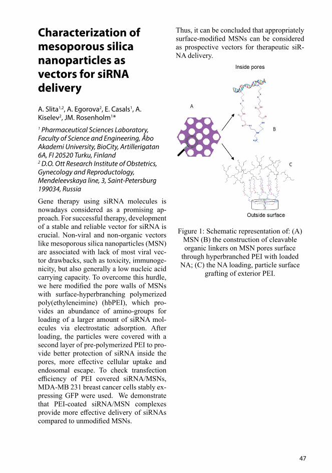
47
Characterization of mesoporous silica nanoparticles as vectors for siRNA delivery
A. Slita1,2, A. Egorova2, E. Casals1, A. Kiselev2, JM. Rosenholm1* 1 Pharmaceutical Sciences Laboratory, Faculty of Science and Engineering, Åbo Akademi University, BioCity, Artillerigatan 6A, FI 20520 Turku, Finland2 D.O. Ott Research Institute of Obstetrics, Gynecology and Reproductology, Mendeleevskaya line, 3, Saint-Petersburg 199034, Russia
Gene therapy using siRNA molecules is nowadays considered as a promising ap-proach. For successful therapy, development of a stable and reliable vector for siRNA is crucial. Non-viral and non-organic vectors like mesoporous silica nanoparticles (MSN) are associated with lack of most viral vec-tor drawbacks, such as toxicity, immunoge-nicity, but also generally a low nucleic acid carrying capacity. To overcome this hurdle, we here modified the pore walls of MSNs with surface-hyperbranching polymerized poly(ethyleneimine) (hbPEI), which pro-vides an abundance of amino-groups for loading of a larger amount of siRNA mol-ecules via electrostatic adsorption. After loading, the particles were covered with a second layer of pre-polymerized PEI to pro-vide better protection of siRNA inside the pores, more effective cellular uptake and endosomal escape. To check transfection efficiency of PEI covered siRNA/MSNs, MDA-MB 231 breast cancer cells stably ex-pressing GFP were used. We demonstrate that PEI-coated siRNA/MSN complexes provide more effective delivery of siRNAs compared to unmodified MSNs.
Thus, it can be concluded that appropriately surface-modified MSNs can be considered as prospective vectors for therapeutic siR-NA delivery.
Figure 1: Schematic representation of: (A) MSN (B) the construction of cleavable organic linkers on MSN pores surface
through hyperbranched PEI with loaded NA; (C) the NA loading, particle surface
grafting of exterior PEI.
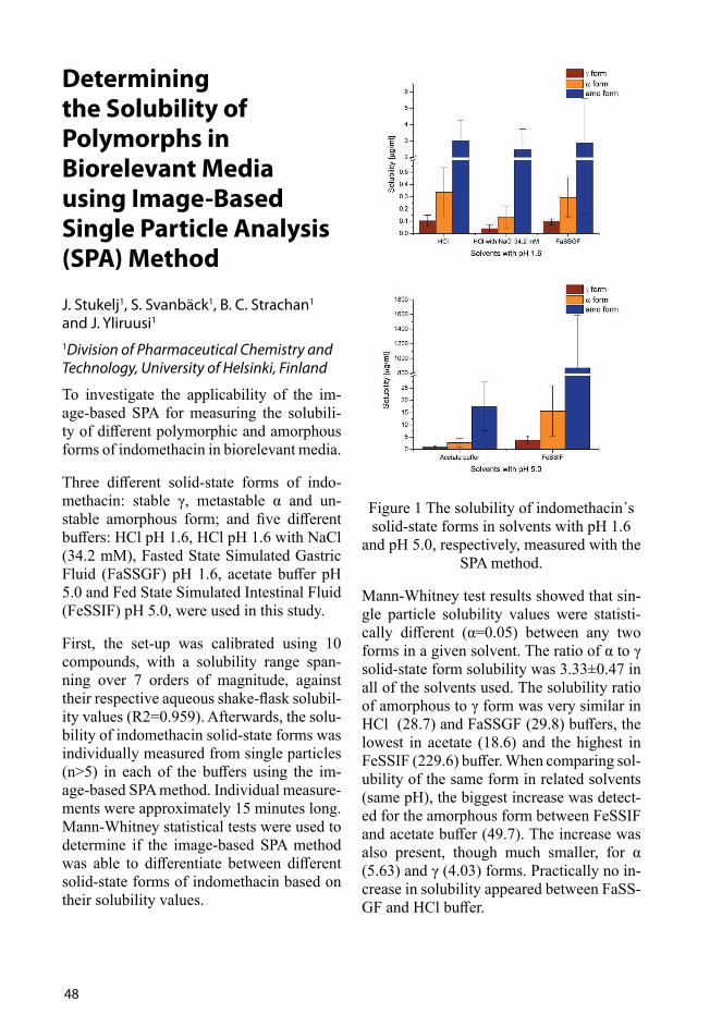
48
Determining the Solubility of Polymorphs in Biorelevant Media using Image-Based Single Particle Analysis (SPA) Method
J. Stukelj1, S. Svanbäck1, B. C. Strachan1 and J. Yliruusi1
1Division of Pharmaceutical Chemistry and Technology, University of Helsinki, Finland
To investigate the applicability of the im-age-based SPA for measuring the solubili-ty of different polymorphic and amorphous forms of indomethacin in biorelevant media.
Three different solid-state forms of indo-methacin: stable γ, metastable α and un-stable amorphous form; and five different buffers: HCl pH 1.6, HCl pH 1.6 with NaCl (34.2 mM), Fasted State Simulated Gastric Fluid (FaSSGF) pH 1.6, acetate buffer pH 5.0 and Fed State Simulated Intestinal Fluid (FeSSIF) pH 5.0, were used in this study.
First, the set-up was calibrated using 10 compounds, with a solubility range span-ning over 7 orders of magnitude, against their respective aqueous shake-flask solubil-ity values (R2=0.959). Afterwards, the solu-bility of indomethacin solid-state forms was individually measured from single particles (n>5) in each of the buffers using the im-age-based SPA method. Individual measure-ments were approximately 15 minutes long. Mann-Whitney statistical tests were used to determine if the image-based SPA method was able to differentiate between different solid-state forms of indomethacin based on their solubility values.
Figure 1 The solubility of indomethacin´s solid-state forms in solvents with pH 1.6
and pH 5.0, respectively, measured with the SPA method.
Mann-Whitney test results showed that sin-gle particle solubility values were statisti-cally different (α=0.05) between any two forms in a given solvent. The ratio of α to γ solid-state form solubility was 3.33±0.47 in all of the solvents used. The solubility ratio of amorphous to γ form was very similar in HCl (28.7) and FaSSGF (29.8) buffers, the lowest in acetate (18.6) and the highest in FeSSIF (229.6) buffer. When comparing sol-ubility of the same form in related solvents (same pH), the biggest increase was detect-ed for the amorphous form between FeSSIF and acetate buffer (49.7). The increase was also present, though much smaller, for α (5.63) and γ (4.03) forms. Practically no in-crease in solubility appeared between FaSS-GF and HCl buffer.

49
The Image-Based SPA method is capable of differentiating between polymorphic forms of indomethacin based on the solubility values of their single particles. Moreover, using the method, a quick estimation of the drug behaviour in the gastrointestinal tract can be achieved, and the effects of surfac-tants on drug solubility studied.
References
[1] J. Stukelj, S. Svanbäck, B. C. Strachan and J. Yliruusi, Apparent Solubility of Drugs in Biorelevant Media Using Image Based Single Particle Analysis, Master´s Thesis at University of Lju-bljana (2017)
50
Hot-melt extrusion of 3D printable isoniazid formulations
H. Öblom1, J. Zhang2, M. Pimparade2, J. Rahman1, M. Repka2, M. Preis1, N. Sandler1 1 Pharmaceutical Sciences Laboratory, Åbo Akademi University, Åbo, Finland2 Department of Pharmaceutics and Drug Delivery, The University of Mississippi,MS, USA
The use of three-dimensional (3D) printers to manufacture flexible and tailored drug delivery systems is an emerging area of in-terest in pharmaceutical research since the precise and additive nature of the process potentially could solve the unmet need for personalized medicine (1,2). The aim of the present study was to produce 3D printable feedstock material possessing properties relevant for oral dosage forms and compare how the subsequent fused deposition mod-eling (FDM) 3D printing affects the final dosage form.
A Thermo Scientific Process 11 (USA) co-rotating twin-screw hot-melt extruder was used to produce 3D printable feedstock materials (filaments) that subsequently were printed into model geometries using a Mak-erBot Replicator 2 FDM 3D printer. High-ly water-soluble isoniazid (TCI America, USA) served as a model drug and polymers with relevant pharmaceutical properties were included in the formulations. Ten dif-ferent hot-melt extruded formulations con-taining various polymer and excipient com-binations and a drug loading of 30% (w/w) were prepared. The printability in an FDM printer of the manufactured filaments was evaluated. Formulations were furthermore analyzed regards to mechanical and sol-id-state properties e.g. by utilizing a texture analyzer (TA-XT2i, Texture Technologies, Hamilton, MA, USA) and x-ray diffrac-tion. In vitro drug release was carried out in
phosphate buffer pH 7.45.
Extruded filaments were evaluated for their printability in a MakerBot 2 Replicator 3D printer. Six formulations were successfully 3D-printed, the other formulations were dis-carded either due to unsuitable filament di-ameter or mechanical properties leading to an unsuccessful loading of the filament into the print-head (Figure 1). Mechanical test-ing of the produced HME filaments showed a fairly good correlation to the printability of the filaments, revealing that filaments that in the 3 point bend test failed to bend 1.5 mm or more were too brittle to feed into the printer.
Solid-state analysis revealed that the drug was present in a crystalline form in all of the hot-melt extruded formulations. Due to the high water-soluble nature of the drug, a complete drug release could be achieved for all hot-melt extruded as well as the printed formulations. The 3D-printed geometry that was designed to have the same geometrical outlines as the filaments showed similar in vitro drug release as the corresponding hot-melt extruded filaments, indicating that 3D printing at elevated temperatures does not seem to affect the drug release behavior (Figure 2). 3D-printed tablets (8x2.5 mm), however, showed a more sustained drug re-lease profile due to the difference in surface area compared to the filaments. This indi-cates that 3D printing may be used to tailor the drug release profile simply by changing the shape of the printed dosage form. The drug-release may furthermore be altered by changing the composition of the extruded filaments. In this study, 3D-printed and hot-melt extruded filaments showed 80% isonia-zid release within 11-85 minutes depending on the polymer and excipient composition of the formulations.
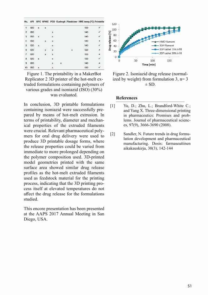
51
Figure 1. The printability in a MakerBot Replicator 2 3D printer of the hot-melt ex-truded formulations containing polymers of various grades and isoniazid (ISO) (30%)
was evaluated.
In conclusion, 3D printable formulations containing isoniazid were successfully pre-pared by means of hot-melt extrusion. In terms of printability, diameter and mechan-ical properties of the extruded filaments were crucial. Relevant pharmaceutical poly-mers for oral drug delivery were used to produce 3D printable dosage forms, where the release properties could be varied from immediate to more prolonged depending on the polymer composition used. 3D-printed model geometries printed with the same surface area showed similar drug release profiles as the hot-melt extruded filaments used as feedstock material for the printing process, indicating that the 3D printing pro-cess itself at elevated temperatures do not affect the drug release for the formulations studied.
This encore presentation has been presented at the AAPS 2017 Annual Meeting in San Diego, USA.
Figure 2. Isoniazid drug release (normal-ized by weight) from formulation 3, n= 3
± SD.
References
[1] Yu, D.; Zhu, L.; Brandford-White C.; and Yang X. Three-dimensional printing in pharmaceutics: Promises and prob-lems. Journal of pharmaceutical scienc-es, 97(9), 3666-3690 (2008).
[2] Sandler, N. Future trends in drug formu-lation development and pharmaceutical manufacturing. Dosis: farmaseuttinen aikakauskirja, 30(3), 142-144

52
Tissue penetrance and toxicity investigations of differently functionalized mesoporous silica nanoparticles in the zebrafish model to understand chemical design-toxicity relationship
I. Paatero1,3, E. Casals4, R. Niemi2,3, E. Özliseli4, J. M. Rosenholm4, C. Sahlgren2,3,5,6
1Department of Cell Biology, Biozentrum, University of Basel, Basel, Switzerland 2Faculty of Science and Engineering, Cell Biology, Åbo Akademi University, Turku, Finland 3Turku Centre for Biotechnology, Åbo Akademi University and University of Turku, Turku, Finland 4Pharmaceutical Sciences Laboratory, Faculty of Science and Engineering, Åbo Akademi University, Turku, Finland5Department of Biomedical Engineering, Technical University of Eindhoven, Eindhoven, The Netherlands 6Institute for Complex Molecular Systems, Eindhoven University of Technology, Eindhoven, The Netherlands
Mesoporous silica nanoparticles (MSNs) have been receiving extensive attention in the last decades as drug delivery systems due to its versatile design flexibility and uniform mesopores for drug molecule loading and release. Surface functionalization of MSNs expands applicability as drug delivery platform by influencing dispersion stabil-ity, biocompatibility, biodistribution, drug release, efficacy and biodegradation [1]. However, their design-toxicity relationship needs to be investigated in the more compli-
cated biological systems relevant to human physiology to able to develop safer nanoth-erapies. In this study, zebrafish (Danio rerio) embryos were selected as model system and their toxicity profiles of differently surface functionalized MSNs were studied. Zebraf-ish model is widely used in toxicity studies and has similar cardiovascular, nervous and digestive systems with mammals. Chorion membrane intact embryos or dechoroniated embryos were incubated or microinjected with amino (NH2-MSNs), polyethylenei-mine (PEI-MSNs), succinic acid (SUCC-MSNs) or polyethyleneglycol (PEG-MSNs) functionalized MSNs and toxicity was eval-uated by embryo viability and cardiovascu-lar function. Different surface functionaliza-tions of MSNs resulted as different surface charges, which is the key parameter for the interactions of nanoparticles with the bio-logical membranes, and therefore influenc-ing cellular internalization, biodistribution, residence time, immune system and toxicity.
NH2-MSNs, SUCC-MSNs and PEG-MSNs did not show any lethality, whereas, 50µg/ml PEI-MSNs induced 100% lethality 48 hours post fertilization (hpf). Dechoroniat-ed embryos were more sensitive and 10µg/ml PEI-MSNs reduced viability to 5% at 96 hpf. Dechoroniation increased the sensitiv-ity for PEG-MSNs and SUCC-MSNs, but not NH2-MSNs. Cardiovascular toxicity was observed prior to lethality by stereomi-croscope and confocal microscopy revealed that PEI-MSNs were penetrated to the em-bryos whereas PEG-MSNs, NH2-MSNs and SUCC-MSNs remained aggregated on the skin surface (Figure 1). Direct exposure of inner organs was tested by microinject-ing NH2-MSNs and PEI-MSNs which were demonstrated the lowest and highest tox-icity previously, displayed similar toxicity indicating that functionalization affects the toxicity profile by influencing penetrance through biological barriers. The data re-veals that functionalization critically de-termines penetrance of biological barriers and resulting toxicity, and emphasize the
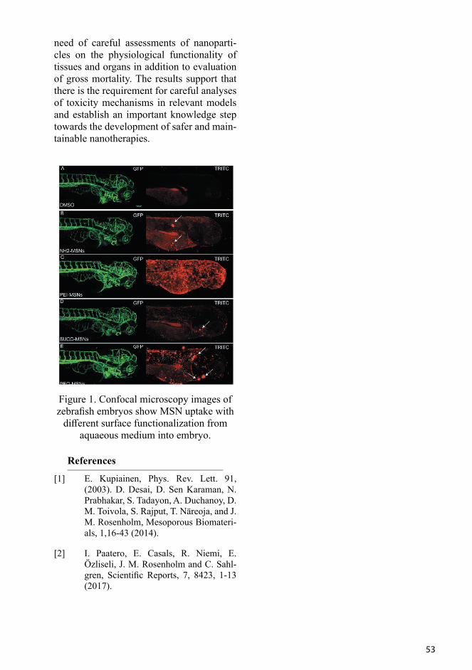
53
need of careful assessments of nanoparti-cles on the physiological functionality of tissues and organs in addition to evaluation of gross mortality. The results support that there is the requirement for careful analyses of toxicity mechanisms in relevant models and establish an important knowledge step towards the development of safer and main-tainable nanotherapies.
Figure 1. Confocal microscopy images of zebrafish embryos show MSN uptake with
different surface functionalization from aquaeous medium into embryo.
References
[1] E. Kupiainen, Phys. Rev. Lett. 91, (2003). D. Desai, D. Sen Karaman, N. Prabhakar, S. Tadayon, A. Duchanoy, D. M. Toivola, S. Rajput, T. Näreoja, and J. M. Rosenholm, Mesoporous Biomateri-als, 1,16-43 (2014).
[2] I. Paatero, E. Casals, R. Niemi, E. Özliseli, J. M. Rosenholm and C. Sahl-gren, Scientific Reports, 7, 8423, 1-13 (2017).

54
When the Society of Physical Pharmacy was founded in 1988 it felt like a dream come true. For a M.Sc. student doing the last year in M.Sc. studies, it was mind-blowing to learn about polymorphs, solid-state prop-erties and physicochemical characterisation of APIs, excipients and products. We were introduced the techniques like X-ray pow-der diffraction and differential scanning cal-orimetry (DSC). Before that pharmaceutical technology was very empirical and the first techniques really opened our mind for mo-lecular interactions and physical phenome-na in pharmaceuticals.
Since then, the number and level of solid state and particle analytical technologies has increased markedly. This is demonstrated with two example areas. When spectroscop-ic technologies like NIR came to use, the concept of process analytical technologies were introduced to pharmaceutical process-es and understanding of APIs, formulations and processes increased one step further. Novel spectroscopic technologies have been introduced to pharmaceutics with increasing tempo in last decades, like FTIR, solid-state NMR, Raman, CARS, tera-hertz spectros-copy and time-gated Raman. As another example, the area of DSC has expanded by introducing the high-performance DSC, StepScan, modulated temperature DSC and for biomolecules techniques like micro-electromechanical systems (MEMS)-DSC, infrared (IR)-heated DSC, gas flow-modu-lated DSC (GFMDSC), parallel-nano DSC (PNDSC), pressure perturbation calorime-
try (PPC) and self-reference DSC (SRDSC) (1).
It seems that the development of analytical technologies has almost become a meaning and area of research itself (and a business). Do we remember what we want to do with these technologies? How can these tech-niques do better medicines for patients? Are we losing the understanding of the mean-ingful parameters for APIs and formulations since we can measure everything, and we do that just to make it sure? As a research scientist we always recommend that one should investigate a phenomenon by mea-suring with more than one analytical meth-od, but how much is enough? Are our meth-ods really relevant or just nice to know and looking good for the publications?
Further, more and more process data are ob-tained from our instrumented manufactur-ing equipment. The systematic study design and statistical and multi-variate modelling of our data has given us more understand-ing of our pharmaceutical processes and systems. However, the amount of data is enormous, processing data of in-line mea-surements combined with analytical in-line and other data gives a huge challenge how to store, handle and process the data to get the valuable information. The human brain might come to a limit in its capacity to han-dle these multidimensional data spaces. Pharmaceutical companies have started to use advanced artificial intelligence in han-dling and processing the data. Utilisation
Commentary Article
Are we measuring too much?
Anne Juppo
Professor in Industrial PharmacyFaculty of Pharmacy, University of Helsinki, Helsinki

55
of augmented reality sounds like science fiction or entertainment, but it is already in use in handling the video data of pharma-ceutical manufacturing area to investigate the line clearance in GMP areas in real time.
Also, in silico tools are already widely used in drug discovery and they will be also utilised even more in formulation devel-opment. In silico tools based on structural informatics will give insight on solid state landscape and assist us in material design and predict the material performance. The prediction capacity of solid state properties with these tools is improving (2).
What will be the needs of physicochemi-cal characterisation of pharmaceuticals in future? Should we start to prepare for pro-cessing of pharmaceuticals in space in grav-ity free areas? Should we start to study the handling and behaviour of powders in those conditions and develop the flowability and compressibility tests to be used in space? This has already been started by the mining industry being understandingly first in line and some research in pharmaceutical pro-duction in space has already been done (3).
Physicochemical characterisation of raw materials will be emphasized even further when continuous processing will increase in order to ensure smooth processes, especial-ly powder flow in production lines. What will be the drug forms used in future? Will the use of biological drugs switch the focus from solid dosage forms towards parenter-als and liquid systems? The physicochem-ical characterisation of biologicals is chal-lenging and requires a different portfolio of techniques than low-weight molecules that we are used to work with. Thus, the topics of the Physical Pharmacy Symposium in 2047 are impossible to predict.
The aim of this article is to provoke criti-cal thinking and discussion of what we are doing and why, and to present wild guesses what might be needed in future. A deeper and true understanding of our APIs, excip-
ients, formulations and processes for the benefit of patients should be our common goal. The use and easier access to synchro-trons, for example, will definitely improve understanding of our systems. A wise use of the techniques available combined with know-how in pharmaceutical materials and processes will be the success factors even for coming decades.
References
[1] P Gill, TT Moghadam, B Ranjbar. Dif-ferential scanning calorimetry tech-niques: applications in biology and na-noscience. J. Biomol. Tech 21 (2010) 167-193.
[2] D A Bardwell, C S. Adjiman, Y A Ar-nautova, E Bartashevich, S X M Boer-rigter, D E Braun et al. Towards crystal structure prediction of complex organic compounds – a report on the fifth blind test. Acta Cryst. Section B 67 (2011) 535-551.
[3] M R Benoit, W Li, L S Stodieck, K S Lam, C L Winther, T M Roane, D M Klaus. Microbial antibiotic production aboard the international space station. Applied Microbiol. Biotech. 70 (2006) 403-411.

56

57
PhD and MSc theses – 2017

58
Multifunctional Nanoparticles for Targeted Drug Delivery and Imaging for Ischemic Myocardial Injury
Ferreira, Mónica
Division of Pharmaceutical Chemistry and TechnologyUniversity of HelsinkiHelsinki, FinlandISBN 978-951-51-3864-4
Currently, there is no major discovery of an effective cure to restore the function of an injured heart, despite the existing and de-veloping therapies. While existing options ameliorate the care of myocardial infarc-tion (MI) and heart failure patients, cardiac stem cell therapy has only recently shown positive results in clinical trials, and thus there is an urgent medical need to develop advanced therapeutic entities to reverse this disease burden. The employment of bioma-terials as potential therapeutics for MI is at the pre-clinical stage. Particulate systems are arising as a promising tool to provide minimally invasive treatment, an important aspect to take into account for clinical trans-lation and patient compliance.
Porous silicon (PSi) and spermine-acetalat-ed dextran (AcDXSp) are emerging bioma-terials for applications in varying biomedi-cal fields. Drug delivery is one of these fields benefiting from the materials’ properties, such as biocompatibility, biodegradability, customized particle preparation, surface functionalization, simple yet efficient drug loading, and tunable release of the therapeu-tic cargos. Therefore, the aim of this thesis was to develop multifunctional PSi and Ac-DXSp platforms for targeted drug delivery to and imaging of the ischemic heart.
Initially, the biocompatibility of PSi-based carriers of different sizes and surface chem-istries was evaluated.
Secondly, three different PSi-based nano-systems were developed, functionalized with a metal chelator for radiolabeling and three different peptides (atrial natriuretic peptide (ANP) and two other heart-homing peptides), with the aim to screen the targ-etability of the nanoparticles to the isch-emic heart. All the nanosystems showed no toxicity up to 50 µg/mL concentration, and cell–nanoparticle interaction studies in cardiomyocytes and non-myocytes re-vealed a preferential cellular interaction with ANP-functionalized nanoparticles in both the cell types, through the natriuretic peptide receptors (NPRs) present at the cell surface.
Thirdly, the ANP-PSi functionalized nanoparticles were PEGylated in order to im-prove the colloidal stability and enhance the circulation time. Upon labeling with radio-isotope Indium-111, the ANP-PSi nanopar-ticles displayed a preferential accumulation and selectivity towards the endocardial layer of the ischemic heart. In vivo deliv-ery of a cardioprotective small drug mole-cule from the ANP-PSi showed attenuation of the extracellular signal-regulated kinase pathway that is involved in the hypertrophic signaling of the injured heart. Lastly, and in parallel, the development of functionalized and dual-loaded AcDXSp nanoparticles for potential application in cellular reprogram-ming was proven successful, by utilizing acidic pH-triggered drug delivery of the two poorly water-soluble cargos. The incubation of non-myocytes with ANP-functionalized AcDXSp nanoparticles showed therapeu-tic modulation of key signaling pathways involved in the direct fibroblast reprogram-ming into cardiomyocytes.

59
Overall, PSi and AcDXSp-based (nano)particulate systems were developed, bring-ing new insights about potential therapeu-tic advances in the applicability of imaging and targeted delivery of relevant pharma-cological molecules to the ischemic heart with a minimally invasive therapeutic ap-proach.
Melanin Binding and Drug Transporters in the Retinal Pigment Epithelium: Insights into Retinal Drug Delivery
Hellinen, Laura
University of Eastern Finland, Faculty of Health SciencesKuopio, FinlandISBN: 978-952-61-2566-4
Diseases affecting the posterior of the eye (retina, choroid) are difficult to treat since there are protective ocular barriers. As the population ages, the number of patients with sight threatening diseases is expected to grow rapidly. Currently, the treatment of retinal diseases involves frequent intra-vitreal injections, but many patients suffer from diseases for which even today there is no effective treatment. The retinal pigment epithelium (RPE) is a single cell layer in the posterior of the eye. The RPE protects the eye from xenobiotics and therefore it affects drug entry into the ocular tissues. It also represents an important drug target since its functions are compromised in some retinal diseases, such as age-related macular de-generation.
One aim of this study was to quantify the drug transporting proteins on the RPE sur-face. These transporters might significantly influence ocular pharmacokinetics (PK),
but the published literature is based on qual-itative data, complicating the prediction of their functionality. In addition, the localiza-tion of the transporters in the RPE is mostly unknown. Thus, another aim was to quanti-fy the transporters separately from the api-cal and basolateral surfaces of the RPE. The localization is important when predicting PK parameters since a drug can enter the RPE from either side, depending on the ad-ministration route. The clinical significance of the efflux transporters was evaluated with simulation models.
RPE is heavily pigmented, i.e. it contains melanin polymer packaged inside intracellu-lar organelles, melanosomes. Melanin binds many drugs, thereby affecting their ocular PK. Since melanin binding can prolong a drug’s action, which would be desirable for many retinal drugs, melanin targeting is an interesting targeting approach. However, many aspects of melanin binding are far from clear. One aim of this study was to de-velop an isolation method for RPE melano-somes so that they could be used in in vitro pigment binding assays. An additional aim was to assay melanin binding with a small compound library to compare melanin bind-ing under similar study conditions.
These experiments revealed that multi-drug-resistance associated proteins (MRPs) are most likely involved in drug efflux in the RPE. Most of the studied drug transporters remained below the detection limit, indicat-ing that passive permeation may be the most prominent permeation mechanism by which many drugs cross the RPE. The majority of the detected transporters were expressed on both surfaces of the RPE. The simulations indicated that efflux proteins hindered the intracellular drug accumulation in the RPE regardless of the localization of the efflux proteins. This finding highlights the impor-tance of RPE efflux proteins for modifying a drug’s PK properties. Therefore, substrates of efflux proteins should be avoided when the RPE is targeted in drug discovery pro-grams. We devised an isolation method that
60
isolated intact and functional melanosomes for further in vitro studies. We also classi-fied 33 compounds based on their melanin binding into low, intermediate and high binders. It was demonstrated that melanin binding and plasma protein binding do not correlate. These two parameters can be used as selection criteria in drug discovery pro-grams aiming to target the pigmented oc-ular tissues from the systemic circulation. This study provided insights into the role of transporters and melanin binding for retinal drug delivery.
Improving the Palatability of Minitablets for Feline Medication
Hautala, Jaana Division of Pharmaceutical Chemistry and TechnologyUniversity of HelsinkiHelsinki, FinlandISBN 978-951-51-3017-4
Product acceptability and administration of drugs on a free choice basis are key factors determining the success of peroral veteri-nary medication, and are particularly im-portant when treating chronic diseases of companion animals. Veterinary medicines found unpalatable and repulsive in odour, taste or form will result in refused voluntary intake. Particularly with cats, the issues re-lated to product unacceptability are difficult to overcome, and specific tailor-made pero-ral dosage forms for cats are currently lack-ing. Both animal treatment compliance and owner treatment bonding are essential for successful veterinary drug therapy. Product dosing should be simple and easy, and it should be performed without any complica-tions. The present study was undertaken to investigate feline peroral drug therapy and to develop novel feline-specific minitablets
with increased palatability supporting the safe, simple, flexible and convenient drug treatment of cats.
The acceptability of minitablets was eval-uated in a new feline behavioural test set-ting with domestic pet cats. The minitablets, developed with a focus on target species characteristics, were found to be more ac-ceptable than non-favoured food. However, improvements in minitablet odour and/or taste were required.
Feline-specific flavours of non-natural ori-gin together with a model substance having a bitter taste were investigated in the minit-ablet formulations. For a pharmaceutical industry point of view, synthetic flavours are considered more suitable over natural substances. In the present study, amino ac-ids such as L-leucine and L-methionine, and thiamine hydrochloride, were considered as suitable candidates for feline minitablet for-mulations.
New flavoured polymer coating formu-lations for feline medication purposes on minitablets were developed. Feline-spe-cific synthetic flavours and their mixtures were incorporated in the aqueous film coat-ings of the polymethacrylate copolymer of Eudragit® E. The film coatings contain-ing meat flavours of 2-acetylpyridine and 2-acetylthiazole in small concentrations were found the most applicable for minitab-let coating and taste-masking purposes.
Atomic layer deposition (ALD) was inves-tigated as a novel ultrathin-coating method for pharmaceutical minitablets and for taste masking applications. The ALD thin coat-ing, however, did not provide effective taste masking (with the coating levels studied) for the bitter tasting minitablets composed of heterogeneous excipients.
In conclusion, the present results support the more cost-effective product development of palatable feline peroral medication. It is ev-ident that pet and owner compliance as well

61
as drug treatment efficacy and safety can be increased with the feline-specific products introduced here. The present results are also likely to be applicable for other veterinary target species, such as pet dogs. However, minitablets as dosage forms would addition-ally be suitable for humans.
Development of Thin Film Formulations for Poorly Soluble Drugs
Korhonen, KristiinaUniversity of Eastern Finland, Faculty of Health Sciences
Kuopio, Finland
ISBN: 978-952-61-2488-9
Polymeric thin films are used to achieve a systemic and local drug effect via the buc-cal, sublingual, ocular, vaginal and trans-dermal routes. Thin films have many advan-tages such as convenient administration, the potential for tailored personalized medica-tion and low unit dose cost. For example as the population ages, many elderly patients experience problems swallowing tablets and thus thin films are interesting alternatives to oral dosage forms. Thin film formulations usually consist of drug, polymers and plasti-cizers. For example, the choice and qualities of the polymer e.g. its molecular weight, can influence the film’s properties such as its mechanical strength, drug release rate and disintegration time. Thin films are usually manufactured by solvent casting or solid extrusion methods. The in vitro evaluation of thin film formulations includes physical, and mechanical testing.
The overall aim of this work was to for-mulate a poorly soluble drug perphenazine (PPZ) into a thin polymeric film where the drug would exist in an amorphous form. The specific aims were: (1) to develop a spraying method to manufacture thin poly-
mer-drug films with good mechanical and drug release properties and to optimize manufacturing conditions by the Design of Experiments, (2) to observe in real time the physical changes occurring in the films during drug dissolution by applying a nov-el multi-parametric surface plasmon res-onance method (MP-SPR), (3) to explore the mechanical and physical properties of the thin films during storage under different conditions of temperature and humidity.
It was found that a pneumatic airbrush can be used to manufacture thin polymer-PPZ-films with good mechanical and release properties. The systematic evaluation of the effect of the formulation and process vari-ables revealed that the amounts of drug and the film thickeners (polyvinylpyrrolidone (PVP), Soluplus®) were the two main vari-ables affecting the mechanical properties of the films. Moreover, it was found that the amount of PVP enhanced the dissolution rate of drug and the release of drug followed a square root of time kinetics. The MP-SPR method was utilized for the first time to ac-quire real-time information about the phys-ical changes occurring in the films during drug dissolution. In addition, this technique can be used to study and optimize drug re-lease from thin drug delivery systems. The physical stability of thin films was evaluated under three different storage conditions with different methods. High temperature and humidity were found to induce drug crystal-lization especially in binary phase systems. Instead, in low temperature and dry condi-tion, the drug remained amorphous. Crys-tallization of the drug was found to have an impact on the films’ mechanical properties and the in vitro drug release from the films.

62
In conclusion, well designed, wide-rang-ing studies are crucial in the manufacture of drug delivery systems. Temperature and relative humidity can destabilize amor-phous drug formulations during manufac-turing and storage, and thus it is important to control these parameters. In addition to traditional methods, new techniques, such as MP-SPR, can be exploited to monitor the changes occurring in thin films during drug dissolution.
Early discovery approaches of biofilm inhibitors from naturally-inspired sources and insights into biofilm models
Manner, SuviÅbo Akademi UniversityPharmaceutical Sciences LaboratoryÅbo, FinlandISBN 978-952-12-3625-9
Biofilm formation complicates diagnosis and treatment of bacterial infections. Bac-terial biofilms can be defined as structur-ally organized communities of bacterial cells embedded in a matrix of extracellular polymeric matrix (EPS). The majority of bacteria exist as biofilms in most natural environments. Biofilm bacteria are highly tolerant to antimicrobials and host immune responses. Conventional antibiotics are in-efficient in the treatment of biofilm-associ-ated infections, especially those occurring in hospitalized patients and associated with the use of medical devices. Moreover, in vitro laboratory methods that have been designed for growing of planktonic bacte-ria and evaluation of antimicrobials against them are not applicable for biofilms. There-fore, alternative methods and models have been developed for investigation of bio-
films and testing of antimicrobials against biofilm-growing bacteria. However, so far, the repertoire of existing antibiofilm agents is extremely limited and thus, there is a great need for the discovery and develop-ment of novel anti-biofilm compounds. In that context, the primary aim of this thesis project was to identify biofilm inhibitors from naturally-inspired sources. Towards this goal, 3570 compounds were screened for biofilm inhibition. Screening campaigns were designed to explore different strate-gies aimed at the discovery of anti-biofilm leads with bactericidal or non-bactericidal effects. In one direction, two synthetic fla-van derivatives as well as the Dtryptophan and the β-cyclohexyl-L-alanine derivatives of (+)-dehydroabietic acid (DHA) were identified as anti-biofilm leads. These leads were characterized as desirable antimicro-bials that displayed both antibacterial and anti-biofilm activity in contrast to conven-tional antibiotics. They were able to prevent biofilm formation and eradicate pre-formed biofilms at micromolar concentrations. A second discovery strategy allowed the iden-tification of two flavone derivatives as Quo-rum Sensing Inhibitors (QSIs). As opposed to the leads identified by the first strategy, these leads did not display any bactericidal activity but interfered with biofilm forma-tion and maturation. Furthermore, given the relevance of biofilm models for drug discovery, a comparative methodological study was also performed. Efficacy testing of conventional antibiotics in prevention of biofilm formation was conducted in two dis-tinct biofilm models, microtiter well plates (MWP) and drip flow reactor (DFR), classi-fied as closed and open systems, respective-ly. The goal was to investigate if the choice of model affects the experimental outcome. The comparative study revealed that bio-films grown under continuous flow viii of nutrients displayed significantly higher anti-microbial tolerance than those grown in the absence of flow. Altogether, this thesis proj-ect led to the identification of anti-biofilm leads, which can serve as starting points for

63
further optimization towards more potent biofilm inhibitors that can be used either as alternatives to conventional antibiotics or as adjunctive agents in combination with con-ventional antibiotics or other antimicrobi-als. Given the complexity of biofilms, it is increasingly understood that no single strat-egy will be sufficient for biofilm control. Thus, complementary strategies aimed at interfering with biofilms in different mech-anisms could offer a promising solution. Further, when selecting the best anti-biofilm compounds, activity of the most promising compounds needs to be confirmed using dif-ferent biofilm models, as the choice of bio-film model was shown to have a profound impact on the experimental outcome.
Design and development of personalized dosage forms by printing technology
Palo, MirjaÅbo Akademi University Pharmaceutical Sciences LaboratoryÅbo, FinlandUniversity of TartuInstitute of PharmacyTartu, EstoniaISBN 978-952-12-3557-3
The development of tailored dosage forms provides a wide range of possibilities for meeting the needs of individual drug thera-py. The personalized dosage forms improve the safety of drug treatment by decreasing the risk of overdosing and adverse reac-tions. Conventional drug preparations with fixed dose strengths are generally produced in large industrial scale. However, the tai-lored dosage forms for individual patients could be manufactured in small batches with specific materials, drug content and re-lease profile. Therefore, alternative fabrica-
tion methods, such as printing technology, are being investigated for the customization of the dosage forms. Printing technology is a flexible method for the on-demand pro-duction of drug preparations with variable doses at the point-of-care. The thesis was aimed at investigating the feasibility of two-dimensional (2D) printing technology for the fabrication of personalized dosage forms. In the 2D printed dosage forms, a pharmaceutical ink is typically deposited and solidified on a planar carrier substrate according to a predefined pattern. The dos-ing accuracy and reproducibility of the ink-jet-printed formulations could be controlled on the single droplet scale. Furthermore, tailoring the properties and the composition of the formulations allows obtaining drug delivery systems (DDS) with controlled drug release profiles and/or with multiple active pharmaceutical ingredients (APIs). The versatility of 2D printing technology was demonstrated by preparing printed for-mulations either by inkjet or flexographic printing on planar edible substrates with different types of pharmaceutical inks. The printed formulations and their components were analyzed to allocate the crucial aspects in the development process and to improve the knowledge about the physicochemical properties, in vitro performance and stabil-ity of the printed APIs. The printability of the inks and the specific printing parame-ters were closely related to the rheological properties of the drug solutions. The solid state of the printed APIs was dependent on the ink composition, the ink incorporation capacity of the substrates, and the physico-chemical properties of the APIs. Solid state analysis of the final dosage forms showed that the APIs were distributed uniformly in a crystalline or molecularly dispersed state. Furthermore, the flexographically prepared solid nanoparticulate systems exhibited an enhanced in vitro drug release due to the spatial distribution of the crystalline nano-suspension inks. The high dosing precision of the inkjet printing process was ensured by the stable jetting of the drug solutions.

64
However, the dosing of nanosuspensions by flexographic imprinting was less accu-rate mainly because of the format of the ink transfer system. The dosing flexibility of the inkjet-printed pharmaceuticals could be regulated by adjusting the printing res-olution or the physical size of the dosage units. Furthermore, the implementation vii of non-destructive attenuated total reflec-tance Fourier transform infrared spectrosco-py with multivariate data analysis showed high applicability for the quantification of printed pharmaceuticals. In addition to ed-ible commercial substrates, the suitability of gelatin-based electrospun fiber matrices as carrier substrates for the fabrication of printed dosage forms was studied. More-over, drug-loaded electrospun fiber mats were produced by stabilizing the amorphous state of a poorly water-soluble drug within the inner structure of these fibers. The use of drug-loaded fibrous substrates present-ed a unique approach for the preparation of dual DDS, where an API was inkjet-printed on the drug-loaded matrices that contained another API. The analysis of the designed combination DDS showed that both drugs exhibited an independent release behavior. The thesis presents an extensive overview on the main aspects of the development of personalized dosage forms by 2D printing technology. The research improves the un-derstanding of the key factors for successful tailoring and manufacturing of the printed dosage forms, elaborates on the quality con-trol aspects of the printing process, and pro-vides an insight into the essential properties and the performance of the printed pharma-ceuticals.
Insights into particle formation and analysis
Pessi, Jenni Division of Pharmaceutical Chemistry and TechnologyUniversity of HelsinkiHelsinki, FinlandISBN 978-951-51-3680-0
This thesis consists of two parts, particle formation and analysis. In the first part, par-ticle formation in microfluidic devices and in devices employing supercritical fluids is investigated, and in the second part, essen-tial issues in analytical methods for deter-mining drug release and solid-state proper-ties are addressed.
Microfluidic technology was employed to produce microcapsules for protein formu-lations. The microcapsules were produced with a biphasic flow to create water-oil-wa-ter double emulsion droplets with ultrathin shells. All the particles were found to be in-tact and with a particle size of 23 - 47 µm. The encapsulation efficiency of bovine se-rum albumin in the microcapsules was 84%. This study demonstrates that microfluidics is a powerful technique for engineering for-mulations for therapeutic proteins.
A new, robust, stable, and reproducible method based on expansion of supercritical solutions using carbon dioxide as a solvent was developed to produce nanoparticles. The method, Controlled Expansion of Su-percritical Solution (CESS), uses controlled mass transfer, flow, pressure reduction, and particle collection in dry ice. CESS offers control over the crystallization process as the pressure in the system is reduced ac-cording to a specific profile. Controlled pressure reduction keeps the particle growth and production process stable. With CESS, we produced piroxicam nanoparticles, 60 mg/h, featuring narrow size distribution (176 ± 53 nm).

65
The Lyophilic Matrix (LM) method was de-veloped for investigating dissolution rates of nanoparticles, powders, and particulate systems. The LM method is based on its ability to discriminate between non-dis-solved particles and the dissolved species. In the LM method, the test substance is em-bedded in a thin lyophilic core-shell matrix. This permits rapid contact with the dissolu-tion medium while inhibiting dispersion of non-dissolved particles without presenting a substantial diffusion barrier. By minimizing method-induced effects on the dissolution profile of nanopowders, the LM method overcomes shortcomings associated with current dissolution tests.
Time-gated Raman spectroscopy was ap-plied for solid-state analysis of fluorescent powder mixtures. A setup with a 128 × (2) × 4 CMOS SPAD detector was used for the quantitative analysis of solid-state forms of piroxicam. Time-gating provides an in-strumental method for rejecting the fluo-rescence signal. This study demonstrated that traditional PLS analysis of time-gated Raman spectra resulted in mean RMSE of 4.1%. The time-gated Raman spectroscopy method shows potential for relatively rou-tine quantitative solid-state analysis of pho-toluminescent pharmaceuticals.
Imitation of biologically relevant oxidation reactions by titanium dioxide photocatalysis: Advances in understanding the mimicking of drug metabolism and the oxidation of phosphopeptides
Ruokolainen, MiinaDivision of Pharmaceutical Chemistry and TechnologyUniversity of HelsinkiHelsinki, FinlandISBN 978-951-51-3714-2
Redox reactions play an important role in human physiology and pathophysiology. For example, oxidative stress and free rad-ical-mediated oxidation of proteins and lip-ids are implicated in several diseases such as Alzheimer’s and Parkinson’s disease. Oxidation reactions belong also to the most important phase I metabolism pathways of drugs, which can give rise to pharmacologi-cally active or toxic metabolites. The estab-lished methods for in vitro drug metabolism studies, e.g. methods using hepatocytes, hu-man liver microsomes (HLMs), and recom-binant enzymes, are relatively time-con-suming and expensive. Thus, the potential of several nonenzymatic oxidation methods, such as those based on metalloporphyrins, electrochemistry (EC), and Fenton reaction, have been explored for metabolism stud-ies. However, new methods need to be de-veloped to enable rapid production of drug metabolite standards and since none of the above nonenzymatic methods allow com-prehensive prediction of phase I drug me-tabolism.

66
The titanium dioxide (TiO2) photocatalysis method was developed and applied to evalu-ate the effect of phosphorylation of tyrosine on the oxidation of (phospho)peptides with the same sequence but different phosphor-ylation states. The results obtained using ultra-high-performance liquid chromatog-raphy – mass spectrometry (UHPLC-MS) show that nonphosphorylated tyrosine was the amino acid most susceptible to hydroxyl radical-initiated oxidation, but oxidation of tyrosine was in most cases inhibited by its phosphorylation.
The feasibility of TiO2 photocatalysis for imitation of in vitro phase I HLM metab-olism of small drug molecules was studied using UHPLC-MS and compared with the electrochemically assisted Fenton reaction (EC-Fenton) and EC. TiO2 photocatalysis, EC-Fenton, and EC imitated 44%, 31%, and 11%, respectively, of the in vitro phase I HLM metabolites of four model compounds. As TiO2 photocatalysis proved most feasi-ble for the imitation of in vitro phase I HLM metabolism, its feasibility for imitation of in vitro phase I HLM metabolism of five anabolic steroids was also examined. TiO2 photocatalysis was able to imitate over half of the hydroxylation and dehydrogenation metabolites, but its imitation of the metab-olites resulting from combinations of these reactions was considerably poorer.
To enable even more rapid experiments to study biologically relevant oxidation re-actions, TiO2-photocatalysis was simply integrated with desorption electrospray ionization (DESI)-MS by using the same TiO2-coated glass wafer for photocatalytic reactions and DESI-MS analysis. This new method enabled high-throughput investiga-tion of photocatalytic oxidation reactions, as demonstrated using 12 model compounds, and imitation of several drug metabolism reactions of three model compounds studied in more detail.
In conclusion, TiO2 photocatalysis proved a feasible method for oxidation of compounds with different polarities. TiO2 photocataly-sis cannot predict drug metabolism compre-hensively, but offers a potential method for rapid, simple, and inexpensive study of oxi-dation reactions of biomolecules and imita-tion of several drug metabolism reactions. Preparative scale synthesis of oxidation products by TiO2 photocatalysis is likely an alternative application of the method, but this remains to be demonstrated.

67
Field-based Proteochemometric Models Derived from 3D Protein Structures: A Novel Approach to Visualize Affinity and Selectivity Features
Subramanian, VigneshwariDivision of Pharmaceutical Chemistry and TechnologyUniversity of HelsinkiHelsinki, FinlandISBN: 978-951-51-2813-3
Designing drugs that are selective is crucial in pharmaceutical research to avoid un-wanted side effects. To decipher selectivity of drug targets, computational approaches that utilize the sequence and structural in-formation of the protein binding pockets are frequently exploited. In addition to methods that rely only on protein information, quan-titative approaches such as proteochemom-etrics (PCM) use the combination of protein and ligand descriptions to derive quantita-tive relationships with binding affinity. PCM aims to explain cross-interactions between the different proteins and ligands, hence fa-cilitating our understanding of selectivity.
The main goal of this dissertation is to de-velop and apply field-based PCM to improve the understanding of relevant molecular in-teractions through visual illustrations. Field-based description that depends on the 3D structural information of proteins enhances visual interpretability of PCM models rela-tive to the frequently used sequence-based descriptors for proteins. In these field-based PCM studies, knowledge-based fields that explain polarity and lipophilicity of the binding pockets and WaterMap-derived fields that elucidate the positions and ener-getics of water molecules are used together
with the various 2D / 3D ligand descriptors to investigate the selectivity profiles of ki-nases and serine proteases.
Field-based PCM is first applied to protein kinases, for which designing selective in-hibitors has always been a challenge, owing to their highly similar ATP binding pockets. Our studies show that the method could be successfully applied to pinpoint the regions influencing the binding affinity and selectiv-ity of kinases. As an extension of the initial studies conducted on a set of 50 kinases and 80 inhibitors, field-based PCM was used to build classification models on a large dataset (95 kinases and 1572 inhibitors) to distin-guish active from inactive ligands. The pre-diction of the bioactivities of external test set compounds or kinases with accuracies over 80% (Matthews correlation coefficient, MCC: ~0.50) and area under the ROC curve (AUC) above 0.8 together with the visual inspection of the regions promoting activity demonstrates the ability of field-based PCM to generate both predictive and visually in-terpretable models. Further, the application of this method to serine proteases provides an overview of the sub-pocket specificities, which is crucial for inhibitor design. Ad-ditionally, alignment-independent Zernike descriptors derived from fields were used in PCM models to study the influence of pro-tein superimpositions on field comparisons and subsequent PCM modelling.

68
Polyamine Analogues as Anticancer Agents
Ucal, SebahatUniversity of Eastern Finland, Faculty of Health SciencesKuopio, FinlandISBN 978-952-61-2600-5
Polyamines are low molecular weight al-iphatic polycations, found in all kingdoms of life. At physiological pH, they are pos-itively charged, enabling them to interact with negatively charged cellular constitu-ents, such as nucleic acids, phospholipids and some acidic protein motifs. It is rec-ognized that polyamines play an important role in fundamental cellular processes such as replication, transcription, translation, cell proliferation and differentiation. Moreover, polyamine metabolism is often dysregulated in cancer, thus it has been, and still is, an attractive target for the development of an-ticancer drugs. The main focus of this thesis was to expand our knowledge in polyamine analogues as potential anticancer agents and to clarify their effects on polyamine metab-olism in detail.
This thesis comprises of three original stud-ies. In the first study, we developed a simple and straightforward synthesis of polyamine analogues such as diacetylated polyamines, N1,N8-diAcSpd and N1,N12-diAcSpm, which are known as early stage cancer bio-markers. In the second study, the effects of the anticancer agent, triethylenetetramine (TETA), on polyamine metabolism were investigated. Biological assays with TETA were performed to evaluate its effects on cell proliferation, polyamine metabolism and uptake in comparison to other Cu(II) chelators, D-penicillamine and tetrathiomo-lybdate in DU145 prostate cancer cells. The results of this study revealed that TETA is a multitargeting drug and its anticancer effect is not only attributable to its property as a selective Cu(II) chelator but also due to its
significant effects on polyamine and energy metabolism.
In the third study, the catabolic path-ways of N-alkylated polyamine ana-logues, N,N′-bis-(3-ethylaminopropyl)butane-1,4-diamine (DESpm), N-(3-ben-zyl-aminopropyl)-N’-(3-ethylamino-propyl)butane-1,4-diamine (BnEtSpm), N,N′-bis-(3-benzylaminopropyl)-bu-tane-1,4-diamine (DBSpm) and their vari-ably deuterated counterparts were tested in vitro with recombinant enzymes. Deu-teration retarded the total reaction rate and changed the preferred cleavage site of both enzymes participating in polyamine catabolism i.e. spermine oxidase (SMO) and acetylpolyamine oxidase (APAO). BnEtSpm was found to be the most cytotox-ic of the evaluated analogues in the tested cancer cell lines, whereas in mouse embry-onic fibroblasts, DBSpm exhibited the high-est cytotoxicity. Our findings showed that the analogues’ antiproliferative efficacies correlated with the induction of SMO. As a result of this study, we undertook targeted polyamine analogue deuteration to demon-strate that the kinetic isotope effect could be applied to redirect analogue catabolism to SMO and APAO. Moreover, total hydro-gen peroxide generation by both catabolic enzymes was decreased with the deuter-ated analogues as compared to the parent non-deuterated analogues. Unexpectedly, their efficacies remained almost the same regardless of deuteration of the analogue, indicating that analogue catabolism plays only a minor role in their antiproliferative action in those cell lines where basal APAO and SMO activities are low.

69
Design and Evaluation of Nanoparticle-Based Delivery Systems: Towards Cancer Theranostics
von Haartman, EvaÅbo Akademi UniversityPharmaceutical Sciences LaboratoryÅbo, FinlandISBN 978-952-12-3507-8
The design, characterization and applica-bility of nanoparticle (NP)-based delivery systems intended for cancer theranostics, are presented in this thesis. Mesoporous sil-ica nanoparticles (MSNs) have been widely established as biocompatible and efficient carriers of hydrophobic molecules, such as drugs for in vitro and in vivo tumor target-ing. Although their intracellular delivery and cargo release have been demonstrated, knowledge of the underlying drug release mechanisms still remain unclear. For fu-ture control and prediction of these param-eters, which from a clinical perspective are imperative to all drug delivery systems (DDSs), the release of hydrophobic cargo from MSNs is studied. In simple aqueous solvents, cargo release is strongly associat-ed with nanocarrier degradation, whereas in media mimicking intracellular conditions, where lipids or hydrophobic structures are present, the physicochemical properties of the cargo molecule itself and its interac-tions with the surrounding medium are the release-governing parameters. For compar-ison, the relationship between intracellular cargo release and degradation of poly(alkyl-cyanoacrylate) (PACA) nanocarriers is also investigated, for which the release is found to be dependent on the biodegradation of the carrier. The influence of NP monomer com-position on intracellular delivery and the role of different endocytosis pathways are also assessed. This thesis moreover pres-
ents a novel multifunctional composite NP for combined optical imaging, tracking and drug delivery. The used approaches include creation and optimization of core-shell nanostructures of photoluminescent (PL) nanodiamonds (NDs) encapsulated within mesoporous silica shells that allow tuning of the composite NP size and loading of hy-drophobic cargo molecules. Through subse-quent surface engineering, efficient passive uptake by endocytosis, followed by intra-cellular release of cargo, is achieved and displayed by optical fluorescence imaging. The approaches presented in this thesis are highly interdisciplinary, placed at the meet-ing point between chemistry, physics, engi-neering, biotechnology and pharmaceutical sciences, and provide a basis for the rational design and evaluation of NP-based DDSs, intended for cancer theranostics, mainly by intravenous (IV) administration.

70
University of Eastern Finland
Biopharmacy
Annala, AdaSiliaarisen neurotrofisen tekijän vaikutus fo-toreseptoridegeneraatioonKokkinen, EssiMesohuokoiset silikamikropartikkelit ja ni-iden hyödyntäminen säädellyssä lääkevapau-tumisessa osa IKorhonen, Sanni-MariBuprenorfiinin farmakokinetiikka: lähem-mässä tarkastelussa transdermaalilaastariKuikka, JohannaCAR-tumareseptori ihmisen energiametabo-lian säätelijänäOjanen, ReettaKuvantaminen silmän farmakokinetiikan tut-kimuksessa ja valoaktivoituvat liposomit in vivoPeltokangas, SoileAkuutin inflammaation vaikutus lääkeainei-den kuljetinproteiinien toimintaan Alzhei-merin taudissaTokola, IlonaNenän limakalvolle annetun fentanyylin farmakokinetiikka synnyttäjillä ja sikiön lääkeainealtistus
Pharmaceutical ChemistryHelisten, PasiSteroidien kvantitatiivinen määritys biologis-esta näytteestä HPLC-MS -menetelmälläHuhtanen, LauriThe mobility guided drug activityPaukkonen, ToukoKuljetinproteiinien ilmentymisen säätely ja sen merkitys keskushermostosairauksissaRuusuniitty, KirsiBRD4-proteiinin dynaamisuus: bromo-domeenien tarkastelu molekyylidynamiikalla
Pharmaceutical Technology
Koponen, AnnaNIR- ja Raman-kuvantamisen periaatteet ja käyttö lääketeollisuudessa sekä NIR-spek-troskopiaan perustuvan kosteusmallin kehitys leijupetirakeistuksen kuivauksen ajalleKyttä, KaisaLääkeaineen vaikutus rakeiden ja tablettien ominaisuuksiin: suorapuristuksen, high shear- ja kaksoisruuvirakeistuksen vertailuNiinikoski, HannesJatkuvatoiminen lääkevalmistus ja viipymäai-kajakaumaSmolander, ElinaOhuiden lääkeainetta sisältävien polymeeri-kalvojen säilyvyys ja tutkiminen

71
University of Helsinki
Pharmaceutical technology
Holländer. Jenny 3D printed UV light cured polydimethylsilox-ane scaffolds for drug delivery
Industrial pharmacy
Hietala, Tarja Impact of twin screw granulation on the compactability of pharmaceutical materialsHeinström, Jennica Alpha-2 adrenoceptor agonists and pheno-thiazines used for minor procedures in dogs - A survey of dog owner experiences and opinionsSaarela, Anna-LeenaUse of telephone and web interface of Inter-active Response Technology at clinical inves-tigator sites in randomization and trial supply management of clinical trialsVarakas, Hanna Apteekkien valmistamat lääkevalmisteet Suomen lapsipotilailleNiittymäki, Johanna Lääkemuotoihin liittyvät ongelmat palvelua-sumisen yksiköissä asuvien iäkkäiden lääke-hoidoissaKinnunen, Roosa Eläinlääkkeiden lanseerauksen menestysteki-jät Euroopassa
Pharmaceutical chemistry
Malmi, KaleviMikrosirumenetelmän kehittäminen ympäristökemikaaleille sytokromi P450 -ent-syymien inhibitiotutkimuksiinMäenpää,TiinaAbietiinihapon johdannaiset ja niiden bioak-tiivisuusVaskela, Mirva -Kahden prosessointiohjelman vertailu lääkeaineiden metaboliittien etsimisessä
Åbo Akademi UniversityAlanko, IdaThe interaction of Staphylococcus aureus coagulase with prothrombin in various or-ganismsBerg, Jannica The impact of twin screw granulation on the compaction behaviour of excipient blends Nyman, JohanCharacterization of pharmaceutical surfaces with white light interferometryPohjavirta, Jennie Development and characterization of bio-adhesive buccal drug delivery systems for improved drug delivery to childrenSolin, SaraPrediction of powder flow: evaluating stan-dard excipients and IBC discharge
University of Turku
Biomedical Imaging
Govardhanam, Narayana Prakirth
Development and evaluation of nanoantibi-otics via in vitro and in vivo imaging.
72
List of participants
Aspiala Markus Orion Pharma
Balasubramanian Vimalkumar Bayer
Bansal Kuldeep Åbo Akademi University
Beckers Detlef Malvern Panalytical
Bohr Adam University of Copenhagen
Boyd Cait GEA
De Beer Thomas Ghent University
Ervasti Tuomas University of Eastern Finland
Fontana Flavia University of Helsinki
Gounani Zarah Åbo Akademi University
Govardhanam Prakirth Åbo Akademi University
Govender Rydvikha AstraZeneca/Chalmers University of Technology
von Haartman Eva Åbo Akademi University
Hakkarainen Enni University of Eastern Finland / University of Tartu
Heikkilä Teemu Orion Pharma
Heinämäki Jyrki University of Tartu
Heljo Petteri Novo Nordisk
Hokkala Emma University of Helsinki
Honkavirta Petra Hosmed
Husman-Piirainen Johanna Orion Pharma
Jaakkola Pia Vitabalans Oy
Jakobsson Ulrika University of Helsinki
Jalkanen Tero Bayer
Jalonen Hannu Orion Pharma
Juppo Anne University of Helsinki
Kaasalainen Martti University of Turku
Karttunen Anssi-Pekka University of Eastern Finland
73
Kashiwagura Yasuharu Åbo Akademi University
Kaukonen Ann Marie Fimea
Kolakovic Ruzica Janssen Pharmaceutica
Korhonen Ossi University of Eastern Finland
Koskela Jaana University of Helsinki
Krogars Karin Fimea
Laitinen Riikka University of Eastern Finland
Lakio Satu Orion Pharma
Lehtovaara Teea Orion Pharma
Leimu Laura Orion Pharma
Limnell Tarja Orion Pharma
Linna Aija Jrs Pharma
Lintunen Juha Orion Pharma
Lipiäinen Tiina University of Helsinki
Majaharju Jukka Vitabalans Oy
Mäkilä Ermei University of Turku
Mäki-Lohiluoma Eero Orion Pharma
Mikkonen Heidi Orion Pharma
Musial Witold Wroclaw Medical University
Nieminen Jouko Malvern Panalytical
Novakovic Dunja University of Helsinki
Nyman Johan Åbo Akademi University
Ojarinta Rami University of Eastern Finland
Paaver Urve University of Tartu
Paavola Anne Fimea
Pappinen Sari Orion Pharma
Rades Thomas University of Copenhagen
Rantanen Krista Svanholm.com

74
Rosenholm Jessica Åbo Akademi University
Saarela Timo Hosmed
Saarinen Jukka University of Helsinki
Salomäki Kirsi Orion Pharma
Sandler Niklas Åbo Akademi University
Santos Hélder University of Helsinki
Sen Karaman Didem Åbo Akademi University
Shevchenko Anna Orion Pharma
Sjöholm Erica Åbo Akademi University
Slita Anna Åbo Akademi University
Strachan Clare University of Helsinki
Stukelj Jernej University of Helsinki
Svanbäck Sami University of Helsinki
Svanholm Bent Svanholm.com
Taipale-Kovalainen Krista Vitabalans Oy
Tajarobi Pirjo AstraZeneca
Tiittanen Saara Orion Pharma
Turunen Elina Orion Pharma
Wickström Henrika Åbo Akademi University
Ylänen Maria Orion Pharma
Öblom Heidi Åbo Akademi University
Özliseli Ezgi Åbo Akademi University
Asiantuntemusta asiakkaan hyväksiwww.hosmed.fi , 020 7756 330, [email protected]
Zetasizer Nano -tuoteperhe• Partikkelikoko
• Zetapotentiaali
• Mikroreologia
• Proteiinien liikkuvuus ja aggregoituminen
OMNISEC• Absoluuttinen molekyylipaino
• Molekyylien koko
• Molekyylien rakenne
• Proteiinien aggregoituminen
Morphologi G3 ja G3-ID• Partikkelien koko ja muoto
• Partikkelien lukumäärä
• Kemiallinen tunnistaminen G3-ID:llä
Malvern analyysilaitteisto biofysikaaliseen karakterisointiin Hosmedilta
Ja monta muuta mahdollisuutta. Kysy lisätietoja Timo Saarela 020 7890 331.
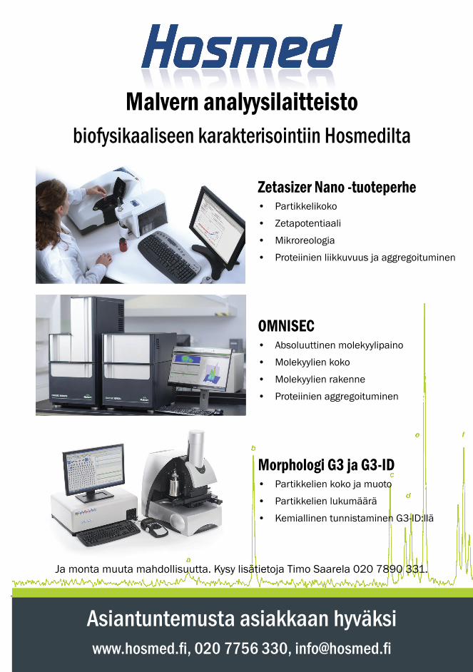
75
Asiantuntemusta asiakkaan hyväksiwww.hosmed.fi , 020 7756 330, [email protected]
Zetasizer Nano -tuoteperhe• Partikkelikoko
• Zetapotentiaali
• Mikroreologia
• Proteiinien liikkuvuus ja aggregoituminen
OMNISEC• Absoluuttinen molekyylipaino
• Molekyylien koko
• Molekyylien rakenne
• Proteiinien aggregoituminen
Morphologi G3 ja G3-ID• Partikkelien koko ja muoto
• Partikkelien lukumäärä
• Kemiallinen tunnistaminen G3-ID:llä
Malvern analyysilaitteisto biofysikaaliseen karakterisointiin Hosmedilta
Ja monta muuta mahdollisuutta. Kysy lisätietoja Timo Saarela 020 7890 331.