Embed Size (px)
Citation preview
Current Research in Food Science 3 (2020) 178–188
Contents lists available at ScienceDirect
Current Research in Food Science
journal homepage: www.editorialmanager.com/crfs/
Research Paper
Pickering emulsions stabilized by colloidal gel particles complexed orconjugated with biopolymers to enhance bioaccessibility and cellular uptakeof curcumin
Andrea Araiza-Calahorra a, Yunqing Wang b, Christine Boesch b, Yansheng Zhao c,Anwesha Sarkar a,*
a Food Colloids and Bioprocessing Group, School of Food Science and Nutrition, University of Leeds, Leeds, LS2 9JT, UKb Nutritional Sciences and Epidemiology Group, School of Food Science and Nutrition, University of Leeds, Leeds, LS2 9JT, UKc School of Food and Biological Engineering, Jiangsu University, Zhenjiang, 212013, China
A R T I C L E I N F O
Keywords:Pickering emulsionMicrogelCurcuminin vitro digestionCaco-2 cellsCellular uptake
* Corresponding author.E-mail address: [email protected] (A. Sarkar
https://doi.org/10.1016/j.crfs.2020.05.0012665-9271/© 2020 The Author(s). Published by Els
A B S T R A C T
The aim of this study was to investigate the fate of curcumin (CUR)-loaded Pickering emulsions with complexinterfaces during in vitro gastrointestinal transit and test the efficacy of such emulsions on improving the bio-accessibility and cellular uptake of CUR. CUR-loaded Pickering emulsions tested were whey protein nanogelparticle-stabilized Pickering emulsions (CUR-EWPN) and emulsions displaying complex interfaces included 1)layer-by-layer dextran sulphate-coated nanogel-stabilized Pickering emulsions (CUR-DxSþEWPN) and 2) pro-teinþdextran-conjugated microgel-stabilized Pickering emulsions (CUR-EWPDxM). The hypothesis was that thepresence of complex interfacial material at the droplet surface would provide better protection to the dropletsagainst physiological degradation, particularly under gastric conditions and thus, improve the delivery of CUR toCaco-2 intestinal cells. The emulsions were characterized using droplet sizing, apparent viscosity, confocal andcryo-scanning electron microscopy, zeta-potential, lipid digestion kinetics, bioaccessibility of CUR as well as cellviability and uptake by Caco-2 cells. Emulsion droplets with modified to complex interfacial composition (i.e.CUR-DxSþEWPN and CUR-EWPDxM) provided enhanced kinetic stability to the Pickering emulsion droplets againstcoalescence in the gastric regime as compared to droplets having unmodified interface (i.e. CUR-EWPN), whereasdroplet coalescence occurred in intestinal conditions irrespective of the initial interfacial materials. A similar rateand extent of free fatty acid release occurred in all the emulsions during intestinal digestion (p > 0.05), whichcorrelated with the bioaccessibility of CUR. Striking, CUR-DxSþEWPN and CUR-EWPDxM significantly improvedcellular CUR uptake as compared to CUR-EWPN (p < 0.05). These results highlight a promising new strategy ofdesigning gastric-stable Pickering emulsions with complex interfaces to improve the delivery of lipophilicbioactive compounds to the cells for the future design of functional foods.
1. Introduction
Curcumin (CUR), a natural polyphenol, is the major curcuminoid(70–80%) present in the rhizomes of turmeric plant Curcuma longa (Goelet al., 2008). Due to its potential health-promoting properties such asantitumor, anti-oxidant, anti-microbial and anti-inflammatory, theincorporation of CUR into functional foods has been of major interest inrecent years to both functional food and pharmaceutical industries(Anand et al., 2007). However, significant research challenges exist withthe incorporation of CUR and its use as a bioactive ingredient due to
).
evier B.V. This is an open access
limited aqueous solubility, high rate of metabolism and low bioavail-ability with rapid clearance of CUR (Tønnesen et al., 2002).
To address these delivery challenges of CUR, several colloidal ap-proaches such as liposomes, vesicles, protein-based complexes, andemulsion-based delivery systems have surfaced in the literature (Amaniet al., 2019; Araiza-Calahorra et al., 2018; Kolter et al., 2019; Sarkar andMackie, 2020). In particular, emulsion-based delivery systems have beenincreasingly used to encapsulate CUR due to their simple processingtechnique, the fact that they can be made entirely from bio-based ma-terials, and that they are suitable for incorporation into a variety of food
article under the CC BY license (http://creativecommons.org/licenses/by/4.0/).

A. Araiza-Calahorra et al. Current Research in Food Science 3 (2020) 178–188
matrices (Kharat and McClements, 2019). Nonetheless, emulsion-baseddelivery vehicles are mostly designed in isolation and often, the fate ofthe encapsulated CUR within these delivery systems during physiologicaltransit remains poorly understood. In particular, the bioaccessibility ofCUR after passing through the gastrointestinal (GI) tract and its perme-ability across the intestinal epithelium are crucial to understand the ef-ficacy of these delivery vehicles, which have been given rare attention inthe literature to date (Zou et al., 2015).
Among the emulsion-based delivery vehicles, Pickering emulsions haveattracted significant recent scientific and industrial interests since theypossess many advantages in terms of high stability against coalescence andOstwald ripening (Rayner et al., 2012, Tzoumaki et al., 2011), andcontrolled digestibility of lipids by preventing competitive displacementby bio-surfactants (bile salts) (Dickinson, 2012; Ruiz-Rodriguez et al.,2014; Sarkar et al., 2016, 2019; Shimoni et al., 2013; Xiao et al., 2015).More specifically, Pickering emulsions stabilized by a wide range of par-ticles, such as protein nanogels, modified starch granules,chitosan-tripolyphosphate complexes, silica, kafirin, ovotransferrin fibrils,nanocellulose and kaolinite (Araiza-Calahorra and Sarkar, 2019a; Asa-buwa Ngwabebhoh et al., 2018; Lu et al., 2019; Marefati et al., 2017; Shahet al., 2016a, 2016b; Tang et al., 2019; Tikekar et al., 2013; Wei et al.,2019) have been recently used as delivery vehicles for CUR. However, onlya few studies have investigated the biofunctionalities of the encapsulatedCUR in these Pickering emulsions after in vitro digestion (Lu et al., 2019;Marefati et al., 2017; Shah et al., 2016b; Tikekar et al., 2013; Wei et al.,2019). Many of these emulsions have been prepared using inorganicparticles, restricting their application in edible formats (Asabuwa Ngwa-bebhoh et al., 2018; Shah et al., 2016a, 2016b; Tang et al., 2019; Tikekaret al., 2013). In addition, literature is scarce on CUR bioaccessibility andpotential cell toxicity as well as uptake of CUR by the cells when CUR isencapsulated in such Pickering emulsions (Lu et al., 2019).
In our previous study, we demonstrated the capacity of whey proteinnanogel-stabilized Pickering emulsions to encapsulate CUR under differentconditions of physiologically relevant different pHs and ionic strengths(Araiza-Calahorra and Sarkar, 2019a). In addition, Pickering emulsionsstabilized by complex interfaces such as dextran sulphate (DxS)-coatednanogel particles (Araiza-Calahorra and Sarkar, 2019b) or conjugatemicrogels in which dextran (Dx) was covalently conjugated to proteinbefore the micro-gelation process (Araiza-Calahorra et al., 2020) havesuccessfully demonstrated higher kinetic stability to coalescence in thegastric phase as compared to that of non-modified simplenanogel-stabilized emulsions. The aim of this work was therefore tocompare Pickering emulsions with complex interfaces (electro-statically-driven protein gel particles þ biopolymer orcovalently-conjugated protein-biopolymer gel particles at the interfaces)over nanogel particles as delivery vehicles for CUR for the first time. Totest the efficacy of these delivery vehicles for curcumin, the fate of thesePickering emulsions loaded with CUR in simulated in vitro gastrointestinaldigestion environment was investigated followed by assessment of cur-cumin bioaccessibility and cellular uptake. To our knowledge, the bio-accessibility and efficacy for delivering CUR to Caco2-cells after in vitrostatic simulated digestion when encapsulated in complexparticle-stabilized Pickering emulsions has not been studied to date. Ourhypothesis was that complex interfacial material can provide a betterbarrier to the droplets in the gastric environment and thus allow efficientrelease in the intestinal phase and therefore enhance the cellular uptake ofCUR. Thus, novel insights from this study would advance the fundamentalunderstanding of how interfacial design of emulsions can be tailored toalter gastrointestinal release and increase intestinal uptake of CUR.
2. Materials and methods
2.1. Materials
Curcumin (CUR) (1,7-bis(4-hydroxy-3-methoxyphenyl)-1,6-hepta-diene-3,5-dione) (�65% purity), dextran (Dx) as well as dextran sulphate
179
(DxS) of molecular weight (MW) 500 kDa were purchased from Sigma-Aldrich Company Ltd (Dorset, UK) and used without any further purifi-cation. Powdered whey protein isolate (WPI) with�90% protein contentwas a kind gift from Fonterra Co-operative Group Limited (Auckland,New Zealand). Miglyol® 812medium-chain triglyceride (MCT) oil with adensity of 945 kg m3 at 20 �C was purchased from Cremer Oleo GmbH &Co (Germany) and was used as the dispersed phase without any furtherpurification. All enzymes i.e. porcine pepsin (P7000, 526 U mg�1 usinghaemoglobin as a substrate), porcine pancreatin (P7545, 8 � USP andtrypsin activity of 6.48 Umg�1 using TAME, N-p-Tosyl-L-arginine methylester hydrochloride, as a substrate) and porcine bile extract B8631 (totalbile salt content 49 wt% with 10–15% glycodeoxycholic acid, 3–9%taurodeoxycholic acid, 0.5–7% deoxycholic acid, 5 wt% phospholipids)were purchased from Sigma-Aldrich Company Ltd. For cell culture ex-periments, human colon adenocarcinoma (Caco-2) cells were purchasedfrom the European Collection of Authenticated Cell Culture (ECACC).Cell culture media and supplements i.e. Dulbecco's Modified Eagle Me-dium (DMEM), fetal bovine serum (FBS), Dulbecco's Phosphate-BufferedSaline (DPBS), non-essential amino acids (NEAA), trypsin EDTA, andpenicillin-streptomycin mixture (5000 U mL-1)) were obtained fromGibco Cell Culture Products, Thermo Fisher Scientific (UK). Neutral redpowder, HPLC-grade methanol, ethanol, and analytical-grade glacialacid, were acquired from Sigma-Aldrich Company Ltd. All solutions wereprepared with Milli-Q water (resistivity of 18.2 MΩ cm at 25 �C) (Milli-Qapparatus, Millipore, Bedford, UK).
2.2. Methods
2.2.1. Preparation of CUR-loaded Pickering emulsion systems
2.2.1.1. Preparation of whey protein nanogel particles (WPN) and wheyprotein isolateþdextran conjugate microgel particles (WPDxM). Whey pro-tein nanogel particles (WPN) were produced based on a previouslydeveloped top-down technique (Araiza-Calahorra and Sarkar, 2019a).Briefly, WPI powder (10 wt%) was dissolved in 20 mM phosphate bufferat pH 7.0 for 2 h and the solution was heated in a temperature-controlledwater bath at 90 �C for 30 min to form a heat-set gel (quiescent). Theresultant WPI gels were pre-homogenized with phosphate buffer (5 wt%protein) using a hand blender (HB724, Kenwood) for 1 min and theresulting whey protein macrogel dispersion (5 wt% protein) was passedthrough a high-pressure homogenizer at 300 bars for two passes to createWPN. The resultant WPN was diluted with buffer to the desired proteinconcentration for the Pickering emulsion preparation.
Whey protein isolateþDx conjugate powder was prepared asdescribed previously (Araiza-Calahorra et al., 2020). The pH of theWPIþDx solution (1:2 w/w ratio) was adjusted to pH 7.0 and gentlystirred for 2 h at 25 �C. TheWPIþDx solution was stored at 4 �C overnightand then frozen at �20 �C for 6 h. Samples were then freeze-dried for24 h and Maillard reaction of the resulting WPIþDx was promoted byincubating the powder in a pre-heated desiccator at 60 �C for 24 h, withrelative humidity (79%) controlled by saturated KBr solution.
Whey protein isolateþDx conjugated microgels (WPDxM) with adegree of conjugation of 10% were produced using the aforementionedmethod used for creating WPN with minor modifications. Briefly, theconjugate powder was dispersed for 2 h in phosphate buffer at pH 7.0 toensure complete dissolution to a final protein concentration of 11.6 wt%.The conjugate solution was heated in a temperature-controlled waterbath at 65 �C for 1 h to form a heat-set gel (quiescent), followed bycooling down for 15 min and stored at 4 �C. The obtained gels were pre-homogenized with phosphate buffer (2 wt%) to create macrogel particlesusing a hand blender (HB724, Kenwood) for 1 min and then passedthrough a high-pressure homogenizer at 300 bars twice to createmicrogel particles. Obtained conjugate microgel particles (WPDxM) werediluted with buffer to the desired protein concentration for the Pickeringemulsion preparation.

A. Araiza-Calahorra et al. Current Research in Food Science 3 (2020) 178–188
2.2.1.2. CUR-loaded Pickering oil-in-water emulsion preparation. The oilphase was prepared by dissolving 2 wt% CUR into heated MCT-oil(60 �C), by magnetically stirring for 30 min, and centrifuging for10 min at 4 �C to remove any undissolved CUR (Araiza-Calahorra andSarkar, 2019a). Oil-in-water Pickering emulsions (80:20 w/w) con-taining CUR i.e. CUR-EWPN or CUR-EWPDxM were prepared using WPNor WPDxM as Pickering stabilizers, respectively. Coarse emulsionswere prepared by homogenizing the MCT-oil containing CUR withfresh WPN or WPDxM aqueous suspension at pH 7.0 (1 wt% finalprotein concentration in all emulsions) using an Ultra Turrax T25homogenizer (IKA-Werke GmbH & Co., Staufen Germany) at 13,500 rpm for 1 min. Fine CUR-EWPN or CUR-EWPDxM droplets wereprepared by passing the coarse emulsions twice through ahigh-pressure homogenizer at 300 bars. For the biopolymer-coatedPickering emulsions, CUR-EWPN (40 wt% MCT-oil) and aqueous dis-persions of DxS of 500 kDa Mw (0.4 wt%) were mixed in 1: 1 w/w atpH 3.0 to allow mutually attractive interaction between the cationicWPN and anionic DxS at the interface, as previously described byAraiza-Calahorra and Sarkar (2019b) and produce CUR-DxS-EWPN(20 wt% MCT, 1 wt% WPN). For comparison purposes, all emulsionscontained the same amount of oil and protein.
2.2.2. Particle and droplet size measurementsLight scattering was used to measure the size distribution of the initial
nanogel/microgel particles (dynamic light scattering, DLS) and freshemulsion droplets (static light scattering, SLS) undergoing in vitrogastrointestinal digestion. Aqueous dispersions of WPN and WPDxM wasmeasured using DLS at 25 �C using a Zetasizer Nano-ZS (Malvern In-struments, Malvern UK) after 100� dilution in phosphate buffer (pH 7.0)at room temperature. Droplet size distributions before and after the invitro digestion of the emulsion samples were determined using SLS at25 �C using Malvern MasterSizer 3000 (Malvern Instruments Ltd, Mal-vern, Worcestershire, UK). The mean particle size of the emulsions wasreported as volume mean diameter (d43) as it is more sensitive to dropletaggregation with systems showing bimodal size distribution. Results arebased on three measurements on triplicate samples.
2.2.3. ζ-potential measurementsThe ζ-potential values of aqueous dispersions of the nanogel and
microgel particles and the three Pickering emulsion samples weredetermined using Zetasizer, Nano ZS series, Malvern Instruments, Wor-cestershire, UK. Samples before and after in vitro digestion were dilutedto 0.01% particle or 0.004 wt% oil in 100� in phosphate buffer (pH 7.0)or SGF buffer (pH 3.0) or SIF buffer (pH 7.0) depending upon the con-dition and added to a folded capillary cell (Model DTS 1070, MalvernInstruments Ltd., Worcestershire, UK). The ζ-potential measurementswere performed for duplicate samples with three readings for each ofthem.
2.2.4. Apparent viscosityThe apparent viscosity of the freshly prepared Pickering emulsions
was measured using a rheometer (Kinexus Ultraþ, Malvern InstrumentsLtd, Worcestershire, UK) equipped with a cone-and-plate geometry(diameter 40 mm, model: CP4/40 SS017SS). About 1.4 mL of theemulsion sample was placed onto the sample plate. Apparent viscositieswere obtained for all the emulsion samples as a function of shear ratesranging from 1 to 1000 s�1 at 37 �C. Data from the flow curves were fittedto Ostwald de Waele fit as shown in Eq. (1):
ηað _γÞ¼K _γn�1 (1)
where ηa is the apparent viscosity, _γ is the shear rate, K is the consistencyindex and n is the flow behaviour index. Linear regression analysis wasapplied to the data to calculate the flow behaviour index and the con-sistency coefficient.
180
2.2.5. In vitro gastrointestinal digestion (static model)A static digestion model was used in the in vitro digestion experiment
employing a slightly adapted version fromMinekus et al. (2014) omittingthe oral step. Exactly 5 mL of the CUR-loaded Pickering emulsions at pH3.0 (pre-incubated at 37 �C, 1 h) were mixed with 5 mL of simulatedgastric fluid (SGF), consisting of 0.257 g L�1 of KCl, 0.061 g L�1 ofKH2PO4, 1.05 g L�1 of NaHCO3, 1.38 g L�1 of NaCl, 0.0122 g L�1 ofMgCl2(H2O)6, 0.024 g L�1 of (NH4)2CO3 and 2000 U/mL pepsin at pH3.0. The mixture was incubated for 2 h at 37 �C under agitation using ashaking water bath (Grant Instruments Ltd, Cambridge, UK) at 100 rpm.
To allow sequential gastrointestinal digestion, after 2 h of incubation,the pH of the sample þ SGF (10 mL) was adjusted to pH 6.8 with 1 MNaOH and mixed with 7.73 mL of simulated intestinal fluid (SIF) elec-trolyte stock solution consisting of 0.254 g L�1 of KCl, 0.054 g L�1 ofKH2PO4, 3.570 g L�1 of NaHCO3, 1.123 g L�1 of NaCl and 0.335 g L�1 ofMgCl2(H2O)6, 1.25 mL fresh bile (10 mM in the final digesta), 20 μL of0.3 M CaCl2 and 1 mL of a pancreatin solution (100 U/mL based ontrypsin activity in the final digesta) made up in SIF electrolyte stock so-lution. The in vitro intestinal digestion was carried out over 3 h at pH 6.8and 37 �C.
During this 5 h in vitro digestion period, samples (sampleþSGF andsampleþSGFþSIF) were periodically collected for characterization.Samples were also prepared where CUR was dispersed in MCT-oilwithout any added protein gel particles and without employing anyemulsification process (no vehicle) and also digested using similar SGFand SIF buffer. To stop the pepsin activity at specific time points, 0.2 Msodium bicarbonate was added to the samples to reach a final pH of 7.0.The pancreatin activity was stopped by adding 0.1 M of 4-(2-aminoethyl)benzenesulfonyl fluoride hydrochloride (Pefabloc©) to the sample (5 mMfinal concentration). Experiments were performed in triplicate and meanvalues were calculated.
2.2.6. Free fatty acid releaseAfter passing through simulated gastric and intestinal conditions, the
free fatty acids (FFAs) released from the CUR-loaded emulsions weremeasured by using an automatic pH-stat titration unit (TIM 856 titrationmanager, Titralab, Radiometer analytical). Noteworthy that for doing thepH stat analysis of FFA release, this was a separate experiment where noaliquots were removed during the sequential gastrointestinal digestion.The pH-stat was used to monitor and control the pH at pH 6.8 for 3 h. Thevolume of added NaOH (0.25 M) was assumed to be equal to the amountof free fatty acids generated by the lipolysis of emulsified triacylglycerols.The amount of free fatty acids released was calculated from the titrationcurves as described by Sarkar et al. (2016). Using a nonlinear regressionmodel, the kinetic parameters for the initial stages of FFA release werederived as described previously (Sarkar et al., 2016, 2019) using Eqs. (2)and (3).
Φt ¼Φmax
�1� exp
��6kMwDnt2
ρod20Γ
max
��(2)
where t is the lipid digestion time in the intestine (min), Φmax is themaximum total FFA level (%), k (mol s�1 m�2) is the conversion rate ofthe lipid per unit area of the emulsion droplet surface, occurring at themaximum lipase surface coverage, Mw is the molecular weight of MCT-oil, do is the initial average diameter of the emulsions (d32) and ρo isthe density of the MCT-oil. Γmax is the maximum coverage of the surfaceby the enzyme, D is the diffusion coefficient of the enzyme in thecontinuous aqueous phase and n donates the molar concentration of thelipase in SIF solution. In addition, the lipolysis half time (t1/2) (minutes)i.e. the time required to achieve half of the maximum extent of lipiddigestion was obtained from Eq. (3) (Sarkar et al., 2016, 2019):
t1=2 ¼ lnð2Þ�doρo6kMw
�(3)
A. Araiza-Calahorra et al. Current Research in Food Science 3 (2020) 178–188
2.2.7. Bioaccessibility of CURThe bioaccessibility of CUR in the Pickering emulsions was deter-
mined after 5 h of sequential in vitro gastrointestinal digestion. Thedigesta obtained at the end of the sequential gastrointestinal digestionprocess was centrifuged at 3000�g for 50 min at 5 �C. The middle layerwas considered to be the “micellar fraction”, in which the CUR wassolubilized. The concentration of CUR in the micelles was analysed usinghigh-performance liquid chromatography (HPLC) analysis. An Agilent1200 series HPLC instrument coupled with DAD detector was used for theanalyses of CUR. The measurement wavelength was 425 nm, and theseparation column was Agilent XDS-C18 (150 mm�4.6 mm, 5μm). Themobile phase A was 0.2% of acetic acid aqueous, and the mobile phase Bwas acetonitrile, with a flow rate of 1 mL/min. The gradient elutionprogram was: 0min, 60% of A; 4–10min: 20% of A. The column tem-perature was maintained at 25 �C. The injection volume was 20 μL, andan external CUR standard was used for quantitative analysis. A calibra-tion curve was prepared with standard CUR in acetonitrile in concen-trations ranging from 0.1 μM to 20.0 μM. Bioaccessibility (%) of CUR wascalculated by dividing the amount of solubilized CUR in the micellarphase by the amount of CUR in the emulsion.
2.2.8. Microstructural characterization
2.2.8.1. Confocal scanning laser microscopy (CLSM). The microstructureof the samples before and after in vitro digestion was imaged using a ZeissLSM 700 CLSM (Carl Zeiss MicroImaging GmbH, Jena, Germany)confocal microscope using an oil immersion 63 � lens and the pinholediameter maintained at 1 Airy Unit to filter out the majority of the lightscattering. A stock solution of Fast Green (1 mg mL─1 in Milli-Q water)was used to stain the protein particles to a final concentration of0.1 mgmL─1, which was excited at a wavelength of 633 nm. The emissionfilter was set at 660–710 nm. Samples were placed on a concave confocalmicroscope slide, secured with a glass coverslip and imaged.
2.2.8.2. Cryogenic-scanning electron microscopy. Cryogenic scanningelectron microscopy (cryo-SEM) of the fresh CUR-loaded emulsionsamples i.e. CUR-EWPN, CUR-DxSþEWPN, and CUR-EWPDxM were con-ducted. Cryo-SEM images were acquired using heptane as the dispersedrather than MCT oil to avoid interference by crystallization of oil duringthe freezing step as used in previous studies by Destribats et al. (2014)and Araiza-Calahorra and Sarkar (2019a). The CUR-EWPN,CUR-DxSþEWPN, and CUR-EWPDxM were mounted on rivets attached tothe sample stub. The samples were plunge-frozen in liquid nitrogen“slush” at �180 �C and then transferred to the cryo-preparation chamberin the SEM. The frozen Pickering emulsion droplets were cleaved andthen etched at �95 �C for 4 min. Next, the samples were coated with5 nm of platinum (Pt). Finally, the Pt-coated samples were transferred tothe SEM for imaging at �135 �C. The heptane-based emulsion sampleswere imaged in a FEI Quanta 200 F ESEM with a Quorum Polar Prep2000 cryo system.
2.2.9. Cell-based assays
2.2.9.1. Cell culture. Human colon adenocarcinoma cells, Caco-2, werecultivated in high glucose DMEM medium with pyruvate, supplementedwith 10% FBS, 1% NEAA, 100 U mL-1 penicillin and 100 μg mL-1 strep-tomycin. Cells were grown under standard conditions at 37 �C with 5%CO2 in a humidified atmosphere and medium was changed every 2–3days. Cells were used for experiments within 10 in-house passages.
2.2.9.2. Cytotoxicity assay. The cytotoxicity of the micellar phases of thePickering emulsions was assessed by the neutral red assay as describedpreviously (Perez-Hernandez et al., 2020). Briefly, Caco-2 cells wereseeded in 24-well plates at a density of 1 � 105 cells cm�2 and, uponreaching min 90% confluence, they were treated with CUR dissolved in
181
DMSO (0.5–6.0 μM), micellar phase of digested CUR-encapsulatedemulsion sample or micellar phase of digested emulsion samplewithout CUR. The medium was removed after 2.5 h and replaced byDMEM containing 40 μg mL�1 neutral red dye which was incubated for2 h. Subsequently, cells were washed with DPBS and the intracellular dyeextracted by distain solution (AcOH/H2O/glacial acid, 50:49:1, v/v/v)for 10–15 min. Absorbance of the neutral red dye was measured at540 nm using a microplate reader (Tecan Spark 10 M, Switzerland).Viability of the Caco-2 cells was calculated as the percentage of controlcells (DMEM medium only). DMSO (5%) was included as control whichlowered the cell viability to 67%. Experiments were conducted over threeindependent passages and performed in triplicates per experiment.
2.2.9.3. Cellular curcumin uptake. The cellular uptake of CUR from themicellar phase of digested CUR-encapsulated Pickering emulsions wasdetermined using HPLC. Caco-2 cells were seeded at a density of2� 106 cells per 10 cm Petri dish. When reaching confluence (min 90%),the cells were exposed to different micellar phase digesta containing1 μM CUR for 2 h under standard conditions. Subsequently, cells werewashed twice with ice-cold DPBS and lysed with methanol. The cellpellets were collected and subjected to extraction, which involved vortex(1 min), sonication (4 �C, 5 min) and centrifugation (10,000�g, 4 �C,10 min). The supernatant was filtered through a 0.2 μmPTFE syringe andprepared for the subsequent curcumin HPLC analysis. Experiments wereconducted in triplicate in subsequent cell passages.
2.3. Statistical analysis
Significant differences between samples were determined by one-wayANOVA and multiple comparison test and Tukey’s adjustment was per-formed using SPSS software (IBM, SPSS statistics, version 24) and thelevel of confidence was 95%. Experiments were conducted at least intriplicate. Results in tables are expressed as mean � standard deviation.Error bars in figures represent standard deviation.
3. Results and discussion
3.1. Characteristics of curcumin Pickering emulsions
Initially, we evaluated the characteristics of the Pickering emulsionsystems (CUR-EWPN, CUR-DxSþEWPN, and CUR-EWPDxM) using dropletsize, size distribution, microstructure, surface morphology, and electricalcharacteristics (ζ) before and after gastric and intestinal digestion steps.Apparent viscosity of the emulsions was also evaluated to understandhow bulk properties might impact digestion behaviour. Fig. 1 shows thesurface morphology of the freshly prepared Pickering emulsion sampleswith particle-laden interfaces probed using cryo-SEM. Fig. 1a1 showsseveral CUR-EWPN emulsion droplets homogeneously distributedthroughout the micrograph with a woolly jacket of WPN attached to thedroplet surface giving a raspberry-like surface appearance. At highermagnification (Fig. 1a2), WPN seemed to have an end-to-end aggregationat the droplet surface, which might be associated with nanogel mergingwith each other at the interface. Besides, sample preparation process (e.g.freeze-fracturing) during the cryo-SEM might had also result in suchaggregation. Similar morphology has been also previously observed incryo-SEM images of Pickering droplets in WPN-stabilized oil dropletswithout containing CUR (Araiza-Calahorra and Sarkar 2019a). Thissuggested that the addition of CUR had limited effects on the surfacemorphology of the droplets.
Pickering emulsion droplets stabilized by a more complex interfacei.e. with WPN electrostatically-coated with DxS of 500 kDa Mw werespherical with an average diameter of ~15 μm (Fig. 1b1). Looking at thesurface of such droplets at higher magnification, it can be observed thatindividual spherical WPN seemed to be aggregated, which can be aneffect of DxS coating electrostatically attracting multiple neighbouring

Fig. 1. Cryo-SEM images of the three Pickering emulsions used for delivering curcumin i.e. a) CUR-EWPN emulsion (magnification of 15,000 � (a1), and 50,000� (a2), respectively), b) CUR-DxSþEWPN (magnification of 25,000 � (b1), and 50,000 � (b2), respectively) and c) CUR-EWPDxM (magnification of 10,000 � (c1),and 20,000 � (c2), respectively). Arrows indicate the nanogel particle in a2, nanogel particle aggregated with dextran sulphate in b2, and conjugate microgel particlesin c2.
Table 1Mean droplet size and ζ-potential of initial emulsions.
Emulsions d43/μm ζ-potential/mV
CUR-EWPN 14.93 � 3.3 �26.9 � 0.5CUR-DxSþEWPN 54.56 � 6.1 �32.33 � 4.8a
CUR-EWPDxM 7.9 � 0.2 �15.36 � 1.1
a Note that the initial emulsion CUR-DxSþEWPN was at pH 3.0 to allow elec-trostatic attraction between WPN and DxS, unlike the other two emulsions,which were at pH 7.0.
A. Araiza-Calahorra et al. Current Research in Food Science 3 (2020) 178–188
WPN within a thread-like network (Fig. 1b2). The other Pickeringemulsion with complex interface i.e. CUR-EWPDxM samples showed thepresence of conjugated microgel particles formed a thin surface layeradopting a more discrete configuration of individual microgel particles(Fig. 1c1 and c2).
The droplet size distribution and mean diameters with representativeconfocal images of the three freshly prepared Pickering emulsion systemsare shown in Table 1 and Fig. 2. The initial droplet size distribution of thethree emulsions, CUR-EWPN, CUR-DxSþEWPN, and CUR-EWPDxM presentedbimodal distributions where the peak in the area of 0.1–1 μm in allsystems correspond to unadsorbed particles in line with the systemspreviously studied without loaded CUR (Araiza-Calahorra and Sarkar2019a, 2019b) and small emulsion droplets, while the peak in the area of1–100 μm corresponds to the bigger emulsion droplets (Fig. 2). TheCUR-EWPN presented oil droplet ranging in size from 1 to 50 μm and anaverage droplet diameter (d43) of 14.93 μm (Table 1), whereasCUR-DxSþEWPN system presented oil droplets ranging in size from 3 to100 μmand an average droplet diameter (d43) of 54.56 μm (Table 1). Thiscan be expected as the electrostatic coating with DxS resulted in dropletflocculation with DxS not only binding to individual droplets but also
182
connecting two or more adjacent droplets as can be clearly observed asflocs in the confocal images (Fig. 2). On the other hand, the conjugatedmicrogel-laden system i.e. CUR-EWPDxM presented a d43 of 7.9 μm(Table 1). From the confocal images in Fig. 2, it is noticeable that all threesystems had proteinaceous particles (stained in green) adsorbed at theinterface acting as a barrier against oil droplet coalescence.
All the Pickering emulsions studied were negatively charged(Table 1). CUR-EWPN presented a high negative charge because theinitial pH of the emulsions was appreciably above the isoelectric point
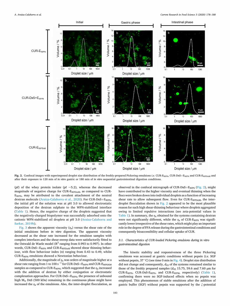
Fig. 2. Confocal images with superimposed droplet size distribution of the freshly-prepared Pickering emulsions i.e. CUR-EWPN, CUR-DxSþEWPN and CUR-EWPDxM andafter their exposure to 120 min of in vitro gastric or 180 min of in vitro sequential gastrointestinal digestion conditions.
A. Araiza-Calahorra et al. Current Research in Food Science 3 (2020) 178–188
(pI) of the whey protein isolate (pI ~5.2), whereas the decreasedmagnitude of negative charge for CUR-EWPDxM, as compared to CUR-EWPN, may be attributed to the covalent attachment of the neutraldextran molecule (Araiza-Calahorra et al., 2020). For CUR-DxSþEWPN,the initial pH of the solution was at pH 3.0 to allowed electrostaticdeposition of the dextran sulphate to the WPN-stabilized interface(Table 1). Hence, the negative charge of the droplets suggested thatthe negatively-charged biopolymer was successfully adsorbed onto thecationic WPN-stabilized oil droplets at pH 3.0 (Araiza-Calahorra andSarkar, 2019b).
Fig. 3 shows the apparent viscosity (ηa) versus the shear rate of theinitial emulsions before in vitro digestion. The apparent viscositydecreased as the shear rate increased for the emulsion samples withcomplex interfaces and the shear-sweep data were satisfactorily fitted tothe Ostwald de Waele model (R2 ranging from 0.993 to 0.997). In otherwords, CUR-DxSþEWPN and CUR-EWPDxM showed shear thinning behav-iour, with flow behaviour index (n) ranging from 0.43 to 0.49, whilstCUR-EWPN emulsions showed a Newtonian behaviour.
Additionally, the magnitude of ηa was orders of magnitude higher at ashear rate ranging from 1 to 100 s�1 for CUR-DxSþEWPN and CUR-EWPDXMsamples as compared to CUR-EWPN, which suggested that the ηa increasedwith the addition of dextran by either conjugation or electrostaticcomplexation approaches. For CUR-DxSþEWPN, the presence of unboundhigh Mw DxS (500 kDa) remaining in the continuous phase might haveincreased the ηa of the emulsions. Also, the inter-droplet flocculation, as
183
observed in the confocal micrograph of CUR-DxSþEWPN (Fig. 2), mighthave contributed to the higher viscosity and eventual thinning when theflocswerebrokendown into individual droplets as a functionof increasingshear rate to allow subsequent flow. Even for CUR-EWPDxM, the inter-droplet flocculation shown in Fig. 2 appeared to be the most plausiblereason for such high shear-thinning behaviour where droplets aggregatedowing to limited repulsive interactions (see zeta-potential values inTable 1). In summary, the ηa obtained for the systems containing dextranwere not significantly different, while the ηa of CUR-EWPN was signifi-cantly lower irrespective of the shear rates,whichmight play an importantrole in the degree of FFA release during the gastrointestinal conditions andconsequently bioaccessibility and cellular uptake of CUR.
3.1. Characteristics of CUR-loaded Pickering emulsions during in vitrogastrointestinal digestion
The kinetic stability and responsiveness of the three Pickeringemulsions was accessed at gastric conditions without pepsin (i.e. SGFwithout pepsin, 37 �C) (see time 0min in Fig. 4). Droplet size distributiondid not change and consequently, d43 of the systems remained similar tothose of the freshly prepared samples (d43 15.75, 59.6 and 7.60 μm forCUR-EWPN, CUR-DxS-EWPN, and CUR-EWPM, respectively) (Table 1),confirming there were no SGF-induced effects when no pepsin wasemployed. This phenomenon of stable emulsions after the addition ofgastric buffer (SGF) without pepsin was supported by the ζ-potential

Fig. 3. Flow curves of freshly prepared Pickering emulsions i.e. CUR-EWPN
(black squares) CUR-DxSþEWPN (blue triangles) and CUR-EWPDxM (red circles) at37 �C. Data points represent the average of at least three measurements ontriplicate sample. Error bars indicate the standard deviations. Solid lines are thebest fits to the experimental data predicted using the Ostwald de Waele model(Eq. (1)). (For interpretation of the references to colour in this figure legend, thereader is referred to the Web version of this article.)
A. Araiza-Calahorra et al. Current Research in Food Science 3 (2020) 178–188
measurements where both CUR-EWPN and CUR-EWPDxM presented acharge reversal from negative to positive due to the protonation of theionizable groups as they move from above to below the isoelectric point(Fig. 4b). Interestingly, there were no significant differences in theζ-potential of CUR-DxSþEWPN after addition of gastric buffer (p > 0.05).
After being exposed to 120min of in vitro gastric digestion stage in thepresence of pepsin, d43 values significantly decreased (p> 0.05) for CUR-DxSþEWPN from 59.6 μm to 52.75 μm, (Fig. 4a), which explained theslight shift to smaller values observed in the droplet distribution with thebreakdown of the droplet flocs (Fig. 2). For CUR-EWPN, the droplet sizedistribution evidenced some coalescence phenomena, which is clearlyshown by a rise in a third peak in the range of 100–1000 μm size range(Fig. 2), whereas the d43 and droplet size distribution remained un-changed for CUR-EWPDxM. This behaviour in CUR-EWPN might be relatedto the peptic hydrolysis of the proteinaceous nanogel particles at thesurface of the emulsion droplets, which might have caused some coa-lescence of the droplets, whereas CUR-EWPDxM presented good physicalstability after the gastric stage. The complex interfaces were indeedsuccessful in providing gastric stability to the droplets. Such desirableresults in the case of CUR-DxSþEWPN and CUR-EWPDxM might be attrib-uted to the polysaccharide coating/conjugation, which restricted theaccess of pepsin to potential cleavage sites of the protein (Araiza-Cala-horra et al., 2020; Araiza-Calahorra and Sarkar, 2019b). On the otherhand, the high bulk viscosity of the CUR-DxSþEWPN and CUR-EWPDxM(Fig. 3) may have also hindered the diffusion of pepsin to the proteina-ceous sides of the particle (Sarkar et al., 2017).
Upon subjecting the emulsions to gastric conditions with pepsin, theζ-potential became less positive for all the samples, with CUR-EWPNpresenting significant differences from þ31.7 to þ16.1 mV after120 min of gastric digestion (Fig. 4b). This reduction in the absolutemagnitude of ζ-potential after in vitro gastric conditions further sup-ported the pepsin-induced hydrolysis of WPN particles when absorbedat the EWPN interface. However, such changes were not seen in theemulsions stabilized by complex interfaces supporting the confocalimages and size distribution data (Fig. 2), highlighting the kinetic sta-bility of these emulsions with complex interfaces to droplet coalescencein the gastric conditions.
Under small intestinal conditions, all samples studied exhibited asignificant increase in the d43 ranging between 60.25 and 69.25 μmcorresponding to droplet coalescence irrespective of the initial interfacial
184
material (Figs. 2 and 4c). From the microscopic analysis, it became clearthat the larger droplets measured by laser diffraction corresponded to thecoalesced oil droplets of similar size after 180min of intestinal conditions(Fig. 2). Also, noteworthy that considerable amount of such coalesced oildroplets might have not been captured during confocal imaging due tomigration of the oil to the top of the microscopic slide caused by thedensity gradient, which might explain why the images in the intestinalphase showed such lower number of droplets and largely empty regions,irrespective of the interfacial material.
Such increase of droplet diameter (Fig. 4c) was more likely caused bythe digestion of lipids and proteins by pancreatic lipase and trypsin,respectively. In the intestinal phase, the particles at the droplet surfacewere most likely hydrolyzed by trypsin creating peptide residues orparticle fragments at the interface, thus making them incapable ofproviding coalescence stability, which can be expected in case of‘enzyme-responsive’ particle-laden interfaces (Sarkar et al., 2019).Particularly, one might expect CUR-DxSþEWPN to behave similarly toEWPN because at intestinal pH (pH 6.8), both DxS and WPN arenegatively-charged hindering any electrostatic attraction of the DxS tothe WPN-coated surface, and hence this particular complex interface i.e.the WPNþDxS did not even existed at near alkaline pH. To our surprise,even CUR-EWPDxM behaved similarly to EWPN highlighting that trypsinwas somehowmore capable of hydrolysing the proteinaceous parts of theconjugated microgel particles as compared to pepsin (Fig. 2). Also, thedilution occurring in the emulsions by the addition of SGF and SIF mighthave reduced any anticipated viscosity-induced benefits in these emul-sions with complex interfaces (Fig. 3). Finally, the lipid digestion prod-ucts such as free fatty acids (FFAs), monoglycerides and diglycerideswere also not capable of forming viscoelastic films to provide stability tothe droplets against coalescence (Salvia-Trujillo et al., 2013b; Sarkar etal., 2019; Singh and Sarkar, 2011; Torres et al., 2019). The electricalcharges on the emulsion samples after the intestinal phase significantlydecreased for all samples indirectly highlighting the presence of lipiddigestion products such as mono- and/or di-glycerides and FFA at thedroplet surface (Fig. 4d). In summary, CUR-DxSþEWPN and CUR-EWPDxMsystems were more stable under gastric conditions, however, with theaddition of the intestinal components (i.e. pancreatin, bile salts, CaCl2),all the Pickering systems behaved similarly irrespective of the initialinterfacial material.
3.2. Influence of emulsion type on lipid digestion kinetics
It was important to understand whether such gastric stability hasany influence on the rate and degree of %FFA release as the latter isknown to be related to the droplet size. All emulsions presented a steepincrease in the amount of %FFA released within the first 5 min afterexposure to neutral pH (pH 6.8), ions, bile salts, and pancreatin. It wasfollowed by a more gradual increase at longer times until a relativelyconstant final value of %FFA was reached (Fig. 5a). Using Eqns (2) and(3), the time required for completion of 50% digestion (t1/2), and thedigestion rate (k) were calculated (inset table in Fig. 5a). Interestingly,there was no significant difference between emulsion samples in termsof the rate (k) and extent (Φmax) of %FFA release (p > 0.05). Thissupports the droplet size results, where no significant difference in thefinal d43 between samples was observed in the intestinal stage, sug-gesting that the addition of dextran either by complexation or conju-gation approaches did not influence the lipolysis rate and extent of thePickering emulsions. However, the time required for 50% of digestion(�) was slight but significantly different between samples (p < 0.05).Similar results were reported on employment of dietary fibres on thedigestion rate of emulsified lipids. A slight decrease in lipolysis ratewith increasing concentrations of polysaccharides (chitosan, pectin ormethylcellulose) was attributed to the interaction of the poly-saccharides with digestion metabolites, such as bile salts, lipases, etc.(Espinal-Ruiz et al., 2014). Lipolysis of Pickering emulsions stabilizedby lactoferrin nanoparticles electrostatically coated with

Fig. 4. Average droplet diameter (d43) (1) and ζ-potential value (2) of CUR-EWPN (black bars), CUR-DxSþEWPN (blue bars with diagonal lines) and CUR-EWPDxM (redbars with horizontal lines) after in vitro gastric digestion (a) and in vitro intestinal digestion (b). 0 min in each case indicates the behaviour of the emulsions in presenceof SGF (a) and SIF (b) buffer without any added enzymes. Error bars represent the standard deviations. (For interpretation of the references to colour in this figurelegend, the reader is referred to the Web version of this article.)
A. Araiza-Calahorra et al. Current Research in Food Science 3 (2020) 178–188
iota-carrageenan rendered elevated rate and extent of lipolysis,whereas the use of alginate as a secondary coating significantlyreduced both the afore-mentioned parameters (Meshulam and Lesmes,2014). Changes in the emulsion lipolysis dynamics was explained bythe physical changes in emulsion properties such as droplet size, andorganization state (e.g. aggregation versus coalescence) due to theaddition of the dietary fibre.
These results suggest that both, electrostatically or covalentlyattached dextran were able to control the initial rate of lipid digestionwithin a simulated in vitro system validating the hypothesis. However,the encapsulated lipid irrespective of the interfacial material weredigested and released to the same extent.
185
3.3. Influence of emulsion type on CUR bioaccessibility
CUR bioaccessibility of the three CUR-loaded Pickering emulsions, aswell as the non-emulsifiedMCT-oil are shown in Fig. 5b. It was found thatcurcumin bioaccessibility was 46.67 � 4.19, 78.18 � 7.97, 81.22 � 4.38and 88.42� 4.77% for CUR in non-emulsified MCT-oil, CUR-EWPN, CUR-DxSþEWPN, and CUR-EWPDxM, respectively. This suggests that CUR bio-accessibility increased when CUR was delivered in Pickering emulsionformat in comparison to the non-emulsified bulk system. Between Pick-ering samples, the conjugation with Dx or the electrostatic deposition ofDxS had no significant benefit over the WPN-stabilized emulsions(p > 0.05) on the bioaccessibility of CUR, which is in agreement with the

Fig. 5. Percentage of free fatty acid (% FFA) released (a) from CUR-EWPN (black squares), CUR-DxSþEWPN (blue triangles) and CUR-EWPDxM (red circles) with insetsrepresenting maximum FFA release (Φmax, %), lipolysis rate constant (k, μmol s�1 m�2) and the time to achieve 50% digestion (t1/2, s), andbioaccessibility (b) of CUR after in vitro gastrointestinal digestion from the micellar phase of the aforementioned emulsions. The solid lines connecting the datapoints in the %FFA cuves (a) are the best fits to the experimental data predicted using mathematical model (Eq. (2). Data presented are mean with standard deviationof three independent experiments. Different letters indicate significant differences. (For interpretation of the references to colour in this figure legend, the reader isreferred to the Web version of this article.)
A. Araiza-Calahorra et al. Current Research in Food Science 3 (2020) 178–188
FFA release results showing no significant difference (p> 0.05) (Fig. 5a).In addition, the in vitro bioaccessibility correlated positively with thetotal amount of FFAs produced at the end of the lipid digestion process ofthe three emulsion systems (Pearson correlation coefficient 0.981,p ¼ 0.0182).
The lack of differences in the bioaccessibility (Fig. 5b) among thedifferent delivery systems can be attributed to the fact that all of theemulsion systems had similar droplet characteristics (Figs. 2 and 4) andconsequently similar %FFA release (Fig. 5a) after the small intestinaldigestion phase. In contrast, CUR in bulk MCT-oil (i.e. non-emulsifiedsample) presented significantly lower bioaccessibility under simulatedintestinal conditions (p < 0.05) as compared to those of the emulsioncounterparts (Fig. 5b). This result suggests that there was a more efficienttransfer of CUR into the mixed micelles when incorporated into a Pick-ering emulsion-based delivery vehicle. The reduced surface area of thebulk oil prevented efficient access of the triglyceride to the lipase. Thus,the CUR remained dissolved within this oil phase and was not extractedeffectively into the micellar phase reducing the bioaccessibility (Sal-via-Trujillo et al., 2013a).
Previous studies on Pickering emulsions for CUR delivery have re-ported a bioaccessibility of 8.8% for kafirin-, 21% and 53% for chitosantripolyphosphate-in medium and long-chain triglyceride, respectively,and 25.3% and 80.8% in modified and un-modified kaolinitenanoparticle-stabilized Pickering emulsions (Shah et al., 2016b; Tanget al., 2019; Xiao et al., 2015). This confirms an improved effect of thePickering emulsion systems designed in this study on the bioaccessibilityof CUR over previous studies and thus supports the hypothesis thateffective particle design and Pickering emulsion formation can enhancethe total amount of CUR that can be made available into the micellarphase after digestion. Interestingly, there were no clear relationship be-tween improved gastric stability of the emulsions and bioaccessibility ofCUR. In other words, gastric-stable emulsions with complex interfaces(CUR-DxSþEWPN, CUR-EWPDxM) were not advantageous over the simplenanogel-particles (CUR-EWPN) in terms of bioaccessibility.
3.4. Cell viability and uptake in the presence of CUR
Next, we aimed to understand whether the increased gastric stabilityof the emulsions designed with complex interfaces had any impact on theviability of Caco-2 cells and benefit in terms of cellular CUR uptake. FromFig. 6a, it can be observed that CUR-encapsulated Pickering emulsionformulations, as well as CUR concentration, had a significant effect on
186
cell viability of Caco-2 cells. It has been previously reported that incu-bation of bile acids with Caco-2 cells reversibly decreases the moleculardiffusion across the intestinal epithelium (Münch et al., 2007; Raimondiet al., 2008). Hence, a control experiment was conducted to evaluate thetoxicity of bile salts in the in vitro digestion medium (i.e. SGF þ SIF)(Supplementary Fig. S1). In addition, CUR in non-emulsified MCT-oil(Fig. 6a) and digested Pickering emulsions without added CUR (blank)were also investigated as controls (Fig. 6a). Digested blank-Pickeringemulsions without CUR exhibited some cytotoxicity to the cells inEWPN and EWPDxM systems with cell viability below 80% at an equivalentCUR concentration of 0.50 μM (Fig. 6a). This might be attributed to thegradual decrease in cell viability with increasing bile salt concentration(Supplementary Fig. S1) which suggests that possibly the digestion me-dium (SGF and SIF) resulted in some degree of cytotoxicity, which mightbe attributed to bile salt-mediated disruption of lipidic cell membranesvia its surfactant-like activity and consequently necrosis and cellularinjury (Perez and Briz, 2009).
In Fig. 6a, it is shown that more than 80% cell viability was retainedfor all Pickering emulsion systems from 0.5 to 2 μM CUR concentrationafter 2 h of incubation but was below 80% for the non-emulsified sampleat 2 μM. With increasing CUR concentration to 4 and 6 μM, the viabilityof Caco-2 cells decreased to below 80%, especially for the non-emulsifiedand CUR-EWPN, which clearly suggests that CUR-DxSþEWPN and CUR-EWPDxM were significantly less toxic at higher CUR concentrations (4 and6 μM) as compared to non-emulsified CUR dissolved in MCT oil. In otherwords, Pickering emulsion systems with complex interfaces were moreeffective in reducing digestion-medium associated alteration of theencapsulated CUR and consequently toxicity to the Caco-2 cells. Giventhe effects of CUR on cell viability, a concentration of 1 μM was used forthe cellular uptake study.
As shown in Fig. 6b, the use of a biopolymer by complexationsignificantly increased the cellular uptake from 6.3 � 1.09% for CUR-EWPN to 7.6 � 1.66% for CUR-DxSþEWPN, whereas conjugation did notincrease the cellular uptake (7.36 � 1.34% for CUR-EWPDxM) signifi-cantly. Previous literature that has assessed the cellular uptake of CUR inCaco-2 cells after in vitro digestion have reported an increased cellularuptake from 4.44 � 0.11 to 10.5 � 0.15% when using whey proteinisolate and whey protein isolate-coated with chitosan nanoemulsions,respectively (Silva et al., 2019). Other studies have recently demon-strated intracellular uptake in Caco-2 cells of CUR encapsulated indifferent nanocarriers such as polymer micelles, nanoemulsion andliposome (Yan et al., 2019). The maximum cellular uptake reported for

Fig. 6. Cell viability (a) of MCT-dissolved CUR (non-emulsified) systems and the three Pickering emulsion-based delivery vehicles (CUR-EWPN, CUR-DxSþEWPN andCUR-EWPDxM) at different concentrations of CUR against Caco-2 cells incubated for 2 h along with the digested blank emulsions without any curcumin (blank bars withdiagonal lines), and cellular uptake (b) of CUR by the Caco-2 cells in the three Pickering emulsion-based delivery vehicles. Data presented are mean with standarddeviation of three independent experiments. Different letters indicate mean significant differences between CUR concentrations (0.5 μM - μM) for cell viability, andsignificant diferences between emulsion types (CUR-EWPN, CUR-DxS-EWPN and CUR-EWPDxM) for cellular uptake.
A. Araiza-Calahorra et al. Current Research in Food Science 3 (2020) 178–188
these systems at a CUR concentration of 100 μM was 3.67 � 0.10,5.55 � 0.13, 3.36 � 0.51 and 6.46 � 0.18% for free CUR, polymer mi-celles, liposomes, and nanoemulsions, respectively. In this study by Yanet al. (2019), the increased uptake was attributed to thepositively-charged multilayer nanoemulsion, which was hypothesized tobe more effectively internalized into the Caco-2 cells.
Our results suggest that the cellular uptake for CUR encapsulated inPickering emulsions was significantly increased as compared to free CUR,polymer micelles, liposomes and nanoemulsions reported in previousstudies and that DxS-coated nanogel-stabilized or Dx-conjugated micro-gel-stabilized Pickering emulsions (7.4–7.6% uptake) were more effec-tive vehicles to deliver CUR to Caco-2 cells. This might be explained byan increase in colonic mucosal permeability caused by dextran (Kitajimaet al., 1999). In addition, it is worth reminding that both these emulsionswith complex interfaces were gastric stable and offered increased cellviability. Therefore, at this stage, we hypothesize that reduced physio-logical degradation of CUR in the gastric phase (Kharat et al., 2017;Tønnesen and Karlsen, 1985; Wang et al., 1997) obtained bygastric-stable emulsions using complex particle-biopolymer interfaces,can be a potential mechanism contributing to the reduced cellulartoxicity at an increased concentration of CUR and enhanced cellularinternalization of CUR, however, the exact mechanism needs furtherinvestigation in the future.
4. Conclusions
The purpose of this study was to evaluate Pickering emulsion systemswith complex interfaces for delivery, bioaccessibility and cellular uptakeof curcumin after in vitro simulated gastric and small intestinal digestionphase. Results show that all curcumin-loaded Pickering emulsions sys-tems can increase the bioaccessibility of curcumin as compared to non-emulsified curcumin dissolved in bulk oil. Also, curcumin-loaded Pick-ering emulsions were significantly less toxic at higher curcumin con-centrations as compared to non-emulsified curcumin dissolved in bulkoil, indicating the importance of delivering curcumin using a deliveryvehicle. Cellular uptake results showed that Pickering emulsions withcomplex interfaces that provided kinetic stability to coalescence in thegastric conditions can enhance the cellular uptake of curcumin in Caco-2 cells. This study suggests that the development of Pickering emulsionswith suitable interfacial engineering can be used as effective templates toincrease bioaccessibility and cellular uptake of curcumin. Further studiesare needed to clearly understand the mechanism behind better cellularinternalization of curcumin in the Pickering emulsions designed withcomplex interfaces and whether or not gastric stability has a direct cor-relation with cell viability.
187
Declaration of Competing Interest
The authors declare no conflicts of interests.
CRediT authorship contribution statement
Andrea Araiza-Calahorra: Writing - original draft, Methodology,Validation, Formal analysis, Investigation, Data curation, Writing - re-view & editing, Visualization, Project administration, Funding acquisi-tion. Yunqing Wang: Formal analysis, Investigation. Christine Boesch:Methodology, Formal analysis, Writing - review & editing, Validation.Yansheng Zhao:Methodology, Formal analysis, Data curation, Writing -review & editing. Anwesha Sarkar: Methodology, Validation, Concep-tualization, Data curation, Writing - review & editing, Visualization,Supervision.
Acknowledgments
Author AAC acknowledges financial support from the Mexican Na-tional Council of Science and Technology (CONACyT) for the award of anAcademic Scholarship for her Ph.D. Authors wish to thank the technicalsupport from Mrs. Sara Viney (School of Food Science and Nutrition,University of Leeds) for the HPLC measurements.
Appendix A. Supplementary data
Supplementary data to this article can be found online at https://doi.org/10.1016/j.crfs.2020.05.001.
References
Amani, S., Mohamadnia, Z., Mahdavi, A., 2019. pH-responsive hybrid magneticpolyelectrolyte complex based on alginate/BSA as efficient nanocarrier for curcuminencapsulation and delivery. Int. J. Biol. Macromol. 141, 1258–1270.
Anand, P., Kunnumakkara, A.B., Newman, R.A., Aggarwal, B.B., 2007. Bioavailability ofcurcumin: problems and promises. Mol. Pharm. 4 (6), 807–818.
Araiza-Calahorra, A., Sarkar, A., 2019a. Pickering emulsion stabilized by protein nanogelparticles for delivery of curcumin: effects of pH and ionic strength on curcuminretention. Food Struct. 21, 100113.
Araiza-Calahorra, A., Sarkar, A., 2019b. Designing biopolymer-coated Pickeringemulsions to modulate in vitro gastric digestion: a static model study. Food Funct. 10(9), 5498–5509.
Araiza-Calahorra, A., Akhtar, M., Sarkar, A., 2018. Recent advances in emulsion-baseddelivery approaches for curcumin: from encapsulation to bioaccessibility. TrendsFood Sci. Technol. 71, 155–169.
Araiza-Calahorra, A., Glover, Z.J., Akhtar, M., Sarkar, A., 2020. Conjugatemicrogel-stabilizedPickering emulsions: role in delaying gastric digestion. FoodHydrocolloids 105, 105794.
Asabuwa Ngwabebhoh, F., Ilkar Erdagi, S., Yildiz, U., 2018. Pickering emulsionsstabilized nanocellulosic-based nanoparticles for coumarin and curcumin

A. Araiza-Calahorra et al. Current Research in Food Science 3 (2020) 178–188
nanoencapsulations: in vitro release, anticancer and antimicrobial activities.Carbohydr. Polym. 201, 317–328.
Destribats, M., Rouvet, M., Gehin-Delval, C., Schmitt, C., Binks, B.P., 2014. Emulsionsstabilised by whey protein microgel particles: towards food-grade Pickeringemulsions. Soft Matter 10 (36), 6941–6954.
Dickinson, E., 2012. Use of nanoparticles and microparticles in the formation andstabilization of food emulsions. Trends Food Sci. Technol. 24 (1), 4–12.
Espinal-Ruiz, M., Parada-Alfonso, F., Restrepo-S�anchez, L.P., Narv�aez-Cuenca, C.E.,McClements, D.J., 2014. Impact of dietary fibers [methyl cellulose, chitosan, andpectin] on digestion of lipids under simulated gastrointestinal conditions. Food Funct.5 (12), 3083–3095.
Goel, A., Kunnumakkara, A.B., Aggarwal, B.B., 2008. Curcumin as “Curecumin”: fromkitchen to clinic. Biochem. Pharmacol. 75 (4), 787–809.
Kharat, M., McClements, D.J., 2019. Recent advances in colloidal delivery systems fornutraceuticals: a case study – delivery by design of curcumin. J. Colloid Interface Sci.557, 506–518.
Kharat, M., Du, Z., Zhang, G., McClements, D.J., 2017. Physical and chemical stability ofcurcumin in aqueous solutions and emulsions: impact of pH, temperature, andmolecular environment. J. Agric. Food Chem. 65 (8), 1525–1532.
Kitajima, S., Takuma, S., Morimoto, M., 1999. Changes in colonic mucosal permeability inmouse colitis induced with dextran sulfate sodium. Exp. Anim. 48 (3), 137–143.
Kolter, M., Wittmann, M., K€oll-Weber, M., Süss, R., 2019. The suitability of liposomes forthe delivery of hydrophobic drugs – a case study with curcumin. Eur. J. Pharm.Biopharm. 140, 20–28.
Lu, X., Li, C., Huang, Q., 2019. Combining in vitro digestion model with cell culturemodel: assessment of encapsulation and delivery of curcumin in milled starch particlestabilized Pickering emulsions. Int. J. Biol. Macromol. 139, 917–924.
Marefati, A., Bertrand, M., Sj€o€o, M., Dejmek, P., Rayner, M., 2017. Storage and digestionstability of encapsulated curcumin in emulsions based on starch granule Pickeringstabilization. Food Hydrocolloids 63, 309–320.
Meshulam, D., Lesmes, U., 2014. Responsiveness of emulsions stabilized by lactoferrinnano-particles to simulated intestinal conditions. Food Funct. 5 (1), 65–73.
Minekus,M.,Alminger,M.,Alvito,P.,Ballance,S.,Bohn,T.,Bourlieu,C.,Carri�ere,F.,Boutrou,R.,Corredig, M., Dupont, D., Dufour, C., Egger, L., Golding, M., Karakaya, S., Kirkhus, B., LeFeunteun, S., Lesmes, U., Macierzanka, A., Mackie, A., Marze, S., McClements, D.J.,M�enard, O., Recio, I., Santos, C.N., Singh, R.P., Vegarud, G.E., Wickham, M.S.J.,Weitschies,W., Brodkorb, A., 2014. A standardised static in vitro digestionmethod suitablefor food – an international consensus. Food Funct. 5 (6), 1113–1124.
Münch, A., Str€om, M., S€oderholm, J.D., 2007. Dihydroxy bile acids increase mucosalpermeability and bacterial uptake in human colon biopsies. Scand. J. Gastroenterol.42 (10), 1167–1174.
Perez, M.J., Briz, O., 2009. Bile-acid-induced cell injury and protection. World J.Gastroenterol. 15 (14), 1677–1689.
Perez-Hernandez, L.M., Nugraheni, K., Benohoud, M., Sun, W., Hern�andez-�Alvarez, A.J.,Morgan, M.R.A., Boesch, C., Orfila, C., 2020. Starch digestion enhancesbioaccessibility of anti-inflammatory polyphenols from Borlotti Beans (Phaseolusvulgaris). Nutrients 12 (295).
Raimondi, F., Santoro, P., Barone, M.V., Pappacoda, S., Barretta, M.L., Nanayakkara, M.,Apicella, C., Capasso, L., Paludetto, R., 2008. Bile acids modulate tight junction structureand barrier function of Caco-2 monolayers via EGFR activation, 294 (4), G906–G913.
Rayner, M., Sj€o€o, M., Timgren, A., Dejmek, P., 2012. Quinoa starch granules as stabilizingparticles for production of Pickering emulsions. Faraday Discuss 158, 139–155.
Ruiz-Rodriguez, P.E., Meshulam, D., Lesmes, U., 2014. Characterization of pickering O/Wemulsions stabilized by silica nanoparticles and their responsiveness to in vitrodigestion conditions. Food Biophys. 9 (4), 406–415.
Salvia-Trujillo, L., Qian, C., Martín-Belloso, O., McClements, D.J., 2013a. Influence ofparticle size on lipid digestion and β-carotene bioaccessibility in emulsions andnanoemulsions. Food Chem. 141 (2), 1472–1480.
Salvia-Trujillo, L., Qian, C., Martín-Belloso, O., McClements, D.J., 2013b. Modulatingβ-carotene bioaccessibility by controlling oil composition and concentration in ediblenanoemulsions. Food Chem. 139 (1), 878–884.
188
Sarkar, A., Mackie, A.R., 2020. Engineering oral delivery of hydrophobic bioactives inreal-world scenarios. Curr. Opin. Colloid Interface Sci. 48, 40–52.
Sarkar, A., Murray, B., Holmes, M., Ettelaie, R., Abdalla, A., Yang, X., 2016. In vitrodigestion of Pickering emulsions stabilized by soft whey protein microgel particles:influence of thermal treatment. Soft Matter 12 (15), 3558–3569.
Sarkar, A., Zhang, S., Murray, B., Russell, J.A., Boxal, S., 2017. Modulating in vitro gastricdigestion of emulsions using composite whey protein-cellulose nanocrystal interfaces.Colloids Surf. B Biointerfaces 158, 137–146.
Sarkar, A., Zhang, S., Holmes, M., Ettelaie, R., 2019. Colloidal aspects of digestion ofPickering emulsions: experiments and theoretical models of lipid digestion kinetics.Adv. Colloid Interface Sci. 263, 195–211.
Shah, B.R., Li, Y., Jin, W., An, Y., He, L., Li, Z., Xu, W., Li, B., 2016a. Preparation andoptimization of Pickering emulsion stabilized by chitosan-tripolyphosphatenanoparticles for curcumin encapsulation. Food Hydrocolloids 52, 369–377.
Shah, B.R., Zhang, C., Li, Y., Li, B., 2016b. Bioaccessibility and antioxidant activity ofcurcumin after encapsulated by nano and Pickering emulsion based on chitosan-tripolyphosphate nanoparticles. Food Res. Int. 89, 399–407.
Shimoni, G., Shani Levi, C., Levi Tal, S., Lesmes, U., 2013. Emulsions stabilization bylactoferrin nano-particles under in vitro digestion conditions. Food Hydrocolloids 33(2), 264–272.
Silva, H.D., Beldíkov�a, E., Poejo, J., Abrunhosa, L., Serra, A.T., Duarte, C.M.M.,Br�anyik, T., Cerqueira, M.A., Pinheiro, A.C., Vicente, A.A., 2019. Evaluating the effectof chitosan layer on bioaccessibility and cellular uptake of curcumin nanoemulsions.J. Food Eng. 243, 89–100.
Singh, H., Sarkar, A., 2011. Behaviour of protein-stabilised emulsions under variousphysiological conditions. Adv. Colloid Interface Sci. 165 (1), 47–57.
Tang, Q., Xie, X., Li, C., Zhen, B., Cai, X., Zhang, G., Zhou, C., Wang, L., 2019. Medium-chain triglyceride/water Pickering emulsion stabilized by phosphatidylcholine-kaolinite for encapsulation and controlled release of curcumin. Colloids Surf. BBiointerfaces 183, 110414.
Tikekar, R.V., Pan, Y., Nitin, N., 2013. Fate of curcumin encapsulated in silicananoparticle stabilized Pickering emulsion during storage and simulated digestion.Food Res. Int. 51 (1), 370–377.
Tønnesen, H.H., Karlsen, J., 1985. Studies on curcumin and curcuminoids. VI. Kinetics ofcurcumin degradation in aqueous solution. Z. Lebensm. Unters. Forsch. 180 (5),402–404.
Tønnesen, H.H., M�asson, M., Loftsson, T., 2002. Studies of curcumin and curcuminoids.XXVII. Cyclodextrin complexation: solubility, chemical and photochemical stability.Int. J. Pharm. 244 (1), 127–135.
Torres, O., Murray, B.S., Sarkar, A., 2019. Overcoming in vitro gastric destabilisation ofemulsion droplets using emulsion microgel particles for targeted intestinal release offatty acids. Food Hydrocolloids 89, 523–533.
Tzoumaki, M.V., Moschakis, T., Kiosseoglou, V., Biliaderis, C.G., 2011. Oil-in-wateremulsions stabilized by chitin nanocrystal particles. Food Hydrocolloids 25 (6),1521–1529.
Wang, Y.-J., Pan, M.-H., Cheng, A.-L., Lin, L.-I., Ho, Y.-S., Hsieh, C.-Y., Lin, J.-K., 1997.Stability of curcumin in buffer solutions and characterization of its degradationproducts. J. Pharmaceut. Biomed. Anal. 15 (12), 1867–1876.
Wei, Z., Zhu, J., Cheng, Y., Huang, Q., 2019. Ovotransferrin fibril–stabilized Pickeringemulsions improve protection and bioaccessibility of curcumin. Food Res. Int. 125,108602.
Xiao, J., Li, C., Huang, Q., 2015. Kafirin nanoparticle-stabilized pickering emulsions asoral delivery vehicles: physicochemical stability and in vitro digestion profile.J. Agric. Food Chem. 63 (47), 10263–10270.
Yan, X., Cao, S., Li, Y., Xiao, P., Huang, Z., Li, H., Ma, Y., 2019. Internalization andsubcellular transport mechanisms of different curcumin loaded nanocarriers acrossCaco-2 cell model. J. Drug Deliv. Sci. Technol. 52, 660–669.
Zou, L., Zheng, B., Liu, W., Liu, C., Xiao, H., McClements, D.J., 2015. Enhancingnutraceutical bioavailability using excipient emulsions: influence of lipid dropletsize on solubility and bioaccessibility of powdered curcumin. J. Funct. Foods 15,72–83.