Embed Size (px)
Citation preview

4/28/11
1
ACR CT Accreditation: The New Phantom
Paradigm Douglas Pfeiffer, MS, DABR
Boulder Community Hospital
Introduction
� The CT Accreditation program has been modified into a modular format
� Most of the application is on-line
� Submission process, particularly for the phantom, has changed significantly
Help?
� Call the ACR (800-770-0145) � Theresa Branham, Dina Hernandez � [email protected]
� Has individuals who want to help
� Process is intended to be educational
� Wants facilities to become accredited
� Don’t be shy - call! (800-770-0145)
Outline
� Changes from old system
� How to scan the phantom
� What to submit
� Problems, pitfalls and solutions
Why the Changes?
� Based on experience with original model � “Mini annual survey” � Low yield evaluations � Not applicable to some situations
� Many facilities have no means to print films
� Subjective evaluations
What has Changed?
� Clinical scans
� Some evaluations eliminated � SMPTE � Positioning � Water CT # vs. kVp � Water CT # vs. image thickness � Spatial resolution
� Low contrast ➙ CNR
� No facility measurements
� Ped Head is now included, if Ped accreditation

4/28/11
2
Important Document Fundamentally Changed!
� Following MRI model � Scan up to 4 protocols � Submit scans on CD in
DICOM format
� Dosimetry has not changed
Phantom Scanning
� Gammex 464 phantom performed by technologist or physicist � If performed by technologist, physicist should check all
images and paperwork
� CTDI portion must be performed by physicist
Phantom Submission
� Examination parameters used for phantom submission � Adult Head � Adult Abdomen
� Pediatric Head (1 year old)
� Pediatric Abdomen (average 5 year old [40-50 lb] technique)
� Use average (typical) techniques; do not use automatic dose reduction options
� Exams will not necessarily match clinical submission
Phantom Submission
� CT Phantom Site Scanning Data Form - online
� Dose spreadsheets - online
� ACR Phantom Images (Scanned by technologist or physicist) - on CD
� CTDI Images (Must be performed by physicist) - on CD
CT Phantom Site Scanning Data Form
� All parameter information should be consistent on all forms and images
� Team approach – work with the technologists and radiologists
� Verify that information given to you are the ones actually used for clinical examinations
� It is not necessary to report ROI values or provide CTDI calculations

4/28/11
3
CT Phantom Site Scanning Data Form – page 1
� Verify that information is complete and correct
� List all kVp stations and slice thickness options
CT Phantom Site Scanning Data Form – page 2
� Old “Table 1” � Complete this form prior to any scanning � List “average” or “typical” parameters used in clinical
practice – 70 kg patient
� Will enter mA, mAs, and effective mAs (if available) � Use definitions from Phantom Testing Instructions � Use IEC definition of Pitch – I/NT � Technologist training may be necessary
It is presumed that the images submitted
by the site are the best available
Phantom Images
� ACR CT Accreditation Program requires scans for up to four clinical exams � Adult Head (if Adult Head/Neck) � Adult Abdomen (ALL SUBMISSIONS) � Pediatric Head (if Ped and Head/Neck) � Pediatric Abdomen (if Ped and Abdomen)
� Must scan phantom using clinical protocols with only a few exceptions
Phantom Images
� Use 21 cm DFOV
� Do NOT use auto mA feature
� Match SFOV to phantom
� Scan entire phantom (0 – 120)
Phantom Images
� Entire DICOM series submitted on CD
� Only primary interpretation reconstruction
� May provide viewer � Ensure that it works on a PC � It is NOT required
� Follows MRAP phantom submission paradigm

4/28/11
4
Phantom Scoring
� Performed by ACR Physicist Reviewers
� Protocol review
� CT # accuracy in Abdomen protocol
� Water CT # accuracy in other protocols
� Contrast-to-noise ratio (in Module 2) - all
� CT # uniformity in Abdomen protocol
� Artifacts – all
� “Best” image will be used for CNR
CT# Accuracy
Material CT # Range
Water -7 to +7 HU
Air -970 to -1005 HU
Teflon (bone) 850 to 970 HU
Polyethylene -107 to -84 HU
Acrylic 110 to 135 HU
But What of the VCT?
40 mm 20 mm 40 mm
…with leading phantom
40 mm
GE VCT CT Numbers

4/28/11
5
Low Contrast Performance
� Not a scanner test, per se, but an evaluation of protocols
� Low contrast test was historically too subjective
� Rod analysis does not apply well to Pediatric protocols
� CNR provides an objective measure of noise-limited performance
Contrast-Noise Ratio
� Values derived empirically
� Adult Head and Body � Measured CNR with protocols yielding 6 mm rods “just
visible” per the Physics Subcommittee
� Set CNR value somewhat lower than that to allow for subjective nature of “just visible”
� Typically will get CNR ~ 1.2
Contrast-Noise Ratio
� Pediatric Head and Body � Values derived from set of pediatric imaging experts � Asked the question: “What is the greatest amount of noise I
can tolerate and be comfortable making a diagnosis?”
� With that answered, scanned the phantom with that technique
� CNR limits set a bit lower than resulted from that technique
Contrast-Noise Ratio
� CNR is very dependent upon reconstruction algorithm
� Required values assume standard reconstruction, as is typical for standard head and abdomen studies
� Required values assume standard image thickness: 3-5 mm
Contrast-Noise Ratio
� Controlling dose is important
� Lowest dose is not always the best dose
� Many facilities have driven technique too low, especially in pediatrics
� Ped Head should pass CNR at a dose of approximately 30-35 mGy
This is currently under discussion. Early data may have been misleading (small sample of
experts), though the premise remains.
CT# Uniformity
All peripheral ROI mean values should be within ± 5 HU of
the central value
4/28/11
6
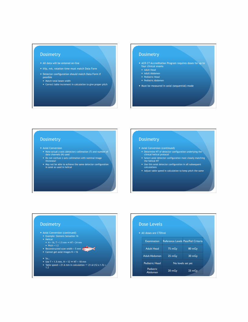
Dosimetry
� All data will be entered on-line
� kVp, mA, rotation time must match Data Form
� Detector configuration should match Data Form if possible � Match total beam width � Correct table increment in calculation to give proper pitch
Dosimetry
� ACR CT Accreditation Program requires doses for up to four clinical exams � Adult Head � Adult Abdomen � Pediatric Head � Pediatric Abdomen
� Must be measured in axial (sequential) mode
Dosimetry
� Axial Conversion � Note actual z-axis (detector) collimation (T) and number of
data channels (N) used � Do not confuse z-axis collimation with nominal image
thickness! � May not be able to achieve the same detector configuration
in axial as used in helical
Dosimetry
� Axial Conversion (continued) � Determine NT of detector configuration underlying the
clinical helical protocol � Select axial detector configuration most closely matching
the helical NT � Use this axial detector configuration in all subsequent
calculations
� Adjust table speed in calculation to keep pitch the same
Dosimetry
� Axial Conversion (continued) � Example: Siemens Sensation 16 � Helical
� N = 16, T = 1.5 mm ⇒ NT = 24 mm � Pitch = 1.2
� Reconstructed scan width = 5 mm � Cannot get axial images N = 16
� So… � Use T = 1.5 mm, N = 12 ⇒ NT = 18 mm � Table speed = 21.6 mm in calculation ➱ 21.6/(12 x 1.5) =
1.2
Dose Levels
� All doses are CTDIvol
Examination Reference Levels Pass/Fail Criteria
Adult Head 75 mGy 80 mGy
Adult Abdomen 25 mGy 30 mGy
Pediatric Head No levels set yet
Pediatric Abdomen 20 mGy 25 mGy

4/28/11
7
Dose P/F Levels
� Based on CTAP data � Head dose limits increased � Abdomen dose limits decreased
� NOTE: Increased head reference dose does not mean that you SHOULD increase dose � A number of scanners could not achieve acceptable image
quality at original value
� MANY SCANNERS CAN � If images are acceptable at 60 mGy limit, leave it
Dosimetry
� DLP (mGy-cm) = CTDIvol (mGy) • total scan length (cm) � For ACR, assume total scan length
� = 17.5 cm for head
� = 25.0 cm for adult abdomen
� = 12.0 cm for pediatric head
� = 15.0 cm for pediatric abdomen
� Effective Dose (E) = k (mSv/mGy-cm) * DLP (mGy-cm) � where k = 0.0021 for head
� = 0.015 for adult abdomen � = .0067 for pediatric head � = 0.020 for pediatric abdomen
Dosimetry
� At least one (1) dosimetry image must be submitted for each phantom protocol submitted � If scanner puts all 6 measurements in one series, submit the
whole series
� If scanner separates each scan into different series, just submit one series
� Whatever is easiest!
Assessment
� Phantom and dose submission will fail if � ≥ 7 minor deficiencies � ≥ 1 major deficiency
Phantom Deficiencies
Deficiency Minor Major
Detector configuration inappropriate X
kVp inappropriate X
Rotation time inappropriate X
Table increment/speed inappropriate X
Reconstruction algorithm inappropriate X
Phantom Deficiencies Deficiency Minor Major
Phantom kVp differs from Data Form X
Phantom mAs different from Data Form
≤10% >10%
Phantom Pitch different from Data Form ≤10% >10%
CT number not in range X
CNR too low X
CT number non-uniformity (N) 5≤N≤7 >7

4/28/11
8
Phantom Deficiencies
Deficiency Minor Major
Artifacts Sub-clinical Super-clinical
Dosimetry scans not axial X
Dosimetry phantom incorrect X
Non-chamber holes not filled X
CTDI kVp does not match Data Form X
Phantom Deficiencies
Deficiency Minor Major
CTDI mAs does not match Data Form X
Dosimetry phantom incorrect X
Detector configuration does not match Data Form* X
Table increment not adjusted to give correct pitch if NT different X
*except for scanner limitations
Clinical Image Quality
� Images are scored for � Technique parameters � Anatomic coverage / display � Filming technique (if hard copy) � Artifacts � Examination identification � Examination protocols
� Radiologist reviewers
� Independent of phantom review
Clinical Image Quality
� Technique factor and anatomic coverage guidelines are given for each exam option
� Artifacts must not hinder interpretation
� Exam identification � If electronic submission, the information should be on the
images or readily accessible in the DICOM header by the reviewer
Pediatric Studies
� “Pediatric Doses – It is very important for you to be aware of the differences between the dose observed for the pediatric abdomen portion of the phantom submission (when a child sized phantom is used) and pediatric doses reported on the scanner (which assume an adult sized phantom). The scanner reported doses (i.e. assuming an adult dose phantom) are approximately 2.4 times lower than the same exact x-ray technique would produce in a pediatric dose phantom. In other words, a dose reported on a pediatric abdomen should ideally be reduced by a factor of 3 or more as compared to adult doses.”

4/28/11
9
Pediatric Studies
� “In other words, a dose reported on a pediatric abdomen should ideally be reduced by a factor of 3 or more as compared to adult doses.” � Applies only when pediatric dose is report in the 32 cm
phantom
� In the 16 cm phantom, doses should be similar
Phantom
� It is not essential for each site to purchase the RMI phantom
� Technologist and physicist QC may be performed using phantom deemed appropriate by the qualified medical physicist
� RMI phantom MUST be used for accreditation scans
Is This Really True?
� Yes!
� The website is slow to be updated
� Documents can get held up in bureaucracy
� Not all reviewers come up to speed at the same rate
� Appeal if necessary
ACR Practice Guidelines and Technical Standards
� The following ACR Practice Guidelines and Technical Standards are pertinent to achieving and maintaining CT Accreditation. These guidelines and standards form the basis of the accreditation program. � ACR Practice Guideline for Imaging Pregnant or Potentially Pregnant Adolescents and Women
with Ionizing Radiation � ACR Practice Guideline for the Use of Intravascular Contrast Media � ACR Practice Guideline for Performing and Interpreting Diagnostic Computed Tomography (CT) � ACR Practice Guideline for the Performance of Computed Tomography (CT) of the Brain � ACR Practice Guideline for the Performance of Computed Tomography (CT) of the Extracranial
Head and Neck in Adults and Children � ACR Practice Guideline for the Performance of Computed Tomography (CT) of the Spine � ACR Practice Guideline for the Performance of Computed Tomography (CT) for the Detection
of Pulmonary Embolism in Adults � Practice Guideline for the Performance of High-Resolution Computed Tomography (HRCT) of
the Lungs in Adults � ACR Practice Guideline for the Performance of Computed Tomography (CT) of the Abdomen
and Computed Tomography (CT) of the Pelvis � ACR Technical Standard for Diagnostic Medical Physics Performance Monitoring of Computed
Tomography (CT) Equipment � ACR Practice Guideline for Communication of Diagnostic Imaging Findings ACR Practice
Guideline for the Performance and Interpretation of Cardiac Computed Tomography (CT)
Help?
� Call the ACR (800-770-0145) � Theresa Branham � [email protected]
� Has individuals who want to help
� Process is intended to be educational
� Wants facilities to become accredited
� Don’t be shy - call! (800-770-0145)
The Future of Accreditation
� CMS can refine any of its requirements at any time � Personnel � QC � Submission
� ACR tries to post such information on its website
� Pay attention to any announcements from ACR or CMS

4/28/11
10
Score Your Phantom
� Use one of the following DICOM viewers
� PC
� K-PACS
� http://www.k-pacs.net/
� Clear Canvas
� http://www.clearcanvas.ca/dnn/
� Mac
� Osirix
� http://www.osirix-viewer.com/
Common Errors
� Table scan parameters do not match those seen on the images
� Incorrect detector configuration used (unless limitation of scanner)
� Artifacts
� Using small or large FOV
� Dose too high
Phantom CD Preparation
� Two series are required for each phantom protocol
� Gammex 464
� Dosimetry
� Must include at least 1 dosimetry image for each protocol
� Burn 2 IDENTICAL phantom discs for each unit on your application
� Separate units must be burned onto separate discs
� Each disc must contain all required images
Phantom and Clinical CD Preparation
� The discs may include an embedded viewer. The following functions are helpful:
� Easy access to the complete DICOM header
� Window/level adjustments
� Distance measurement
� Region of interest (area, pixel mean and standard deviation)
� More importantly, be aware of proprietary formats! (Philips iSite)
� Verify that the images can be imported
CD Labeling
� DO NOT PUT LABEL DIRECTION ON CD!
CD1 label
CD2 label
Adult Head Gammex Dosimetry
Adult Abdomen Gammex Dosimetry
CD1
CTAP #xxxx-01
Adult Head Gammex Dosimetry
Adult Abdomen Gammex Dosimetry
CD2
CTAP #xxxx-01
Place ACR label on case Write CD# and CTAP # on disk
Anything Weird?
� Call the ACR
� Sometimes it is impossible to exactly meet the requirements (e.g. dosimetry)
� Put a note in with submission
� Explain the issue
� Describe actions taken
� Lets reviewers know what’s going on
� Image quality requirements are not negotiable

4/28/11
11
Questions?