Embed Size (px)
DESCRIPTION
CT and MRI FINDINGS IN LOCALIZED NASOPHARYNGEAL AMYLODOSIS : A CASE REPORT. I. GANZOUI, Y. AROUS, R. AOUINI, M. LANDOLSI, S. KOUKI, H. BOUJEMAA, N. BEN ABDALLAH Radiology Department, Military Hospital of Tunis, Montfleury, Tunis, Tunisia. HN4. INTRODUCTION. - PowerPoint PPT Presentation
Citation preview
CT and MRI FINDINGS IN LOCALIZED NASOPHARYNGEAL
AMYLODOSIS : A CASE REPORT
I. GANZOUI, Y. AROUS, R. AOUINI, M. LANDOLSI, S. KOUKI, H. BOUJEMAA, N. BEN ABDALLAH
Radiology Department, Military Hospital of Tunis, Montfleury, Tunis, Tunisia
HN4
INTRODUCTION
Amyloidosis is a group of disorders that result from the deposition of amyloid proteins in organs and tissues.
Localized form of amyloidosis in the head and neck is extremely rare.
the involvement of the nasopharynx is still more uncommon.
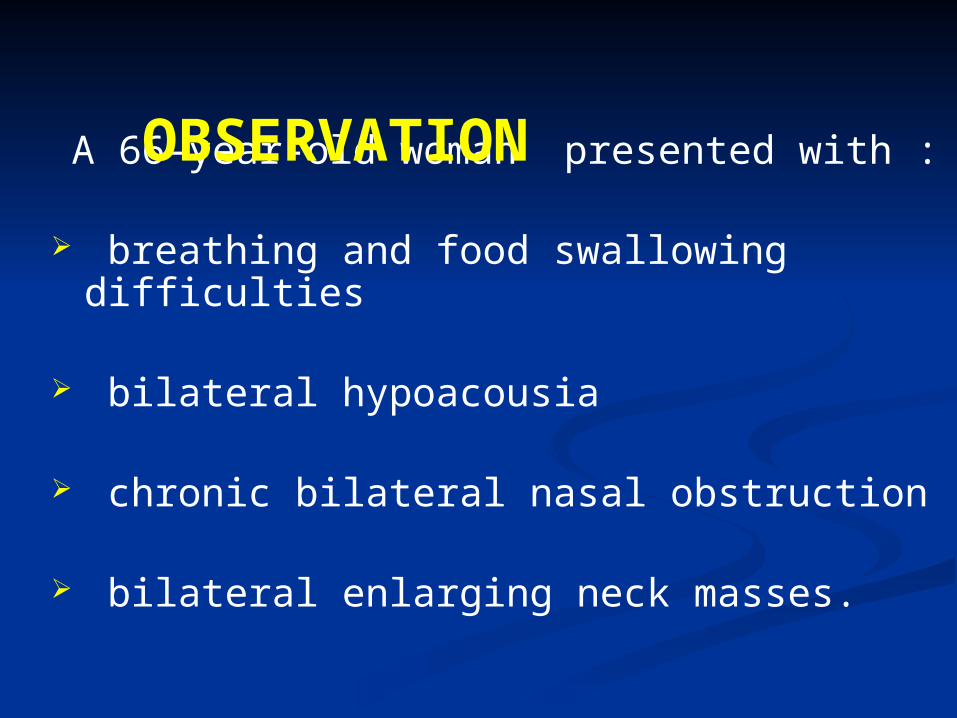
A 66-year-old woman presented with :
breathing and food swallowing difficulties
bilateral hypoacousia
chronic bilateral nasal obstruction
bilateral enlarging neck masses.
OBSERVATION
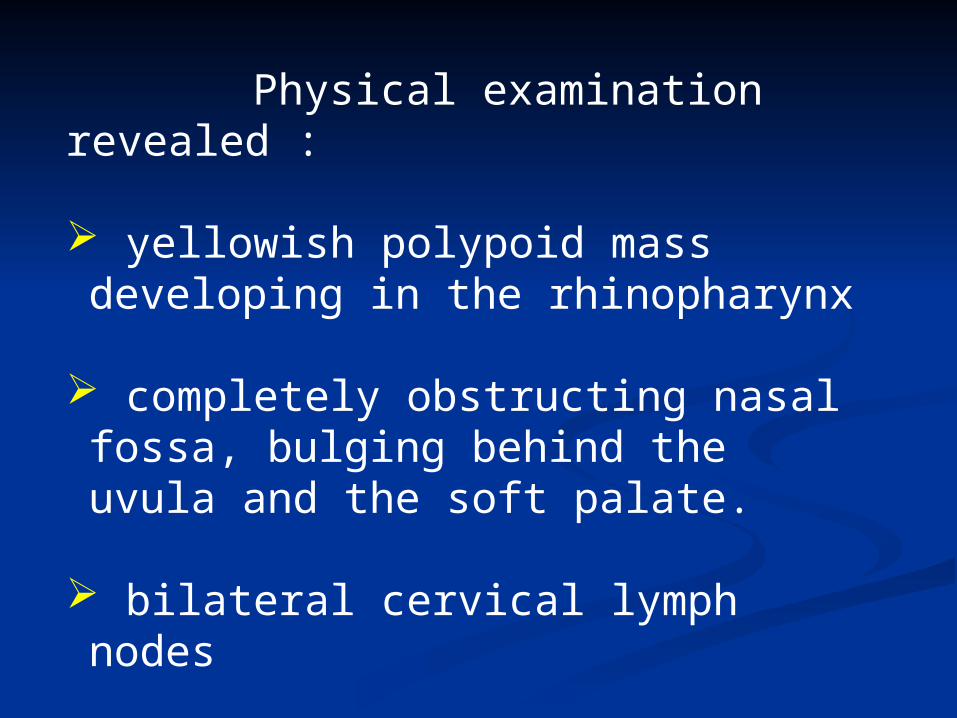
Physical examination revealed :
yellowish polypoid mass developing in the rhinopharynx
completely obstructing nasal fossa, bulging behind the uvula and the soft palate.
bilateral cervical lymph nodes
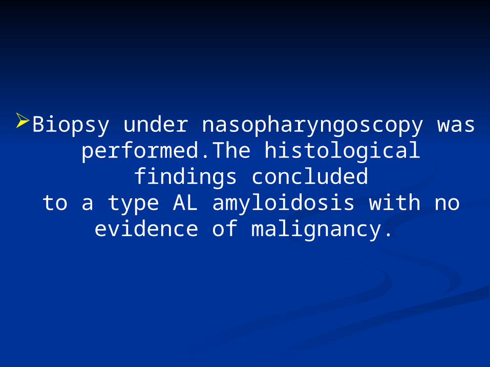
Biopsy under nasopharyngoscopy wasperformed.The histological findings concludedto a type AL amyloidosis with no evidence of
malignancy.
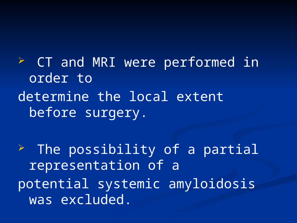
CT and MRI were performed in order to determine the local extent before surgery.
The possibility of a partial representation of apotential systemic amyloidosis was excluded.
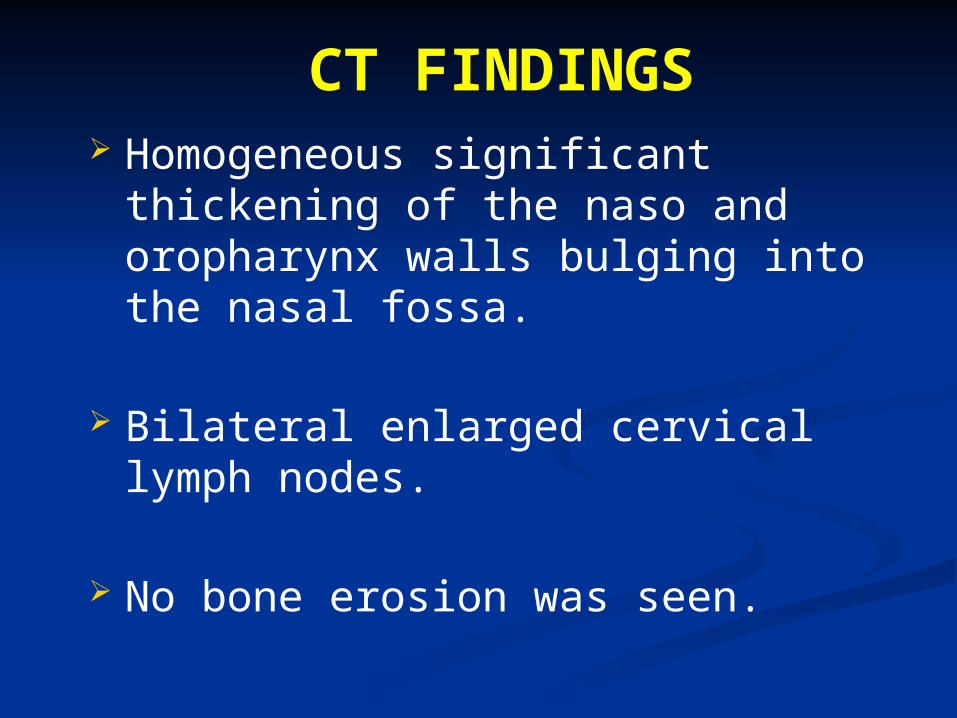
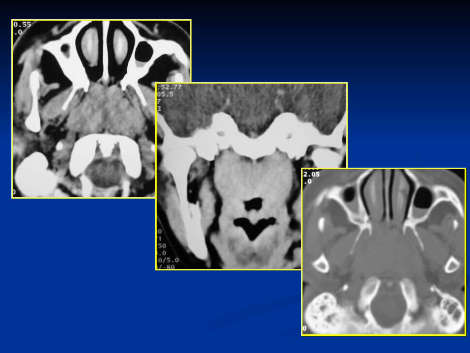
CT FINDINGS Homogeneous significant thickening of the
naso and oropharynx walls bulging into the nasal fossa.
Bilateral enlarged cervical lymph nodes.
No bone erosion was seen.
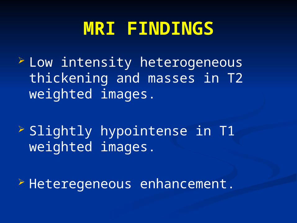
MRI FINDINGS Low intensity heterogeneous thickening and
masses in T2 weighted images.
Slightly hypointense in T1 weighted images.
Heteregeneous enhancement.
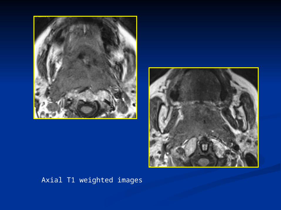
Axial T1 weighted images
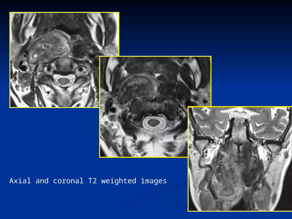
Axial and coronal T2 weighted images
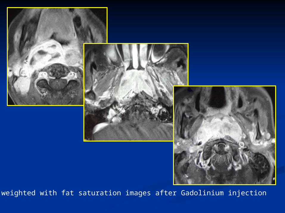
Axial T1 weighted with fat saturation images after Gadolinium injection
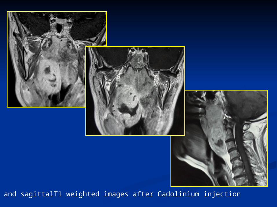
Coronal and sagittalT1 weighted images after Gadolinium injection
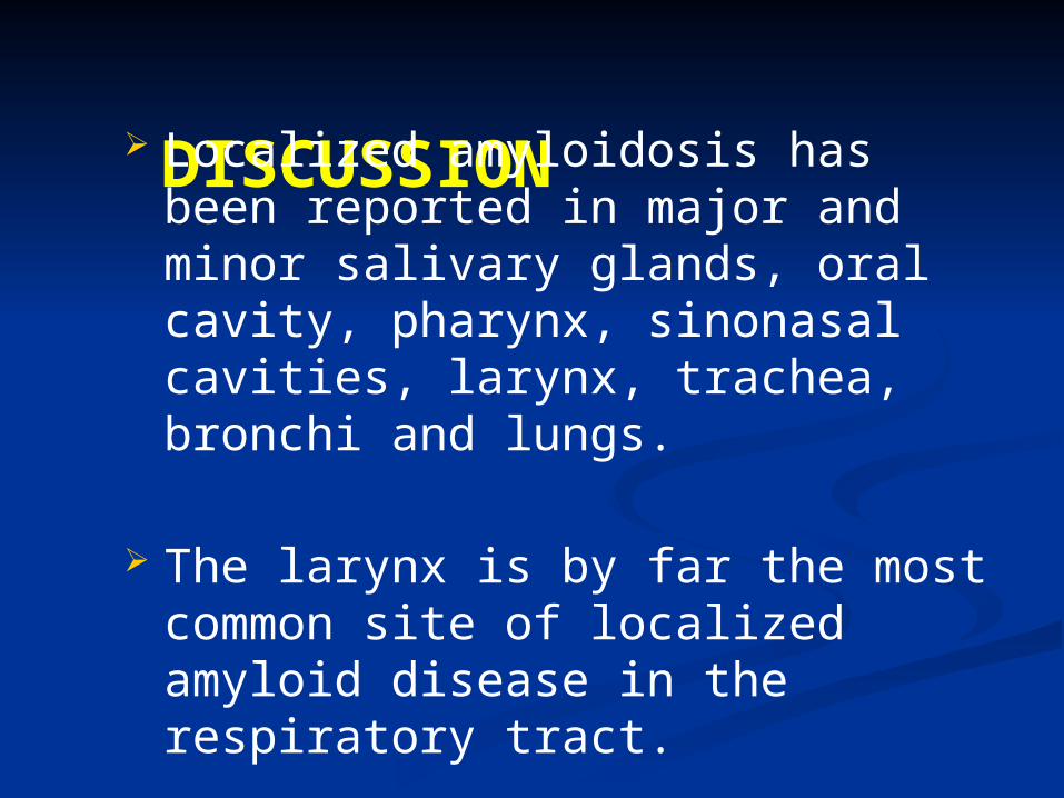
DISCUSSION Localized amyloidosis has been reported in
major and minor salivary glands, oral cavity, pharynx, sinonasal cavities, larynx, trachea, bronchi and lungs.
The larynx is by far the most common site of localized amyloid disease in the respiratory tract.
Localized amyloidosis in the nasopharynx is rare.
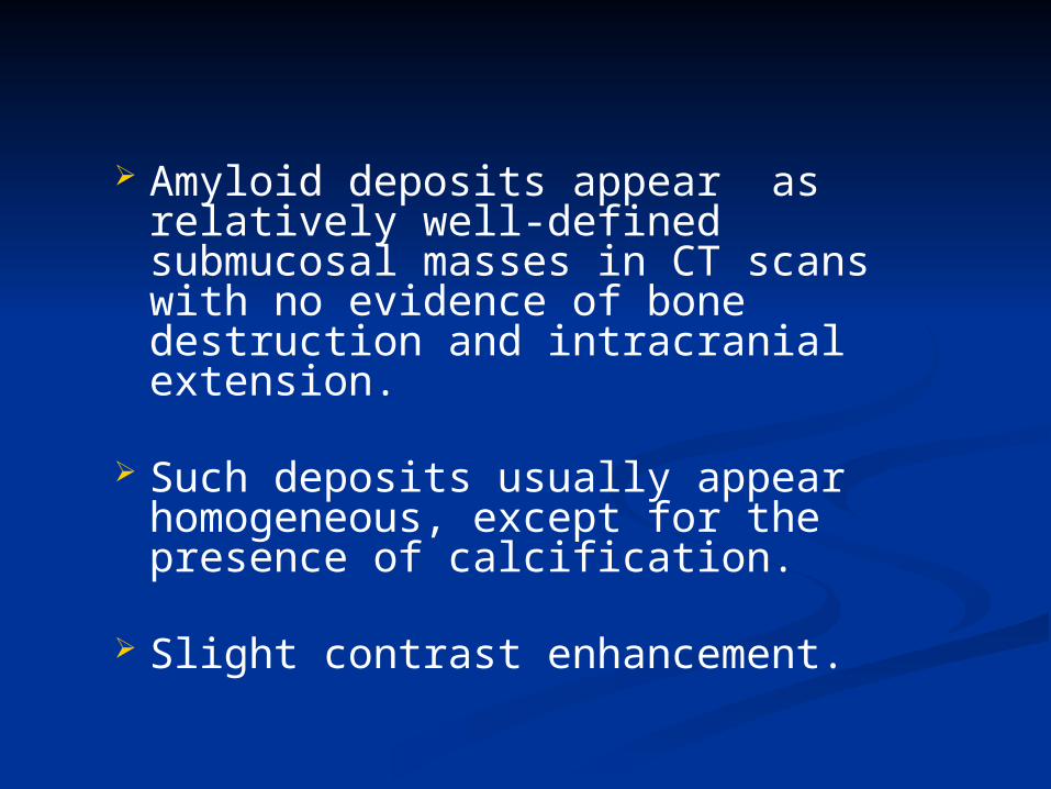
Amyloid deposits appear as relatively well-defined submucosal masses in CT scans with no evidence of bone destruction and intracranial extension.
Such deposits usually appear homogeneous,
except for the presence of calcification.
Slight contrast enhancement.
The presence of calcification helps to narrow the differential diagnosis.
In nasopharyngeal amyloidosis with neck calcification on CT scans, the possibility of neck amyloid deposition should be considered.
It is well known that such a lesion shows low intensity on a T2-weighted image of MRI.
Nasopharyngeal amyloidosis shows slight or no enhancement.
Being a hypocellular lesion, it should show delayed enhancement rather than early enhancement on dynamic contrast enhanced examination.
Some reports state that plasmacytes are occasionally observed within a mass of amyloid deposit and might produce amyloid-related proteins.
It is important to know that plasmacyte infiltration, which occasionally occurs with amyloidosis, shows high intensity on T2 weighted images and remarkable early enhancement on dynamic MRI.
CONCLUSION Although rare, localized amyloidosis
should be considered in the differential diagnosis of head and neck masses.
CT and MRI help in delineating the local extent.
Calcification on CT and low intensity on T2 weighted images are suggestive of the diagnostic