Embed Size (px)
Citation preview

�������� ����� ��
Crystallographic orientation and electrode nature are key factors for electriccurrent generation by Geobacter sulfurreducens
Beatriz Maestro, Juan M. Ortiz, German Schrott, Juan P. Busalmen,Vıctor Climent, Juan M. Feliu
PII: S1567-5394(14)00035-8DOI: doi: 10.1016/j.bioelechem.2014.02.001Reference: BIOJEC 6726
To appear in: Bioelectrochemistry
Received date: 20 December 2013Revised date: 18 February 2014Accepted date: 19 February 2014
Please cite this article as: Beatriz Maestro, Juan M. Ortiz, German Schrott, Juan P.Busalmen, Vıctor Climent, Juan M. Feliu, Crystallographic orientation and electrodenature are key factors for electric current generation by Geobacter sulfurreducens, Bioelec-trochemistry (2014), doi: 10.1016/j.bioelechem.2014.02.001
This is a PDF file of an unedited manuscript that has been accepted for publication.As a service to our customers we are providing this early version of the manuscript.The manuscript will undergo copyediting, typesetting, and review of the resulting proofbefore it is published in its final form. Please note that during the production processerrors may be discovered which could affect the content, and all legal disclaimers thatapply to the journal pertain.

ACC
EPTE
D M
ANU
SCR
IPT
ACCEPTED MANUSCRIPT
1
Crystallographic orientation and electrode nature are key factors for
electric current generation by Geobacter sulfurreducens
Beatriz Maestroa1*
, Juan M. Ortiza2
, Germán Schrottb, Juan P. Busalmen
b, Víctor
Climenta and Juan M. Feliu
a
a Instituto Universitario de Electroquímica. Universidad de Alicante. Apdo. 99, Alicante E-03080, Spain.
b Área de electroquímica y corrosión, INTEMA (CONICET). Juan B. Justo 4302,
B7608FDQ Mar del Plata, Argentina. 1Present address: Instituto de Biología Molecular y Celular, Universidad Miguel Hernández. Avda Universidad s/n,
Elche, 03202, Spain. 2Present address: Aqualia Integrated Water Management SA. Madrid, Spain.
* Corresponding author. E-mail: [email protected]. Telephone: (+34) 966658474
Fax: +34 966658758
E-mail addresses: [email protected] (J.M. Ortiz), [email protected] (G.
Schrott), [email protected] (J.P. Busalmen), [email protected] (V. Climent),
[email protected] (J.M. Feliu).
Abstract
We have investigated the influence of electrode material and crystallographic structure
on electron transfer and biofilm formation of Geobacter sulfurreducens. Single-crystal
gold - Au(110), Au(111), Au(210) - and platinum - Pt(100), Pt(110), Pt(111), Pt(210) -
electrodes were tested and compared to graphite rods. G. sulfurreducens
electrochemically interacts with all these materials with different attachment kinetics
and final current production, although redox species involved in the electron transfer to
the anode are virtually the same in all cases. Initial bacterial colonization was fastest on
graphite up to the monolayer level, whereas gold electrodes led to higher final current
densities. Crystal geometry showed to have an important influence, with Au(210)
sustaining a current density of up to 1442 (±101) µA cm-2
at the steady state, over
Au(111) with 961 (±94) µA cm-2
and Au(110) with 944 (±89) µA cm-2
. On the other
hand, the platinum electrodes displayed the lowest performances, including Pt(210).
Our results indicate that both crystal geometry and electrode material are key
parameters for the efficient interaction of bacteria with the substrate and should be
considered for the design of novel materials and microbial devices to optimize energy
production.
Keywords: Geobacter sulfurreducens; single-crystal electrode; biofilm; electron
transport; cytochrome

ACC
EPTE
D M
ANU
SCR
IPT
ACCEPTED MANUSCRIPT
2
1. Introduction
The mechanisms for current production by electro-active microorganisms
colonizing anode surfaces are the subject of thorough investigation due to their
implication in the development of bioelectrochemical systems such as the promising
technology of microbial fuel cells (MFC) [1]. In the anode of these devices, dissolved
organic matter is oxidized by electro-active microorganisms that possess the
outstanding ability of using an electrode as the final electron acceptor. After extracting
the energy required for growth, these cells transfer the produced electrons to outer
membrane cytochromes (OMC's), and finally to the anodic surface through an
extracellular electron transfer chain whose structure and function remain to be
elucidated in detail [2]. Several bacterial genera have the ability to colonize electrodes
and use them as electron acceptors [3]. Among such microorganisms, strains from the
Geobacter genera typically dominate the natural bacterial populations colonizing
electrodes [4]. Geobacter sulfurreducens is the most efficient current producer
described so far (see [4] for a review) and is one of the few microbes that have been
found to completely oxidize organic compounds to carbon dioxide using an electrode as
the sole electron acceptor [5].
Many molecular biology studies have focused on the external electron transfer
(EET) mechanisms in G. sulfurreducens biofilms, providing relevant information about
participating molecules and identifying OmcZ, OmcB and OmcS cytochromes as
playing a relevant role [6-8]. Moreover, electrochemical and spectroelectrochemical
approaches have demonstrated that external cytochromes in this strain are responsible
for the reaction of charge transfer to the electrode surface [9, 10], but the full identity of
the molecules acting in this step is still unknown. For the octaheme cytochrome OmcZ,
a formal potential of -0.22 V vs. standard hydrogen electrode (SHE) (corresponding to -
0.42 V vs. Ag/AgCl) has been estimated [11] with a very broad potential window from -
0.42 to -0.06 V vs. SHE arising from its multiheme composition. The hexaheme
cytochrome OmcS has an apparent equilibrium potential of -0.21 V vs. SHE
(corresponding to -0.41 V vs. Ag/AgCl) with a reduction potential range from -0.36 to -
0.04 V vs. SHE [12]. Finally, the redox potential of the dodecaheme OmcB has been
estimated to be centered at -0.19 V vs. SHE (corresponding to -0.39 V vs Ag/AgCl)
[13].
G. sulfurreducens does not require soluble mediators to generate a high current
density, but external cytochromes must establish a molecular interaction with the
electrode to allow direct electron transfer (DET) [9, 10]. In DET of isolated protein
systems, an optimal electronic coupling between the electrode and the molecular redox
center is necessary for electron tunnelling to occur. Among many factors, this coupling
is mostly dependent on the orientation of adsorbed proteins [14], and in turn on
variables such as pH [15], ionic strength [16] and electrode surface topography [17] and
charge [18]. Therefore, it should not be surprising that the structure, topology,
functionalization and surface charge of the anode material could play a decisive
influence on the initial bacterial attachment step and on the electrochemical function of
the biofilm. On the other hand, the influence of the mineral (oxyhydr)oxide composition
and crystallinity on reduction rates by Geobacter and Shewanella species has been a
subject of controversy. Roden [19] suggested that oxide reduction is not strongly
controlled by oxide crystal thermodynamic properties, but mainly by the surface area.
However, other authors have highlighted the impact of chemical and crystallographic

ACC
EPTE
D M
ANU
SCR
IPT
ACCEPTED MANUSCRIPT
3
orientation of the surface [20-22]. In these studies, the authors present the respiration
rates by normalized area, and show that poorly crystalline materials such as amorphous
Fe(III) (oxyhydr)oxide or feroxyhyte are better electron acceptors than other minerals
which present a more crystalline structure, as goethite and hematite. Moreover, they
indicate that minerals with the same chemical composition differ on Fe(III) reduction
rates in the same way, depending on their relative crystallinity.
Single-crystal electrodes may provide an unequivocal answer to the question of
whether the structural properties of the electrode surface affect DET in Geobacter
systems. These electrodes are characterized by a precise atomic arrangement on their
surface, leading to a well-distinct electrochemical reactivity. The structure of the
interfacial region between these electrode surfaces and the electrolytic solution has been
characterized with great detail in the past and, therefore, they offer an excellent starting
point for the study of the influence of electrode structural properties on biofilm
electroactivity. Among the different properties characterizing the electrochemical
behaviour of metal surfaces, the potential of zero charge (pzc) plays a very important
role [23]. This parameter represents the value of the potential where the charge
separation at the interphase vanishes, therefore allowing the definition of a rational
correlation between the electrode potential and the sign and magnitude of charge at the
interface. As a last advantage, single-crystal surfaces present a well-defined surface area
that allows an unambiguous determination of area-dependent variables, such as current
density and charge accumulation.
In this work, we have comparatively tested gold and platinum single-crystals
with different properties in terms of topology, defectiveness and zero charge potential,
aiming at clarifying the influence of the electrode material and surface structure on
bacterial attachment, maximum current density generation and the activity of redox
species involved in the DET process. Besides, we compare these results to those
obtained on graphite, which is the typically used material for analyzing Geobacter
electrochemistry. Our results show that gold electrodes, and especially Au(210),
produce the highest steady state current densities, demonstrating that both material type
and surface geometrical configuration exert a strong influence on the EET process.
2. Materials and Methods
2.1. Culture preparation
All reagents were analytical and biochemical grade and were obtained from
Sigma-Aldrich. G. sulfurreducens used as inoculum to the electrochemical cell was
grown in batch mode in culture medium containing 50 mM NaHCO3, 9.4 mM NH4Cl,
2.6 mM NaH2PO4, 30 mM KCl, 10 mL L-1
trace vitamins solution [24] and 10 mL
L-1
mineral solution [24]. The pH was adjusted to 7.0, and the medium was
supplemented with 0.1 mM sodium citrate, 20 mM sodium acetate and 40 mM sodium
fumarate (acceptor limitation conditions), purged with N2/CO2 (80 %/20 %) and
sterilized in bottles caped with butyl rubber stoppers and secured with a crimped
aluminium cap (Bellco Glass Inc). Anaerobic culture was promoted by 1/10 (v/v)
inoculum and supported at 30 ºC without shaking for 48 h, until O.D. 600nm was around
0.6. In the case of culture medium used into the electrochemical cell, no electrode
acceptor was added except the polarized electrode.

ACC
EPTE
D M
ANU
SCR
IPT
ACCEPTED MANUSCRIPT
4
2.2. Electrode preparation
Three gold single-crystal: Au(111), Au(110), Au(210), four Pt single-crystal:
Pt(100), Pt(110), Pt(111), Pt(210) and a graphite rod (TED Pella, Grade 1) were tested
as working electrodes. The single-crystals were prepared by melting a high purity gold
or platinum wire (99.9998%, Alfa-Aesar), and were subsequently oriented, cut and
polished as described elsewhere [25]. Prior to each experiment, each single-crystal
electrode was flame-annealed and quenched in ultra-pure water. To check the electrode
surface, cyclic voltammetry (CV) was performed in 100 mM HClO4 , and the
voltammetric profile was compared to those reported in the literature [26, 27]. In order
to completely remove the biofilm from the electrodes after each experiment, the gold
single-crystals were cleaned by applying a potential of 10 V in 100 mM H2SO4 during
10 s, washed with ultra-pure water, then inserted into 1 M HCl and washed again with
ultra-pure water. The process was repeated twice. After this, the gold single-crystals
were heated in an oven at 860 ºC for 12 h. In the case of platinum electrodes, after
overnight washing in nitric acid, they were flame annealed and cooled into clean nitric
acid twice, then washed with ultra-pure water, flamed again and quenched in ultra-pure
water. The electrode structure was checked by CV each time before a new experiment.
Graphite rods (3 mm diameter, Grade 1 Spec-Pure, TED Pella, Inc., Reddong,
CA) (2 cm2 immersed) were polished using an abrasive disc P1200 (Buehler, Illinois,
USA), sonicated in ultra-pure water for 10 min, kept in 1 M NaOH for 30 min, and in 1
M HCl overnight. After each step, graphite rods were washed extensively with ultra-
pure water.
2.3. Cell configuration and electrochemical experiments
A custom-made, single-chamber, three-electrode reactor was used to grow
biofilms and for all the electrochemical experiments. The counter electrode was a
platinum wire and the reference was an Ag/AgCl (3 M NaCl) electrode (RE-5B BASi,
USA). The potential of Ag/AgCl (3 M NaCl) electrode vs SHE was checked (+ 0.2 V).
The reactor had several holes in which the different working electrodes were fitted to
allow biofilm growth on them simultaneously. The single-crystal electrodes were
positioned in a meniscus configuration ensuring that only the well-defined surface with
the selected crystallographic orientation was making contact with the electrolyte. After
assembling and sterilizing the whole system, the tank containing the feeding medium
and the reactor were purged overnight with N2/CO2 (80 %/20 %) previously filtered
through an oxygen filter (Agilent Technologies). The cell was filled with 80 mL growth
medium containing 20 mM acetate as the electron donor, and no acceptor other than the
polarized electrode. Inoculation was performed by adding 25 % (v/v) of a G.
sulfurreducens early stationary phase culture (O.D.600 nm 0.6). All the electrodes were
simultaneously and independently polarized at +0.2 V using a 8-channel potentiostat
(CHI1030B CH Instruments) and current evolution was recorded over time. The system
was batch operated for the first 25 h before turning on circulation of the feeding
medium at 0.2 mL/min for continuous mode. Temperature was controlled at 30 C
during all the experiment. The electrochemical cell was continuously purged with
N2/CO2 (80 %/20 %) in the headspace and the medium in the reactor was continuously
stirred at low rate. Conductivity at 25 ºC of the initial medium was 4 mS cm-1
. Four
independent experiments were performed.

ACC
EPTE
D M
ANU
SCR
IPT
ACCEPTED MANUSCRIPT
5
2.4. Electrochemical measurements
At different times of growth, the electrochemical activity of adsorbed bacteria
was tested by cyclic voltammetry, starting in the anodic direction at +0.2 V and
covering a potential range from 0.4 to -0.6 V. While doing this, stirring and pumping of
the medium were stopped. The scan rate was 10 mV s-1
. A control CV was performed
before inoculation to check the absence of current.
2.5. Laser scanning confocal microscopy (LSCM)
At the end of each experiment, the electrodes were carefully removed, washed
with fresh medium twice and fluorescently stained with the LIVE/DEAD Baclight
bacterial viability kit (Invitrogen) as previously described [28]. In brief, 3 µL of a
mixture 1:1 of SYTO 9:propidium iodide was added to 1 mL of culture medium.
Electrodes were exposed to this mixture during 15 min at room temperature in the dark
before washing with culture medium. Confocal images were captured using an inverse
laser scanning confocal microscope (Leika DM IRE2) with 60x oil-immersion lens. In
order to obtain a three-dimensional image of the biofilm, multiple optical slices were
recorded. The excitation/emission wavelengths for SYTO 9 and propidium iodide were
488/500-550 nm and 543/600-670 nm, respectively. Total (red plus green fluorescent)
and metabolically active (green fluorescent) thicknesses of the biofilm were directly
measured at four different fields of view and subsequently averaged in each experiment.
3. Results and discussion
3.1. Microbial electricity production and voltammetric characterization of the biofilm
on the single-crystal electrodes
Single-crystal electrodes provides a suitable system that allows the precise
determination of area-dependent variables, such as current density and charge
accumulation, which may be valuable to characterize the electrochemical behaviour of
Geobacter species. Chronoamperometry of a representative experiment is shown in Fig.
1A. Current production from all electrodes started as soon as bacteria were added, but
initial current production was higher and bacteria grew faster on the graphite electrode,
showing a doubling time for current production of about 6 h in agreement with previous
reports [29]. The high amount of functional groups present at the graphite surface, such
as quinones, carboxylic acids and alcohols, can mimic humic substances present in the
natural environment of Geobacter, facilitating the rapid arrangement of bacteria [30].
After 25 h of incubation, the current produced on graphite and gold electrodes is
indicative of a monolayer stage of growth, as determined by Marsili et al. [29]. Among
the metallic materials, gold electrodes demonstrated a better performance than platinum
ones, with Au(210) displaying the fastest current increase (Fig. 1A), with a doubling
time of about 7 h, compared to 10 and 12 h shown by Au(110) and Au(111),
respectively. It should be noted that the (210) crystal possesses a very low surface
atomic density and the lowest pzc within the gold surfaces [23], thus bearing the most
positive charge at +0.2 V, which may facilitate the interaction with negatively charged
bacteria. On the other hand, current production on platinum electrodes was very small at

ACC
EPTE
D M
ANU
SCR
IPT
ACCEPTED MANUSCRIPT
6
this stage (Fig. 1A, inset), suggesting that bacterial adhesion is hampered by some
negative effect that delays the electrode colonization.
Here Table 1
Here Fig. 1
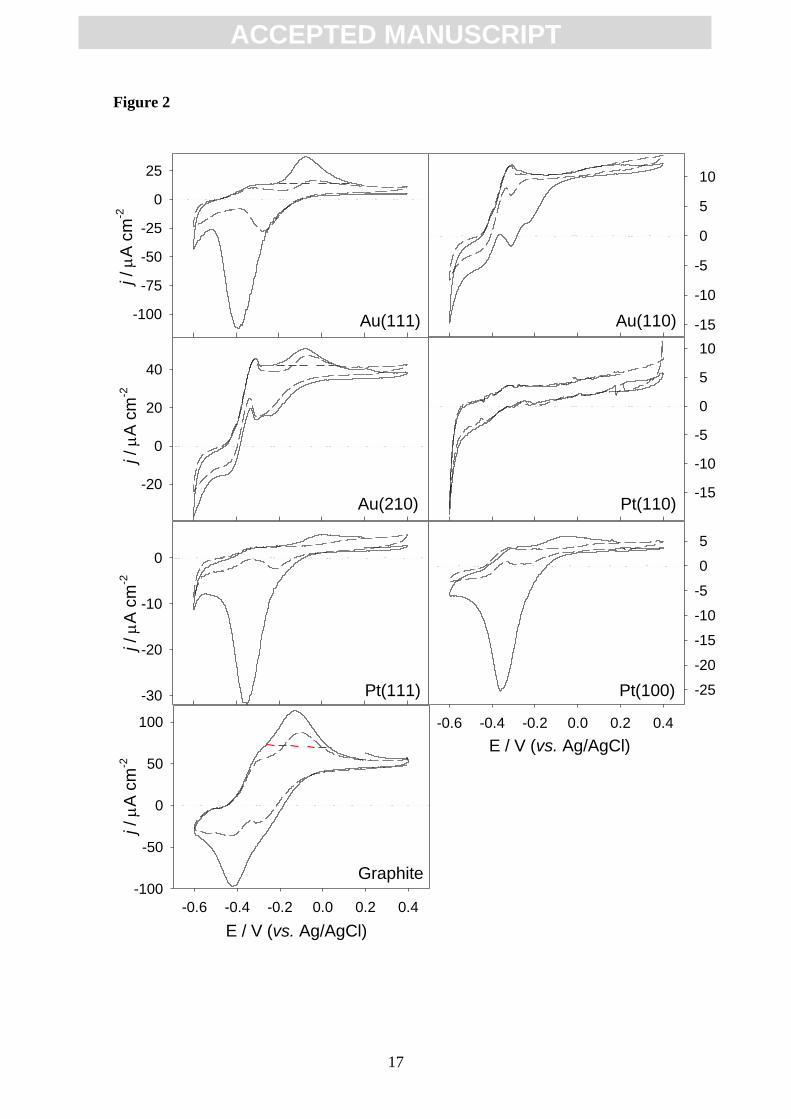
The electrochemical characterization of biofilms developed on the electrodes
was performed by cyclic voltammetry at increasing times of growth. The analysis of
voltammograms registered at the end of the batch culture step (25 h of growth) shows
the turnover signal characteristic of metabolically active cells together with non-
catalytic signals with mid-peak potentials of about -0.2 V that change in size and
position upon cycling, depending on the electrode material (Fig. 2). The occurrence of
these non-catalytic signals is intriguing because they seem to be associated to
cytochromes participating in the EET to the electrode [31], but without playing a role in
electrode respiration. The expression and presence of these cytochromes at the interface
is thought to be a consequence of a programmed response of cells, not directly related to
current production. Taking the steady state current level produced by the biofilm as a
baseline, a transferred charge of 28, 120 and of 390 µC cm-2
can be calculated from the
area below non-catalytic oxidation peaks in the first cycle of CV on Au(210), graphite
and Au(111) (Fig. 2), respectively. Considering a mean storing capacity of 8 electrons
per fully reduced cytochrome (e.g., OmcZ) and a maximum surface coverage of 2.7
10-12
mol of proteins per cm2 of electrode surface [32], a surface coverage density of 3.6
10-11
, 1.5 10-10
and 5 10-10
mol of proteins per cm2 can be calculated,
corresponding to 13, 56 and 185 densely packed cytochrome layers on Au(210),
graphite and Au(111), respectively. These values are on the same order of magnitude
than those described by Bonanni et al [32] and seem to follow an inverse relation to the
current of the biofilms on each material. This gives support to the charge storing role for
these molecules and indeed suggests that their expression and accumulation at the
interface depends in some way on the suitability of the electrode material as an electron
acceptor. On the other hand, changes in the peak size and position of these non-catalytic
signals may be the result of variations in the position of external cytochromes as a
consequence of the potential scan, which would decrease the number of protein
molecules suitably coupled with the electrode. Alternatively, taking into account that
microbial cells have been described to partially desorb from the electrode surface upon
potential cycling (Kuzume et al, unpublished results), the occurrence of irreversible
processes like protein unfolding and/or reductive desorption can not be ruled out either.
Here Fig. 2
Regarding the turnover signals, the comparison between the different metallic
electrodes shows that the catalytic wave is much more evident at 25 h on Au(210) and
Au(110) than on Au(111), or Pt(100) and Pt(111), consistently with the low current
production shown at this stage by platinum electrodes (Fig. 1A). No redox signals were
detected on Pt(110),. Finally, the voltammogram recorded on graphite is more
reversible, displaying a higher non-catalytic oxidation current than on the Au(210)
crystal, in spite of producing a turnover current similar to that measured on this metallic
electrode (Fig. 2, see below).
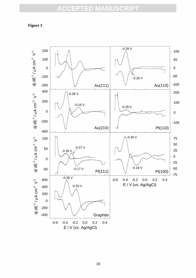
The first derivative analysis of the second cycle of voltammograms in all
materials (Fig. 2) reveals one dominant inflection point at around -0.39 V,

ACC
EPTE
D M
ANU
SCR
IPT
ACCEPTED MANUSCRIPT
7
corresponding to the catalytic process in current production, and additional smaller
signals at about -0.28 and -0.16 V, the position of which slightly varies with the
electrode material (Fig. 3). The presence of more than one redox center has been
previously described [31, 33]. The results suggest that, in all cases, there is a major
redox species involved in EET, together with appended, smaller signals which present
slight differences between materials, probably due to minor deviations on the reaction
involved.
Here Fig. 3
After 25 h of operation in batch, cultures were changed to continuous mode and
operated up to a total of 140 h. A representative current evolution profile is displayed in
Fig. 1B to show that the relative performance of electrodes changed with time upon
biofilm growth. Although the graphite electrode presented the fastest kinetics in initial
current generation (Fig. 1A), it was also the first in reaching the saturation current, at
approximately 660 µA cm-2
(Table 1). Moreover, all gold electrodes reached
appreciable higher values of current density at the steady state, outperforming graphite
(Fig. 1B; Table 1). This suggests that, in spite of the initial advantage presented by
graphite as electron aceptor up to the monolayer level, highly ordered surfaces such as
those of gold electrodes may represent in the longer term better substrates for biofilm
structuring and current production (Fig. 1B). In this context crystal geometry turned out
to be decisive, with Au(210) sustaining a current density of up to 1442 µA cm-2
(Table
1). It should be remarked that the experiments were repeated four times, and in the case
of Au(210) three different electrodes were tested.
Platinum electrodes reached the steady state current at longer times and showed
the poorest performance in current production (Fig. 1B; Table 1). In spite of this, it is
worth to mention that they also served as respiratory substrate for G. sulfurreducens.
Very few reports have previously analyzed the use of this metal for bacterial
attachment. Yi et al [34] showed that the maximum current density obtained by G.
sulfurreducens on graphite is bigger than on platinum electrodes, an observation that
these authors attributed to a higher surface area of the unpolished graphite. However, in
our experiments, Pt(111) and Pt(100) crystals demonstrated nearly the same
performance efficiency as graphite when the system reached the steady state. The poor
performance of Pt(110) compared to the other platinum anodes demonstrates that crystal
geometry effectively affects the bacterial electrochemical behaviour. The remarkably
slow kinetics and low efficiency of biofilm growth on platinum, when compared with
gold single-crystals of the same geometry, suggests a negative effect of the former metal
on the bacterial metabolism. As a first explanation, the superior reactivity of platinum in
surface catalysis for example, may be deleterious for cell functions by inducing
irreversible oxidation of cellular molecules/structures.
In order to test the general influence of the (210) surface geometry we checked
the current density obtained with a Pt(210) electrode. Supplementary Fig. 1S shows that
current density evolution on Pt(210), although somehow slower than on Pt(100), does
not lead to significant differences in the steady state current when compared with the
other platinum electrodes (Supplementary Fig. 1S, Table 1). This demonstrates that not
only the structure but also the type of electrode material affects both the adhesion and
efficient interaction with the substrate.

ACC
EPTE
D M
ANU
SCR
IPT
ACCEPTED MANUSCRIPT
8
The cyclic voltammograms recorded from approximately 47 h of biofilm growth
show the same sigmoid shape on all materials, which is indicative of the catalysis of
acetate oxidation by cells. Fig. 4 shows the CV obtained at the end of the experiment
(140 h), when the current density was stabilized. The onset potential appears to be the
same for all electrodes (around -0.48 V) attaining in all cases a limiting current of about
-0.25 V. The first derivative analysis of the voltammograms shows a symmetrical
maximum in all cases, initially centered around -0.39 V for gold and platinum
electrodes, and shifting to about -0.35 V upon saturation (Table 1). In the case of
graphite, the maximum initially appears at -0.36 V after 47 h of growth, evolving to -
0.33 V after 140 h. These results show that the mechanism of electron transfer from
cytochromes to the electrode does not change significantly along biofilm growth and
that it is typically the same irrespective of the electrode material. Slight changes in the
calculated midpoint potential could be ascribed to variations in local conditions at the
interface (e.g.: pH decrease). Besides, the calculated midpoint potential of the catalytic
redox species (around -0.35 V) is compatible with the participation of any of the
external cytochromes of Geobacter, i.e. with OmcZ, OmcS and/or OmcB [11-13] that
have all been shown to play some role in the electron transfer process (reviewed by
Lovley, [2]), thus not allowing their separate electrochemical identification.
Here Fig.4
3.2. LSCM visualization of the biofilm
Once the maximum current density was reached, all the anodes were harvested
and biofilms were characterized by LSCM. For this purpose, the anodes were
independently stained with the LIVE/DEAD BacLight bacterial viability kit. After
labelling, bacteria with intact cell membrane emit green light, whereas those with
compromised membrane emit red light. An homogeneous and tightly attached biofilm
covered the total area of the anode surface (Fig. 5). Intriguingly, a layer of cells with
compromised membrane was observed in almost all cases in the proximity of the
electrode surface, underlying a thick layer of healthy cells (green in colour). This
phenomenon has been previously shown by other authors [7, 35], but the reasons behind
this phenomenon are still unclear. The loss of cell viability does not seem to be related
to a negative effect of the CV scans, as the effect also appeared after control
experiments without performed voltammetries (data not shown). In any case, membrane
compromise does not seem to alter the conductive function of cell envelopes and the
surrounding matrix, as indicated by the production of current at high rate on almost all
the materials (Fig. 1B). This would confirm previous suggestions that electrons may be
transported from outer layers of the biofilm by a molecular pathway (putatively
composed by cytochromes) external to the cell membrane, which would be associated to
the extracellular polysaccharide network [36]. In this sense, the analysis of the C-
terminal moiety of OmcZ by the Phyre2 server, which identifies distant homologs in the
database of proteins with known three-dimensional structure [37], suggests that this
protein displays structural similarity with the polysaccharide-binding domain of a sugar
binding protein from Bacteroides ovatus (PDB code: 3ORJ) (data not shown).
Therefore, the OmcZ cytochrome might be important in configuring an effective
electron network in vivo despite the finding that the C-terminal domain is excised upon
in vitro purification of the protein [11]. The fact that reactivity is sensitive to surface
structure even for a thick biofilm indicates that this inner layer stratum, even with

ACC
EPTE
D M
ANU
SCR
IPT
ACCEPTED MANUSCRIPT
9
compromised cells, plays a fundamental role in the electron transfer process from the
electrode to the active bacteria.
Here Fig. 5
According to LSCM determinations, the thickness of the biofilm developed on
graphite anodes was about 35 µm, much bigger than that on Au(110) and Au(111), that
was around 10 µm, and in the same order than that observed on Au(210) (around 30
µm) (Table 1). On platinum anodes biofilms were thinner than those on the other
materials, in agreement with the production of lower currents (Table 1). Noteworthily,
the produced current is related to the total biofilm thickness in single-crystal electrodes
(Supplementary Fig. 2S), displaying a better correlation if only the measured thickness
of the metabolically active portion of the biofilm is considered. In both cases the current
seems to become independent of thickness beyond approximately 1000 µA cm-2
.
3.3. Final remarks
The variables determining the efficiency of a given material as an electronic
acceptor in Geobacter-based electrochemical systems are mostly unknown. It is well
known that the structure of the surface strongly affects the properties of the interphase.
In particular, under electrochemical conditions, it has a strong influence on the state of
charge of the metal electrode as reflected in the variation of the pzc with the
crystallographic orientation (Table 1). In our system, the correlation between pzc and
the bacterial electrochemical performance is different for platinum and gold. As shown
in Table 1, the most efficient crystal, Au(210), has the more negative potential among
the gold electrodes, whereas Pt(111) shows the most positive one within the platinum
samples. The effect of electrode charge seems to play a more significant role during the
initial stages of bacterial attachment, as the (111) crystals, with the most positive pzc,
should encounter more difficulties in attracting the negatively charged bacteria, and
accordingly they display the lowest current densities at short times (Fig. 1).
Our results demonstrate that electrode material and surface crystallography are
key factors for current generation. One possible explanation is that these factors
modulate the correct positioning of the bacterial outer cytochromes. The few previously
described reports on the interaction of proteins with defined, single-crystal electrodes
support this hypothesis. Thus, it has been demonstrated that insulin binding critically
depends on the Au-surface atom topology [38], yielding surface specific interaction
patterns in which the more open and reactive Au(110) surface structure supports a much
higher adsorption of the protein, as compared to Au(111) and Au(100) [38]. This is in
accordance with other authors who have simulated the binding of an engineered gold-
binding peptide with gold, also showing that molecular interaction depends on the
specific crystallographic surface [39]. In our case, the most efficient surfaces are the
gold electrodes, and more specifically the Au(210) crystal. An appreciable chemical
effect derives from the low coordination number of surface atoms on the more open
surfaces such as the (210) crystals. This low coordination usually implies a higher
reactivity that might affect adsorption energies and also rate of electron transfer between
redox active proteins and electrode surface. The existence of a chemical effect on
protein attachment might also explain the difference between gold and platinum as well
as the different effect of the crystallographic orientation on both metals. On the other

ACC
EPTE
D M
ANU
SCR
IPT
ACCEPTED MANUSCRIPT
10
hand, steric effects are likely to exert a considerable influence in the accommodation on
the electrode of a higher number of catalytic cytochromes in a favourable position per
unit surface. The Au(210) plane is the single-crystal plane with the lowest packing
density, leading to a very open and reactive structure [40] that it is probably more suited
to better associate to the irregular, flexible structure of the cytochrome protein
molecules than other smoother gold surfaces. This, in turn, would improve the
heterogeneous electron transfer process and consequently the respiration of cells by
increasing the number of oxidized cytochromes acting as electron acceptors from the
cell in the biofilm, allowing the growth of thicker biofilms.
4. Conclusions
Using a single-crystal approach we have unequivocally demonstrated a direct,
area-independent relationship between electrode composition and crystallographic
structure on Geobacter electric power generation. Although graphite is initially more
competent, gold electrodes represent in the long term the most efficient substrates for
biofilm structuring and current production. In particular, the Au(210) electrode
sustained a maximum current density average value as high as 1442 (±101) µA cm-2
.
Biofilms grown on platinum substrates showed the slowest current increase and the
poorest performance when compared with gold electrodes of the same geometry,
suggesting that this material is not optimal for the electron transfer process. On the other
hand, the voltammograms recorded at different times of biofilm growth suggest that the
kind of cytochromes involved in the electron transfer to the anode are the same in all
cases. Further studies on the interaction between OMC's and defined electrode surfaces
may open up a wide panoply of possibilities to boost the performance of Geobacter-
based electrochemical devices by modulating both material type and surface
crystallography.
Acknowledgements
We are grateful to Dr. Abraham Esteve-Núñez, who generously provided us the
strain Geobacter sulfurreducens DL-1, and for excellent scientific discussions. This
work was supported by the European Union though the BacWire FP7 Collaboration
project (contract #: NMP4-SL-2009-229337).

ACC
EPTE
D M
ANU
SCR
IPT
ACCEPTED MANUSCRIPT
11
References
[1] B.E. Logan, Microbial Fuel Cells, first ed., Wiley and Sons, New Jersey, 2008.
[2] D.R. Lovley, Electromicrobiology, Annu. Rev. Microbiol. 66 (2012) 391-409.
[3] B.E. Logan, Exoelectrogenic bacteria that power microbial fuel cells, Nat. Rev.
Microbiol. 7 (2009) 375-381.
[4] D.R. Lovley, T. Ueki, T. Zhang, N.S. Malvankar, P.M.Shrestha, K.A. Flanagan, M.
Aklujkar, J.E. Butler, L. Giloteaux, A.E. Rotaru, D.E. Holmes, A.E. Franks, R.
Orellana, C. Risso, K.L. Nevin, Geobacter: the microbe electric’s physiology, ecology,
and practical applications, Adv. Microb. Physiol. 59 (2011) 1-100.
[5] D.R. Bond, D.R. Lovley, Electricity production by Geobacter sulfurreducens
attached to electrodes, Appl. Environ Microbiol. 69 (2003) 1548-1555.
[6] K. Inoue, C. Leang, A.E. Franks, T.L. Woodard, K.P. Nevin, D.R. Lovley, Specific
localization of the c-type cytochrome OmcZ at the anode surface in current-producing
biofilms of Geobacter sulfurreducens, Environ. Microbiol. Rep. 3 (2011) 211-217.
[7] K.P. Nevin, B.C. Kim, R.H. Glaven, J.P. Johnson, T.L. Woodard, B.A. Methé, Jr.
R.J. DiDonato, S.F. Covalla, A.E. Franks, A. Liu, D.R. Lovley, Anode biofilm
transcriptomics reveals outer surface components essential for high density current
production in Geobacter sulfurreducens fuel cells, PLoS ONE 4 (2009) e5628.
[8] H. Richter, K.P. Nevin, H. Jia, D.A. Lowy, D.R. Lovley, L.M. Tender, Cyclic
voltammetry of biofilms of wild type and mutant Geobacter sulfurreducens on fuel cell
anodes indicates possible roles of OmcB, OmcZ, type IV pili, and protons in
extracellular electron transfer, Energy and Environ. Sci. 2 (2009) 506-516.
[9] J.P. Busalmen, A. Esteve-Núñez, A. Berna, J.M. Feliu, C-type cytochromes wire
electricity-producing bacteria to electrodes, Angew. Chem. Int. Ed. 47 (2008) 4874-
4877.
[10] D. Millo, F. Harnisch, S.A. Patil, H.K. Ly, U. Schröder, P. Hildebrandt, In situ
spectroelectrochemical investigation of electrocatalytic microbial biofilms by surface-
enhanced resonance Raman spectroscopy, Angew. Chem. Int. Ed. 50 (2011) 2625-2627.
[11] K. Inoue, X. Qian, L. Morgado, B.C. Kim, T. Mester, M. Izallalen, C.A. Salgueiro,
D.R. Lovley, Purification and characterization of OmcZ, an outer-surface, octaheme c-
type cytochrome essential for optimal current production by Geobacter sulfurreducens,
Appl. Environ. Microbiol. 76 (2010) 3999-4007.
[12] X. Qian, T. Mester, L. Morgado, T. Arakawa, M.L. Sharma, K. Inoue, C. Joseph,
C.A. Salgueiro, M.J. Maroney, D.R. Lovley, Biochemical characterization of purified
OmcS, a c-type cytochrome required for insoluble Fe(III) reduction in Geobacter
sulfurreducens, Biochim. Biophys. Acta 1807 (2011) 404-412.

ACC
EPTE
D M
ANU
SCR
IPT
ACCEPTED MANUSCRIPT
12
[13] T.S. Magnuson, N. Isoyoma, A.L. Hodges-Myerson, G. Davidson, M.J. Maroney,
G.G. Geesey, D.R. Lovley, Isolation, characterization and gene sequence analysis of a
membrane associated 89 kDa Fe(III) reducing cytochrome c from Geobacter
sulfurreducens, Biochem. J. 359 (2001) 147-152.
[14] L. Krzeminski, S. Cronin, L. Ndamba, G.W. Canters, T.J. Aartsma, S.D. Evans,
L.J.C. Jeuken, Orientational control over nitrite reductase on modified gold electrode
and its effects on the interfacial electron transfer, J. Phys. Chem. B 115 (2011) 12607-
12614.
[15] M.T. Groot, M. Merkx, M.T.M. Koper, Reorganization of immobilized horse and
yeast cytochrome c induced by pH changes or nitric oxide binding, Langmuir 23 (2007)
3832-3839.
[16] H. Yue, D.H. Waldeck, Understanding interfacial electron transfer to monolayer
protein assemblies, Curr. Opin. Solid State Mater. Sci.9 (2005) 28-36.
[17] M.C. Leopold, E.F. Bowden, Influence of gold substrate topography on the
voltammetry of cytochrome c adsorbed on carboxylic acid terminated self-assembled
monolayers, Langmuir 18 (2002) 2239-2245.
[18] X. Chen, R. Ferrigno, J. Yang, G.M. Whitesides, Redox properties of cytochrome c
adsorbed on self-assembled monolayers: a probe for protein conformation and
orientation, Langmuir 18 (2002) 7009-7015.
[19] E.E. Roden, Fe(III) oxide reactivity toward biological versus chemical reduction,
Environ. Sci. Technol. 37 (2003) 1319-1324.
[20] R.S. Cutting, V.S. Coker, J.W. Fellowes, J.R. Lloyd, D.J. Vaughan, Mineralogical
and morphological constraints on the reduction of Fe(III) minerals by Geobacter
sulfurreducens, Geochim. Cosmochim. Acta 73 (2009) 4004-4022.
[21] D.R. Lovley, E.J.P. Phillips, Organic matter mineralization with reduction of ferric
iron in anaerobic sediments, Appl. Environ. Microbiol. 51 (1986) 683-689.
[22] D.R. Lovley, E.J.P. Phillips, Novel mode of microbial energy metabolism: organic
carbon oxidation coupled to dissimilatory reduction of iron or manganese, Appl.
Environ. Microbiol. 54 (1988) 1472-1480.
[23] S. Trasatti, E. Lust, The potential of zero charge, in: R.E. White, J.O´M. Bockris,
B.E. Conway (Eds.), Modern Aspects of Electrochemistry 33, Kluwer Academic
Publishers, New York, 1999, pp 1-215.
[24] W.E. Balch, R.S. Wolfe, Specificity and biological distribution of coenzyme M (2-
mercaptoethanesulfonic acid), J. Bacteriol. 137 (1979) 256-263.
[25] J. Clavilier, D. Armand, S.G. Sun, M. Petit, Electrochemical adsorption behaviour
of platinum stepped surfaces in sulphuric acid solutions, J. Electroanal. Chem. 205
(1986) 267-277.

ACC
EPTE
D M
ANU
SCR
IPT
ACCEPTED MANUSCRIPT
13
[26] N. García-Araez, V. Climent, J. Feliu, Potential-dependent water orientation on
Pt(111), Pt(100), and Pt(110), as inferred from laser-pulsed experiments, Electrostatic
and chemical effects, J. Phys. Chem. C. 113 (2009) 9290-9304.
[27] A. Rodes, E. Herrero, J.M. Feliu, A. Aldaz, Structure sensitivity of irreversibly
adsorbed tin on gold single-crystal electrodes in acid media, J. Chem. Soc., Faraday
Trans. 92 (1996) 3769-3776.
[28] K.P. Nevin, H. Richter, S.F. Covalla, J.P. Johnson, T.L. Woodard, A.L. Orloff, H.
Jia, M. Zhang, D.R. Lovley, Power output and columbic efficiencies from biofilms of
Geobacter sulfurrerducens comparable to mixed community microbial fuel cells,
Environ. Microbiol. 10 (2008) 2505-2514.
[29] E. Marsili, J. Sun., D.R. Bond, Voltammetry and growth physiology of Geobacter
sulfurreducens biofilms as a function of growth stage and imposed electrode potential,
Electroanalysis 22 (2010) 865-874.
[30] S.R. Crittenden, C.J. Sund, J.J. Summer, Mediating electron transfer from bacteria
to a gold electrode via a self-assembled monolayer, Langmuir 22 (2006) 9473-9476.
[31] A. Jain, G. Gazzola, A. Panzera, M. Zanoni, E. Marsili, Visible
spectroelectrochemical characterization of Geobacter sulfurreducens biofilms on
optically transparent indium tin oxide electrode, Electrochim. Acta 56 (2011) 10776-
10885.
[32] P.S. Bonanni, G.D. Schrott, L. Robuschi, J.P. Busalmen, Charge accumulation and
electron transfer kinetics in Geobacter sulfurreducens biofilms, Energy and Environ.
Sci. 5 (2012) 6188-6195.
[33] K.P. Katuri, P. Kavanagh, S. Rengaraj, D. Leech, Geobacter sulfurreducens
biofilms developed under different growth conditions on glassy carbon electrodes:
insights using cyclic voltammetry, Chem. Commun. 46 (2010) 4758-4760.
[34] H. Yi, K.P. Nevin, B.C. Kim, A.E. Franks, A. Klimes, L.M. Tender, D.R. Lovley,
Selection of a variant of Geobacter sulfurreducens with enhanced capacity for current
production in microbial fuel cells, Biosens. Bioelectron. 24 (2009) 3498-3503.
[35] H. Richter, K. McCarthy, K. Nevin, J.P. Johnson, V.M. Rotello, D.R. Lovley,
Electricity generation by Geobacter sulfurreducens attached to gold electrodes,
Langmuir 24 (2008) 4376-4379.
[36] J.B. Rollefson, C.S. Stephen, M. Tien, D.R. Bond, Identification of an extracellular
polysaccharide network essential for cytochrome anchoring and biofilm formation in
Geobacter sulfurreducens, J. Bacteriol. 193 (2011) 1023-1033.
[37] L.A. Kelley, M.J.E. Sternberg, Protein structure prediction on the Web: a case
study using the Phyre server, Nat. Protoc. 4 (2009) 363-371.

ACC
EPTE
D M
ANU
SCR
IPT
ACCEPTED MANUSCRIPT
14
[38] A.C. Welinder, J. Zhang, D.B. Steensgaard, J. Ulstrup, Adsorption of human
insulin on single-crystal gold surfaces investigated by in situ scanning tunnelling
microscopy and electrochemistry, Phys. Chem. Chem. Phys. 12 (2010) 9999-10011.
[39] R. Braun, M. Sarikaya, K. Schulten, Genetically engineered gold-binding
polypeptides: structure prediction and molecular dynamics, J. Biomater. Sci. Poymer.
Edn. 13 (2002) 747-757.
[40] J.F. Nicholas, An atlas of models of crystal surfaces, Gordon and Breach Science
Publishers, Inc., London, 1965.

ACC
EPTE
D M
ANU
SCR
IPT
ACCEPTED MANUSCRIPT
15
Figure captions
Fig. 1. Current density (in µA cm-2
) generated by G. sulfurreducens on the different
electrode surfaces polarized at +0.2 V vs. Ag/AgCl at early times after inoculation (A)
and for the total experiment time (B). Inset panel A: a closer view of current generated
on platinum electrodes. The batch culture was kept during 25 h and from this time new
growth medium was constantly added. Data corresponding to one out of four
independent experiments are shown.
Fig. 2. Typical cyclic voltammograms taken at 10 mV s-1
after 25 h of in batch culture
of the G. sulfurreducens on the different anode materials. In each voltammogram
dashed lines represent the second cycle. The red dashed lines indicate the steady state
current levels that were taken as baselines for calculating the transferred charges. Data
corresponding to one out of four independent experiments are shown.
Fig. 3. First derivative plots of cyclic voltammograms (second cycle) shown in figure 2
displaying the midpoint potential of the catalytic redox species involved in the electron
transfer process and additional features of the G. sulfurreducens biofilm grown on the
different anodes after 25 h.
Fig. 4. Cyclic voltammograms taken at 10 mV s-1
at 140 h of biofilm growth polarized
at +0.2 V vs. Ag/AgCl on the different electrodes. Data corresponding to one out of four
independent experiments are shown.
Fig. 5. Laser scanning confocal microscopy images of G. sulfurreducens biofilms on the
different anode surfaces. Working anodes were fluorescently stained using the kit
LIVE/DEAD Baclight bacterial viability kit (Invitrogen). Bacteria with intact cell
membrane stain green whereas those with damaged membrane fluoresce red. The lateral
images are the cross section views along the z-axis reconstructed from the set of optical
slides collected at different heights, and the electrode surface would be located on the
bottom and right side of each one.

ACC
EPTE
D M
ANU
SCR
IPT
ACCEPTED MANUSCRIPT
16
Figure 1
A
0 5 10 15 20 25 30
j /
A c
m-2
0
10
20
30
40
50
60
70
Graphite
Au(210)
Au(110)
Au(111)
0 5 10 15 20 25
0
1
2
3 Pt(100)
Pt(110)
Pt(111)
B
t / h
0 20 40 60 80 100 120 140 160
j /
A c
m-2
0
200
400
600
800
1000
1200
1400 Au(210)
Au(110)
Au(111)
Graphite
Pt(111)Pt(100)
Pt(110)
ACC
EPTE
D M
ANU
SCR
IPT
ACCEPTED MANUSCRIPT
17
Figure 2
Pt(111)
j /
A c
m-2
-30
-20
-10
0
Pt(100)
E / V (vs. Ag/AgCl)
-0.6 -0.4 -0.2 0.0 0.2 0.4
-25
-20
-15
-10
-5
0
5
Graphite
E / V (vs. Ag/AgCl)
-0.6 -0.4 -0.2 0.0 0.2 0.4
j /
A c
m-2
-100
-50
0
50
100
Au(111)
j /
A c
m-2
-100
-75
-50
-25
0
25
Au(110) -15
-10
-5
0
5
10
Au(210)
j /
A c
m-2
-20
0
20
40
Pt(110)-15
-10
-5
0
5
10
ACC
EPTE
D M
ANU
SCR
IPT
ACCEPTED MANUSCRIPT
18
Figure 3
Pt(100)
E / V (vs. Ag/AgCl)
-0.6 -0.4 -0.2 0.0 0.2 0.4
-75
-50
-25
0
25
50
75-0.39 V
-0.18 V
Graphite
E / V (vs. Ag/AgCl)
-0.6 -0.4 -0.2 0.0 0.2 0.4
dj dE
-1 /
A c
m-2
V
-1
-400
-200
0
200
400
600-0.36 V
-0.20 V
Pt(111)dj dE
-1 /
A c
m-2
V
-1
-50
0
50
100
-0.39 V
-0.17 V
-0.27 V
Au(111)dj dE
-1 /
A c
m-2
V
-1
-200
-100
0
100
200
Au(110) -100
-50
0
50
100
-0.28 V
-0.39 V
-100
0
100
200
-0.39 V
Pt(110)Au(210)dj dE
-1 /
A c
m-2
V
-1
-400
-200
0
200
400-0.38 V
-0.16 V

ACC
EPTE
D M
ANU
SCR
IPT
ACCEPTED MANUSCRIPT
19
Figure 4
E / V (vs. Ag/AgCl)
-0.6 -0.4 -0.2 0.0 0.2 0.4 0.6
j / A
cm
-2
-200
0
200
400
600
800
1000
1200
1400
1600
Au(111)
Au(210)
Au(110)
Graphite
Pt(111)
Pt(100)
Pt(110)

ACC
EPTE
D M
ANU
SCR
IPT
ACCEPTED MANUSCRIPT
20
Figure 5
Au(210) Pt(110)
75 µm
Au(110)
10 µm
Au(111)
10 µm
5 µm
Pt(100)
10 µm
Pt(111)
10 µm
Graphite
75 µm

ACC
EPTE
D M
ANU
SCR
IPT
ACCEPTED MANUSCRIPT
21
Table 1. Representative data of the bioelectrocatalytic activity of G. sulfurreducens
biofilms grown on gold and platinum single-crystals and on a graphite roda
a Data are the average of four independent experiments.
b jmax, maximum current density.
c The zero charge potential (pzc) for gold crystals has been taken from [23]. For
platinum electrodes, the determination of the pzc is complicated by the existence of
adsorption processes and the potential of maximum entropy (pme) as determined from
laser induced temperature jump experiment represent the best estimation of the pzc if it
is assumed that pzc nearly coincides with the pme (for a detailed discussion see [26]). In
this case, we use the pme as determined from laser induced temperature jump
experiment [26] and extrapolated to the pH of the present study by considering the pH
dependence reported in the same work.
d Emidp, major midpoint potential.
e dbiofilm, measured biofilm thickness.
Electrode jmax
b /
µA cm-2
pzcc or pme
c
± 0.01 V /
V (vs. Ag/AgCl)
Emidpd ± 0.01 /
V (vs. Ag/AgCl)
dbiofilme /µm
25 h 47 h 140 h Total Active
Au(210) 1442 ± 101 -0.09 -0.38 -0.38 -0.35 30 ± 5 25 ± 4
Au(111) 961 ± 94 +0.36 -- -0.39 -0.35 9 ± 2 4 ± 1
Au(110) 944 ± 89 -0.01 -0.39 -0.39 -0.35 11 ± 4 4 ± 1
Pt(111) 658 ± 65 +0.09 -0.39 -0.38 -0.36 8 ± 2 4 ± 1
Pt(100) 518 ± 37 -0.06 -0.39 -0.37 -0.36 9 ± 3 3 ± 1
Pt(110) 343 ± 31 -0.38 -0.39 -0.39 -0.36 7 ± 2 2 ± 1
Graphite 662 ± 37 -- -0.37 -0.36 -0.33 35 ± 8 15 ± 3

ACC
EPTE
D M
ANU
SCR
IPT
ACCEPTED MANUSCRIPT
22
Highlights
1. Electrode crystallographic orientation and nature affect Geobacter current generation
2. Gold single-crystals are better substrates than platinum for current production
3. The highest current density is attained with Au(210) electrodes (1442 ±101 µA cm-2
)
4. The active molecules wiring the biofilm to the electrode are the same in all cases

ACC
EPTE
D M
ANU
SCR
IPT
ACCEPTED MANUSCRIPT
23
Graphical Abstract

ACC
EPTE
D M
ANU
SCR
IPT
ACCEPTED MANUSCRIPT
24
Brief author CV
Beatriz Maestro received her PhD in Pharmacy in 2001 from the
Complutense University in Madrid (Spain). She spent a postdoctoral
stay at the Institute for Molecular and Cellular Biology in the Miguel
Hernández University (Elche, Spain) developing studies on protein
structure, engineering and function. She was awarded the 5th
SALVAT prize for the best medical sciences project in 2006. She then joined the
Institute of Electrochemistry in the University of Alicante to develop several research
lines involving the construction of novel microbial fuel cells and enzymatic electrodes.
Juan Manuel Ortiz received his PhD in Chemical Engineering from
the University of Alicante (Spain) (UA) in 2009. After a postdoctoral
stay at the Institute of Electrochemistry (UA), working on the
microbial fuel cells subjects, he is currently spending a stay as IISIS
project researcher, in the Aqualia Integrated Water Management, S.A.
private company. His scientific interests are focused on the electrochemical processes
for environmental protection, water desalination, water treatment by electrochemical
methods, microbial cells, bioelectrogenesis and sustainable engineering.
Germán Schrott completed his MSc in Biological Sciences
(National University of Mar del Plata, Argentina). He is currently a
PhD student at the Research Institute for Materials Science and
Technology (INTEMA, Mar del Plata, Argentina). His scientific
interests are focused on the components of external respiratory chain
in electrogenic microorganisms.
Juan Pablo Busalmen obtained his PhD in Biological Sciences from
the National University of Mar del Plata (Argentina) in 2000. After a
postdoctoral stay at the University of Alicante supported by a Marie
Curie postodoctoral fellowship, he is now an independent researcher
at the Research Institute for Materials Science and Technology
(INTEMA, Mar del Plata, Argentina), where he is the head of the Bioelectrochemistry
Laboratory. His research lines involve several studies on bioelectrogenesis, and more

ACC
EPTE
D M
ANU
SCR
IPT
ACCEPTED MANUSCRIPT
25
specifically the study of the electrochemical interaction between the bacteria and the
electrode in processes such as corrosion or microbial fuel cells.
Victor Climent received his PhD in Chemistry from the University
of Alicante (Spain) (UA) in 2000. He was awarded a Marie Curie
fellowship from the European Council to perform a postdoctoral stay
at Oxford University (U.K.) and returned to the UA with the finantial
support of the European Council (2002-2004), and the Ramón y Cajal
program from the Spanish Ministery of Science (2004-2009). He then became
Professor. His scientific interest is focused on the electrochemical characterization of
redox proteins immovilized onto well-defined electrode surfaces as well as on the
microbial fuel cell technology.
Juan M. Feliu received his PhD in Chemistry in 1978 from the
University of Barcelona. He is now full professor of Physical
Chemistry at the University of Alicante. Among other posts he was
director of the Institute of Electrochemistry of the University of
Alicante, elected president of the International Society of
Electrochemistry and secretary of the Electrochemistry commision in
the IUPAC. He is the author of more than 200 publications in the most important
electrochemical journals and editor of the Electroanalytical journal. The research lines
he currently leads involve the electrochemical reactivity within the surface
electrochemistry and electrocatalysis fields.


![Crystallographic orientation inhomogeneity-Pubmanpubman.mpdl.mpg.de/pubman/item/escidoc:1825136... · diffraction (EBSD) [3]. ... The nanostructure of biocrystals has also been investigated](https://img.pdfslide.us/doc/110x75/5b5a6a997f8b9a2d458b8b02/crystallographic-orientation-inhomogeneity-1825136-diffraction-ebsd-3.jpg)