Embed Size (px)
Citation preview

Crystal engineering to fabricate twin boundary induced
highly strained network of Au doped Ag nanorod with
excellent catalytic efficiency: Bridging application from
catalysis to sensing for early detection of dengue serotype-2
and its related metabolites in human serum
Sandip Kumar De, Dulal Senapati
Chemical Sciences Division, HBNI,
Saha Institute of Nuclear PhysicsSaha Institute of Nuclear Physics
Kolkata-700064
Different sized Au doped Ag nanorod has been synthesized in aqueous
medium by a new methodology using CTAC as surfactant at elevated
temperature below the boiling point. The length of the nanorod was
controlled by varying the amount of CTAC.
It was found that the longest (NPR840, approximately 840 nm in length)
bimetallic nanorod has the maximum strain within it as confirmed from
XRD broadening. Each nanorod is directed towards {110} facet along the
length where in the side wise direction was along {111} as confirmed from
HRTEM analysis.
The catalytic activity of these nanorods have been checked by
electrochemical performances in different systems & it was found that
NPR840 has the maximum catalytic efficacy than the shorter ones. The
catalytic activity of these nanorods was further improved by cross linking
them through thiol chemistry. Au-Ag network (NPRnet) was formed which
comprises multiple low coordinated atomic sites like steps, kinks, edges,
terraces etc,.
The Au-Ag network was then used as an electrode material for sensing of
Dengue serotype related metabolites.

A & D are the SEM
images of bimetallic
Au-Ag nanorod of
different length like
120 and 840 nm. The
nanorods are highly
monodispersed in
nature. B & E are the
TEM images where
in C and F are the
HRTEM images.
G represents the
SEM images of
highly strained &
porous Au-Ag
network, where as H
is the HAADF image.
The elemental
mapping is shown in
I which clearly shows
the existence of Au
and Ag.
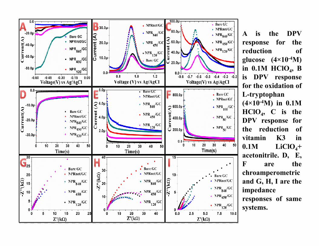
A is the DPV
response for the
reduction of
glucose (4×10⁻⁴M)
in 0.1M HClO4, B
is DPV response
for the oxidation of
L-tryptophan
(4×10⁻⁴M) in 0.1M
HClO4, C is the
DPV response forDPV response for
the reduction of
vitamin K3 in
0.1M LiClO4+
acetonitrile. D, E,
F are the
chroamperometric
and G, H, I are the
impedance
responses of same
systems.

High current and low Impedance of NPRnet compared to other systems in electrochemical
performances encourages us to use it as an electrode material for detection of Dengue
serotype-II related metabolites.
Oxidation of metabolites
in 0.1M NaOH medium:
(A) Ascorbic Acid , (B)(A) Ascorbic Acid , (B)
Creatinine (C)
Dopamine(DA), (D)
thiamine, (E) riboflavin, (F)
Pantothenic acid (PA) (G)
urea (H) uric acid (UA), (I)
inositol.

A-I: linear fitting
for different
water soluble
human
metabolites as
mentioned in the
figure during
their catalytic
oxidation at theiroxidation at their
physiological
concentration
level.
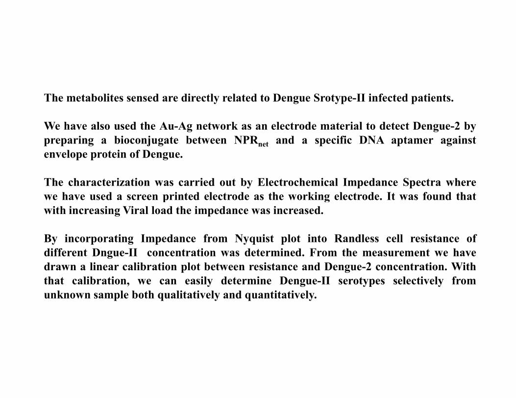
The metabolites sensed are directly related to Dengue Srotype-II infected patients.
We have also used the Au-Ag network as an electrode material to detect Dengue-2 by
preparing a bioconjugate between NPRnet and a specific DNA aptamer against
envelope protein of Dengue.
The characterization was carried out by Electrochemical Impedance Spectra where
we have used a screen printed electrode as the working electrode. It was found thatwe have used a screen printed electrode as the working electrode. It was found that
with increasing Viral load the impedance was increased.
By incorporating Impedance from Nyquist plot into Randless cell resistance of
different Dngue-II concentration was determined. From the measurement we have
drawn a linear calibration plot between resistance and Dengue-2 concentration. With
that calibration, we can easily determine Dengue-II serotypes selectively from
unknown sample both qualitatively and quantitatively.

A and B are the
Nyquist plot for
DENV-II and
DENV-III where
all the virus
concentrations are
in PFU/mL unit. C
is the calibration
plot for DENV-II.
D is the cross-
reactivity check by
impedance
response in a response in a
mixture of DENV-
II+ DENV-III. E is
the ∆RCT of
DENV-II in a
presence of other
DENV serotypes
and bacteria. For
all the samples we
used the
concentration at
101 PFU/mL.

Conclusion:
A low cost biosensor has been developed for early detection of Dengue-II &
Related metabolites.
The results obtained from our developed techniques are highly reproducible.
This developed method can easily replace the traditional spectrometric,
fluoremetric methods in terms od cost, accuracy, and time efficiency.
We are now studying real samples for further applications.