Embed Size (px)
Citation preview
Introduction Despite major advances in research and ther-apy, cancer continues to be the second cause of death in the United States, with 1 in 4 deaths due to cancer [1]. Primary tumors rarely have deadly consequences, while metastatic disease accounts for around 90% of the mortality due to solid tumors [2]. Therefore, the development of new sensitive methods that allow the detection of cancer dissemination, most notably in the common carcinomas, before full blown clinically detectable gross metastatic deposits are estab-lished is of tremendous utility to help physicians in treatment decisions. During the first stages of the metastatic cas-cade, cancer cells escape from a primary tumor mass and intravasate allowing their lympho-hematogenous dissemination to distant sites of the body. Most of these “circulating tumor cells” (CTCs) that depart the primary tumor will die, whereas as few as 0.01% of CTCs are likely to give rise to metastases, as suggested by pre-clinical models [3, 4]. Once cancer cells extrava-sate in anatomically distant organs, they can be found as single cells or small number of clus-tered cells referred to as “disseminated tumor cells” (DTCs). Although bone marrow (BM) is a frequent site for DTCs derived from many carci-nomas metastatic to bone or even other organs
[5], many studies failed to demonstrate an inde-pendent prognostic value of DTCs in BM [6, 7, 8, 9, 10, 11], while a meta-analysis in early breast cancer patients has reported its proven statistical utility [12]. Discrepancies between different studies could be explained on the ba-sis of the distinct sensitivity of the assays used to detect DTCs, or the biological function of those DTCs that potentially could evolve over time into overt metastases or remain as indo-lent or dormant cancer cells. Independent of the actual clinical significance of DTCs in BM, their collection involves painful BM aspirates that usually allow the detection of just a few DTCs in cancer patients with no evidence of overt metastases, even with the best validated techniques available (i.e. immunocytochemistry) [13]. In view of these obstacles and current ex-tensive studies showing the prognostic value of CTCs in patients with metastatic disease, the analysis of circulating rather than disseminated cells has been lately proposed as the method of choice to replace invasive BM sampling for the detection of occult non-hematologic cancer cells. CTCs can be easily obtained from periph-eral blood for which frequent sampling is usually accepted by patients and their treating physi-cians, reason by which this now called “liquid biopsy” holds significant promise in this regard. Recent studies tend to focus on CTCs in pa-tients with metastatic disease, in whose blood
Am J Cancer Res 2011;1(6):740-751 www.ajcr.us /ISSN:2156-6976/ajcr0000065
Review Article Circulating tumor cells: finding the needle in the haystack Xiaoning Zhe1, Michael L. Cher 1,2,3, R. Daniel Bonfil1,2,3
Departments of 1Urology and 2Pathology, Wayne State University School of Medicine, and 3The Barbara Ann Karmanos Cancer Institute, Detroit, Michigan, USA. Received April 29, 2011; accepted May 27, 2011; Epub June 1, 2011; Published June 30, 2011 Abstract: Localized and metastatic cancers give rise to circulating tumor cells (CTCs) which are detectable in the bloodstream. Recent studies have highlighted the prognostic significance of the presence and number of CTCs, par-ticularly in patients with metastatic disease. Future studies are designed not only to detect CTCs, but also to charac-terize them. This review discusses current and developing methodologies for the isolation and characterization of CTCs as well as recent studies focusing on the clinical relevance of CTC detection and characterization. Keywords: Circulating tumor cells, detection technique, enrichment technique, immunomagnetic separation, CTCs, cell capture and enumeration
Circulating tumor cells
741 Am J Cancer Res 2011;1(6):740-751
CTCs are present in high numbers, and thus are easier to identify and characterize. Here, we will review the most common techniques used to detect and enrich CTCs, and will summarize some of the results obtained in clinical studies. Methods used for CTC detection The identification of CTCs is achieved through different techniques that are typically used in combination with CTC enrichment procedures (see next section). These methodologies, sum-marized in recent reviews [14, 15, 16], are es-sentially constructed on antibody-based or nu-cleic acid-based approaches. In the first case, antibodies targeted to epithe-lial-specific antigens (e.g., epithelial cell adhe-sion molecule [EpCAM], or cytokeratins [CKs]), tissue-specific antigens (e.g., prostate specific antigen [PSA] in prostate cancer), or tumor-associated antigens (e.g., mammaglobin in breast cancer, or carcinoembryonic antigen [CEA] in colon cancer) are employed, since tu-mor-specific antigens have not been identified in most cancers. As a consequence of this, anti-body-based techniques usually have low speci-ficity, and anti-CD45 antibodies are frequently used to distinguish potentially contaminating leukocytes. One of the advantages of these techniques is that they do not require cell lysis, allowing morphological characterization, enu-meration, and molecular characterization (e.g., fluorescent in situ hybridization [FISH]) of CTCs. However, these methods are hampered by the low concentration of CTCs in blood. Despite this drawback, flow cytometry and laser scanning cytometry (LSC®, Compucyte Corporation, Cam-bridge, MA) are currently being used to detect CTCs, since these methods are rapid, quantita-tive, can simultaneously analyze multiple pa-rameters, such as size, DNA content, and spe-cific antigens, and identify viable from non-viable cells [17, 18, 19]. Despite their high specificity, and the advantage of LSC® over flow cytometry in allowing morphological analysis through automated fluorescence microscopy, both methods have low sensitivity, requiring large sample volumes to detect a few CTCs if no previous enrichment is done. The fiber-optic array scanning technology (FAST) is an ultra-speed technology that can scan up to 3x10⁵ cells per second and can detect CTCs with fluo-rescently labeled antibodies directly on a slide, without CTC enrichment required [20].
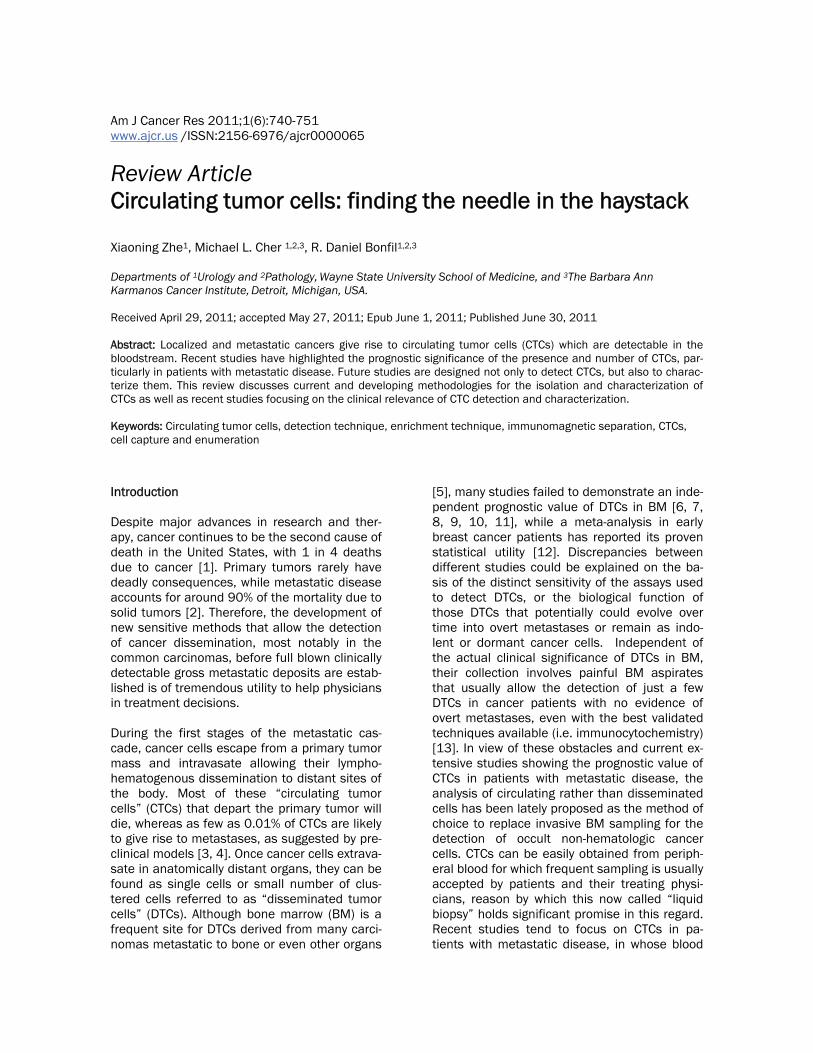
Another antibody-based method potentially use-ful for CTC detection is EPISPOT (epithelial im-munospot), an assay based on the enzyme-linked immunosorbent spot (ELISPOT) assay, which is a common method for monitoring im-mune responses. EPISPOT is based on the iden-tification of specific soluble proteins secreted by single viable epithelial tumor cells, such as cathepsin D and mucin-1 in breast cancer [21], and full-length CK19 in different cancers [22]. This assay is usually used with enrichments methods, and allows the indirect identification of viable epithelial cancer cells cultured for up to 48 hours via specific immunocapture of the proteins released by unlabeled antibodies im-mobilized on the bottom of the well, and then by addition of biotinylated or fluorochrome-conjugated antibodies. Each immunospot de-tected by either immunohistochemistry or im-munofluorescence, is considered the fingerprint left only by one viable cancer cell releasing the protein under analysis [23] (Figure 1). Despite being the only assay available that can detect cancer cells on the basis of secreted/shed pro-teins, it has not been tested until now in large clinical trials.
Nucleic acid-based methods are considered to be more sensitive than cytometry and immuno-cytochemistry methods [24, 25], achieving specificity through oligonucleotide primers de-
Figure 1. Identification of RANKL-shedding cells in SAOS-2 cells transfected with MT1-MMP using the ELISPOT assay. Each immunospot identifies single viable cells shedding RANKL.

Circulating tumor cells
742 Am J Cancer Res 2011;1(6):740-751
signed for detection of genes of interest. Al-though polymerase chain reaction (PCR) can be used for CTC detection, free DNA from dying cancer cells can be found in peripheral blood, thus affecting the accuracy of this method. Con-sequently, the more unstable mRNA, which is rapidly degraded in blood, is currently consid-ered as a better target for detection of CTCs using reverse-transcriptase PCR (RT-PCR) or real-time RT-PCR. A variety of epithelium-specific or more cancer-specific transcripts, such as CK19, mammaglobin, mucin 1, or CEA, have been used to amplify mRNA by RT-PCR techniques and distinguish the presence of CTCs from other blood elements [26, 27]. However, despite the high sensitivity of RT-PCR, this technique has low specificity due to expression of some of the transcripts in normal blood cells, leading to false positives. This obstacle could be circum-vent using combined real-time RT-PCR for more than one single transcript, which results in en-hanced specificity and sensitivity and allows relative quantification of message levels [28, 29, 30]. The major drawback of PCR-based techniques is the need of cell lysis, which im-pedes morphological analysis and enumeration of CTCs. CTC enrichment methods The existence of CTCs was first reported in 1869 by the Australian pathologist Thomas Ashworth in the peripheral blood a patient who died of cancer [31]. These rare cells, found at a concentration of about 1 in about 5x10⁹ eryth-rocytes and 107 leukocytes per milliliter of blood in advanced cancer patients [32, 33], were de-tected in the 1990s using RT-PCR aimed at identifying tissue-specific genes in prostate can-cer and melanoma patients [34, 35]. To date, cell enrichment techniques have emerged mainly based on physical properties or antigenic characteristics that distinguish CTCs from nor-mal blood cells. Enrichment methods based on physical proper-ties Based on their lower buoyant density with re-spect to most blood cells (with a density >1.077 g/ml), CTCs can be enriched through gradient centrifugation using Ficoll-Hypaque or other similar density gradient media. However, de-spite being easy and economical, this technique has low CTC recovery and specificity, as other
low density mononuclear cells such as mono-cytes and lymphocytes, and platelets, are lay-ered together with CTCs. OncoQuick® (Greiner Bio-One, Frickenhausen, Germany), a density gradient-based method that adds a porous membrane that prevents cross contamination of the mononuclear fraction by blood cells with higher buoyant density, has shown to reduce the number of co-enriched mononuclear cells without compromising CTC recovery rate [36]. Other CTC enrichment methods rely on the small diameter of the majority of blood cells, ranging from 8 to 11 µm, and the assumption that, in general, tumor cells have relative larger size (e.g., around 30 µm in breast cancer cells) [37]. Based on these physical characteristics, CTCs can be enriched by filtration of blood through polycarbonate membranes with 8 µm-pores. The first filter-based method described was ISET® (Isolation by Size of Epithelial Tumor cells) (Rarecells, Paris, France), which allows the separation of fixed tumor cells using a dis-posable block containing the pored membrane in an automated filtration device [38]. Although it has been reported that an average of 0.0002% of leukocytes from blood are retained in the pored membrane [38], we must be aware that this represents about 2,000 leukocytes per ml of blood [39], diminishing the specificity of ISET® for CTC enrichment. Fortunately, CTCs enriched through this rapid and simple method can be easily identified using cytological stain-ing or immunolabeling [38, 39]. Recently, the ScreenCell® filtration device (ScreenCell, Paris, France) was developed based on the same prin-ciple than ISET®, with the added advantage that can be used to retrieve live CTCs for cell culture needs in addition to the traditional use for stain-ing, cell enumeration, immunolabeling, and mo-lecular biology techniques [40], and no auto-mated filtration machine is needed. In experi-ments in which 2 or 5 cultured tumor cells were spiked in whole peripheral blood, 74 and 91% recovery of the cancer cells was achieved re-spectively [40], confirming the high sensitivity of this methodology. Antibody-based enrichment methods The differential expression of specific antigens on the surface of epithelial cancer and mononu-clear peripheral blood cells is used for CTC en-richment methods using either positive or nega-tive selection. Most of the antibody-based tech-

Circulating tumor cells
743 Am J Cancer Res 2011;1(6):740-751
niques used to enrich CTCs employ antibodies that target epithelial markers (positive selec-tion) of hematopietic cell markers (negative se-lection) and are coupled to magnetic particles (beads or ferrofluids). The most commonly used antigen to directly isolate epithelial CTCs is Ep-CAM [41], while the depletion of leukocytes is usually achieved targeting CD45 [42]. In the magnetic activated cell sorting system MACS® (Miltenyi Biotec GmbH, Bergisch-Gladbach, Germany) epithelial tumor cells are usually separated from blood components by incubation with ferromagnetic microbeads cou-pled to anti-EpCAM antibodies, followed by transferring to a column placed in a strong mag-netic field. Using this system, cells not recog-nized by antibody-coated ferromagnetic mi-crobeads flow through, whereas cells express-ing EpCAM remain trapped within the column and are then recovered by removing the mag-netic field. A similar methodology involves the use of Dynabeads® (Invitrogen, Carlsbad, CA), which also depends on the attraction by a mag-netic field of cells recognized by antibodies cou-pled to ferromagnetic beads, but does not re-quire a separation column [43]. Studies using either positive or negative sorting of CTCs has been described, but the cell recovery rates are inconsistent, diminishing the value of these techniques for CTC enrichment [5]. In general, negative selection using these methods leads to low purity due to deficient removal of interfering cells [44]. The CellSearch® semiautomated system (Veridex, Raritan, NJ) has become the most popular method for CTC enrichment and detec-tion. This technology combines positive selec-tion of cells with epithelial markers and nega-tive selection of leukocytes, and is the only cur-rently US Food and Drug Administration (FDA)-approved system for the detection of CTCs in patients with metastatic breast, prostate, and colorectal cancer [33, 45, 46, 47, 48]. In the CellSearch® system, after an automated separa-tion of cells from plasma obtained from 7.5 ml of blood, CTCs are magnetically captured trough a ferrofluid-coupled antibody against EpCAM, and then sequentially permeabilized, fixed, and labeled with the fluorescent nuclear dye DAPI and fluorescent antibodies to the leukocyte marker CD45 and to epithelial markers cy-tokeratins (CK) 8, 18, and 19. The treated sam-ple is then loaded into a cartridge where a
strong magnetic force attracts the immunomag-netically-labeled cells for analysis by the Cell-Tracks AnalyzerVR, a semi-automated fluores-cence microscope that scans the sample at four different wavelengths, records the fluorescent events, and automatically presents images in a gallery format for classification by trained opera-tors. CTCs are identified as cells that are DAPI- and CK-8/18/19-positive and CD45-negative. This method allows accurate and sensitive counting of CTCs up to 72 hours after blood drawing using tubes containing a special pre-servative, and has shown very low interlabora-tory variability when identical samples were analyzed in different centers [49]. Recently, the new CellSearch® Profile kit (instead of the Cell-Search® Epithelial kit) represents an improve-ment in CTC isolation [50], and allows molecular profiling, flow cytometry, and FISH [30] (Figure 2).
Another immunomagnetic platform used to en-rich CTCs is the “MagSweeper” technology. Ba-sically, 6-mm-diameter magnetic rods covered with a 25-µm non-adherent plastic sheet are swept in concentric circular loops within wells of a 6-well plate with CTC-containing diluted blood
Figure 2. Sensitivity of RT-PCR analysis to detect genes of interest in cultured prostate cancer cells spiked in blood. Peripheral blood of a healthy volun-teer was spiked with different numbers of LNCaP prostate cancer cells transfected with wild-type MT1-MMP (LNCaP-MT1wt), and processed using the Cell-Search® Profile kit. PCR amplification products ob-tained after a two-cycle amplification process in EpCAM-positive fractions demonstrate the feasibility of molecular analysis for diffferent genes in samples enriched using the CellSearch® platform. Peripheral blood mononuclear cells (PBMC) of the same volun-teer were used as control.
Circulating tumor cells
744 Am J Cancer Res 2011;1(6):740-751
samples previously labeled with anti-EpCAM antibodies coupled to ferromagnetic beads. After 45- to 60-min sweeping of the whole area of the well, the rods are washed with a buffer solution, and disengaged from their plastic sleeves. CTCs initially captured by the magnetic rod are then released from the plastic sheet by external magnets located under the wells, and freed of most contaminated cells by a second round of capture-wash-release [51]. Spiking experiments with breast cancer cells in blood of normal volunteers have demonstrated around 60% capture of CTCs, with almost 100% purity. Through this method, CTCs could be isolated from 47 of 47 blood samples obtained from metastatic breast cancer patients [51]. Other methods based on the identification of epithelial markers on the surface of CTCs do not count on magnetic platforms but on microfluidic systems. One of such methods involves the novel microfluidic-based technology known as “CTC-chip”, which offers both very high sensitiv-ity and preservation of viability of isolated CTCs. This platform consists of an array of 78,000 microspots coated with antibodies to EpCAM aligned on a chip the size of a standard micro-scope slide. Whole EDTA-treated blood from patients is pneumatically forced to flow over the surface of the chip and between the microspots, maximizing the capture of CTCs by anti-EpCAM antibodies [52]. CTCs can be identified and quantitated in situ by automatic scanning of the microchip after immunolabeling with fluorescent anti-CK and anti-CD45 antibodies and DAPI, taking in consideration only those cells which are CK+, DAPI+, and CD45-. The CTC-chip was used for enumeration of CTCs in blood from patients with non-small cell lung, prostate, breast, pancreatic, and colon cancer, with aver-age CTC counts ranging between 79 and 196 per ml of blood, and more than 99% detection in cancer patients, as opposed to 0% in healthy individuals [52]. Despite the higher sensitivity of this method to detect CTCs as compared to Cell-Search®, the test is currently useful in the re-search setting as no studies have been done yet to prove its clinical significance. Recently, a re-fined methodology called “herringbone-chip”, or "HB-Chip," was developed to provide an en-hanced platform for CTC isolation [53]. Briefly, it operates on the basis of the CTC-chip except that the flat upper wall of the microfluidic device is replaced by ridges or herringbones that gen-erate microvortices and disrupt the laminar flow streamlines that cells travel, thus increasing the
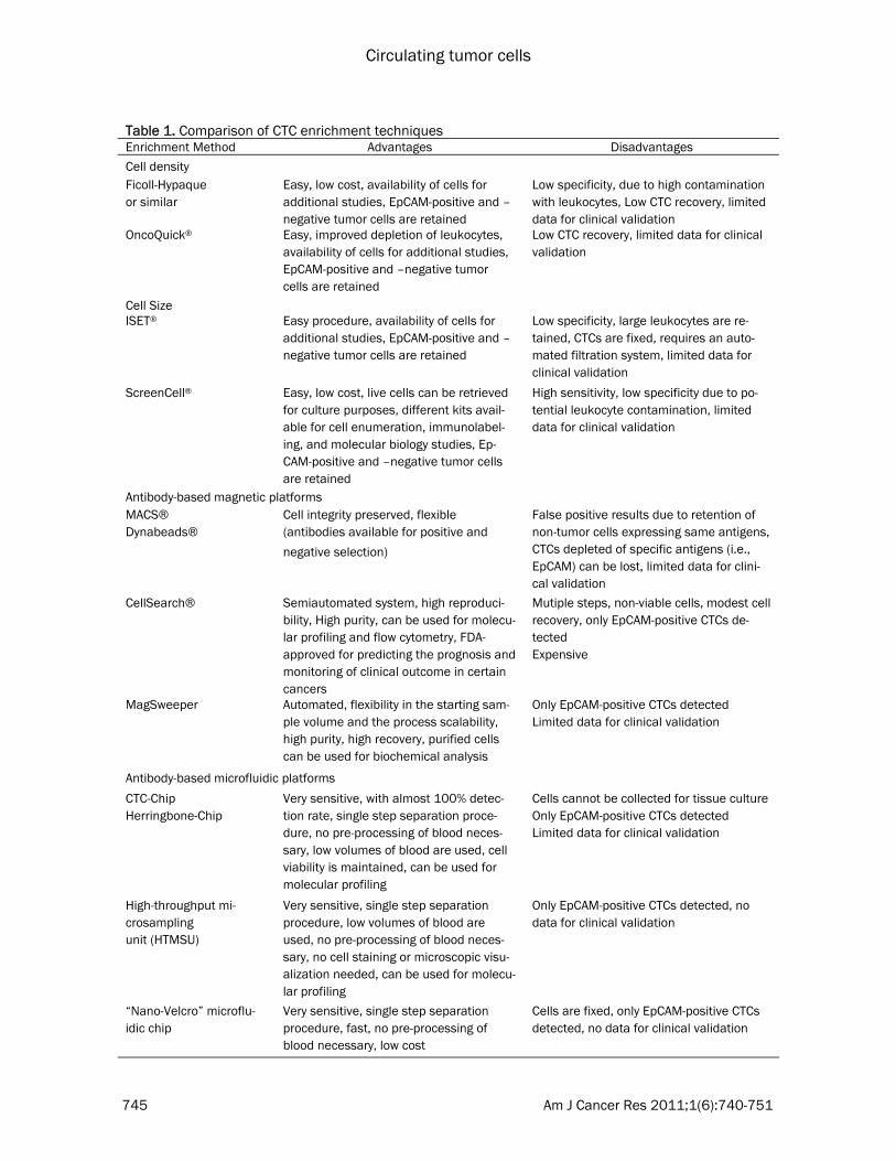
collisions between CTCs and the anti-EpCAM-coated device and the average capture effi-ciency. Another microfluidic chip, though integrated to an electrokinetic enrichment device, is the “HTMSU” or “high-throughput microsampling unit”. This technology consists of microstruc-tures replicated in a polymeric substrate con-taining 51 sinusoidal channels coated with anti-EpCAM antibodies. The channels have a width (35 µm) similar to CTCs’ diameters (15-30 µm), leading to high cell recovery (~97%). CTCs cap-tured from whole blood flowing at 2 mm/sec in each channel are released through enzymatic dissociation of the EpCAM/anti-EpCAM antibody complexes, and then counted one at a time via an integrated conductivity sensor that specifi-cally detects CTCs via their electrical signatures without the necessity of cell staining or micro-scopic analysis [54, 55]. Recently, the HTMSU has proved to be useful to profile point muta-tions in genomic DNA of CTCs spiked into blood [56]. To date, there are no data published for clinical validation of this method. Using “Nano-Velcro” technology, a new microflu-idic device was recently reported. This CTC-capture method involves a silicon nanopillar substrate coated with streptavidin and bioti-nylated anti-EpCAM antibodies and a serpentine chaotic mixing channel sandwiched together in a 2.5 cm x 5.0 cm device. The nanopillar-covered silicon chip binds to microvilli and filo-podia of CTCs in a very efficient way, creating an effect much like the top and bottom of Velcro that significantly increases the capture of Ep-CAM-expressing cells compared to other existing methods. In addition, the overlaid microfluidic channel architecture increases mixing of the fluid flowing over the nanopillar substrate, maxi-mizing the contact frequency of CTCs with anti-EpCAM antibodies. Peripheral blood from pros-tate cancer patients have been analyzed side-by-side using this method and CellSearch®, con-firming the greater capacity of the “Nano-Velcro” chip to capture CTCs [57]. This microflu-idic system, similarly to CellSearch®, used three-color immunofluorescence for detection of CTCs in fixed samples. Table 1 summarizes the differ-ent CTC-enrichment methods described above. Clinical relevance Although BM sampling is frequently performed in patients with leukemia or lymphoma to moni-
Circulating tumor cells
745 Am J Cancer Res 2011;1(6):740-751
Table 1. Comparison of CTC enrichment techniques Enrichment Method Advantages Disadvantages
Cell density Ficoll-Hypaque or similar
Easy, low cost, availability of cells for additional studies, EpCAM-positive and –negative tumor cells are retained
Low specificity, due to high contamination with leukocytes, Low CTC recovery, limited data for clinical validation
OncoQuick® Easy, improved depletion of leukocytes, availability of cells for additional studies, EpCAM-positive and –negative tumor cells are retained
Low CTC recovery, limited data for clinical validation
Cell Size ISET® Easy procedure, availability of cells for
additional studies, EpCAM-positive and –negative tumor cells are retained
Low specificity, large leukocytes are re-tained, CTCs are fixed, requires an auto-mated filtration system, limited data for clinical validation
ScreenCell® Easy, low cost, live cells can be retrieved for culture purposes, different kits avail-able for cell enumeration, immunolabel-ing, and molecular biology studies, Ep-CAM-positive and –negative tumor cells are retained
High sensitivity, low specificity due to po-tential leukocyte contamination, limited data for clinical validation
Antibody-based magnetic platforms MACS® Dynabeads®
Cell integrity preserved, flexible (antibodies available for positive and
negative selection)
False positive results due to retention of non-tumor cells expressing same antigens, CTCs depleted of specific antigens (i.e., EpCAM) can be lost, limited data for clini-cal validation
CellSearch®
Semiautomated system, high reproduci-bility, High purity, can be used for molecu-lar profiling and flow cytometry, FDA-approved for predicting the prognosis and monitoring of clinical outcome in certain cancers
Mutiple steps, non-viable cells, modest cell recovery, only EpCAM-positive CTCs de-tected Expensive
MagSweeper
Automated, flexibility in the starting sam-ple volume and the process scalability, high purity, high recovery, purified cells can be used for biochemical analysis
Only EpCAM-positive CTCs detected Limited data for clinical validation
Antibody-based microfluidic platforms
CTC-Chip Herringbone-Chip
Very sensitive, with almost 100% detec-tion rate, single step separation proce-dure, no pre-processing of blood neces-sary, low volumes of blood are used, cell viability is maintained, can be used for molecular profiling
Cells cannot be collected for tissue culture Only EpCAM-positive CTCs detected Limited data for clinical validation
High-throughput mi-crosampling unit (HTMSU)
Very sensitive, single step separation procedure, low volumes of blood are used, no pre-processing of blood neces-sary, no cell staining or microscopic visu-alization needed, can be used for molecu-lar profiling
Only EpCAM-positive CTCs detected, no data for clinical validation
“Nano-Velcro” microflu-idic chip
Very sensitive, single step separation procedure, fast, no pre-processing of blood necessary, low cost
Cells are fixed, only EpCAM-positive CTCs detected, no data for clinical validation
Circulating tumor cells
746 Am J Cancer Res 2011;1(6):740-751
tor residual disease, this painful procedure is usually not well accepted by treating physicians and their patients with solid tumors. The idea of a “liquid biopsy” obtained by regular blood drawing is definitively more attractive, reason by which different groups are currently assessing the utility of CTCs as prognostic markers and predictors of responses to therapy. Here, we will refer to some of the studies in which the Cell-Search® platform was used, considering that it is the only CTC-enrichment system currently approved by the FDA for clinical use in some cancers, and most studies using other combina-tion of enrichment and detection methods still have not been performed in large trials and, consequently, do not have sufficient statistical power for translation to regular clinical practice. Among the studies that led to the approval of the CellSearch® system as a tool of prognostic and predictive value in certain cancer patient populations, we cannot omit mentioning those performed in breast, prostate, and colorectal cancers. In a multicenter study with patients with metastatic breast cancer, Cristofanilli et al. found that women with 5 or more CTCs per 7.5 ml of peripheral blood before therapy have a statistically shorter overall survival (OS) and progression-free survival (PFS) than patients with less than 5 CTCs per 7.5 ml of blood (median of 10.1 versus 18 months for OS, and of 2.7 versus 7 months for PFS, respectively) [33]. Moreover, CTCs predicted clinical outcome after therapy with women with CTC counts ≥ 5 showing significant worse OS and PFS [33], and proved to be a better predictor of treatment response than current radiologic assessment [58]. Similar results were obtained by de Bono et al. in studies with patients with metastatic castrate-resistant prostate cancer (mCRPC), where those with baseline CTC counts ≥ 5 showed significantly worse OS (median of 11.5 months in the later versus 21.7 months in mCRPC patients with baseline CTC counts < 5) [48]. Interestingly, they also found that CTC counts represent a more accurate and inde-pendent predictor of OS than PSA decrements before and after treatment in mCRPC patients [48]. As for colorectal cancer, Cohen et al. also found that higher numbers of CTCs were associ-ated with worst prognosis [45]. However, in this study the threshold number of CTCs identified for stratification in groups of better or worst prognosis was 3: OS and PFS for patients with metastatic colorectal cancer with baseline CT
counts ≥ 3 compared with <3 were significantly shorter (median of 4.4 versus 7.8 months for OS, and of 9.4 versus 20.6 months for PFS, re-spectively) [45, 59]. Molecular characterization of CTCs may also be useful for the assessment of predictive bio-markers in real-time and for the development of tailored therapies. This is clearly illustrated by studies in which immunomagnetically-enriched CTCs obtained from patients with metastatic breast cancer were found to be HER2-positive using FISH analysis; despite their primary tu-mors were HER2-negative at diagnosis [60]. Interestingly, treatment with anti-HER2 therapy in some of the patients with HER-2 amplification in their CTCs resulted in complete or partial re-sponse [60], though the actual significance of this finding remains to be confirmed in studies with larger number of patients. Recently, in a study in which FISH has been applied to CTCs enriched with the CellSearch® system to analyze androgen receptor (AR) gene amplification in patients with mCRPC, high-level chromosomal AR amplification was found in 50% of patients with CTC counts ≥ 10 [61], while different AR mutations, many of which are associated with resistance to androgen-directed therapies, were detected in CTCs captured using the same sys-tem [62]. These two studies demonstrate the feasibility of molecular profiling of CTCs to ana-lyze the significance of AR amplification/mutations in prognosis and response to therapy in mCRPC patients. In another study in patients with metastatic non-small cell lung carcinoma, DNA obtained from CTCs captured using the “CTC chip” was analyzed for specific mutations in the epidermal growth factor receptor (EGFR) gene. The authors found that 11 of 12 patients with specific EGFR mutations in their primary tumors also showed the mutations in the CTCs isolated, and that the activating mutation T790M, which confers drug resistance, was prevalent in a group of patients who received tyrosine kinase inhibitor treatment and showed clinical progression [63]. This exemplifies how CTCs could be used in the clinical setting to monitor molecular changes potentially useful to predict the response to therapy in cancer pa-tients. Concluding remarks Several studies have demonstrated the prog-nostic and predictive value of CTC quantitation

Circulating tumor cells
747 Am J Cancer Res 2011;1(6):740-751
using the CellSearch® platform in breast, pros-tate, and colorectal cancer, while new data are emerging in other cancer types. Despite many advantages of this FDA-approved combined enrichment/detection system, based mainly on its high reproducibility across institutions and its validation in large patient cohorts, one of its limitations involve the low cell recovery. For ex-ample, CellSearch® identifies CTCs in only 60% of the metastatic breast cancer patients [64]. Although other enrichment methods, such as many of the new antibody-based microfluidic techniques, show higher cell recovery without compromising cell purity sensitivity, they still need to be validated in studies with large num-ber of patients to confirm their clinical value. Moreover, there is concern that different enrich-ment/detection methods could detect distinct subsets of the heterogeneous CTC population, which have different biological roles and clinical relevance. In that sense, uniform methodologies should be used for every single detection plat-form across the different laboratories, and tested in a large number of patients, so that meaningful interpretations are being drawn by comparing different CTC enrichment/detection methods. Even in methods based on similar principles, such as the majority of antibody-based techniques that use EpCAM expression for positive selection of CTCs, their different sensitivities and cell recovery rates will result in the establishment of different threshold number of CTCs for stratification in groups of better or worst prognosis in each case. This is also true when the focus is on real-time assessment of predictive biomarkers and development of tai-lored therapies based on molecular characteri-zation of CTCs. Nevertheless, with the exception of CTC-enriching methods based on cell density or size, most antibody-based techniques depend on capture of CTCs based on expression of EpCAM, known to be present on the cell surface of the vast majority of carcinomas. However, a recent study revealed that “normal-like” breast cancer cells, which usually display an aggressive phe-notype, express low expression of EpCAM and are not detected by the CellSearch® test [65]. Moreover, a retrospective study that involved 292 patients with metastatic breast cancer has shown that 36% of them showed an undetect-able CTC status, which could be due, at least in part, to an underestimation of CTCs by the Cell-Search® test due to CTC undergoing epithelial-
mesenchymal transition (EMT) [66]. In fact, EMT is characterized by downregulation of certain CKs and gain of mesenchymal markers such as vimentin and fibronectin, which could result in an inefficient identification by anti-CK antibod-ies used in many of the CTC-detection tech-niques. Indeed, these putative CTCs could have an aggressive phenotype, bearing in mind that EMT is considered to enable cancer cells to en-ter new tissues through extravasation [67], and has been linked to the generation of cells with tumor initiating ability and drug resistant pheno-type [68]. In conclusion, multiple methodologies for en-richment and detection of CTCs have emerged in the last years, with one of them already ap-proved by FDA for certain clinical uses. A signifi-cant optimization and integration of the avail-able techniques is still needed to obtain an en-richment and detection sensitive enough to dis-criminate CTCs going through EMT from epithe-lial CTCs and normal mesenchymal blood cells, with clear clinical applications and uses for mo-lecular studies with translational potential. These advances will have important implica-tions for a better understanding of the mecha-nisms operating during metastatic dissemina-tion and an improved treatment of cancer pa-tients. Address correspondence to: Dr. R. Daniel Bonfil, De-partments of Urology and Pathology, Wayne State University School of Medicine, 540 E. Canfield, Room # 9105, Detroit, MI 48201, USA. Tel: (313)-577-2879; Fax: (313)-577-0057; E-mail: [email protected] References [1] Jemal A, Siegel R, Xu J and Ward E. Cancer
statistics, 2010. CA Cancer J Clin 2010; 60: 277-300.
[2] Gupta GP and Massague J. Cancer metasta-sis: building a framework. Cell 2006; 127: 679-695.
[3] Fidler IJ. Metastasis: guantitative analysis of distribution and fate of tumor embolilabeled with 125 I-5-iodo-2'-deoxyuridine. J Natl Can-cer Inst 1970; 45: 773-782.
[4] Langley RR and Fidler IJ. The seed and soil hypothesis revisited-The role of tumor-stroma interactions in metastasis to different organs. Int J Cancer 2011; 128: 2527-2535.
[5] Lin H, Balic M, Zheng S, Datar R and Cote RJ. Disseminated and circulating tumor cells: Role in effective cancer management. Crit Rev On-col Hematol 2011; 77: 1-11.
Circulating tumor cells
748 Am J Cancer Res 2011;1(6):740-751
[6] Cote RJ, Rosen PP, Lesser ML, Old LJ and Osborne MP. Prediction of early relapse in patients with operable breast cancer by detec-tion of occult bone marrow micrometastases. J Clin Oncol 1991; 9: 1749-1756.
[7] Mansi JL, Gogas H, Bliss JM, Gazet JC, Berger U and Coombes RC. Outcome of primary-breast-cancer patients with micrometastases: a long-term follow-up study. Lancet 1999; 354: 197-202.
[8] Funke I and Schraut W. Meta-analyses of stud-ies on bone marrow micrometastases: an independent prognostic impact remains to be substantiated. J Clin Oncol 1998; 16: 557-566.
[9] Gebauer G, Fehm T, Merkle E, Jaeger W and Mitze M. Micrometastases in axillary lymph nodes and bone marrow of lymph node-negative breast cancer patients--prognostic relevance after 10 years. Anticancer Res 2003; 23: 4319-4324.
[10] Gerber B, Krause A, Muller H, Richter D, Reimer T, Makovitzky J, Herrnring C, Jeschke U, Kundt G and Friese K. Simultaneous immu-nohistochemical detection of tumor cells in lymph nodes and bone marrow aspirates in breast cancer and its correlation with other prognostic factors. J Clin Oncol 2001; 19: 960-971.
[11] Landys K, Persson S, Kovarik J, Hultborn R and Holmberg E. Prognostic value of bone marrow biopsy in operable breast cancer pa-tients at the time of initial diagnosis: Results of a 20-year median follow-up. Breast Cancer Res Treat 1998; 49: 27-33.
[12] Braun S, Vogl FD, Naume B, Janni W, Osborne MP, Coombes RC, Schlimok G, Diel IJ, Gerber B, Gebauer G, Pierga JY, Marth C, Oruzio D, Wiedswang G, Solomayer EF, Kundt G, Strobl B, Fehm T, Wong GY, Bliss J, Vincent-Salomon A and Pantel K. A pooled analysis of bone marrow micrometastasis in breast cancer. N Engl J Med 2005; 353: 793-802.
[13] Slade MJ, Payne R, Riethdorf S, Ward B, Zaidi SA, Stebbing J, Palmieri C, Sinnett HD, Kulin-skaya E, Pitfield T, McCormack RT, Pantel K and Coombes RC. Comparison of bone mar-row, disseminated tumour cells and blood-circulating tumour cells in breast cancer pa-tients after primary treatment. Br J Cancer 2009; 100: 160-166.
[14] Alunni-Fabbroni M and Sandri MT. Circulating tumour cells in clinical practice: Methods of detection and possible characterization. Meth-ods 2010; 50: 289-297.
[15] Allan AL and Keeney M. Circulating tumor cell analysis: technical and statistical considera-tions for application to the clinic. J Oncol 2010; 2010: 426218.
[16] Ross JS and Slodkowska EA. Circulating and disseminated tumor cells in the management of breast cancer. Am J Clin Pathol 2009; 132:
237-245. [17] Cruz I, Ciudad J, Cruz JJ, Ramos M, Gomez-
Alonso A, Adansa JC, Rodriguez C and Orfao A. Evaluation of multiparameter flow cytometry for the detection of breast cancer tumor cells in blood samples. Am J Clin Pathol 2005; 123: 66-74.
[18] Pachmann K, Heiss P, Demel U and Tilz G. Detection and quantification of small numbers of circulating tumour cells in peripheral blood using laser scanning cytometer (LSC). Clin Chem Lab Med 2001; 39: 811-817.
[19] Zabaglo L, Ormerod MG, Parton M, Ring A, Smith IE and Dowsett M. Cell filtration-laser scanning cytometry for the characterisation of circulating breast cancer cells. Cytometry A 2003; 55: 102-108.
[20] Krivacic RT, Ladanyi A, Curry DN, Hsieh HB, Kuhn P, Bergsrud DE, Kepros JF, Barbera T, Ho MY, Chen LB, Lerner RA and Bruce RH. A rare-cell detector for cancer. Proc Natl Acad Sci U S A 2004; 101: 10501-10504.
[21] Alix-Panabieres C, Brouillet JP, Fabbro M, Yssel H, Rousset T, Maudelonde T, Choquet-Kastylevsky G and Vendrell JP. Characteriza-tion and enumeration of cells secreting tumor markers in the peripheral blood of breast can-cer patients. J Immunol Methods 2005; 299: 177-188.
[22] Alix-Panabieres C, Vendrell JP, Slijper M, Pelle O, Barbotte E, Mercier G, Jacot W, Fabbro M and Pantel K. Full-length cytokeratin-19 is released by human tumor cells: a potential role in metastatic progression of breast can-cer. Breast Cancer Res 2009; 11: R39.
[23] Alix-Panabieres C, Riethdorf S and Pantel K. Circulating tumor cells and bone marrow mi-crometastasis. Clin Cancer Res 2008; 14: 5013-5021.
[24] Ring AE, Zabaglo L, Ormerod MG, Smith IE and Dowsett M. Detection of circulating epithelial cells in the blood of patients with breast can-cer: comparison of three techniques. Br J Can-cer 2005; 92: 906-912.
[25] Smith BM, Slade MJ, English J, Graham H, Luchtenborg M, Sinnett HD, Cross NC and Coombes RC. Response of circulating tumor cells to systemic therapy in patients with me-tastatic breast cancer: comparison of quanti-tative polymerase chain reaction and immuno-cytochemical techniques. J Clin Oncol 2000; 18: 1432-1439.
[26] Berois N, Varangot M, Aizen B, Estrugo R, Zarantonelli L, Fernandez P, Krygier G, Si-monet F, Barrios E, Muse I and Osinaga E. Molecular detection of cancer cells in bone marrow and peripheral blood of patients with operable breast cancer. Comparison of CK19, MUC1 and CEA using RT-PCR. Eur J Cancer 2000; 36: 717-723.
[27] Xenidis N, Ignatiadis M, Apostolaki S, Perraki M, Kalbakis K, Agelaki S, Stathopoulos EN,

Circulating tumor cells
749 Am J Cancer Res 2011;1(6):740-751
Chlouverakis G, Lianidou E, Kakolyris S, Geor-goulias V and Mavroudis D. Cytokeratin-19 mRNA-positive circulating tumor cells after adjuvant chemotherapy in patients with early breast cancer. J Clin Oncol 2009; 27: 2177-2184.
[28] Ignatiadis M, Kallergi G, Ntoulia M, Perraki M, Apostolaki S, Kafousi M, Chlouverakis G, Stathopoulos E, Lianidou E, Georgoulias V and Mavroudis D. Prognostic value of the molecu-lar detection of circulating tumor cells using a multimarker reverse transcription-PCR assay for cytokeratin 19, mammaglobin A, and HER2 in early breast cancer. Clin Cancer Res 2008; 14: 2593-2600.
[29] Hayes DC, Secrist H, Bangur CS, Wang T, Zhang X, Harlan D, Goodman GE, Houghton RL, Persing DH and Zehentner BK. Multigene real-time PCR detection of circulating tumor cells in peripheral blood of lung cancer pa-tients. Anticancer Res 2006; 26: 1567-1575.
[30] Sieuwerts AM, Kraan J, Bolt-de Vries J, van der Spoel P, Mostert B, Martens JW, Gratama JW, Sleijfer S and Foekens JA. Molecular charac-terization of circulating tumor cells in large quantities of contaminating leukocytes by a multiplex real-time PCR. Breast Cancer Res Treat 2009; 118: 455-468.
[31] Ashworth TR. A case of cancer in which cells similar to those in the tumors were seen in the blood after death. Aust Med J 1869; 14: 146-149.
[32] Yu M, Stott S, Toner M, Maheswaran S and Haber DA. Circulating tumor cells: approaches to isolation and characterization. J Cell Biol 2011; 192: 373-382.
[33] Cristofanilli M, Budd GT, Ellis MJ, Stopeck A, Matera J, Miller MC, Reuben JM, Doyle GV, Allard WJ, Terstappen LW and Hayes DF. Cir-culating tumor cells, disease progression, and survival in metastatic breast cancer. N Engl J Med 2004; 351: 781-791.
[34] Katz AE, Olsson CA, Raffo AJ, Cama C, Perlman H, Seaman E, O'Toole KM, McMahon D, Benson MC and Buttyan R. Molecular stag-ing of prostate cancer with the use of an en-hanced reverse transcriptase-PCR assay. Urol-ogy 1994; 43: 765-775.
[35] Smith B, Selby P, Southgate J, Pittman K, Bradley C and Blair GE. Detection of mela-noma cells in peripheral blood by means of reverse transcriptase and polymerase chain reaction. Lancet 1991; 338: 1227-1229.
[36] Rosenberg R, Gertler R, Friederichs J, Fuehrer K, Dahm M, Phelps R, Thorban S, Nekarda H and Siewert JR. Comparison of two density gradient centrifugation systems for the enrich-ment of disseminated tumor cells in blood. Cytometry 2002; 49: 150-158.
[37] Meng S, Tripathy D, Frenkel EP, Shete S, Naf-talis EZ, Huth JF, Beitsch PD, Leitch M, Hoover S, Euhus D, Haley B, Morrison L, Fleming TP,
Herlyn D, Terstappen LW, Fehm T, Tucker TF, Lane N, Wang J and Uhr JW. Circulating tumor cells in patients with breast cancer dormancy. Clin Cancer Res 2004; 10: 8152-8162.
[38] Vona G, Sabile A, Louha M, Sitruk V, Romana S, Schutze K, Capron F, Franco D, Pazzagli M, Vekemans M, Lacour B, Brechot C and Pater-lini-Brechot P. Isolation by size of epithelial tumor cells: a new method for the immuno-morphological and molecular characterization of circulatingtumor cells. Am J Pathol 2000; 156: 57-63.
[39] Paterlini-Brechot P and Benali NL. Circulating tumor cells (CTC) detection: clinical impact and future directions. Cancer Lett 2007; 253: 180-204.
[40] Desitter I, Guerrouahen BS, Benali-Furet N, Wechsler J, Janne PA, Kuang Y, Yanagita M, Wang L, Berkowitz JA, Distel RJ and Cayre YE. A new device for rapid isolation by size and characterization of rare circulating tumor cells. Anticancer Res 2011; 31: 427-441.
[41] Trzpis M, McLaughlin PM, de Leij LM and Harmsen MC. Epithelial cell adhesion mole-cule: more than a carcinoma marker and ad-hesion molecule. Am J Pathol 2007; 171: 386-395.
[42] Lara O, Tong X, Zborowski M and Chalmers JJ. Enrichment of rare cancer cells through deple-tion of normal cells using density and flow-through, immunomagnetic cell separation. Exp Hematol 2004; 32: 891-904.
[43] Neurauter AA, Bonyhadi M, Lien E, Nokleby L, Ruud E, Camacho S and Aarvak T. Cell isola-tion and expansion using Dynabeads. Adv Biochem Eng Biotechnol 2007; 106: 41-73.
[44] Sitar G, Brambati B, Baldi M, Montanari L, Vincitorio M, Tului L, Forabosco A and Ascari E. The use of non-physiological conditions to isolate fetal cells from maternal blood. Exp Cell Res 2005; 302: 153-161.
[45] Cohen SJ, Punt CJ, Iannotti N, Saidman BH, Sabbath KD, Gabrail NY, Picus J, Morse M, Mitchell E, Miller MC, Doyle GV, Tissing H, Terstappen LW and Meropol NJ. Relationship of circulating tumor cells to tumor response, progression-free survival, and overall survival in patients with metastatic colorectal cancer. J Clin Oncol 2008; 26: 3213-3221.
[46] Danila DC, Heller G, Gignac GA, Gonzalez-Espinoza R, Anand A, Tanaka E, Lilja H, Schwartz L, Larson S, Fleisher M and Scher HI. Circulating tumor cell number and prognosis in progressive castration-resistant prostate cancer. Clin Cancer Res 2007; 13: 7053-7058.
[47] Olmos D, Arkenau HT, Ang JE, Ledaki I, Attard G, Carden CP, Reid AH, A'Hern R, Fong PC, Oomen NB, Molife R, Dearnaley D, Parker C, Terstappen LW and de Bono JS. Circulating tumour cell (CTC) counts as intermediate end points in castration-resistant prostate cancer
Circulating tumor cells
750 Am J Cancer Res 2011;1(6):740-751
(CRPC): a single-centre experience. Ann Oncol 2009; 20: 27-33.
[48] de Bono JS, Scher HI, Montgomery RB, Parker C, Miller MC, Tissing H, Doyle GV, Terstappen LW, Pienta KJ and Raghavan D. Circulating tumor cells predict survival benefit from treat-ment in metastatic castration-resistant pros-tate cancer. Clin Cancer Res 2008; 14: 6302-6309.
[49] Riethdorf S, Fritsche H, Muller V, Rau T, Schindlbeck C, Rack B, Janni W, Coith C, Beck K, Janicke F, Jackson S, Gornet T, Cristofanilli M and Pantel K. Detection of circulating tumor cells in peripheral blood of patients with me-tastatic breast cancer: a validation study of the CellSearch system. Clin Cancer Res 2007; 13: 920-928.
[50] Flores LM, Kindelberger DW, Ligon AH, Capel-letti M, Fiorentino M, Loda M, Cibas ES, Janne PA and Krop IE. Improving the yield of circulat-ing tumour cells facilitates molecular charac-terisation and recognition of discordant HER2 amplification in breast cancer. Br J Cancer 2010; 102: 1495-1502.
[51] Talasaz AH, Powell AA, Huber DE, Berbee JG, Roh KH, Yu W, Xiao W, Davis MM, Pease RF, Mindrinos MN, Jeffrey SS and Davis RW. Iso-lating highly enriched populations of circulat-ing epithelial cells and other rare cells from blood using a magnetic sweeper device. Proc Natl Acad Sci U S A 2009; 106: 3970-3975.
[52] Nagrath S, Sequist LV, Maheswaran S, Bell DW, Irimia D, Ulkus L, Smith MR, Kwak EL, Digumarthy S, Muzikansky A, Ryan P, Balis UJ, Tompkins RG, Haber DA and Toner M. Isola-tion of rare circulating tumour cells in cancer patients by microchip technology. Nature 2007; 450: 1235-1239.
[53] Stott SL, Hsu CH, Tsukrov DI, Yu M, Miyamoto DT, Waltman BA, Rothenberg SM, Shah AM, Smas ME, Korir GK, Floyd FP, Jr., Gilman AJ, Lord JB, Winokur D, Springer S, Irimia D, Na-grath S, Sequist LV, Lee RJ, Isselbacher KJ, Maheswaran S, Haber DA and Toner M. Isola-tion of circulating tumor cells using a micro-vortex-generating herringbone-chip. Proc Natl Acad Sci U S A 2010; 107: 18392-18397.
[54] Adams AA, Okagbare PI, Feng J, Hupert ML, Patterson D, Gottert J, McCarley RL, Nikitopou-los D, Murphy MC and Soper SA. Highly effi-cient circulating tumor cell isolation from whole blood and label-free enumeration using polymer-based microfluidics with an inte-grated conductivity sensor. J Am Chem Soc 2008; 130: 8633-8641.
[55] Dharmasiri U, Witek MA, Adams AA and Soper SA. Microsystems for the capture of low-abundance cells. Annu Rev Anal Chem (Palo Alto Calif) 2010; 3: 409-431.
[56] Dharmasiri U, Njoroge SK, Witek MA, Adebiyi MG, Kamande JW, Hupert ML, Barany F and Soper SA. High-throughput selection, enu-
meration, electrokinetic manipulation, and molecular profiling of low-abundance circulat-ing tumor cells using a microfluidic system. Anal Chem 2011; 83: 2301-2309.
[57] Wang S, Liu K, Liu J, Yu ZT, Xu X, Zhao L, Lee T, Lee EK, Reiss J, Lee YK, Chung LW, Huang J, Rettig M, Seligson D, Duraiswamy KN, Shen CK and Tseng HR. Highly Efficient Capture of Circulating Tumor Cells by Using Nanostruc-tured Silicon Substrates with Integrated Cha-otic Micromixers. Angew Chem Int Ed Engl 2011; 50: 3084-3088.
[58] Budd GT, Cristofanilli M, Ellis MJ, Stopeck A, Borden E, Miller MC, Matera J, Repollet M, Doyle GV, Terstappen LW and Hayes DF. Circu-lating tumor cells versus imaging--predicting overall survival in metastatic breast cancer. Clin Cancer Res 2006; 12: 6403-6409.
[59] Cohen SJ, Punt CJ, Iannotti N, Saidman BH, Sabbath KD, Gabrail NY, Picus J, Morse MA, Mitchell E, Miller MC, Doyle GV, Tissing H, Terstappen LW and Meropol NJ. Prognostic significance of circulating tumor cells in pa-tients with metastatic colorectal cancer. Ann Oncol 2009; 20: 1223-1229.
[60] Meng S, Tripathy D, Shete S, Ashfaq R, Haley B, Perkins S, Beitsch P, Khan A, Euhus D, Os-borne C, Frenkel E, Hoover S, Leitch M, Clif-ford E, Vitetta E, Morrison L, Herlyn D, Terstap-pen LW, Fleming T, Fehm T, Tucker T, Lane N, Wang J and Uhr J. HER-2 gene amplification can be acquired as breast cancer progresses. Proc Natl Acad Sci U S A 2004; 101: 9393-9398.
[61] Leversha MA, Han J, Asgari Z, Danila DC, Lin O, Gonzalez-Espinoza R, Anand A, Lilja H, Heller G, Fleisher M and Scher HI. Fluores-cence in situ hybridization analysis of circulat-ing tumor cells in metastatic prostate cancer. Clin Cancer Res 2009; 15: 2091-2097.
[62] Jiang Y, Palma JF, Agus DB, Wang Y and Gross ME. Detection of androgen receptor mutations in circulating tumor cells in castration-resistant prostate cancer. Clin Chem 2010; 56: 1492-1495.
[63] Maheswaran S, Sequist LV, Nagrath S, Ulkus L, Brannigan B, Collura CV, Inserra E, Died-erichs S, Iafrate AJ, Bell DW, Digumarthy S, Muzikansky A, Irimia D, Settleman J, Tompkins RG, Lynch TJ, Toner M and Haber DA. Detec-tion of mutations in EGFR in circulating lung-cancer cells. N Engl J Med 2008; 359: 366-377.
[64] Cristofanilli M, Hayes DF, Budd GT, Ellis MJ, Stopeck A, Reuben JM, Doyle GV, Matera J, Allard WJ, Miller MC, Fritsche HA, Hortobagyi GN and Terstappen LW. Circulating tumor cells: a novel prognostic factor for newly diag-nosed metastatic breast cancer. J Clin Oncol 2005; 23: 1420-1430.
[65] Sieuwerts AM, Kraan J, Bolt J, van der Spoel P, Elstrodt F, Schutte M, Martens JW, Gratama
Circulating tumor cells
751 Am J Cancer Res 2011;1(6):740-751
JW, Sleijfer S and Foekens JA. Anti-epithelial cell adhesion molecule antibodies and the detection of circulating normal-like breast tumor cells. J Natl Cancer Inst 2009; 101: 61-66.
[66] Mego M, De Giorgi U, Dawood S, Wang X, Va-lero V, Andreopoulou E, Handy B, Ueno NT, Reuben JM and Cristofanilli M. Characteriza-tion of metastatic breast cancer patients with nondetectable circulating tumor cells. Int J Cancer 2010;
[67] Thiery JP. Epithelial-mesenchymal transitions in tumour progression. Nat Rev Cancer 2002; 2: 442-454.
[68] Mani SA, Guo W, Liao MJ, Eaton EN, Ayyanan A, Zhou AY, Brooks M, Reinhard F, Zhang CC, Shipitsin M, Campbell LL, Polyak K, Brisken C, Yang J and Weinberg RA. The epithelial-mesenchymal transition generates cells with properties of stem cells. Cell 2008; 133: 704-715.


![BOXPARK CROYDON - Bars€¦ · BOXPARK CROYDON. INTRODUCTION [03] THE CONCEPT [05] BXPARKO SHOREDITCH [07] CROYDON [16] BXPARKO CROYDON [20] DETAILS [31] CONTENTs. ELUTIONTH EvO Of](https://img.pdfslide.us/doc/110x75/5f105a3e7e708231d448afca/boxpark-croydon-bars-boxpark-croydon-introduction-03-the-concept-05-bxparko.jpg)