Embed Size (px)
Citation preview
CroniconO P E N A C C E S S EC ENDOCRINOLOGY AND METABOLIC RESEARCH
Review Article
Endocrine Disrupting Chemicals, Transgenerational Epigenetics and Metabolic Diseases
Aliya Feroe1, Richard Broene2, David Albuquerque3 and Patricia Ruiz4*1Harvard Medical School, Boston, MA, USA2Department of Chemistry, Bowdoin College, Brunswick, ME, USA3Research Center for Anthropology and Health, University of Coimbra, Coimbra, Portugal4Division of Toxicology and Human Health Sciences, Agency for Toxic Substances and Disease Registry, Atlanta, Georgia
*Corresponding Author: Patricia Ruiz, Division of Toxicology and Human Health Sciences, Agency for Toxic Substances and Disease Registry, Atlanta, Georgia.
Citation: Aliya Feroe., et al. “Endocrine Disrupting Chemicals, Transgenerational Epigenetics and Metabolic Diseases”. EC Endocrinology and Metabolic Research 2.1 (2017): 31-51.
Received: October 24, 2017; Published: November 07, 2017
AbstractExposure to environmental chemicals can produce effects on the endocrine system through epigenetic mechanisms. These can
considerably decrease or increase the sensitivity of multiple hormones depending on the dose, route, or time of exposure. The expo-sure of endocrine disrupting chemicals (EDCs) during the in utero period could be a critical window, altering the epigenome profile. Recently, several researchers suggest a role of EDCs in the obesity epidemic. In this brief review, we focused on how four EDCs (bi-sphenol A, dichlorodiphenyltrichloroethane, di-(2-ethylhexyl) phthalate and tributyltin) may underlay transgenerational epigenetic effects. We also discuss the adipogenesis signaling pathway and the impact of exposure to individual or mixtures of EDCs on the developing endocrine system. Understanding the molecular determinants of epigenetic memory across generations will provide es-sential insight into how environmental exposure can affect the health of individuals, as well as subsequent generations.
Keywords: Endocrine Disrupting Chemicals; In Utero; Obesity; Epigenetics; Obesogen
IntroductionThe prevalence of obesity and related non-communicable diseases has grown significantly, imposing a significant burden on human
health at the individual and public health levels [1,2]. Over 36% of the adult U.S. population and 13% of the population worldwide is clini-cally obese (body mass index [BMI] ≥ 30) [3,4]. Furthermore, the proportion of overweight adults in the U.S. (BMI ≥ 25) is predicted to increase from 68% to 86% by 2020 [3]. While BMI measurements do not differentiate between subcutaneous adiposity, visceral adipos-ity, and muscularity, the trend of increasing BMI rates has been significantly associated with an increase in visceral adiposity [5]. Visceral adiposity describes the abdominal fat linked to cardiovascular disease, metabolic syndrome, and diabetes [6]. Furthermore, this epidemic has spread to children with current childhood obesity rates around 17% for children among 2- to- 19 years old [4]. The increasing obesity rate is expected to worsen health outcomes, shorten life expectancy, and increase health-related costs in the coming years [7-9].
In recent decades, researchers have obtained persuasive evidence for the role of several epigenetics mechanisms in the obesity epi-demic due to in utero exposure(s) to adverse maternal environments affected by nutrition, smoking, stress, alcohol, various industrial chemicals, etc [10-13]. This review briefly focuses on whether exposure to four endocrine disruptor chemicals (EDCs)-bisphenol A, di-chlorodiphenyltrichloroethane, di-(2-ethylhexyl) phthalate, and tributyltin-may induce transgenerational epigenetic inheritance of obe-
32
Endocrine Disrupting Chemicals, Transgenerational Epigenetics and Metabolic Diseases
Citation: Aliya Feroe., et al. “Endocrine Disrupting Chemicals, Transgenerational Epigenetics and Metabolic Diseases”. EC Endocrinology and Metabolic Research 2.1 (2017): 31-51.
sity. These EDCs were selected for this study since they are commonly detected in the environment, and in human tissues. Based upon the data from the epidemiological and data mining literature noted below, they are also linked with metabolic diseases such as diabetes and obesity. We also examine the adverse effects of exposure to individual or mixtures of EDCs during the development of different organ systems.
Epigenetic regulation of gene expression
Epigenetics refers to the study of heritable changes that are mitotically and/or meiotically stable and affect gene expression without altering the underlying DNA sequence [14,15]. The principals epigenetic mechanisms are DNA methylation, histone modifications, and noncoding RNAs [14]. All these mechanisms can alter the folding of DNA into a three-dimensional structure which may promote or inhibit gene expression [16]. Furthermore, epigenetic marks are tissue-specific and can change during our lifetime [14].
The most studied method in humans, DNA methylation, consists of the addition of a methyl group at the carbon-5 position of cytosine (C) neighboring by guanine (G) nucleotides, in the context of the CpG dinucleotides, and generally acts to repress transcription through the modification of chromatin structure to regulate the binding of proteins in the DNA major groove; these hyper methylated genomic regions generally decreased gene expression [17,18]. DNA methylation at CpG sites has been shown to be relatively stable, resulting in early-life DNA methylation to persist throughout an individual’s life [15].
In contrast to the stability of DNA methylation, histone modifications are a more dynamic epigenetic mechanism that tighten or loosen the packing of DNA around histone protein complexes [13]. Tightly packaged chromatin inhibits the binding of transcription factors, leading to reduced transcriptional activity; loosely packaged chromatin is more active since transcriptional protein complexes can better access the chromatin.
There are several post-translational modifications to histone proteins, including acetylation, methylation, phosphorylation, ubiqui-tination, and sumoylation, which induce changes of the chromatin structure that ultimately affect gene expression [13]. For instance, histone acetylation involves the transfer of acetyl groups onto lysines on the N-terminal tails of histones by histone acetyl transferases (HATs), removing the positive charge of lysine. This acetylation is associated with transcriptional activation of specific chromosomal re-gions as the elimination of the positive charge relaxes the chromatin to facilitate access of transcription factors to promoter sequences. In contrast, histone methylation can lead to either the activation or repression of chromatin transcription depending on the location where a methyl group is transferred to the ε-amino group of lysine and arginine often on the histone 3 and histone 4 tails [19]. For an extensive review on the other histone modifications [19,20].
MicroRNA (miRNA) is another important epigenetic mechanism that contributes to the regulation of the epigenome. miRNAs are small noncoding RNA molecules that are usually 20 to 30 nucleotides long [14]. They can regulate gene expression at the post-transcriptional level by imperfect complementarity with a target mRNA, usually resulting in their silencing via translational repression or target degrada-tion [1,21-26]. These epigenetic mechanisms promote heritable multi- and trans-generational effects that affect subsequent phenotypic expression.
Transgenerational inheritance of epigenetic changes
DNA methylation and histone modifications are epigenetic mechanisms that develop predominantly during early stages of mamma-lian embryonic development and throughout life [27]. During embryonic development, prior DNA methylation modifications are erased from the genome and are restoring the methylation patterns in later developmental stages to maintain accurate imprint reprogramming through this method of non-Mendelian inheritance [28,29]. During the process of demethylation, the genome is more sensitive to environ-mental factors that may induce de novo methylation changes, altering the imprinting pattern and, subsequently, changing the expression of particular genes [30].
33
Endocrine Disrupting Chemicals, Transgenerational Epigenetics and Metabolic Diseases
Citation: Aliya Feroe., et al. “Endocrine Disrupting Chemicals, Transgenerational Epigenetics and Metabolic Diseases”. EC Endocrinology and Metabolic Research 2.1 (2017): 31-51.
A potential mechanism in which epigenetic changes induce transgenerational phenotypic alterations is suggested through the associa-tion between DNA methylation and transgenerational gene expression [31]. Laboratory rodent exposures to various conditions-chemicals and maternal nutrition-can alter the cytosine methylation pattern at metastable epialleles-alleles known to be particularly vulnerable to environmental influences [32] (i.e. Agouti yellow allele)-to induce certain phenotypes, like obesity [16]. Evidence in laboratory rodents is possibly relevant to human fetus exposures, representing the potential for similar changes in the epigenome. Furthermore, studies of transgenerational inheritance of diseases and particular phenotype variations have found DNA sequence motifs, such as zinc finger binding regions and guanine quadruplex sequences, in differential DNA methylation regions in sperm [33,34]. The interaction of these sequences with molecular factors could alter chromatin structure and accessibility of proteins with DNA methyltransferases-enzymes that facilitate DNA methylation-altering de novo DNA methylation patterns [32,33].
Another assumed transgenerationally inherited epimutation (heritable change in gene activity that is not associated with a DNA mu-tation but rather with gain or loss of DNA methylation or other heritable modifications of chromatin) is the 5’ hydroxymethylation of cytosines (5-hmeC) [35]. Accumulating evidence associates 5-hmeC with stem cell differentiation, especially with regard to controlling development [17,31,36]. Furthermore, 5-hmeC is associated with demethylation, suggesting its critical role in DNA methylation [37]. The similar roles of 5-hmeC epimutations and DNA methylation in stem cells suggests a central role in gene expression and heritable charac-teristics in stem cell differentiation [37]. These hypotheses continue to gain support but evidence remains scarce.
New approach on obesity epidemic
While significant evidence supports the thermodynamic model of fat accumulation balancing caloric intake and caloric expenditure, researchers are now investigating other influences [38]; diet and physical activity alone do not appear to explain the rapid worldwide increase in the prevalence of adult overweight and obesity reaching values around 29% in 1980 to 37% in 2013 [39]. Several studies pointed out an important genetic component contributing to the risk of developing obesity [40]. However, until now, all genetic loci identified can only explain part of obesity heritability, accounting for around 2 - 3% of the total genetic variance in BMI. This value is far from the BMI heritability estimates around 40 - 70% [40]. As such, a mechanism beyond the thermodynamic model and genetics must be implicated in the growing obesity epidemic.
It is now generally accepted that gene expression is altered by epigenetic factors in response to environmental exposures throughout the lifetime. Furthermore, it is also true that human diet suffered profound changes in the last century, marked by innovations in food technology, novel ingredients, and bioactive molecules altering the human diet which can interfere in epigenetic changes [14]. With the advances in the field of epigenetics, many scholars suggest that epigenetic factors may play a greater role than genetics in the develop-ment of obesity due in part to our new lifestyle [14,40]. Furthermore, these different kinds of epigenetic modifications were suggested to be one possible source for the non-genetic heritability that still needs to be explained in obesity.
For instance, animals living in proximity to humans-pets or laboratory animals-demonstrated considerable higher rates of obesity when comparing to their counterparts in the wild [41]. The study examined twenty-four animal populations from eight different species showing an increase in weight over the past several decades, suggesting that a change in environmental exposures may lead to obesity in both humans and animals [41]. The authors had suggested that, sheer chance likely could not explain this trend in weight gain, nor can a possible change in the treatment of animals since experimental design strictly controls for potential confounding factors, such as physical activity and feeding. Thus, a recent hypothesis suggests that this increasing trend of obesity is rooted in the prenatal environment, which may predispose infants to increased fat accumulation throughout their lives [42].
Endocrine disrupting chemicals (EDCs) and obesity
In the past several decades, the increasing obesity epidemic has correlated with the increased use of industrial chemicals [43]. In-evitably, these chemicals subsequently leach into the environment, including food and water supplies. A considerable portion of these
34
Endocrine Disrupting Chemicals, Transgenerational Epigenetics and Metabolic Diseases
Citation: Aliya Feroe., et al. “Endocrine Disrupting Chemicals, Transgenerational Epigenetics and Metabolic Diseases”. EC Endocrinology and Metabolic Research 2.1 (2017): 31-51.
chemicals are known as endocrine disrupting chemicals (EDCs) [43]. The World Health Organization (WHO) and the United Nations En-vironment Programme (UNEP) define an EDC as “an exogenous substance or mixture that alters function(s) of the endocrine system and consequently causes adverse health effects in an intact organism, or its progeny, or (sub)populations” [44]. In short, EDCs are chemicals that disrupt normal hormone function [45]. An increasing number of studies are linking EDCs with the obesity epidemic [33,46,47]. These substances are being termed “obesogens” or molecules that induce adverse effects on lipid metabolism and adipogenesis, which can lead to obesity [45,48-50].
The obesogen hypothesis
Blumberg and colleagues [42] proposed the obesogen hypothesis in 2006, and defined obesogens as chemicals that increase the num-ber of fat cells and/or the storage of fat into existing adipocytes to directly stimulate obesity. Obesogens can also promote obesity through altering basal metabolic rate and hormonal control of appetite and satiety [42,51,52], increasing the proportion of calorie storage, and inducing food storage via gut microbiota [53]. Recent studies have identified several obesogenic EDCs, including dichlorodiphenyltrichlo-roethane (DDT) [54,55], bisphenol A (BPA) [56,57], tributyltin (TBT) [58,59], diethylstilbestrol (DES) [60], perfluorooctanoic acid (PFOA) [61], and phthalates [62]. Recent literature has extensively compiled the evidence for the obesogen hypothesis and will not be further addressed in this review (for further information [50]).
One of the most compelling components to the obesogen hypothesis is the developmental origins of health and disease hypothesis, which suggests that humans develop predispositions to various diseases, such as obesity, when exposed to a particular in utero environ-ment during a critical window of fetal development [2,21,63,64]. While this review focuses on in utero EDC exposures that promote obesity, other factors that alter the in utero environment are critical in understanding the etiology of obesity. For instance, several studies have examined the association between maternal malnutrition and the onset of obesity through the up-regulation of the glucocorticoid receptor [65,66]. However, this review will not address the non-EDC in utero obesogens. For future studies, researchers may consider these other risk factors for obesity determined by the in utero environment when performing and analyzing their experiments around developmental EDC exposures.
A recent concern around obesogens stems from evidence of the long-lasting effects of in utero exposure to EDCs and the subsequent onset of childhood or later-life obesity [67]. Because early development requires precise timing of hormonal action to support proper coordination of tissue and organ growth, alterations in hormonal activity induced by EDCs can hinder normal endogenous activities and induce various endocrine-related disorders in humans [21]. Focusing obesogenic research on in utero exposures will target one of the most vulnerable stages in a human’s life, which can help curb certain predispositions towards obesity [52].
Overview of adipogenesis
Adipogenesis describes the differentiation of mesenchymal stem cells (MSCs) and their more lineage-restricted derivatives into adi-pocytes during both development and adulthood to produce and maintain fat cell numbers [68,69]. MSCs inhabit the perivascular region of many organs [70] and stimulate the development of many cells types (i.e. adipose, bone, and muscle) following treatment by specific differentiation-stimulating mixtures in vitro. Further research is required to determine whether differing tissue localization of MSCs in vivo will demonstrate the same lineage potential as these in vitro results, or whether tissue localization restricts lineage potential [71].
Although there is a strong understanding of how other cells that have committed to the adipocyte lineage differentiate into mature adi-pocytes, little is known about the mechanisms through which MSCs commit to the adipocyte lineage and what environmental influences affect this commitment [72]. Recent studies have identified important signals in the adipocyte commitment lineage, such as Wnt (proteins used for cell-cell communication), bone morphogenic proteins (BMPs), and PI3K/Akt signaling, that are likely regulated by the expression of certain genes, including TCF7-like 1 [68], Zfp423 [73], Zfp521 [74], and S6K1 [75]. Mediation of PPARγ-the master regulator of adipo-genesis [76,77]-controls the mutually exclusive cell commitment into either adipocyte or osteoblast lineages [78]; up-regulation of PPARγ
35
Endocrine Disrupting Chemicals, Transgenerational Epigenetics and Metabolic Diseases
Citation: Aliya Feroe., et al. “Endocrine Disrupting Chemicals, Transgenerational Epigenetics and Metabolic Diseases”. EC Endocrinology and Metabolic Research 2.1 (2017): 31-51.
induces commitment to the adipogenic lineage, while down-regulation of PPARγ through Wnt signaling stimulates MSC commitment to the osteogenic lineage [68]. Thus, active BMP/TGF-β [79] and PI3K/Akt signaling [75] coupled with the repression of Wnt-3a/10b [80-82] and Wnt-5a [82] signaling induces MSC adipogenesis. Increasingly, non-coding RNAs are also being associated with the promotion of adipogenesis [83]. Future research will be critical to understanding the commitment of MSCs to adipogenesis and how various EDCs may affect the manifestation of adipocytes in the development of excess fat, and, possibly, obesity.
EDCs and reprogramming MSCs
Despite the multiple signaling pathways influencing adipogenesis in MSCs that EDCs could potentially disrupt, only a few studies have examined the effect of EDCs on the programming of MSCs. One study found that in vitro treatment of 3T3-L1 preadipocytes with the EDC obesogen TBT and pharmaceutical obesogen rosiglitazone (ROSI) induced adipocyte differentiation [64]. In the same study, prena-tal exposure of pregnant mice to TBT or ROSI induced higher fat deposition at birth. The authors concluded that prenatal TBT or ROSI exposure led to adipogenesis in a PPARγ-dependent manner, significantly shifting MSC commitment from osteogenic to adipogenic [63]. The in vitro reprogramming of cell lineage commitment is evident in vivo as well, wherein treatment with TBT or ROSI in rats evidenced increased expression of adipogenic markers and decreased expression of osteogenic markers, as well as larger adipocytes and adipose depots where fat is stored [58].
Recent studies have suggested transgenerational effects of various EDC exposures following MSC programming [24,54,55]. Transgen-erational effects describe genomic changes observed in the F3 or later generations that have not been directly exposed to a particular chemical. Multigenerational effects, in contrast, describe the F1 and F2 genomes following direct exposure to the chemicals in utero [24,54,55]. Therefore, observed genomic alterations in F3 generations stemming from F1 in utero obesogen exposure are due to genetic, or more likely, aforementioned epigenetic, modifications [24,54,55]. Prenatal TBT exposure in pregnant F0 animals increased fat depot size and MSC adipogenesis through the F3 generation [58]. Thus, prenatal TBT exposure likely causes heritable alterations in the germ cell genome of the directly exposed F1 fetuses, increasing the commitment of MSCs to adipocytes rather than the osteoblast lineage. This finding implies an in utero programming event that causes permanent MSC adipogenesis, subsequently manifesting as observable adult phenotypes; although, currently there are no studies that explain the heritable effects of obesogen exposures on MSC differentiation [69].
Beyond the transgenerational effects discussed, further work has shown that BPA, diethylhexyl phthalate (DEHP), and dibutyl phthal-ate (DBP) [62], and the widespread pesticide, DDT, induce transgenerational effects in rats [54,55]. Section 2.4 contains a deeper discus-sion of these studies, which are summarized in table 1. Several groups, including Blumberg and Skinner, are actively investigating the EDCs influence on programming MSC adipogenic fate, as well as the underlying epigenetic mechanisms of multi- and trans-generational effects [33,69].

36
Endocrine Disrupting Chemicals, Transgenerational Epigenetics and Metabolic Diseases
Citation: Aliya Feroe., et al. “Endocrine Disrupting Chemicals, Transgenerational Epigenetics and Metabolic Diseases”. EC Endocrinology and Metabolic Research 2.1 (2017): 31-51.
Chemical Dose Species Treatment period
Obesity measure
F1 observed epigenetic effect (direct exposure)
F3 observed epigenetic effect
(transgenerational exposure)
Suggested mechanism Gene expression changes Reference
BPA 50 μg/kg/day Wistar rats
GD0-wean-ing
Insulin and glucose; DNA methylation
Week 3: No significant
changes; Week 21: Serum insulin
and HOMA-IR increased; insulin sensitivity index
and hepatic glycogen storage
decreased
N/A Hypermethylation of Gck Decreased Gck expression; Alteration in DNA meth-ylation occurring in early development persists as the animals grow older
[47]
BPA 40 μg/kg/day SD rats GD0-PND21
Insulin and glucose Hypermethylation of Gck
F0 exposure dur-ing gestation and
lactation can affect glucose and insulin tolerance in the F2
offspring
Hypermethylation of Gck Decreased Gck expression as mechanism underlying glucose and insulin intol-erance in F2 BPA-treated offspring; a permanent
epigenetic change in male germline that transmits
multigenerational epigen-etic alterations
[46]
p,p’-DDT 25 or 50 mg/kg/day SD rats GD8-14 Abdominal adiposity and body
weight; TUNEL assay
No observed change
LD males and fe-males: 50% obese;
ND males: 75% obese
Females: male/both parental germline trans-mission; Males: female germline transmission
Tubb3, Slc4a4, Carm1 epi-mutations in F3 sperm to
increase visceral adiposity
[55]
DEHP 1, 10, or 100 mg/kg/day
Wistar rats
GD9-21 Glucose and insulin F1 predisposed to glucometabolic
dysfunction at adulthood by
down-regulation of Glut4
N/A Glut4 epigenetic altera-tions via DNA methylation
of CpG islands; Glut4 as primary GLUT in adipose
and muscle tissues
Hypermethylation of Glut4 promoter at MYOD-bind-ing sites with decreased GLUT4 expression; nega-tive correlation between
Glut4 expression and CpG island methylation
[84]
TBT 5.42,
54.2, or 542 nM
Mice 7 days before
mating; GD0-wean-
ing
WAT and BAT weight; adipocyte size and number
Males: Increased perirenal and
interscapular WAT depot weight and
adipocyte size/number; Females:
Increased WAT depot weight and
adipocyte size. Minimal weight gain in male and female but de-creased BAT
Males: Increased all WAT depot weights and adipocyte size/number; Females:
Adipocyte number/size increased in all WAT depots besides
interscapular and ovarian. LD exposure led to increase in AT for ≥ 3 generations
Transgenerational repro-gramming of MSCs to favor
the adipocyte lineage
Sharp increase in expres-sion of all adipogenic
markers and decreased expression of Pref-1
(adipocyte differentiation inhibitor)
[58]
Mixture (BPA, DEHP, DBP)
50 (BPA), 750 (DEHP), 66 (DBP)
mg/kg/day
SD rats GD8-14 Body weight and abdominal adiposity
No observed change
Increased obesity in F3 females and F3 LD
males
Gdnf and Esrra DNA meth-ylation
Hypermethylation to de-crease expression of Gdnf
and Esrra
[62]
Abbreviations: GD: Gestation Day; PN: Post-Natal; MeDIP: methylated DNA Fragments; QPCR, MeDIP: Quantitative Polymerase Chain Reaction; LD: Low Dose; ND: Normal Dose; SD: Sprague Dawley; N/A: Non-Avail-
able; BPA: Bisphenol A; DDT: Dichlorodiphenyltrichloroethane; DEHP: Di-(2-ethylhexyl) Phthalate; TBT: Tributylin.
Table 1: Studies conducting epigenetic analyses of EDC-induced obesogenic effects.
37
Endocrine Disrupting Chemicals, Transgenerational Epigenetics and Metabolic Diseases
Citation: Aliya Feroe., et al. “Endocrine Disrupting Chemicals, Transgenerational Epigenetics and Metabolic Diseases”. EC Endocrinology and Metabolic Research 2.1 (2017): 31-51.
Underlying mechanisms of multi- and trans-generational epigenetic effects of obesogenic EDC exposure
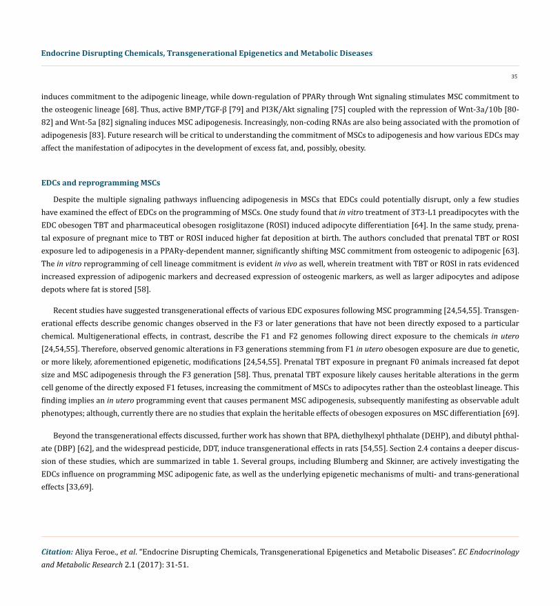
Several studies have identified potential mechanisms in which in utero exposures to various obesogenic EDCs produce multi- and trans-generational epigenetic effects in vivo [46,47,54,55,58,62,84]. Table 1 summarizes these critical findings and proposed mecha-nisms, while a discussion is provided in the subsequent subsections for each individual chemical. A short discussion of chemical mixtures will follow, although more research is needed to elucidate the epigenetic effects associated with mixtures of multiple obesogenic EDCs. Studies on EDC mixtures will better represent the complex reality, where humans are exposed to hundreds of chemicals every day-not a single chemical in isolation [16,85,86]. The articles cited in this literature review were obtained from a comprehensive PubMed search, and subsequently filtered using the method outlined in figure 1. The studies in table 1 were selected as the papers offering evidence for strictly obesity and obesity-related phenotypes (i.e. type II diabetes) inherited through potential epigenetic mechanisms following in utero EDC exposure. The rest of the articles obtained from this literature search were excluded from the specific study, but used to supple-ment and contextualize these studies. The final excluded full texts provided current information on EDC epigenetic regulation of other diseases and/or EDC-related heritable phenotypes. This review excluded the human epidemiological studies and experimental animal studies associating these EDCs with obesity since they have already undergone exhaustive review [87].
Figure 1: Flowchart of study selection. One reviewer to determine the eligibility and relevance of the literature to the literature review screened articles. Articles were deemed relevant if they were full-text and discussed any experiments or mechanisms involving epigenetics, obesity, and
obesogenic chemicals.
38
Endocrine Disrupting Chemicals, Transgenerational Epigenetics and Metabolic Diseases
Citation: Aliya Feroe., et al. “Endocrine Disrupting Chemicals, Transgenerational Epigenetics and Metabolic Diseases”. EC Endocrinology and Metabolic Research 2.1 (2017): 31-51.
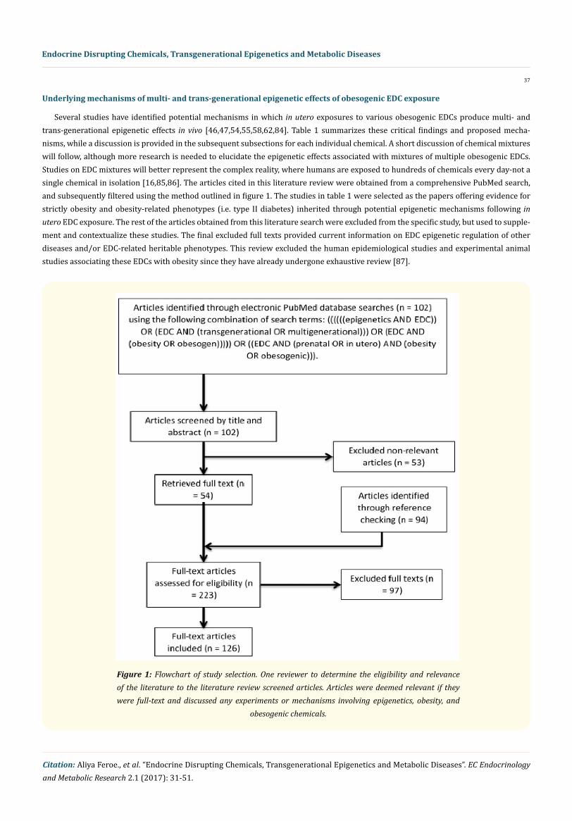
For each EDC, this literature review examined the epigenetic studies in animals, including the physiological changes associated with obesity, proposed epigenetic mechanisms, and potential multi- or trans-generational epigenetic alterations of the EDC. These studies differed based on whether the affected generation had direct exposure to the original environmental chemical or metabolite exposure. When a pregnant mother (F0) is exposed to an EDC, her child (F1) could be affected by the same EDC throughout gestation exposure. Furthermore, because the germ cells of the F1 offspring are developing throughout gestation, the grandchildren (F2) are also directly exposed. Effects observed in the F2 generation would be considered multigenerational. In contrast, effects observed in the F3 generation that had no direct exposure to the original EDC would be transgenerational. When the EDCs’ exposure occurs through the F0 father, the transgenerational effects are observed in the F2 generation, as the only other generation directly exposed to the original exposure is the future F1 offspring, which is exposed as a germ cell (Figure 2).
Figure 2: Potential multi- or trans-generational epigenetic alterations.
Bisphenol A
Bisphenol A (BPA) is a monomer used in the manufacturing of polycarbonate plastics and epoxy resins produce a multitude of con-sumer products, food and drink containers, and medical devices [88]. BPA has demonstrated the ability to be transferred both transpla-centally and lactationally into the fetus or infant from the mother [89,90]. Mechanistically, BPA mimics estrogen both in vitro and in vivo, binding to the estrogen receptors ERα and ERβ, although with relatively weak binding affinities [91].
39
Endocrine Disrupting Chemicals, Transgenerational Epigenetics and Metabolic Diseases
Citation: Aliya Feroe., et al. “Endocrine Disrupting Chemicals, Transgenerational Epigenetics and Metabolic Diseases”. EC Endocrinology and Metabolic Research 2.1 (2017): 31-51.
In utero exposure to “safe” or lower than safe doses of BPA (as defined by the current EPA standard of 50 ug/kg/day) has been as-sociated with increased DNA methylation levels, resulting in impaired regulation of insulin and glucose control [46]. Following direct F0 rat exposure to BPA during gestation and lactation, the F1 offspring demonstrated no significant changes in glucose/insulin tolerance by week 3 beyond birth; but by week 21, rat pups showed increased serum insulin and HOMA-IR levels (Homeostatic Model Assessment of Insulin Resistance) as evidenced by analysis of hepatic tissue (Table 1). Additionally, insulin sensitivity and hepatic glycogen storage decreased [47]. In rat pups, significant promoter hypermethylation of Gck gene was observed in the liver of F1 at weeks 3 and 21 when compared with the control [47]. Gck is a key regulator of glucose use in hepatocytes as Gck initiates glucose metabolism through glucose phosphorylation [92]. Previously, decreased Gck activity was associated with type 2 diabetes mellitus [93], and mutations in the Gck gene have been linked to maturity-onset diabetes of the young (MODY)-a form of diabetes diagnosed in late childhood [94]. Thus, Gck is con-sidered a diabetes susceptibility gene. Furthermore, this alteration in DNA methylation during early development was shown to persist throughout animal maturation [92].
A subsequent study supported that in utero BPA exposure induced the hypermethylation of Gck, and further identified the multigener-ational inheritance of these epigenetic mutations into the F2 offspring (Table 1) [46]. F0 exposure during gestation and lactation affected glucose and insulin tolerance in the F2 offspring, predisposing the offspring of each generation to diabetes. This decreased Gck expression provides a possible mechanism underlying glucose and insulin intolerance in subsequent BPA-treated generations since global DNA hy-pomethylation in liver was previously associated with diabetes in rats [95]. These examples demonstrate multigenerational inheritance of epigenetic alterations that cause diabetes-a primary risk factor for obesity-but further research is needed to study the potential transgen-erational inheritance of these epimutations. One important avenue of research is studying germline mutations, such as the methylation status of the Gck promoter region in sperm following BPA exposure, as a vehicle for transgenerational inheritance.
Dichlorodiphenyltrichloroethane
Dichlorodiphenyltrichloroethane (DDT) is historically one of the most commonly used insecticides [96]. While DDT has been banned in the U.S. since 1972 and by the UN for most uses since 2001, many countries have continued spraying DDT for malaria control [96-98]. Subsequently, due to the globalization of food supplies, DDT contaminated food is circumventing the U.S. ban and reaching U.S. consumers [96]. Estimates of global DDT use for disease vector control fluctuate between 4,000 to 5,000 metric tons per year [99]. Furthermore, evi-dence is accumulating for the association between exposure to DDT and its metabolite dichlorodiphenyldichloroethylene (DDE), diabetes and obesity [100-103]. While transgenerational phenomena have been demonstrated in humans [104], rodents [105], worms [106], flies [107], and plants [108] for DDT-induced diseases, only one study today has examined underlying epigenetic mechanisms of DDT-expo-sures contributing to the obesity epidemic [54,55].
Ancestral DDT exposure during a critical window of germline development promoted epigenetic transgenerational inheritance of obesity into the F3 rat generation (Table 1) [54,55]. Broadly, the study found some differential DNA methylation regions (DMR) in TUBB3, SLC4A4, and CARM1 genes in F3 sperm that resulted in increased visceral adiposity following both lower dose (25 mg/kg/day) and higher dose (50 mg/kg/day) F0 exposures during gestational development. Both the lower and higher doses of DDT used were consistent with wildlife and human environmental exposure levels [109]. The F2 generation (those directly exposed in utero) were not found to develop obesity, but developed kidney, prostate, and ovarian disease and tumor development as adults. In the F3 generation, 50% of lower dose males and females developed obesity and 75% of the higher dose males (although not females) developed obesity [54,55], suggesting transgenerational transmission of disease through both the female and male germlines-the egg and sperm, respectively. In contrast, the F1 obesity pathology was due to direct exposure of fetal somatic cells and was distinct from the germline exposure mechanism of the F3 generation [110,111]. Lower density CpG regions, also called “CpG deserts”, were identified within DMRs [110]. The CpG desert possesses less methylation capabilities due to a lack of cytosines available to accept methyl groups, and it was suggested to be involved in transgen-erational inheritance [110]. However, further studies are needed to determine the functional significance of CpG deserts in epigenetics.
40
Endocrine Disrupting Chemicals, Transgenerational Epigenetics and Metabolic Diseases
Citation: Aliya Feroe., et al. “Endocrine Disrupting Chemicals, Transgenerational Epigenetics and Metabolic Diseases”. EC Endocrinology and Metabolic Research 2.1 (2017): 31-51.
Di-(2-ethylhexyl) phthalate
Di-(2-ethylhexyl) phthalate (DEHP) is widely used as a plasticizer in everyday consumer products. Estimated annual DEHP production is approximately 2 million tons [112]. As a ubiquitous environmental pollutant, DEHP has been found in human amniotic fluid, umbili-cal cord blood, milk, semen and saliva [112]. Exposures to DEHP have been recently associated with energy imbalance and metabolic disorders [113] making DEHP a concern in the obesity epidemic. DEHP reduces blood glucose utilization and hepatic glycogenesis and glycogenolysis in rats [114]. DEHP-exposed rats also have reduced muscle glucose and lactate transport, reductions in muscle hexokinase and hepatic glucokinase, and glycogen synthesis [115], as well as disrupted pancreas and whole-body glucose homeostasis [116]. Fur-thermore, recent epidemiological studies identified a positive correlation between increased urinary phthalate metabolites, abdominal obesity and insulin resistance in both adolescent and adult males [117-119]. To date, only one study has examined the epigenetic effects of in utero DEHP exposures on obesity and obesity-related disorders, primarily type 2 diabetes [84]. However, no studies have yet focused their attention on transgenerational epigenetic alterations associated with these diseases.
One group studied the effects of gestational DEHP exposure on insulin signaling molecules and glucose transporter 4 (Glut4 (SLC2A4)) and its epigenome in the gastrocnemius muscle of F1 rat offspring (Table 1) [84]. They found that DEHP-exposed F1 offspring were predisposed to glucometabolic dysfunction at adulthood due to down-regulated Glut4 as a result of hypermethylation of the Glut4 gene promoter. They found a negative correlation between Glut4 expression and methylation level of the CpG islands. These Glut4 epigenetic alterations suggest a mechanism for multigenerational inheritance of obesity and obesity-related phenotypes since Glut4 is the primary GLUT-family gene in adipose and muscle tissues that acts as a major transporter protein for insulin-mediated whole-body glucose uptake [120]. In short, DEHP-induced epigenetic alterations in Glut4 gene appear to be a key component of the observed disposition towards metabolic abnormality, but more studies are needed to identify the potential transgenerational effects and underlying mechanisms lead-ing to obesity.
Tributyltin
Tributyltin (TBT), a marine paint additive, is an obesogenic EDC banned by many international agencies (i.e. International Maritime Organization in 2008) but persists in our water and soil today [121]. TBT directly mediates adipogenesis through the RXR/PPARγ path-way [59,122]. In utero exposures to TBT have been observed to predispose MSCs to the adipocyte lineage at the expense of the osteogenic lineage, through epigenetic imprinting, both in vitro and in vivo [63]. To date, only one group to has studied the transgenerational epigen-etic effects of TBT in promoting obesity (Table 1) [58]. They exposed pregnant mice to three different concentrations of TBT in drinking water to deliver approximately 50-fold lower (5.42 nM), 5-fold lower (54.2 nM), and 2-fold higher (542 nM) doses compared with the established no observable adverse effect level (NOAEL) of 25 µg/kg/day for mice [123]. This threshold is comparable to the established human tolerable daily intake of 250 ng/kg/day, derived by applying a 100-fold safety factor to the mouse NOAEL [124,125].
In these experiments, F2 and F3 mice exhibited increased adipose depot (fat storage) weight, had larger adipocyte size, increased adipocyte number, and greater differentiation of MSCs toward the adipocyte lineage [58]. These obesogenic effects occurred despite regu-lated caloric consumption, suggesting that these results were not attributable to a high caloric diet. Specifically, F1 males demonstrated increased perirenal and interscapular white adipose tissue (WAT) depot weight and adipocyte size and number. F1 females demonstrated increased WAT depot weight and adipocyte size. Both F1 males and females demonstrated minimal weight gain likely due to decreased brown adipose tissue (BAT-associated with high-energy metabolism). In the F3 generation, males showed increased WAT depot weights and adipocyte size and number, and females demonstrated increased adipocyte number and size in all WAT depots except interscapular and ovarian. In all 3 concentrations, prenatal exposure led to increased adipose tissue for at least 3 generations, indicating permanent, transgenerational obesogenic effects through germline alterations [58]. Further research is needed to better understand the mechanisms of TBT-induced adipocyte differentiation.
41
Endocrine Disrupting Chemicals, Transgenerational Epigenetics and Metabolic Diseases
Citation: Aliya Feroe., et al. “Endocrine Disrupting Chemicals, Transgenerational Epigenetics and Metabolic Diseases”. EC Endocrinology and Metabolic Research 2.1 (2017): 31-51.
Mixtures
While the aforementioned studies focus on the epigenetic mechanisms underlying the obesogenic effects of developmental EDC ex-posures, those studies examine only single chemicals. Arguably, the most significant results will come from experimental studies on the adverse health effects of mixtures, which would more realistically represent daily human exposure to the large number of EDCs in the environment. To date, only one group has studied the epigenetic transgenerational inheritance of obesity via exposure to a chemical mixture (Table 1) [62]. They studied the mixture of BPA, DEHP, and dibutyl phthalate (DBP)-another common plasticizer used in latex adhesives, cosmetics, cellulose plastics, and as a solvent for dyes [126]. These three chemicals have been found in humans [127,128], and are all derived from various plastic products [129]. In this study, rats were exposed to BPA, DEHP, and DBP during gestational develop-ment at similar concentrations to single component EDC studies (Table 1). Increased obesity was observed in both F3 females and males, without obesogenic effects in the F1 offspring [62]. The observation that obesity was not seen in the directly exposed F1 generation and only the indirectly exposed F3 generation implies the involvement of a transgenerational mechanism. Analysis of DNA methylation in the altered sperm epigenome identified several epimutations on Gdnf and Esrra genes promoters that have also been previously associated with obesity [130,131]. Further studies are needed to confirm that BPA, DEHP, and DBP exposure will influence Gdnf and Esrra obeso-genic effects in adipocytes. Furthermore, studies investigating a number of other EDC mixtures are needed to determine the combined epigenetic mechanisms fueling obesogenic effects relevant to our daily exposures. For instance, recent work integrated environmental, epidemiologic, genomic, and bioinformatics approaches to better apply laboratory data to the general population and identify the com-mon environmental and molecular risk factors between breast cancer and endometriosis [132]. More integrated approaches as outlined by this methodological framework could be used to more effectively apply laboratory studies to a more realistic population burden of EDC exposures.
The present review aims to stimulate experimental laboratory research by identifying important biological pathways and EDCs candi-dates for investigation. Specific avenues of laboratory research might include in vitro and in vivo studies that should be conducted using exposure to selected endocrine-disrupting chemicals either individually or as mixtures. Specific receptors or pathway nodes of interest identified using an integrated or combined in vitro, in vivo, bioinformatics, computational systems biology/chemistry/or toxicology ap-proaches could be technically evaluated by application of genomic, proteomic, or metabolomics methods. Thus, researchers could exam-ine potential linkages for combined exposures to specific EDCs, cellular pathway alterations, and metabolic disturbances related to the development of important clinical outcomes [133].
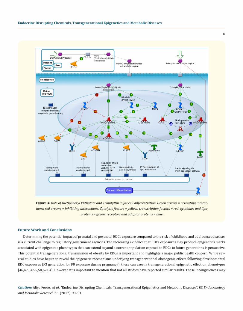
For example, DEHP is converted after ingestion to mono-(2-ethylhexyl)phthalate (active metabolite of DEHP) which directly activates PPAR-gamma, induces its co-repressor N-CoR release and promotes recruitment of coactivators TRIP2 and PPARGC1 (PGC1-alpha). Mono (2-ethylhexyl) phthalate-induced fat cell differentiation may contribute to the development of obesity and associated metabolic disorders [113]. It is known that Tributyltin induces fat cell differentiation by direct activation of PPAR-gamma and, possibly, RXR-alpha and pro-motes recruitment of the coactivators NCOA2 (GRIP1/TIF2) [63]. Activated PPAR-gamma, RXR-alpha and/or PPAR-gamma/RXR-alpha dimers stimulate transcription of early adipogenic markers A-FABP [59,63], PPAR-gamma and the late marker Leptin. In addition, Tribu-tyltin inhibits expression of adipogenesis inhibitor DLK via an unknown pathway [63].
Mono (2-ethylhexyl) phthalate and tributyltin-dependent expression of PPAR-gamma and RXR-alpha targets leads to fat cell differen-tiation [63,113]. DEHP and tributyltin are considered obesogens, which carry out their action via stimulation of PPAR-gamma (Figure 3). By integrating available information and bridging the gap between toxicology, epidemiology, computational methods, bioinformatics and chemistry within the world of disease mechanisms, scientific community can look further beyond the primary target of the individual or combined chemicals by several steps down the relevant pathway [133].
42
Endocrine Disrupting Chemicals, Transgenerational Epigenetics and Metabolic Diseases
Citation: Aliya Feroe., et al. “Endocrine Disrupting Chemicals, Transgenerational Epigenetics and Metabolic Diseases”. EC Endocrinology and Metabolic Research 2.1 (2017): 31-51.
Figure 3: Role of Diethylhexyl Phthalate and Tributyltin in fat cell differentiation. Green arrows = activating interac-tions; red arrows = inhibiting interactions. Catalytic factors = yellow; transcription factors = red; cytokines and lipo-
proteins = green; receptors and adaptor proteins = blue.
Future Work and ConclusionsDetermining the potential impact of prenatal and postnatal EDCs exposure compared to the risk of childhood and adult onset diseases
is a current challenge to regulatory government agencies. The increasing evidence that EDCs exposures may produce epigenetics marks associated with epigenetic phenotypes that can extend beyond a current population exposed to EDCs to future generations is persuasive. This potential transgenerational transmission of obesity by EDCs is important and highlights a major public health concern. While sev-eral studies have begun to reveal the epigenetic mechanisms underlying transgenerational obesogenic effects following developmental EDC exposures (F3 generation for F0 exposure during pregnancy), these can exert a transgenerational epigenetic effect on phenotypes [46,47,54,55,58,62,84]. However, it is important to mention that not all studies have reported similar results. These incongruences may
43
Endocrine Disrupting Chemicals, Transgenerational Epigenetics and Metabolic Diseases
Citation: Aliya Feroe., et al. “Endocrine Disrupting Chemicals, Transgenerational Epigenetics and Metabolic Diseases”. EC Endocrinology and Metabolic Research 2.1 (2017): 31-51.
be related to differences in the administration route of EDC exposure or experimental protocols, which may influence the transgenera-tional epigenetic effect. More research needs to be completed in an integrated approach to disease etiology, especially around realistic concentrations of environmentally relevant EDCs or EDC mixtures in humans and animals. The initial integrated studies are those that examine mixtures relevant to actual human exposures, as opposed to single EDCs in isolation. Subsequent studies need to incorporate en-vironmental, epidemiologic, genomic, and bioinformatics studies into conclusions about the effects of EDC mixture exposures on obesity. Certainly, determination of environmentally relevant EDC concentrations that humans may be exposed to daily would be a helpful next step in these EDC or EDCs mixture studies, as they will be key to illustrating the distinction between multigenerational versus transgen-erational epigenetic inheritance.
Understanding the molecular determinants of established epigenetic memory across generations would provide essential insight into how environmental changes can affect the health of exposed individuals, as well as subsequent generations. Studying the epigenetic inheritance of EDC-induced obesity and other diseases may reveal findings that might help curb the health effects associated with EDCs exposure. A better understanding of the network and biological pathway mechanisms underlying how EDC exposures affect adipogenesis, transgenerational inherence, and obesity are still needed. Acceptance, implementation, as well as new methods and new requirements for deep sequencing-based expression analysis, will show major advances in robustness and inter-lab transferability over microarray platforms for gene expression analysis. Chemical-gene-disease interaction information can also be applied for designing animal and cell-based laboratory experiments that can continue to investigate the established transgenerational obesity hypotheses more effectively. Fur-thermore, there is a need to understand the windows of susceptibility of different vulnerable populations’ exposure to EDCs and the as-sociated effects. Very little is known about associations between dietary intakes and potential obesogenic-EDCs exposure. Future research needs to focus on investigating the comparative contributions of genetic and epigenetic changes to transgenerational effects of EDCs.
Innovative methods of analysis such as deep sequence, microarrays, machine learning, bioinformatics, and computational tools are available and can be used to identify specific outcome pathways from complex data. The use of these methods or integration of these methods may help identify particular and sensitive biomarkers. Cluster identification of biomarkers as signatures of individual or com-bined exposure may help advance the development of EDC mixtures risk assessment methods. Biomonitoring and epidemiological studies need to assess important markers related to metabolic diseases such as obesity. The sensitivity and specificity of these available biomark-ers that are influenced by a range of modifying factors (chemical mixture components, diet, genetics, age, sex, ethnicity, diseases, gut biota, etc.) can be studied using multiple sophisticated techniques. Innovative biomarkers could be developed for their use in human population studies, disease prevention and clinical use to detect individual or multiple chemical exposures.
The body burden of multiple chemicals, particularly those EDCs considered obesogenic chemicals should be considered within the larger framework for obesity and other chronic diseases prevention. Further investigations carried out to study the influence of factors such as transgenerational effects, multiple chemical exposures, nutrition, age, gender, ethnicity and genetic variations will help develop personalized specific treatment protocols for these complex diseases.
AcknowledgementsThe findings and conclusions in this report are those of the author(s) and do not necessarily represent the official position of the Cen-
ters for Disease Control and Prevention/the Agency for Toxic Substances and Disease Registry. Mention of trade names is not an endorse-ment of any commercial product. The authors declare they have no actual or potential financial interests. David Albuquerque is recipient of a post-doctoral grant (SFRH/BPD/109043/2015) from Fundação para a Ciência e a Tecnologia (FCT).
DisclaimerThe findings and conclusions in this report are those of the author(s) and do not necessarily represent the official position of the Cen-
ters for Disease Control and Prevention/the Agency for Toxic Substances and Disease Registry.
44
Endocrine Disrupting Chemicals, Transgenerational Epigenetics and Metabolic Diseases
Citation: Aliya Feroe., et al. “Endocrine Disrupting Chemicals, Transgenerational Epigenetics and Metabolic Diseases”. EC Endocrinology and Metabolic Research 2.1 (2017): 31-51.
Bibliography
1. Koletzko B., et al. “Early Nutrition Programming of Long-Term Health”. Proceedings of the Nutrition Society 71.3 (2012): 371-378.
2. Zama AM and M Uzumcu. “Epigenetic Effects of Endocrine-Disrupting Chemicals on Female Reproduction: An Ovarian Perspective”. Frontiers in Neuroendocrinology 31.4 (2010): 420-439.
3. Flegal KM., et al. “Prevalence of Obesity and Trends in the Distribution of Body Mass Index among Us Adults, 1999-2010”. JAMA 307.5 (2012): 491-497.
4. Ogden CL., et al. “Prevalence of Childhood and Adult Obesity in the United States, 2011-2012”. JAMA 311.8 (2014): 806-814.
5. Lustig RH. “Childhood Obesity: Behavioral Aberration or Biochemical Drive? Reinterpreting the First Law of Thermodynamics”. Na-ture Clinical Practice Endocrinology and Metabolism 2.8 (2006): 447-458.
6. Freedland E. S. “Role of a Critical Visceral Adipose Tissue Threshold (Cvatt) in Metabolic Syndrome: Implications for Controlling Dietary Carbohydrates: A Review”. Nutrition & Metabolism (Lond) 1.1 (2004): 12.
7. Swinburn BA., et al. “The Global Obesity Pandemic: Shaped by Global Drivers and Local Environments”. Lancet 378.9793 (2011): 804-814.
8. Finucane MM., et al. “National, Regional, and Global Trends in Body-Mass Index since 1980: Systematic Analysis of Health Examina-tion Surveys and Epidemiological Studies with 960 Country-Years and 9.1 Million Participants”. Lancet 377.9765 (2011): 557-567.
9. NCD-RisC. “Trends in Adult Body-Mass Index in 200 Countries from 1975 to 2014: A Pooled Analysis of 1698 Population-Based Mea-surement Studies with 19.2 Million Participants”. Lancet 387.10026 (2016): 1377-1396.
10. Hill JO and JC Peters. “Environmental Contributions to the Obesity Epidemic”. Science 280.5368 (1998): 1371-1374.
11. Power C and BJ Jefferis. “Fetal Environment and Subsequent Obesity: A Study of Maternal Smoking”. International Journal of Epide-miology 31.2 (2002): 413-419.
12. Braithwaite EC., et al. “Maternal Prenatal Depressive Symptoms Predict Infant Nr3c1 1f and Bdnf Iv DNA Methylation”. Epigenetics 10.5 (2015): 408-417.
13. Frias AE and KL Grove. “Obesity: A Transgenerational Problem Linked to Nutrition During Pregnancy”. Seminars in Reproductive Medicine 30.6 (2012): 472-478.
14. Albuquerque D., et al. “Epigenetics of Human Obesity: A Link between Genetics and Nutrition”. In: Molecular Mechanisms Underpin-ning the Development of Obesity. Eds. Nóbrega, Clévio and Raquel Rodriguez-López. Cham: Springer International Publishing (2014): 101-127.
15. Felsenfeld Gary. “A Brief History of Epigenetics”. Cold Spring Harbor Perspectives in Biology 6.1 (2014): a018200.
16. Stel J and J Legler. “The Role of Epigenetics in the Latent Effects of Early Life Exposure to Obesogenic Endocrine Disrupting Chemi-cals”. Endocrinology 156.10 (2015): 3466-4372.
17. Bernal AJ and RL Jirtle. “Epigenomic Disruption: The Effects of Early Developmental Exposures”. Birth Defects Research Part A: Clini-cal and Molecular Teratology 88.10 (2010): 938-944.
18. Jones PA and D Takai. “The Role of DNA Methylation in Mammalian Epigenetics”. Science 293.5532 (2001): 1068-1070.
45
Endocrine Disrupting Chemicals, Transgenerational Epigenetics and Metabolic Diseases
Citation: Aliya Feroe., et al. “Endocrine Disrupting Chemicals, Transgenerational Epigenetics and Metabolic Diseases”. EC Endocrinology and Metabolic Research 2.1 (2017): 31-51.
19. Sadakierska-Chudy A and M Filip. “A Comprehensive View of the Epigenetic Landscape. Part Ii: Histone Post-Translational Modifica-tion, Nucleosome Level, and Chromatin Regulation by Ncrnas”. Neurotoxicity Research 27.2 (2015): 172-197.
20. Sadakierska-Chudy A., et al. “A Comprehensive View of the Epigenetic Landscape Part I: DNA Methylation, Passive and Active DNA Demethylation Pathways and Histone Variants”. Neurotoxicity Research 27 (2015): 84-97.
21. Xin F., et al. “Multigenerational and Transgenerational Effects of Endocrine Disrupting Chemicals: A Role for Altered Epigenetic Regu-lation?” Seminars in Cell and Developmental Biology 43 (2015): 66-75.
22. Castel SE and RA Martienssen. “Rna Interference in the Nucleus: Roles for Small Rnas in Transcription, Epigenetics and Beyond”. Nature Reviews Genetics 14.2 (2013): 100-112.
23. Mercer Tim R and John S Mattick. “Structure and Function of Long Noncoding Rnas in Epigenetic Regulation”. Nature Structural and Molecular Biology 20.3 (2013): 300-307.
24. Remely M., et al. “Obesity: Epigenetic Regulation - Recent Observations”. BioMolecular Concepts 6.3 (2015): 163-175.
25. Bhandari RK., et al. “Transgenerational Effects from Early Developmental Exposures to Bisphenol a or 17alpha-Ethinylestradiol in Medaka, Oryzias Latipes”. Science Reports 5 (2015): 9303.
26. LaRocca J., et al. “First-Trimester Urine Concentrations of Phthalate Metabolites and Phenols and Placenta Mirna Expression in a Cohort of U.S. Women”. Environmental Health Perspectives (2015).
27. Reik W., et al. “Epigenetic Reprogramming in Mammalian Development”. Science 293.5532 (2001): 1089-1093.
28. Liu FL., et al. “Epigenetic Regulation and Related Diseases During Placental Development”. Yi Chuan 39.4 (2017): 263-275.
29. Maccani MA and CJ Marsit. “Epigenetics in the Placenta”. American Journal of Reproductive Immunology 62.2 (2009): 78.
30. Jaenisch R and A Bird. “Epigenetic Regulation of Gene Expression: How the Genome Integrates Intrinsic and Environmental Signals”. Nature Genetics 33 (2003): 245-254.
31. Zhang X and SM Ho. “Epigenetics Meets Endocrinology”. Journal of Molecular Endocrinology 46.1 (2011): R11-R32.
32. Dolinoy DC., et al. “Metastable Epialleles, Imprinting, and the Fetal Origins of Adult Diseases”. Pediatric Research 61.5 (2007): 30R-37R.
33. Skinner MK., et al. “Environmentally Induced Epigenetic Transgenerational Inheritance of Sperm Epimutations Promote Genetic Mutations”. Epigenetics 10.8 (2015): 762-771.
34. Guerrero-Bosagna C ., et al. “Identification of Genomic Features in Environmentally Induced Epigenetic Transgenerational Inherited Sperm Epimutations”. PLoS One 9.6 (2014): e100194.
35. Chamorro-Garcia R and B Blumberg. “Transgenerational Effects of Obesogens and the Obesity Epidemic”. Current Opinion in Pharma-cology 19 (2014): 153-158.
36. Nilsson EE and MK Skinner. “Environmentally Induced Epigenetic Transgenerational Inheritance of Disease Susceptibility”. Trans-lational Research 165.1 (2015): 12-17.
37. Guibert S and M Weber. “Functions of DNA Methylation and Hydroxymethylation in Mammalian Development”. Current Topics in De-velopmental Biology 104 (2013): 47-83.
46
Endocrine Disrupting Chemicals, Transgenerational Epigenetics and Metabolic Diseases
Citation: Aliya Feroe., et al. “Endocrine Disrupting Chemicals, Transgenerational Epigenetics and Metabolic Diseases”. EC Endocrinology and Metabolic Research 2.1 (2017): 31-51.
38. Hall KD., et al. “Energy Balance and Its Components: Implications for Body Weight Regulation”. The American Journal of Clinical Nutri-tion 95.4 (2012): 989-994.
39. Ng M., et al. “Global, Regional, and National Prevalence of Overweight and Obesity in Children and Adults During 1980-2013: A Sys-tematic Analysis for the Global Burden of Disease Study 2013”. Lancet 384.9945 (2014): 766-781.
40. Albuquerque D., et al. “Current Review of Genetics of Human Obesity: From Molecular Mechanisms to an Evolutionary Perspective”. Molecular Genetics and Genomics 290.4 (2015): 1191-221.
41. Klimentidis YC., et al. “Canaries in the Coal Mine: A Cross-Species Analysis of the Plurality of Obesity Epidemics”. Proceedings Biologi-cal Sciences 278.1712 (2011): 1626-1632.
42. Janesick A and B Blumberg. “Endocrine Disrupting Chemicals and the Developmental Programming of Adipogenesis and Obesity”. Birth Defects Research part C Embryo Today 93.1 (2011): 34-50.
43. Baillie-Hamilton PF. “Chemical Toxins: A Hypothesis to Explain the Global Obesity Epidemic”. Journal of Alternative and Complemen-tary Medicine 8.2 (2002): 185-192.
44. WHO/UNEP. “State of the Science of Endocrine Disrupting Chemicals -- 2012” (2013).
45. Diamanti-Kandarakis E., et al. “Endocrine-Disrupting Chemicals: An Endocrine Society Scientific Statement”. Endocrine Reviews 30.4 (2009): 293-342.
46. Li G., et al. “F0 Maternal Bpa Exposure Induced Glucose Intolerance of F2 Generation through DNA Methylation Change in Gck”. Toxi-cology Letters 228.3 (2014): 192-199.
47. Ma Y., et al. “Hepatic DNA Methylation Modifications in Early Development of Rats Resulting from Perinatal Bpa Exposure Contribute to Insulin Resistance in Adulthood”. Diabetologia 56.9 (2013): 2059-2067.
48. Kelishadi., et al. “Role of Environmental Chemicals in Obesity: A Systematic Review on the Current Evidence”. International Journal of Environmental Research and Public Health 2013 (2013): 896789.
49. Elobeid MA., et al.”Endocrine Disruptors and Obesity: An Examination of Selected Persistent Organic Pollutants in the Nhanes 1999-2002 Data”. International Journal of Environmental Research and Public Health 7.7 (2010): 2988-3005.
50. de Cock M and M van de Bor. “Obesogenic Effects of Endocrine Disruptors, What Do We Know from Animal and Human Studies?” Environment International 70 (2014): 15-24.
51. La Merrill M and LS Birnbaum. “Childhood Obesity and Environmental Chemicals”. Mount Sinai Journal of Medicine 78.1 (2011): 22-48.
52. Janesick and Blumberg. “Obesogens, Stem Cells and the Developmental Programming of Obesity”. International Journal of Andrology 35.3 (2012): 437-448.
53. Snedeker SM and AG Hay. “Do Interactions between Gut Ecology and Environmental Chemicals Contribute to Obesity and Diabetes?” Environmental Health Perspectives 120.3 (2012): 332-339.
54. Skinner MK., et al. “Epigenetic Transgenerational Actions of Environmental Factors in Disease Etiology”. Trends in Endocrinology and Metabolism 21.4 (2010): 214-222.
55. Skinner MK., et al. “Ancestral Dichlorodiphenyltrichloroethane (Ddt) Exposure Promotes Epigenetic Transgenerational Inheritance of Obesity”. BMC Medicine 11 (2013): 228.
47
Endocrine Disrupting Chemicals, Transgenerational Epigenetics and Metabolic Diseases
Citation: Aliya Feroe., et al. “Endocrine Disrupting Chemicals, Transgenerational Epigenetics and Metabolic Diseases”. EC Endocrinology and Metabolic Research 2.1 (2017): 31-51.
56. Rubin BS. “Bisphenol A: An Endocrine Disruptor with Widespread Exposure and Multiple Effects”. Journal of Steroid Biochemistry and Molecular Biology 127.1-2 (2011): 27-34.
57. Rubin BS., et al. “Perinatal Exposure to Low Doses of Bisphenol a Affects Body Weight, Patterns of Estrous Cyclicity, and Plasma Lh Levels”. Environmental Health Perspectives 109.7 (2001): 675-680.
58. Chamorro-Garcia R., et al. “Transgenerational Inheritance of Increased Fat Depot Size, Stem Cell Reprogramming, and Hepatic Steato-sis Elicited by Prenatal Exposure to the Obesogen Tributyltin in Mice”. Environmental Health Sciences 121.3 (2013): 359-366.
59. Grun F., et al. “Endocrine-Disrupting Organotin Compounds Are Potent Inducers of Adipogenesis in Vertebrates”. Molecular Endocri-nology 20.9 (2006): 2141-2155.
60. Newbold RR., et al. “Perinatal Exposure to Environmental Estrogens and the Development of Obesity”. Molecular Nutrition nd Food Research 51.7 (2007): 912-917.
61. Hines EP., et al. “Phenotypic Dichotomy Following Developmental Exposure to Perfluorooctanoic Acid (Pfoa) in Female Cd-1 Mice: Low Doses Induce Elevated Serum Leptin and Insulin, and Overweight in Mid-Life”. Molecular Cellular Endocrinology 304.1-2 (2009): 97-105.
62. Manikkam M., et al. “Plastics Derived Endocrine Disruptors (Bpa, Dehp and Dbp) Induce Epigenetic Transgenerational Inheritance of Obesity, Reproductive Disease and Sperm Epimutations”. PLOS One 8.1 (2013): e55387.
63. Kirchner S., et al. “Prenatal Exposure to the Environmental Obesogen Tributyltin Predisposes Multipotent Stem Cells to Become Adi-pocytes”. Molecular Endocrinology 24.3 (2010): 526-539.
64. Thayer KA., et al. “Role of Environmental Chemicals in Diabetes and Obesity: A National Toxicology Program Workshop Review”. Environmental Health Perspectives 120.6 (2012): 779-789.
65. Budge H., et al. “Maternal Nutritional Programming of Fetal Adipose Tissue Development: Long-Term Consequences for Later Obe-sity”. Birth Defects Research Part C: Embryo Today 75.3 (2005): 193-199.
66. Correia-Branco AE., et al. “Maternal Undernutrition and Fetal Developmental Programming of Obesity: The Glucocorticoid Connec-tion”. Reproductive Sciences 22.2 (2015): 138-145.
67. Boekelheide K., et al. “Predicting Later-Life Outcomes of Early-Life Exposures”. Environmental Health Sciences 120.10 (2012): 1353-1361.
68. Cristancho Ana G and Mitchell A Lazar. “Forming Functional Fat: A Growing Understanding of Adipocyte Differentiation”. Nature Re-views Molecular Cell Biology 12.11 (2011): 722-734.
69. Janesick AS., et al. “Transgenerational Inheritance of Prenatal Obesogen Exposure”. Molecular and Cellular Endocrinology 398.1-2 (2014): 31-35.
70. Crisan M., et al. “A Perivascular Origin for Mesenchymal Stem Cells in Multiple Human Organs”. Cell Stem Cell 3.3 (2008): 301-313.
71. Bianco P. “Back to the Future: Moving Beyond “Mesenchymal Stem Cells”. Journal of Cellular Biochemistry 112.7 (2011): 1713-1721.
72. Rosen ED and BM Spiegelman. “What We Talk About When We Talk About Fat”. Cell 156.1-2 (2014): 20-44.
73. Gupta RK., et al. “Zfp423 Expression Identifies Committed Preadipocytes and Localizes to Adipose Endothelial and Perivascular Cells”. Cell Metabolism 15.2 (2012): 230-239.
48
Endocrine Disrupting Chemicals, Transgenerational Epigenetics and Metabolic Diseases
Citation: Aliya Feroe., et al. “Endocrine Disrupting Chemicals, Transgenerational Epigenetics and Metabolic Diseases”. EC Endocrinology and Metabolic Research 2.1 (2017): 31-51.
74. Kang S., et al. “Regulation of Early Adipose Commitment by Zfp521”. PLOS Biology 10.11 (2012): e1001433.
75. Carnevalli LS., et al. “S6k1 Plays a Critical Role in Early Adipocyte Differentiation”. Developmental Cell 18.5 (2010): 763-774.
76. Remillard RB and NJ Bunce. “Linking Dioxins to Diabetes: Epidemiology and Biologic Plausibility”. Environmental Health Perspectives 110.9 (2002): 853-858.
77. Kersten S. “Peroxisome Proliferator Activated Receptors and Obesity”. European Journal of Pharmacology 440.2-3 (2002): 223-234.
78. Shockley KR., et al. “Ppargamma2 Regulates a Molecular Signature of Marrow Mesenchymal Stem Cells”. PPAR Research 2007 (2007): 81219.
79. Zamani N and CW Brown. “Emerging Roles for the Transforming Growth Factor-{Beta} Superfamily in Regulating Adiposity and En-ergy Expenditure”. Endocrine Reviews 32.3 (2011): 387-403.
80. Kang S., et al. “Wnt Signaling Stimulates Osteoblastogenesis of Mesenchymal Precursors by Suppressing Ccaat/Enhancer-Binding Protein Alpha and Peroxisome Proliferator-Activated Receptor Gamma”. Journal of Biological Chemistry 282.19 (2007): 14515-14524.
81. Kawai M., et al. “Wnt/Lrp/Beta-Catenin Signaling Suppresses Adipogenesis by Inhibiting Mutual Activation of Ppargamma and C/Ebpalpha”. Biochemical and Biophysical Research Communications 363.2 (2007): 276-282.
82. Takada I., et al. “Suppression of Ppar Transactivation Switches Cell Fate of Bone Marrow Stem Cells from Adipocytes into Osteoblasts”. Annals of the New York Academy of Sciences 1116 (2007): 182-195.
83. Sun L., et al. “Long Noncoding Rnas Regulate Adipogenesis”. Proceedings of the National Academy of Sciences of the United States of America 110.9 (2013): 3387-3392.
84. Rajesh P and K Balasubramanian. “Phthalate Exposure in Utero Causes Epigenetic Changes and Impairs Insulin Signalling”. Journal of Endocrinology 223.1 (2014): 47-66.
85. Biemann RB., et al. “Adipogenic Effects of a Combination of the Endocrine-Disrupting Compounds Bisphenol a, Diethylhexylphthalate, and Tributyltin”. Obesity Facts 7.1 (2014): 48-56.
86. Lubrano C., et al. “ Obesity and Metabolic Comorbidities: Environmental Diseases?” Oxidative Medicine and Cellular Longevity 2013 (2013): 640673.
87. Gore AC., et al. “Edc-2: The Endocrine Society’s Second Scientific Statement on Endocrine-Disrupting Chemicals”. Endocrine Reviews 36.6 (2015): E1-E150.
88. Chianese R., et al. “Bisphenol a in Reproduction: Epigenetic Effects”. Current Medicinal Chemistry (2017).
89. vom Saal FS., et al. “Chapel Hill Bisphenol a Expert Panel Consensus Statement: Integration of Mechanisms, Effects in Animals and Potential to Impact Human Health at Current Levels of Exposure”. Reproductive Toxicology 24.2 (2007): 131-138.
90. Vandenberg LN., et al. “Bisphenol-a and the Great Divide: A Review of Controversies in the Field of Endocrine Disruption”. Endocrine Reviews 30.1 (2009): 75-95.
91. Calafat AM., et al. “Exposure of the U.S. Population to Bisphenol a and 4-Tertiary-Octylphenol: 2003-2004”. Environmental Health Perspectives 116.1 (2008): 39-44.
92. Walker DG and S Rao. “The Role of Glucokinase in the Phosphorylation of Glucose by Rat Liver”. Biochemical Journal 90.2 (1964): 360-368.
49
Endocrine Disrupting Chemicals, Transgenerational Epigenetics and Metabolic Diseases
Citation: Aliya Feroe., et al. “Endocrine Disrupting Chemicals, Transgenerational Epigenetics and Metabolic Diseases”. EC Endocrinology and Metabolic Research 2.1 (2017): 31-51.
93. Caro JF., et al. “Liver Glucokinase: Decreased Activity in Patients with Type Ii Diabetes”. Hormone and Metabolic Research 27.1 (1995): 19-22.
94. Flack JR., et al. “Gck Monogenic Diabetes and Gestational Diabetes: Possible Diagnosis on Clinical Grounds”. Diabetic Medicine 32.12 (2015): 1596-601.
95. Williams KT., et al. “Type I Diabetes Leads to Tissue-Specific DNA Hypomethylation in Male Rats”. Journal of Nutrition 138.11 (2008): 2064-2069.
96. Sadasivaiah S., et al. “Dichlorodiphenyltrichloroethane (Ddt) for Indoor Residual Spraying in Africa: How Can It Be Used for Malaria Control?” American Journal of Tropical Medicine and Hygiene 77.6 (2007): 249-263.
97. Packard RM. “The Making of a Tropical Disease: A Short History of Malaria.” Johns Hopkins University Press (2007).
98. Guimaraes RM., et al. “Ddt Reintroduction for Malaria Control: The Cost-Benefit Debate for Public Health”. Cadernos de Saúde Pública 23.12 (2007): 2835-2844.
99. Kabasenche WP and MK Skinner. “Ddt, Epigenetic Harm, and Transgenerational Environmental Justice”. Environmental Health 13 (2014): 62.
100. Valvi D., et al. “Prenatal Concentrations of Polychlorinated Biphenyls, Dde, and Ddt and Overweight in Children: A Prospective Birth Cohort Study”. Environmental Health Perspectives 120.3 (2012): 451-457.
101. Heggeseth B., et al. “Detecting Associations between Early-Life Ddt Exposures and Childhood Growth Patterns: A Novel Statistical Approach”. PLoS One 10.6 (2015): e0131443.
102. Warner M., et al. “In Utero Ddt and Dde Exposure and Obesity Status of 7-Year-Old Mexican-American Children in the Chamacos Cohort”. Environmental Health Perspectives 121.5 (2013): 631-636.
103. Warner M., et al. “Prenatal Exposure to Dichlorodiphenyltrichloroethane and Obesity at 9 Years of Age in the Chamacos Study Co-hort”. American Journal of Epidemiology 179.11 (2014): 1312-1322.
104. Pembrey ME. “Male-Line Transgenerational Responses in Humans”. Human Fertility 13.4 (2010): 268-271.
105. Anway MD., et al. “Epigenetic Transgenerational Actions of Endocrine Disruptors and Male Fertility”. Science 308.5727 (2005): 1466-1469.
106. Greer EL., et al. “Transgenerational Epigenetic Inheritance of Longevity in Caenorhabditis Elegans”. Nature 479.7373 (2011): 365-371.
107. Ruden DM and X Lu. “Hsp90 Affecting Chromatin Remodeling Might Explain Transgenerational Epigenetic Inheritance in Drosoph-ila”. Current Genomics 9.7 (2008): 500-508.
108. Hauser MT., et al. “Transgenerational Epigenetic Inheritance in Plants”. Biochimica et Biophysica Acta 1809.8 (2011): 459-468.
109. ATSDR. “Toxicological Profile for 4,4′-Ddt, 4,4′-Dde, 4, 4′-Ddd (Update)”. ATSDR: Agency for Toxic Substances and Diseases Registry (ATSDR)/US Public Health Service 1994 (2015).
110. Skinner MK and C Guerrero-Bosagna. “Role of Cpg Deserts in the Epigenetic Transgenerational Inheritance of Differential DNA Methylation Regions”. BMC Genomics 15 (2014): 692.
111. Jirtle RL and M Skinner. “Environmental Epigenomics and Disease Susceptibility”. Nature Reviews Genetics 8.4 (2007): 253-262.
50
Endocrine Disrupting Chemicals, Transgenerational Epigenetics and Metabolic Diseases
Citation: Aliya Feroe., et al. “Endocrine Disrupting Chemicals, Transgenerational Epigenetics and Metabolic Diseases”. EC Endocrinology and Metabolic Research 2.1 (2017): 31-51.
112. Faniband Moosa., et al. “Human Biological Monitoring of Suspected Endocrine-Disrupting Compounds”. Asian Journal of Andrology 16.1 (2014): 5-16.
113. Desvergne B., et al. “Ppar-Mediated Activity of Phthalates: A Link to the Obesity Epidemic?” Molecular and Cellular Endocrinology 304.1-2 (2009): 43-48.
114. Mushtaq M., et al. “Effect of Di-2-Ethylhexyl Phthalate (Dehp) on Glycogen Metabolism in Rat Liver”. Toxicology 16.2 (1980): 153-161.
115. Martinelli MI., et al. “Dietary Di(2-Ethylhexyl)Phthalate-Impaired Glucose Metabolism in Experimental Animals”. Human and Experi-mental Toxicology 25.9 (2006): 531-538.
116. Lin Y., et al. “Developmental Exposure to Di(2-Ethylhexyl) Phthalate Impairs Endocrine Pancreas and Leads to Long-Term Adverse Effects on Glucose Homeostasis in the Rat”. American Journal of Physiology Endocrinology and Metabolism 301.3 (2011): E527-E538.
117. Stahlhut Richard W., et al. “Concentrations of Urinary Phthalate Metabolites Are Associated with Increased Waist Circumference and Insulin Resistance in Adult U.S. Males”. Environmental Health Perspectives 115.6 (2007): 876-882.
118. Hatch EE., et al. “Association of Urinary Phthalate Metabolite Concentrations with Body Mass Index and Waist Circumference: A Cross-Sectional Study of Nhanes Data, 1999-2002”. Environmental Health 7 (2008): 27.
119. Trasande L., et al. “Urinary Phthalates and Increased Insulin Resistance in Adolescents”. Pediatrics 132.3 (2013): e646-e655.
120. Yokomori N., et al. “DNA Demethylation During the Differentiation of 3t3-L1 Cells Affects the Expression of the Mouse Glut4 Gene”. Diabetes 48.4 (1999): 685-690.
121. Antizar-Ladislao Blanca. “Environmental Levels, Toxicity and Human Exposure to Tributyltin (Tbt)-Contaminated Marine Environ-ment. A Review”. Environment International 34.2 (2008): 292-308.
122. Li XJ Ycaza and B Blumberg. “The Environmental Obesogen Tributyltin Chloride Acts Via Peroxisome Proliferator Activated Recep-tor Gamma to Induce Adipogenesis in Murine 3t3-L1 Preadipocytes”. The Journal of Steroid Biochemistry and Molecular Biology 127.1-2 (2011): 9-15.
123. Vos JG., et al. “Immunotoxicity of Bis(Tri-N-Butyltin)Oxide in the Rat: Effects on Thymus-Dependent Immunity and on Nonspecific Resistance Following Long-Term Exposure in Young Versus Aged Rats”. Toxicology and Applied Pharmacology 105.1 (1990): 144-155.
124. Airaksinen R., et al. “Organotin Intake through Fish Consumption in Finland”. Environmental Research 110.6 (2010): 544-547.
125. Fristachi A., et al. “Using Probabilistic Modeling to Evaluate Human Exposure to Organotin in Drinking Water Transported by Poly-vinyl Chloride Pipe”. Risk Analysis 29.11 (2009): 1615-1628.
126. Heudorf U., et al. “Phthalates: Toxicology and Exposure”. International Journal of Hygiene and Environmental Health 210.5 (2007): 623-634.
127. Talsness CE., et al. “Components of Plastic: Experimental Studies in Animals and Relevance for Human Health”. Philosophical Trans-actions of the Royal Society B: Biological Sciences 364.1526 (2009): 2079-2096.
128. Maffini MV., et al. “Endocrine Disruptors and Reproductive Health: The Case of Bisphenol-A”. Molecular and Cellular Endocrinology 254-255 (2006): 179-186.
51
Endocrine Disrupting Chemicals, Transgenerational Epigenetics and Metabolic Diseases
Citation: Aliya Feroe., et al. “Endocrine Disrupting Chemicals, Transgenerational Epigenetics and Metabolic Diseases”. EC Endocrinology and Metabolic Research 2.1 (2017): 31-51.
129. Simoneau C., et al. “Identification and Quantification of the Migration of Chemicals from Plastic Baby Bottles Used as Substitutes for Polycarbonate”. Food Additives and Contaminants: Part A: Chemistry, Analysis, Control, Exposure and Risk Assessment 29.3 (2012): 469-480.
130. Baudry C., et al. “Diet-Induced Obesity Has Neuroprotective Effects in Murine Gastric Enteric Nervous System: Involvement of Leptin and Glial Cell Line-Derived Neurotrophic Factor”. Journal of Physiology 590.3 (2012): 533-544.
131. Slocum N., et al. “Responses of Brown Adipose Tissue to Diet-Induced Obesity, Exercise, Dietary Restriction and Ephedrine Treat-ment”. Experimental and Toxicologic Pathology 65.5 (2013): 549-557.
132. Roy D., et al. “Integrated Bioinformatics, Environmental Epidemiologic and Genomic Approaches to Identify Environmental and Mo-lecular Links between Endometriosis and Breast Cancer”. International Journal of Molecular Sciences 16.10 (2015): 25285-25322.
133. Ruiz P., et al. “A Systems Biology Approach Reveals Converging Molecular Mechanisms That Link Different Pops to Common Meta-bolic Diseases”. Environmental Health Perspectives 124.7 (2016): 1034-1041.
Volume 2 Issue 1 November 2017© All rights reserved by Patricia Ruiz., et al.