Embed Size (px)
Citation preview
CRANIAL IMPLANTS
Christopher J. White M.I.M.F.T.
Maxillofacial Prosthetist & Technologist The Norman Rowe Maxillofacial Unit
Queen Mary’s University Hospital Roehampton
London SW15 5PN
Lambeth College Tower Bridge Centre School of Science & Dental Technology London BTEC Higher National Diploma 1998
Contents Preface 3 Acknowledgements 4 Glossary of Terms Used 5 Introduction 7 Ancient history of cranioplasty 7 Early history 9 Aetiology of cranial defects 10 Indications for cranioplasty 11 General biomaterial considerations for cranioplasty 11 Material selection 12 Methods of cranioplasty 13 Autogenous bone 14 Tibia 14 Cranium 14 Fascia 15 Rib 15 Allograft 15 Xenograft 16 Alloplastic grafts 16 Metals 17 Aluminium, gold, and silver 17 Lead and platinum 17 Vitallium and ticonium 18 Tantalum 18 Stainless steel 18 Titanium 18 Non-metallic alloplastic materials 19 Celluloid 19 Other substances 19 Autopolymerizing acrylic resin 19 Heat polymerising methyl acrylic resin 20 Polyethylene and silicon rubber 20 Hydroxyapatite 21 Prefabricated cranial implants 21 Indirect impression technique 21 Three-dimensional reduced master cast 23 Titanium cranioplasty implant design and fabrication 24 De-contaminating cranioplasty 27 Cranioplasty installation 27 CT-Stereolithography method 31 Imaging the facial skeleton-CT imaging and reformation 32 Prosthesis fabrication 33 Future trends 33 Summary 33 References 35
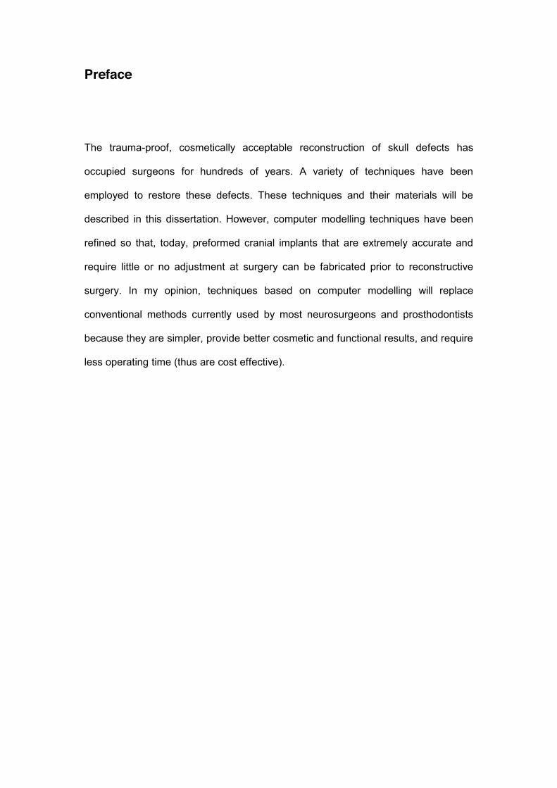
Preface The trauma-proof, cosmetically acceptable reconstruction of skull defects has
occupied surgeons for hundreds of years. A variety of techniques have been
employed to restore these defects. These techniques and their materials will be
described in this dissertation. However, computer modelling techniques have been
refined so that, today, preformed cranial implants that are extremely accurate and
require little or no adjustment at surgery can be fabricated prior to reconstructive
surgery. In my opinion, techniques based on computer modelling will replace
conventional methods currently used by most neurosurgeons and prosthodontists
because they are simpler, provide better cosmetic and functional results, and require
less operating time (thus are cost effective).

Acknowledgements My grateful thanks are due to these who helped me with what is contained in this
dissertation. This took several forms. The staff at Lambeth College, London, who
assisted in completing my Higher National Diploma in Dental Technology and
Atkinson Morley’s Hospital, London, for allowing me to photograph in their theatres. I
must also thank Neil Maffery and Darrin Hawkins in the Medical Photography
Department, Queen Mary’s University Hospital, London, for photographing items of
my work.
I would like to thank my colleague Mr Brian Conroy who generously allowed me to
show examples of his work Figs 25a & 25b, and for supporting me during my
studies.
Finally a mention must also be made to The Norman Rowe Educational Trust*,
which financed my course.
* Foundation for the advancement of Craino-Maxillofacial Surgery, Prosthetics and Technology and co-operation and understanding between Surgery and Technology.
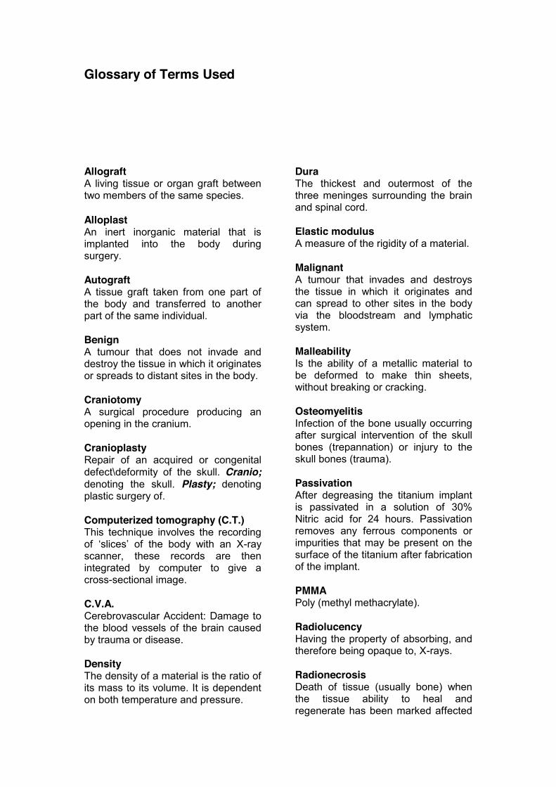
Glossary of Terms Used Allograft A living tissue or organ graft between two members of the same species. Alloplast An inert inorganic material that is implanted into the body during surgery. Autograft A tissue graft taken from one part of the body and transferred to another part of the same individual. Benign A tumour that does not invade and destroy the tissue in which it originates or spreads to distant sites in the body. Craniotomy A surgical procedure producing an opening in the cranium. Cranioplasty Repair of an acquired or congenital defect\deformity of the skull. Cranio; denoting the skull. Plasty; denoting plastic surgery of. Computerized tomography (C.T.) This technique involves the recording of ‘slices’ of the body with an X-ray scanner, these records are then integrated by computer to give a cross-sectional image. C.V.A. Cerebrovascular Accident: Damage to the blood vessels of the brain caused by trauma or disease. Density The density of a material is the ratio of its mass to its volume. It is dependent on both temperature and pressure.
Dura The thickest and outermost of the three meninges surrounding the brain and spinal cord. Elastic modulus A measure of the rigidity of a material. Malignant A tumour that invades and destroys the tissue in which it originates and can spread to other sites in the body via the bloodstream and lymphatic system. Malleability Is the ability of a metallic material to be deformed to make thin sheets, without breaking or cracking. Osteomyelitis Infection of the bone usually occurring after surgical intervention of the skull bones (trepannation) or injury to the skull bones (trauma). Passivation After degreasing the titanium implant is passivated in a solution of 30% Nitric acid for 24 hours. Passivation removes any ferrous components or impurities that may be present on the surface of the titanium after fabrication of the implant. PMMA Poly (methyl methacrylate). Radiolucency Having the property of absorbing, and therefore being opaque to, X-rays. Radionecrosis Death of tissue (usually bone) when the tissue ability to heal and regenerate has been marked affected
by radiotherapy treatment for a tumour. Thermal expansion The linear coefficient of thermal expansion of a material is the change in length per unit length when the temperature is raised by 1oC. Trepanation The use of a trepan or Trephine surgical instrument. A tubular or circular cutting surgical instrument that is used to cut or ‘drill’ holes into the skull and remove a disk or ‘Rondelle’ of bone. This usually precludes a ‘Craniotomy’ surgical procedure to the skull. Ultimate tensile strength The U.T.S. is the maximum stress that a material can undergo in compression or tension without fracture. Xenograft A living tissue graft that is made from an animal of one species to another of a different species. Yield A material reaches a yield point when there is a rapid increase in strain without a corresponding increase in stress; the deformation is now completely plastic. REFERENCE 1. Oxford Reference (1994) Concise Medical Dictionary 4th edn. Oxford
2. Combe E. C. (1986) Notes on Dental Materials, 4th edn. Edinburgh: Churchill
INTRODUTION Cranioplasty is almost as ancient as trephination, yet it’s fascinating history has been neglected. The technique of cranioplasty combines art and surgical science. Simply filling the cranial defect is not sufficient. There must be a respect for reconstruction of the complex topography of the cranium's surface. Many materials have been tried, many techniques tested, with none achieving universal adoption. The persistence of trauma, war and Neurosurgical procedures continues to produce fertile ground for the progress of this fascinating field. Cranioplasty is defined as the repair of a defect or deformity of the cranium. Grant and Norcross, 21 first clarified the reason for cranioplasty and was recently summarised by Prolo48 as follows: x cerebral protection; x physical appearance; x normalisation of intracranial
pressure relationships; and x to ensure normal cranial growth
and development of intracranial structures in the young.
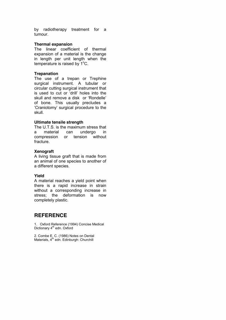
Fig 1a Ancient Incan skull showing three trephination defects in various stages of healing.
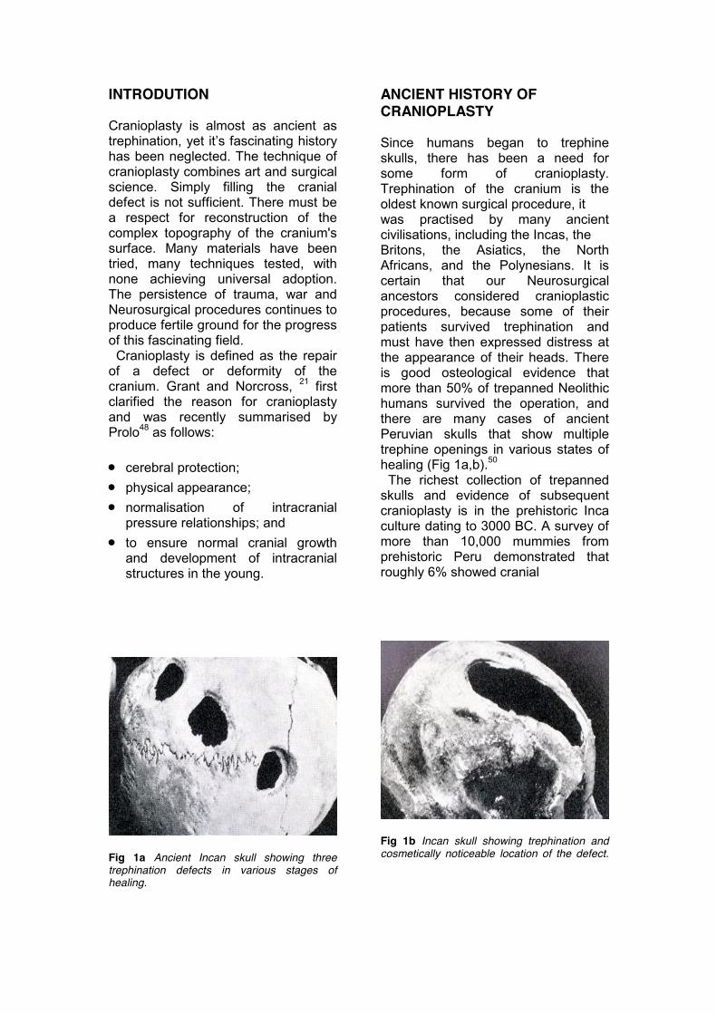
ANCIENT HISTORY OF CRANIOPLASTY Since humans began to trephine skulls, there has been a need for some form of cranioplasty. Trephination of the cranium is the oldest known surgical procedure, it was practised by many ancient civilisations, including the Incas, the Britons, the Asiatics, the North Africans, and the Polynesians. It is certain that our Neurosurgical ancestors considered cranioplastic procedures, because some of their patients survived trephination and must have then expressed distress at the appearance of their heads. There is good osteological evidence that more than 50% of trepanned Neolithic humans survived the operation, and there are many cases of ancient Peruvian skulls that show multiple trephine openings in various states of healing (Fig 1a,b).50 The richest collection of trepanned skulls and evidence of subsequent cranioplasty is in the prehistoric Inca culture dating to 3000 BC. A survey of more than 10,000 mummies from prehistoric Peru demonstrated that roughly 6% showed cranial
Fig 1b Incan skull showing trephination and cosmetically noticeable location of the defect.
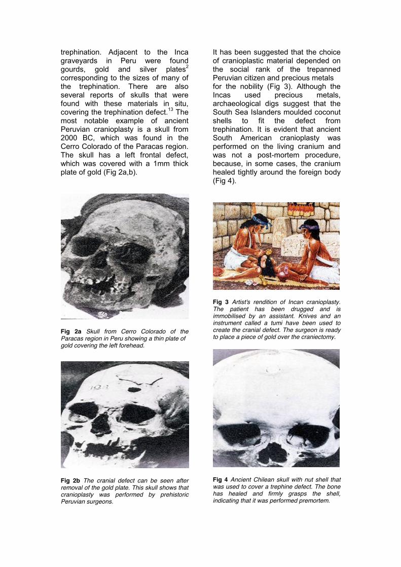
trephination. Adjacent to the Inca graveyards in Peru were found gourds, gold and silver plates2 corresponding to the sizes of many of the trephination. There are also several reports of skulls that were found with these materials in situ, covering the trephination defect.13 The most notable example of ancient Peruvian cranioplasty is a skull from 2000 BC, which was found in the Cerro Colorado of the Paracas region. The skull has a left frontal defect, which was covered with a 1mm thick plate of gold (Fig 2a,b).
Fig 2a Skull from Cerro Colorado of the Paracas region in Peru showing a thin plate of gold covering the left forehead.
Fig 2b The cranial defect can be seen after removal of the gold plate. This skull shows that cranioplasty was performed by prehistoric Peruvian surgeons.
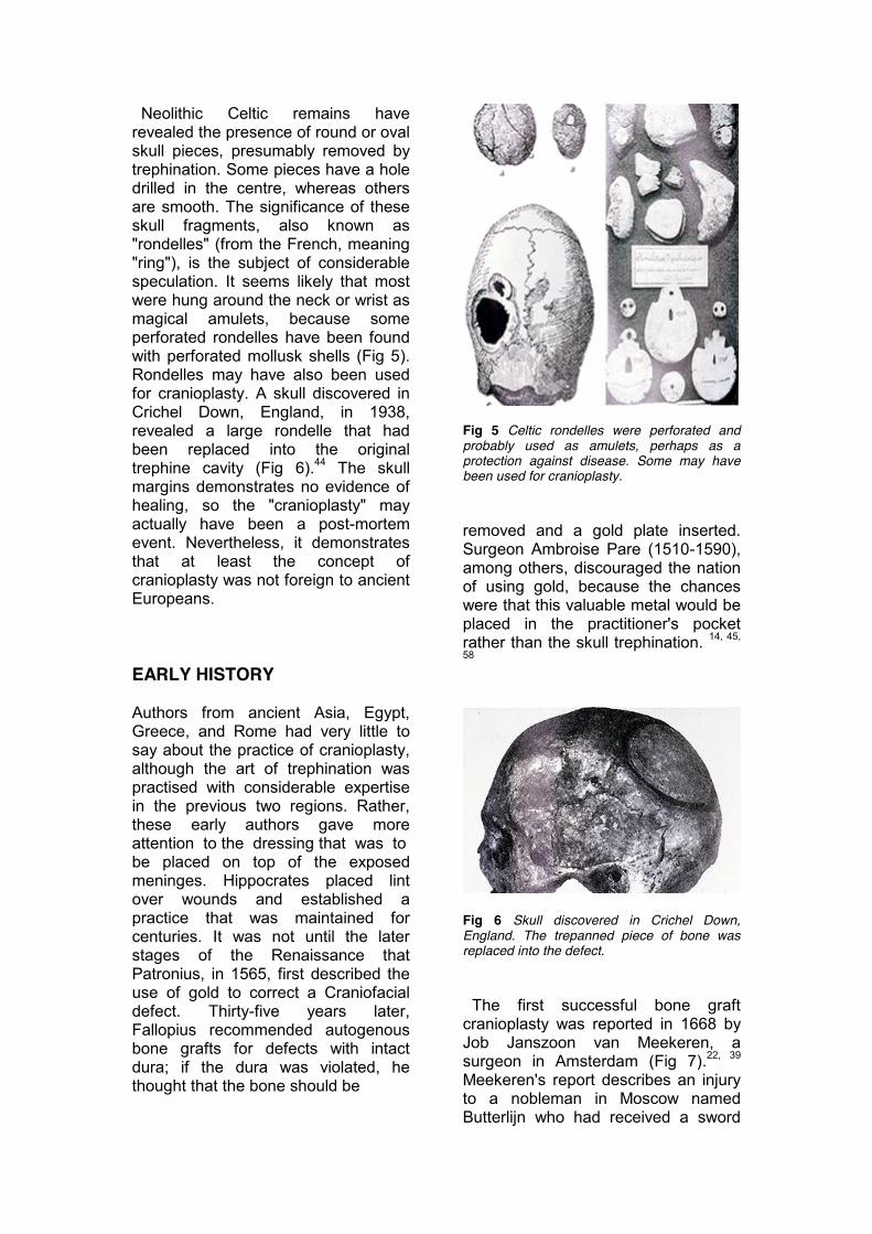
It has been suggested that the choice of cranioplastic material depended on the social rank of the trepanned Peruvian citizen and precious metals for the nobility (Fig 3). Although the Incas used precious metals, archaeological digs suggest that the South Sea Islanders moulded coconut shells to fit the defect from trephination. It is evident that ancient South American cranioplasty was performed on the living cranium and was not a post-mortem procedure, because, in some cases, the cranium healed tightly around the foreign body (Fig 4).
Fig 3 Artist's rendition of Incan cranioplasty. The patient has been drugged and is immobilised by an assistant. Knives and an instrument called a tumi have been used to create the cranial defect. The surgeon is ready to place a piece of gold over the craniectomy.
Fig 4 Ancient Chilean skull with nut shell that was used to cover a trephine defect. The bone has healed and firmly grasps the shell, indicating that it was performed premortem.
Neolithic Celtic remains have revealed the presence of round or oval skull pieces, presumably removed by trephination. Some pieces have a hole drilled in the centre, whereas others are smooth. The significance of these skull fragments, also known as "rondelles" (from the French, meaning "ring"), is the subject of considerable speculation. It seems likely that most were hung around the neck or wrist as magical amulets, because some perforated rondelles have been found with perforated mollusk shells (Fig 5). Rondelles may have also been used for cranioplasty. A skull discovered in Crichel Down, England, in 1938, revealed a large rondelle that had been replaced into the original trephine cavity (Fig 6).44 The skull margins demonstrates no evidence of healing, so the "cranioplasty" may actually have been a post-mortem event. Nevertheless, it demonstrates that at least the concept of cranioplasty was not foreign to ancient Europeans. EARLY HISTORY Authors from ancient Asia, Egypt, Greece, and Rome had very little to say about the practice of cranioplasty, although the art of trephination was practised with considerable expertise in the previous two regions. Rather, these early authors gave more attention to the dressing that was to be placed on top of the exposed meninges. Hippocrates placed lint over wounds and established a practice that was maintained for centuries. It was not until the later stages of the Renaissance that Patronius, in 1565, first described the use of gold to correct a Craniofacial defect. Thirty-five years later, Fallopius recommended autogenous bone grafts for defects with intact dura; if the dura was violated, he thought that the bone should be
Fig 5 Celtic rondelles were perforated and probably used as amulets, perhaps as a protection against disease. Some may have been used for cranioplasty. removed and a gold plate inserted. Surgeon Ambroise Pare (1510-1590), among others, discouraged the nation of using gold, because the chances were that this valuable metal would be placed in the practitioner's pocket rather than the skull trephination. 14, 45,
58
Fig 6 Skull discovered in Crichel Down, England. The trepanned piece of bone was replaced into the defect. The first successful bone graft cranioplasty was reported in 1668 by Job Janszoon van Meekeren, a surgeon in Amsterdam (Fig 7).22, 39 Meekeren's report describes an injury to a nobleman in Moscow named Butterlijn who had received a sword
wound to the head. The blow not only stripped the scalp but also removed a section of the cranium. The repair of the defect was performed using a section from the cranium of a dead dog. The shape and size to that which had been taken off by the sword, and he adapted it to the wounded place. However the operation did not pass the scrutiny of a church that regarded a Christian head too pure to be graced with the bone of a dog. Excommunicated and seeking to regain entry into the Church, Butterlijn ordered his surgeon to remove the graft. The surgeon, however, was helpless, because the canine bone had firmly united with the human cranium. Butterlijn then left Russia because of this.
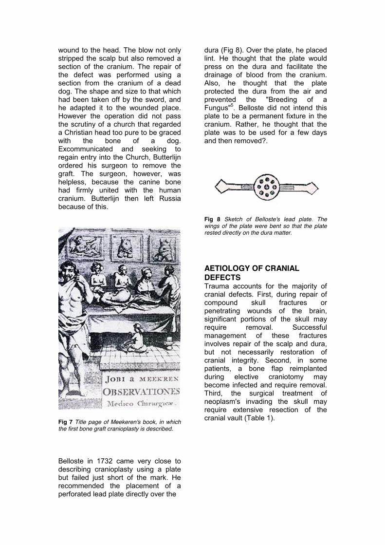
Fig 7 Title page of Meekeren's book, in which the first bone graft cranioplasty is described. Belloste in 1732 came very close to describing cranioplasty using a plate but failed just short of the mark. He recommended the placement of a perforated lead plate directly over the
dura (Fig 8). Over the plate, he placed lint. He thought that the plate would press on the dura and facilitate the drainage of blood from the cranium. Also, he thought that the plate protected the dura from the air and prevented the "Breeding of a Fungus"5. Belloste did not intend this plate to be a permanent fixture in the cranium. Rather, he thought that the plate was to be used for a few days and then removed?.
Fig 8 Sketch of Belloste's lead plate. The wings of the plate were bent so that the plate rested directly on the dura matter. AETIOLOGY OF CRANIAL DEFECTS Trauma accounts for the majority of cranial defects. First, during repair of compound skull fractures or penetrating wounds of the brain, significant portions of the skull may require removal. Successful management of these fractures involves repair of the scalp and dura, but not necessarily restoration of cranial integrity. Second, in some patients, a bone flap reimplanted during elective craniotomy may become infected and require removal. Third, the surgical treatment of neoplasm's invading the skull may require extensive resection of the cranial vault (Table 1).
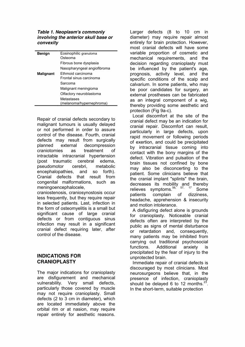
Table 1. Neoplasm's commonly involving the anterior skull base or convexity Benign Eosinophilic granuloma Osteoma Fibrous bone dysplasia Nasopharyngeal angiofibroma Malignant Ethmoid carcinoma Frontal sinus carcinoma Sarcoma Malignant meningioma Olfactory neuroblastoma Metastases
(melanoma/hypernephroma) Repair of cranial defects secondary to malignant tumours is usually delayed or not performed in order to assure control of the disease. Fourth, cranial defects may result from surgically planned external decompression craniotomies as treatment of intractable intracranial hypertension (post traumatic cerebral edema, pseudomotor cerebri, metabolic encephalopathies, and so forth). Cranial defects that result from congenital malformations, such as meningoencephalocele, craniostenosis, craniosynostosis occur less frequently, but they require repair in selected patients. Last, infection in the form of osteomyelitis is a small but significant cause of large cranial defects or from contiguous sinus infection may result in a significant cranial defect requiring later, after control of the disease. INDICATIONS FOR CRANIOPLASTY The major indications for cranioplasty are disfigurement and mechanical vulnerability. Very small defects, particularly those covered by muscle may not require cranioplasty. Small defects (2 to 3 cm in diameter), which are located immediately above the orbital rim or at nasion, may require repair entirely for aesthetic reasons.
Larger defects (8 to 10 cm in diameter) may require repair almost entirely for brain protection. However, most cranial defects will have some variable proportion of cosmetic and mechanical requirements, and the decision regarding cranioplasty must be influenced by the patient's age, prognosis, activity level, and the specific conditions of the scalp and calvarium. In some patients, who may be poor candidates for surgery, an external prostheses can be fabricated as an integral component of a wig, thereby providing some aesthetic and protection (Fig 9a-c). Local discomfort at the site of the cranial defect may be an indication for cranial repair. Discomfort can result, particularly in large defects, upon rapid movement or following periods of exertion, and could be precipitated by intracranial tissue coming into contact with the bony margins of the defect. Vibration and pulsation of the brain tissues not confined by bone may also be disconcerting to the patient. Some clinicians believe that the cranial implant "splints" the brain, decreases its mobility and thereby relieves symptoms.56, 57, 17 Some patients complain of dizziness, headache, apprehension & insecurity and motion intolerance. A disfiguring defect alone is grounds for cranioplasty. Noticeable cranial defects often are interpreted by the public as signs of mental disturbance or retardation and, consequently, many patients may be inhibited from carrying out traditional psychosocial functions. Additional anxiety is precipitated by the fear of injury to the unprotected brain. Immediate repair of cranial defects is discouraged by most clinicians. Most neurosurgeons believe that, in the presence of infection, cranioplasty should be delayed 6 to 12 months.23. In the short-term, suitable protection
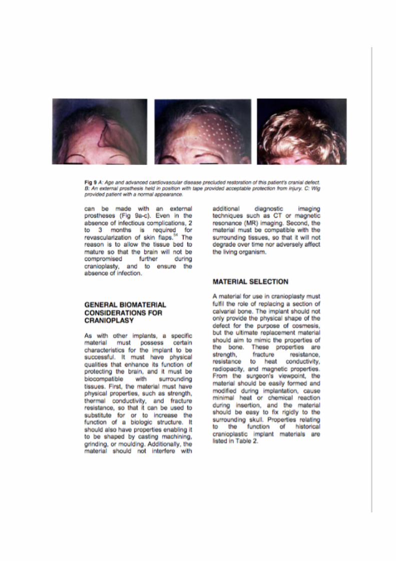
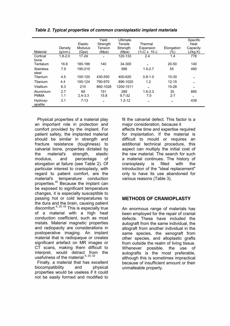
Table 2. Typical properties of common cranioplastic implant materials Material
Density (g/cm3)
Elastic Modulus (Gpa)
Yield Strength Tension (Mpa)
Ultimate Tensile Strength (Mpa)
Thermal Expansion (1/oC x 10-5)
Elongation (%)
Specific Heat Capacity (J/kg K)
Cortical bone
1.8-2.0 17-24 - 120-133 2.4 1.4 778
Tantalum 16.6 180-186 140 34-300 - 20-50 140 Stainless steel
7.9 190-210 - 586 1.5-2.7 55 490
Titanium 4.5 100-120 430-550 400-620 0.8-1.0 15-30 - Titanium 4.4 100-124 790-970 896-1020 1.2 12-15 - Vitallium 8.3 210 860-1028 1200-1511 - 15-28 - Aluminium 2.7 69 151 289 1.6-2.3 35 895 PMMA 1.1 2.4-3.3 15.8 9.7-32 7.0 2-7 - Hydroxy- apatite
3.1 7-13 - 1.2-12 - - 439
Physical properties of a material play an important role in protection and comfort provided by the implant. For patient safety, the implanted material should be similar in strength and fracture resistance (toughness) to calvarial bone, properties dictated by the material's strength, elastic modulus, and percentage of elongation at failure (see Table 2). Of particular interest to cranioplasty, with regard to patient comfort, are the material's temperature conduction properties.45 Because the implant can be exposed to significant temperature changes, it is especially susceptible to passing hot or cold temperatures to the dura and the brain, causing patient discomfort.6, 25, 43 This is especially true of a material with a high heat conduction coefficient, such as most metals. Material magnetic properties and radiopacity are considerations in postoperative imaging. An implant material that is radiopaque or creates significant artefact on MR images or CT scans, making them difficult to interpret, would detract from the usefulness of the material.6, 25, 52 Finally, a material that has excellent biocompatibility and physical properties would be useless if it could not be easily formed and modified to
fit the calvarial defect. This factor is a major consideration, because it affects the time and expertise required for implantation. If the material is difficult to mould or requires an additional technical procedure, this aspect can multiply the initial cost of the raw material. The search for such a material continues. The history of cranioplasty is filled with the introduction of the "ideal replacement" only to have its use abandoned for various reasons (Table 3). METHODS OF CRANIOPLASTY An enormous range of materials has been employed for the repair of cranial defects. These have included the autograft from the same individual, the allograft from another individual in the same species, the xenograft from other species, and alloplastic grafts from outside the realm of living tissue. Whenever possible, the use of autografts is the most preferable, although this is sometimes impractical because of insufficient amount or their unmalleable property.
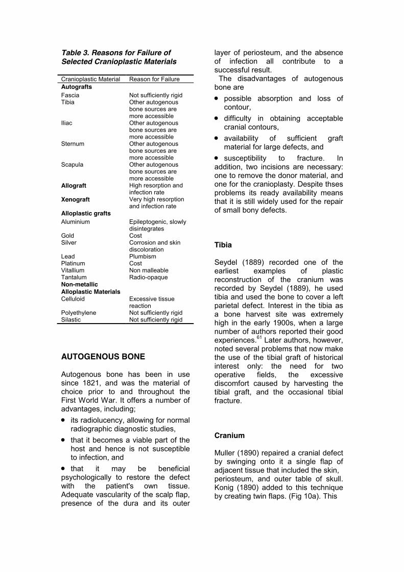
Table 3. Reasons for Failure of Selected Cranioplastic Materials Cranioplastic Material Reason for Failure Autografts Fascia Not sufficiently rigid Tibia Other autogenous
bone sources are more accessible
Iliac Other autogenous bone sources are more accessible
Sternum Other autogenous bone sources are more accessible
Scapula Other autogenous bone sources are more accessible
Allograft High resorption and infection rate
Xenograft Very high resorption and infection rate
Alloplastic grafts Aluminium Epileptogenic, slowly
disintegrates Gold Cost Silver Corrosion and skin
discoloration Lead Plumbism Platinum Cost Vitallium Non malleable Tantalum Radio-opaque Non-metallic Alloplastic Materials
Celluloid Excessive tissue reaction
Polyethylene Not sufficiently rigid Silastic Not sufficiently rigid AUTOGENOUS BONE Autogenous bone has been in use since 1821, and was the material of choice prior to and throughout the First World War. It offers a number of advantages, including; x its radiolucency, allowing for normal
radiographic diagnostic studies, x that it becomes a viable part of the
host and hence is not susceptible to infection, and
x that it may be beneficial psychologically to restore the defect with the patient's own tissue. Adequate vascularity of the scalp flap, presence of the dura and its outer
layer of periosteum, and the absence of infection all contribute to a successful result. The disadvantages of autogenous bone are x possible absorption and loss of
contour, x difficulty in obtaining acceptable
cranial contours, x availability of sufficient graft
material for large defects, and x susceptibility to fracture. In addition, two incisions are necessary: one to remove the donor material, and one for the cranioplasty. Despite thses problems its ready availability means that it is still widely used for the repair of small bony defects. Tibia Seydel (1889) recorded one of the earliest examples of plastic reconstruction of the cranium was recorded by Seydel (1889), he used tibia and used the bone to cover a left parietal defect. Interest in the tibia as a bone harvest site was extremely high in the early 1900s, when a large number of authors reported their good experiences.61 Later authors, however, noted several problems that now make the use of the tibial graft of historical interest only: the need for two operative fields, the excessive discomfort caused by harvesting the tibial graft, and the occasional tibial fracture. Cranium Muller (1890) repaired a cranial defect by swinging onto it a single flap of adjacent tissue that included the skin, periosteum, and outer table of skull. Konig (1890) added to this technique by creating twin flaps. (Fig 10a). This
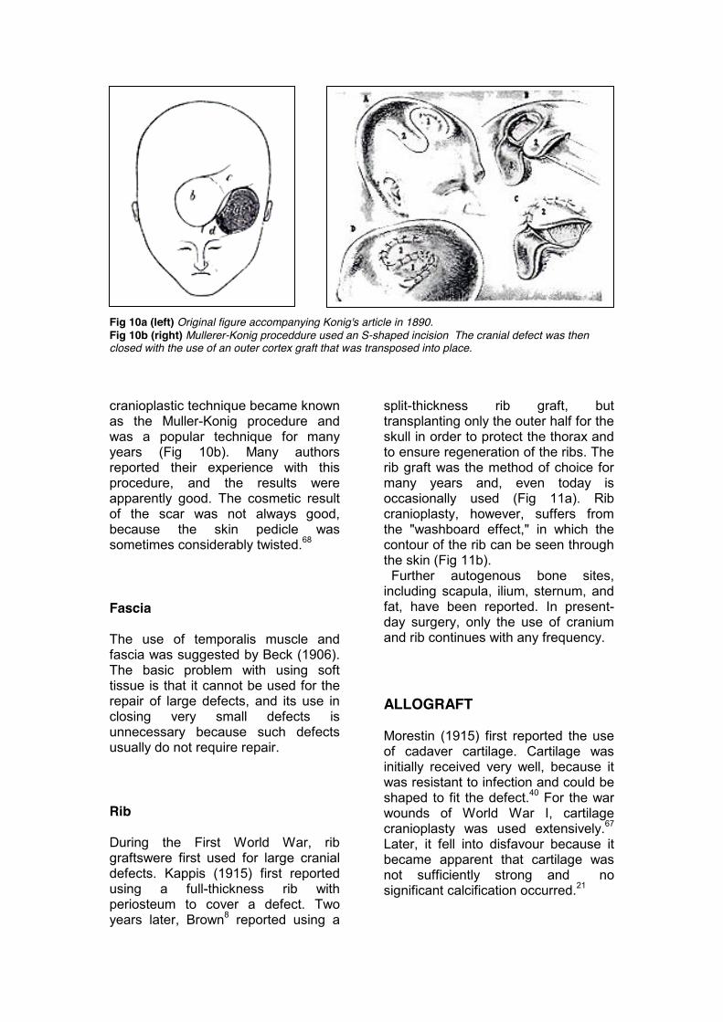
Fig 10a (left) Original figure accompanying Konig's article in 1890. Fig 10b (right) Mullerer-Konig proceddure used an S-shaped incision The cranial defect was then closed with the use of an outer cortex graft that was transposed into place. cranioplastic technique became known as the Muller-Konig procedure and was a popular technique for many years (Fig 10b). Many authors reported their experience with this procedure, and the results were apparently good. The cosmetic result of the scar was not always good, because the skin pedicle was sometimes considerably twisted.68 Fascia The use of temporalis muscle and fascia was suggested by Beck (1906). The basic problem with using soft tissue is that it cannot be used for the repair of large defects, and its use in closing very small defects is unnecessary because such defects usually do not require repair. Rib During the First World War, rib graftswere first used for large cranial defects. Kappis (1915) first reported using a full-thickness rib with periosteum to cover a defect. Two years later, Brown8 reported using a
split-thickness rib graft, but transplanting only the outer half for the skull in order to protect the thorax and to ensure regeneration of the ribs. The rib graft was the method of choice for many years and, even today is occasionally used (Fig 11a). Rib cranioplasty, however, suffers from the "washboard effect," in which the contour of the rib can be seen through the skin (Fig 11b). Further autogenous bone sites, including scapula, ilium, sternum, and fat, have been reported. In present-day surgery, only the use of cranium and rib continues with any frequency. ALLOGRAFT Morestin (1915) first reported the use of cadaver cartilage. Cartilage was initially received very well, because it was resistant to infection and could be shaped to fit the defect.40 For the war wounds of World War I, cartilage cranioplasty was used extensively.67 Later, it fell into disfavour because it became apparent that cartilage was not sufficiently strong and no significant calcification occurred.21
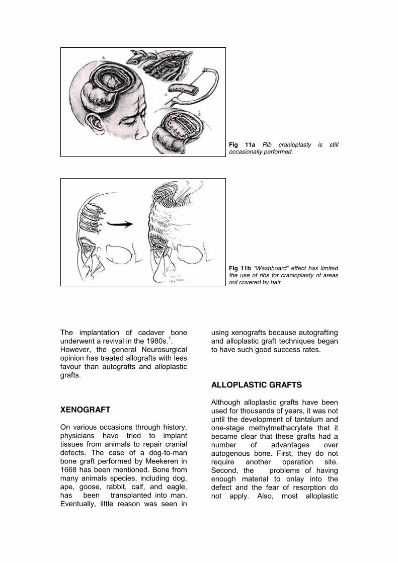
Fig 11a Rib cranioplasty is still occasionally performed.
Fig 11b “Washboard” effect has limited the use of ribs for cranioplasty of areas not covered by hair
The implantation of cadaver bone underwent a revival in the 1980s.1. However, the general Neurosurgical opinion has treated allografts with less favour than autografts and alloplastic grafts. XENOGRAFT On various occasions through history, physicians have tried to implant tissues from animals to repair cranial defects. The case of a dog-to-man bone graft performed by Meekeren in 1668 has been mentioned. Bone from many animals species, including dog, ape, goose, rabbit, calf, and eagle, has been transplanted into man. Eventually, little reason was seen in
using xenografts because autografting and alloplastic graft techniques began to have such good success rates. ALLOPLASTIC GRAFTS Although alloplastic grafts have been used for thousands of years, it was not until the development of tantalum and one-stage methylmethacrylate that it became clear that these grafts had a number of advantages over autogenous bone. First, they do not require another operation site. Second, the problems of having enough material to onlay into the defect and the fear of resorption do not apply. Also, most alloplastic
materials can be moulded to the contour of the defect at the operating table and thus provide for a superior cosmetic result. Finally, the tensile strength of many alloplastic grafts is similar to that of bone. The major disadvantages to using alloplastic materials is that they are foreign bodies. Infection is the most common reason for failure of these types of grafts. Alloplastic grafts placed in the frontal-orbital region are at particular risk for infection, and dislocation of the implant METALS Numerous metals and alloys have been employed historically for restoration of cranial defects. Ideally, the metal should be light in weight; strong enough to resist trauma, yet sufficiently malleable to allow easy alteration at surgery; inevitable so as not to provoke tissue reactions; and moderately radiolucent to permit normal radiologic examination. Aluminium, gold, and silver Aluminium was the first metal in recent history to be used as a bone substitute for a cranial defect. In 1893, Booth and Curtis7 first used a thin aluminium plate to close a defect, 19 days after the removal of a tumour. They reported, however, that on the fifth postoperative day, the patient was febrile and had convulsions, and there was a "milky pus" around the incision. The aluminium was removed. The patient died the following mouth. One hundred cases of gold cranioplasty were reported by Estor (1916), with two deaths from infection and two plate revisions because of infection. Estor supported the use of gold and noted that patients were very
satisfied with the metal. However, the use of gold did not become popular because of its high cost and the softness of the pure metal. Silver was used by Sebileau (1903). Its use was later abandoned, because silver oxide reacted with the surrounding tissues and sometimes discoloured the overlying scalp. Also, pure silver was too soft and could not withstand even minor trauma. Lead and platinum Mauclaire (1908), and Rouvillois (1908), used lead and as expected, the patients developed lead intoxication that required plate removal. Cornioly (1929) used platinum and reported an absence of tissue reaction. Cost precluded platinum's widespread use. Vitallium and ticonium The use of alloys instead of pure metals for bone replacement was suggested by the experimental work of Jones and Lieberman (1936). They implanted metal tacks into the leg bones of dogs and noted that nickel alloys produce less bone necrosis and metal corrosion than pure metals. Vitallium, an alloy of cobalt, chromium, and molybdenum, was suggested as a cranioplastic alloy by Venable (1937). On electrolytic tests on various metals, it was revealed that vitallium was very resistant to corrosion.61. Vitallium continued to be used in the early 1940s but was eventually discarded because it lacked malleability. The alloy had to be cast from a mould, and further shaping at surgery was difficult. Ticonium, an alloy composed of cobalt, chromium, nickel, and molybdenum, was used as a cranioplastic material in 1941 and was a direct competitor to vitallium.10
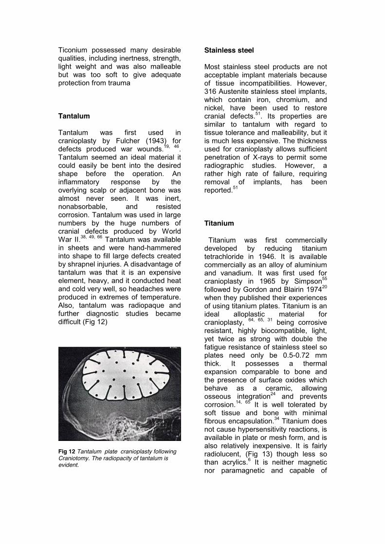
Ticonium possessed many desirable qualities, including inertness, strength, light weight and was also malleable but was too soft to give adequate protection from trauma Tantalum Tantalum was first used in cranioplasty by Fulcher (1943) for defects produced war wounds.19, 46. Tantalum seemed an ideal material it could easily be bent into the desired shape before the operation. An inflammatory response by the overlying scalp or adjacent bone was almost never seen. It was inert, nonabsorbable, and resisted corrosion. Tantalum was used in large numbers by the huge numbers of cranial defects produced by World War II.38, 49, 66 Tantalum was available in sheets and were hand-hammered into shape to fill large defects created by shrapnel injuries. A disadvantage of tantalum was that it is an expensive element, heavy, and it conducted heat and cold very well, so headaches were produced in extremes of temperature. Also, tantalum was radiopaque and further diagnostic studies became difficult (Fig 12)
Fig 12 Tantalum plate cranioplasty following Craniotomy. The radiopacity of tantalum is evident.
Stainless steel Most stainless steel products are not acceptable implant materials because of tissue incompatibilities. However, 316 Austenite stainless steel implants, which contain iron, chromium, and nickel, have been used to restore cranial defects.51. Its properties are similar to tantalum with regard to tissue tolerance and malleability, but it is much less expensive. The thickness used for cranioplasty allows sufficient penetration of X-rays to permit some radiographic studies. However, a rather high rate of failure, requiring removal of implants, has been reported.51 Titanium Titanium was first commercially developed by reducing titanium tetrachloride in 1946. It is available commercially as an alloy of aluminium and vanadium. It was first used for cranioplasty in 1965 by Simpson55 followed by Gordon and Blairin 197420 when they published their experiences of using titanium plates. Titanium is an ideal alloplastic material for cranioplasty, 64, 65, 31 being corrosive resistant, highly biocompatible, light, yet twice as strong with double the fatigue resistance of stainless steel so plates need only be 0.5-0.72 mm thick. It possesses a thermal expansion comparable to bone and the presence of surface oxides which behave as a ceramic, allowing osseous integration24 and prevents corrosion.14, 65 It is well tolerated by soft tissue and bone with minimal fibrous encapsulation.34 Titanium does not cause hypersensitivity reactions, is available in plate or mesh form, and is also relatively inexpensive. It is fairly radiolucent, (Fig 13) though less so than acrylics.6 It is neither magnetic nor paramagnetic and capable of
modification in the operating theatre. Titanium can be safely used in the dental/maxillofacial laboratory. To summarise metals are strong, malleable, and sterilizable. They require only one incision because there is no donor site so this significantly reduces operating time. However, they also corrode, conduct heat, become infected, are relatively radiopaque, and may be epileptogenic. The inability to obtain clear cranial X-rays with the use of metal cranioplasty was a big handicap in the era before computed tomography, and this was the prime reason many surgeons sought non-metallic alloplastic materials.
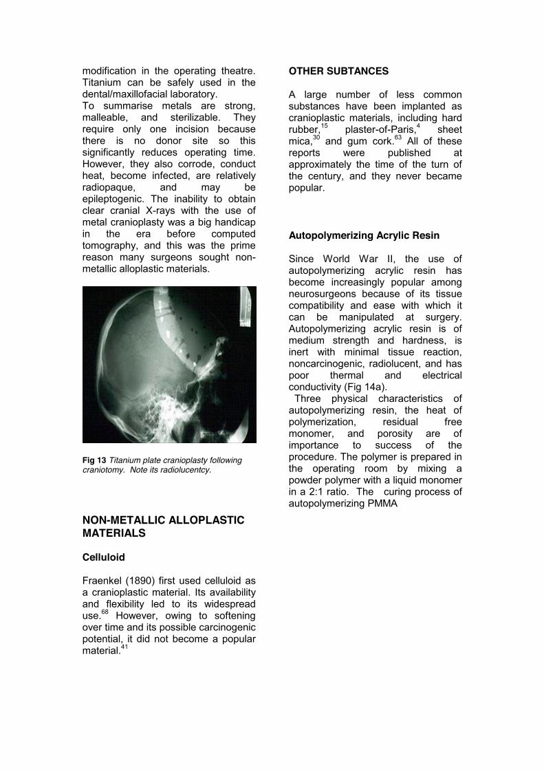
Fig 13 Titanium plate cranioplasty following craniotomy. Note its radiolucentcy. NON-METALLIC ALLOPLASTIC MATERIALS Celluloid Fraenkel (1890) first used celluloid as a cranioplastic material. Its availability and flexibility led to its widespread use.68 However, owing to softening over time and its possible carcinogenic potential, it did not become a popular material.41
OTHER SUBTANCES A large number of less common substances have been implanted as cranioplastic materials, including hard rubber,15 plaster-of-Paris,4 sheet mica,30 and gum cork.63 All of these reports were published at approximately the time of the turn of the century, and they never became popular. Autopolymerizing Acrylic Resin Since World War II, the use of autopolymerizing acrylic resin has become increasingly popular among neurosurgeons because of its tissue compatibility and ease with which it can be manipulated at surgery. Autopolymerizing acrylic resin is of medium strength and hardness, is inert with minimal tissue reaction, noncarcinogenic, radiolucent, and has poor thermal and electrical conductivity (Fig 14a). Three physical characteristics of autopolymerizing resin, the heat of polymerization, residual free monomer, and porosity are of importance to success of the procedure. The polymer is prepared in the operating room by mixing a powder polymer with a liquid monomer in a 2:1 ratio. The curing process of autopolymerizing PMMA
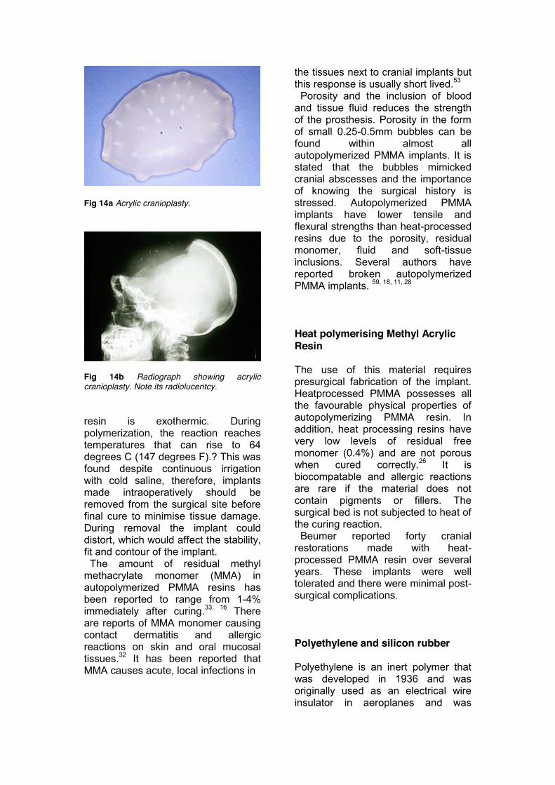
Fig 14a Acrylic cranioplasty.
Fig 14b Radiograph showing acrylic cranioplasty. Note its radiolucentcy. resin is exothermic. During polymerization, the reaction reaches temperatures that can rise to 64 degrees C (147 degrees F).? This was found despite continuous irrigation with cold saline, therefore, implants made intraoperatively should be removed from the surgical site before final cure to minimise tissue damage. During removal the implant could distort, which would affect the stability, fit and contour of the implant. The amount of residual methyl methacrylate monomer (MMA) in autopolymerized PMMA resins has been reported to range from 1-4% immediately after curing.33, 16 There are reports of MMA monomer causing contact dermatitis and allergic reactions on skin and oral mucosal tissues.32 It has been reported that MMA causes acute, local infections in
the tissues next to cranial implants but this response is usually short lived.53 Porosity and the inclusion of blood and tissue fluid reduces the strength of the prosthesis. Porosity in the form of small 0.25-0.5mm bubbles can be found within almost all autopolymerized PMMA implants. It is stated that the bubbles mimicked cranial abscesses and the importance of knowing the surgical history is stressed. Autopolymerized PMMA implants have lower tensile and flexural strengths than heat-processed resins due to the porosity, residual monomer, fluid and soft-tissue inclusions. Several authors have reported broken autopolymerized PMMA implants. 59, 18, 11, 28 Heat polymerising Methyl Acrylic Resin The use of this material requires presurgical fabrication of the implant. Heatprocessed PMMA possesses all the favourable physical properties of autopolymerizing PMMA resin. In addition, heat processing resins have very low levels of residual free monomer (0.4%) and are not porous when cured correctly.26 It is biocompatable and allergic reactions are rare if the material does not contain pigments or fillers. The surgical bed is not subjected to heat of the curing reaction. Beumer reported forty cranial restorations made with heat-processed PMMA resin over several years. These implants were well tolerated and there were minimal post-surgical complications. Polyethylene and silicon rubber Polyethylene is an inert polymer that was developed in 1936 and was originally used as an electrical wire insulator in aeroplanes and was
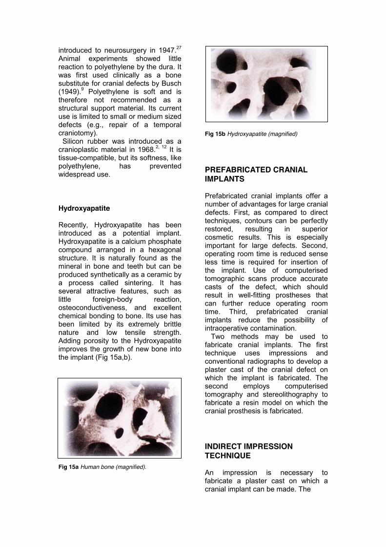
introduced to neurosurgery in 1947.27 Animal experiments showed little reaction to polyethylene by the dura. It was first used clinically as a bone substitute for cranial defects by Busch (1949).9 Polyethylene is soft and is therefore not recommended as a structural support material. Its current use is limited to small or medium sized defects (e.g., repair of a temporal craniotomy). Silicon rubber was introduced as a cranioplastic material in 1968.2, 12 It is tissue-compatible, but its softness, like polyethylene, has prevented widespread use. Hydroxyapatite Recently, Hydroxyapatite has been introduced as a potential implant. Hydroxyapatite is a calcium phosphate compound arranged in a hexagonal structure. It is naturally found as the mineral in bone and teeth but can be produced synthetically as a ceramic by a process called sintering. It has several attractive features, such as little foreign-body reaction, osteoconductiveness, and excellent chemical bonding to bone. Its use has been limited by its extremely brittle nature and low tensile strength. Adding porosity to the Hydroxyapatite improves the growth of new bone into the implant (Fig 15a,b). Fig 15a Human bone (magnified).
Fig 15b Hydroxyapatite (magnified) PREFABRICATED CRANIAL IMPLANTS Prefabricated cranial implants offer a number of advantages for large cranial defects. First, as compared to direct techniques, contours can be perfectly restored, resulting in superior cosmetic results. This is especially important for large defects. Second, operating room time is reduced sense less time is required for insertion of the implant. Use of computerised tomographic scans produce accurate casts of the defect, which should result in well-fitting prostheses that can further reduce operating room time. Third, prefabricated cranial implants reduce the possibility of intraoperative contamination. Two methods may be used to fabricate cranial implants. The first technique uses impressions and conventional radiographs to develop a plaster cast of the cranial defect on which the implant is fabricated. The second employs computerised tomography and stereolithography to fabricate a resin model on which the cranial prosthesis is fabricated. INDIRECT IMPRESSION TECHNIQUE An impression is necessary to fabricate a plaster cast on which a cranial implant can be made. The
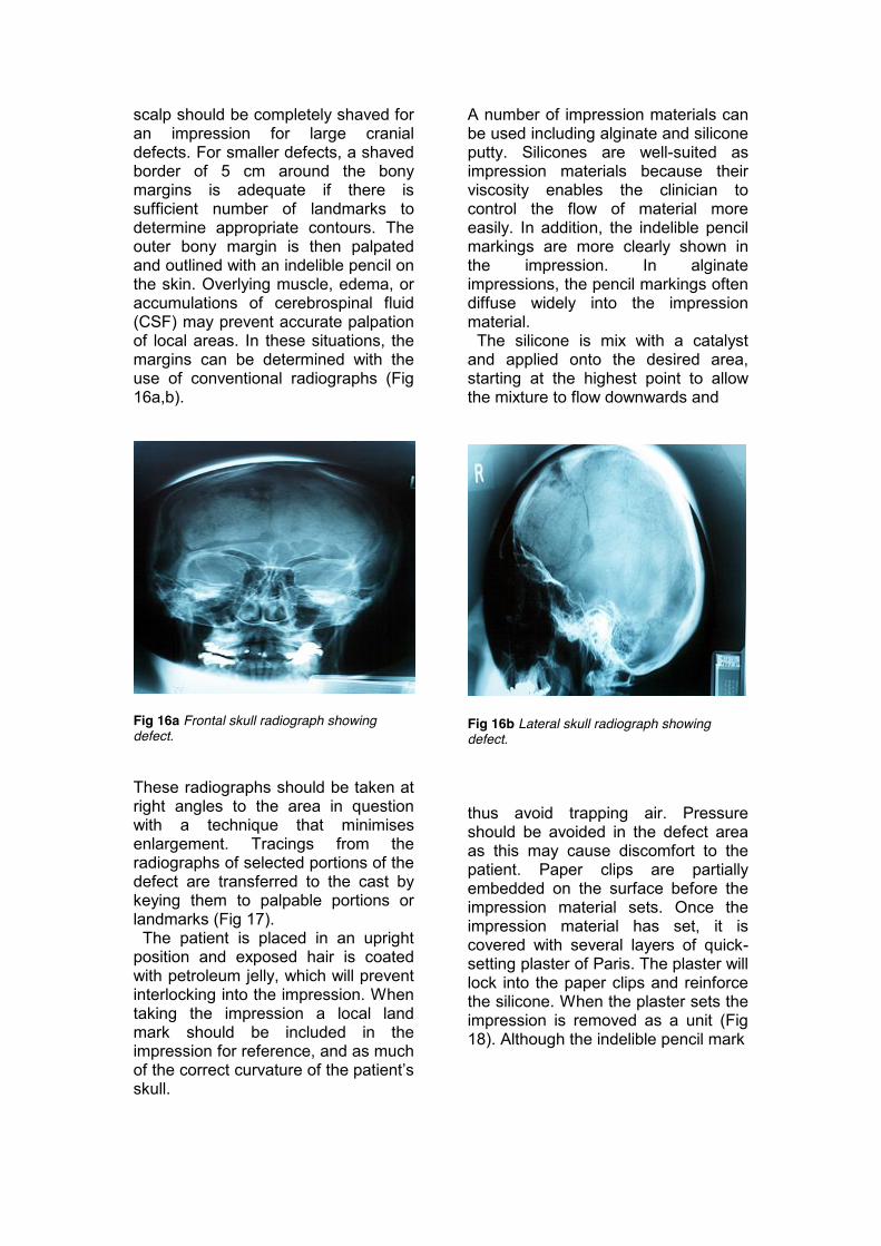
scalp should be completely shaved for an impression for large cranial defects. For smaller defects, a shaved border of 5 cm around the bony margins is adequate if there is sufficient number of landmarks to determine appropriate contours. The outer bony margin is then palpated and outlined with an indelible pencil on the skin. Overlying muscle, edema, or accumulations of cerebrospinal fluid (CSF) may prevent accurate palpation of local areas. In these situations, the margins can be determined with the use of conventional radiographs (Fig 16a,b).
Fig 16a Frontal skull radiograph showing defect. These radiographs should be taken at right angles to the area in question with a technique that minimises enlargement. Tracings from the radiographs of selected portions of the defect are transferred to the cast by keying them to palpable portions or landmarks (Fig 17). The patient is placed in an upright position and exposed hair is coated with petroleum jelly, which will prevent interlocking into the impression. When taking the impression a local land mark should be included in the impression for reference, and as much of the correct curvature of the patient’s skull.
A number of impression materials can be used including alginate and silicone putty. Silicones are well-suited as impression materials because their viscosity enables the clinician to control the flow of material more easily. In addition, the indelible pencil markings are more clearly shown in the impression. In alginate impressions, the pencil markings often diffuse widely into the impression material. The silicone is mix with a catalyst and applied onto the desired area, starting at the highest point to allow the mixture to flow downwards and
Fig 16b Lateral skull radiograph showing defect. thus avoid trapping air. Pressure should be avoided in the defect area as this may cause discomfort to the patient. Paper clips are partially embedded on the surface before the impression material sets. Once the impression material has set, it is covered with several layers of quick-setting plaster of Paris. The plaster will lock into the paper clips and reinforce the silicone. When the plaster sets the impression is removed as a unit (Fig 18). Although the indelible pencil mark
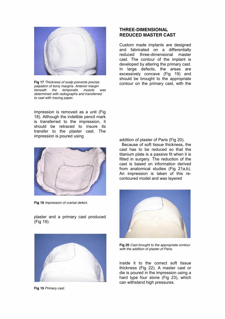
Fig 17 Thickness of scalp prevents precise palpation of bony margins. Anterior margin beneath the temporalis muscle was determined with radiographs and transferred to cast with tracing paper. impression is removed as a unit (Fig 18). Although the indelible pencil mark is transferred to the impression, it should be retraced to insure its transfer to the plaster cast. The impression is poured using
Fig 18 Impression of cranial defect. plaster and a primary cast produced (Fig 19).
Fig 19 Primary cast.
THREE-DIMENSIONAL REDUCED MASTER CAST Custom made implants are designed and fabricated on a differentially reduced three-dimensional master cast. The contour of the implant is developed by altering the primary cast. In large defects, the areas are excessively concave (Fig 19) and should be brought to the appropriate contour on the primary cast, with the
addition of plaster of Paris (Fig 20). Because of soft tissue thickness, the cast has to be reduced so that the titanium plate is a passive fit when it is fitted in surgery. The reduction of the cast is based on information derived from anatomical studies (Fig 21a,b). An impression is taken of this re-contoured model and wax layered
Fig 20 Cast brought to the appropriate contour with the addition of plaster of Paris. inside it to the correct soft tissue thickness (Fig 22). A master cast or die is poured in the impression using a hard type four stone (Fig 23), which can withstand high pressures.
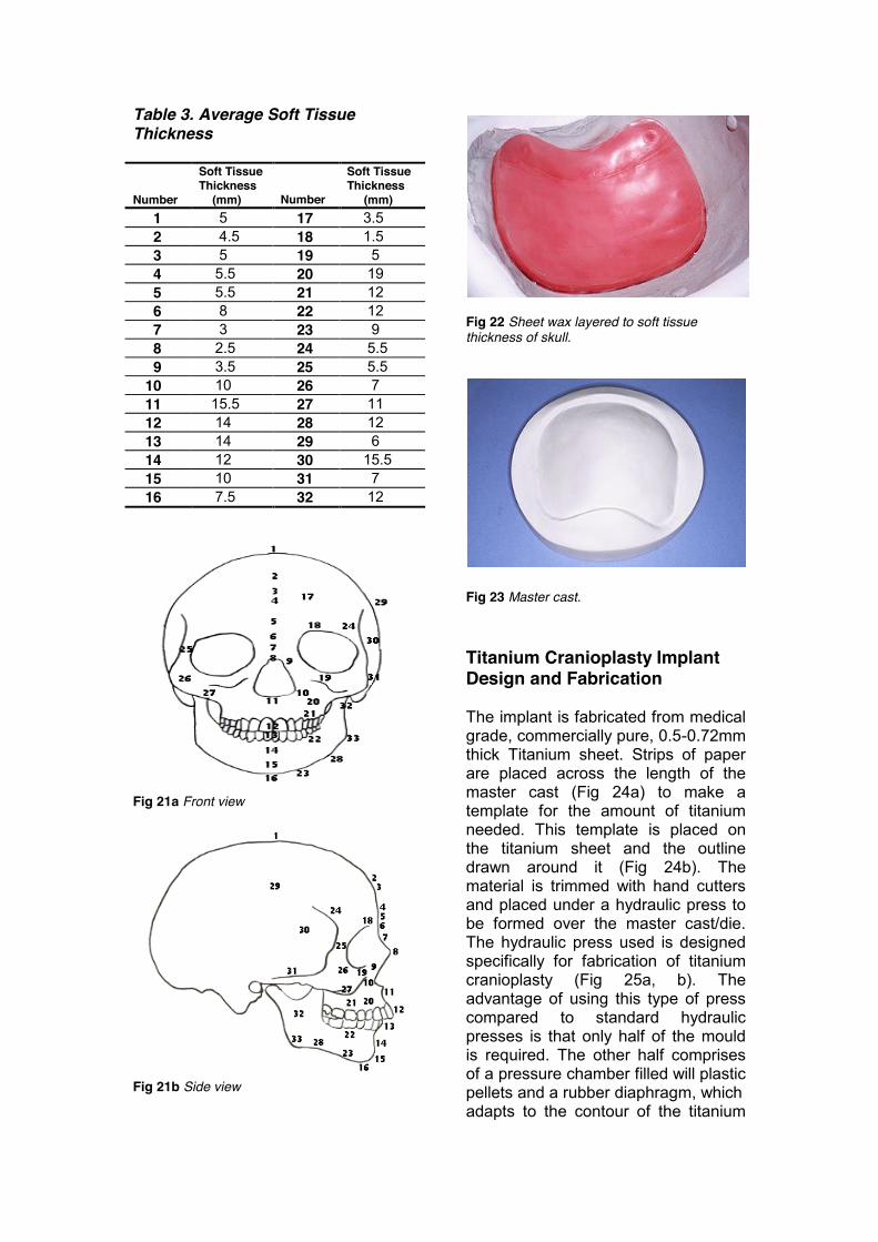
Table 3. Average Soft Tissue Thickness Number
Soft Tissue Thickness (mm)
Number
Soft Tissue Thickness (mm)
1 5 17 3.5 2 4.5 18 1.5 3 5 19 5 4 5.5 20 19 5 5.5 21 12 6 8 22 12 7 3 23 9 8 2.5 24 5.5 9 3.5 25 5.5 10 10 26 7 11 15.5 27 11 12 14 28 12 13 14 29 6 14 12 30 15.5 15 10 31 7 16 7.5 32 12
Fig 21a Front view
Fig 21b Side view
Fig 22 Sheet wax layered to soft tissue thickness of skull.
Fig 23 Master cast. Titanium Cranioplasty Implant Design and Fabrication The implant is fabricated from medical grade, commercially pure, 0.5-0.72mm thick Titanium sheet. Strips of paper are placed across the length of the master cast (Fig 24a) to make a template for the amount of titanium needed. This template is placed on the titanium sheet and the outline drawn around it (Fig 24b). The material is trimmed with hand cutters and placed under a hydraulic press to be formed over the master cast/die. The hydraulic press used is designed specifically for fabrication of titanium cranioplasty (Fig 25a, b). The advantage of using this type of press compared to standard hydraulic presses is that only half of the mould is required. The other half comprises of a pressure chamber filled will plastic pellets and a rubber diaphragm, which adapts to the contour of the titanium
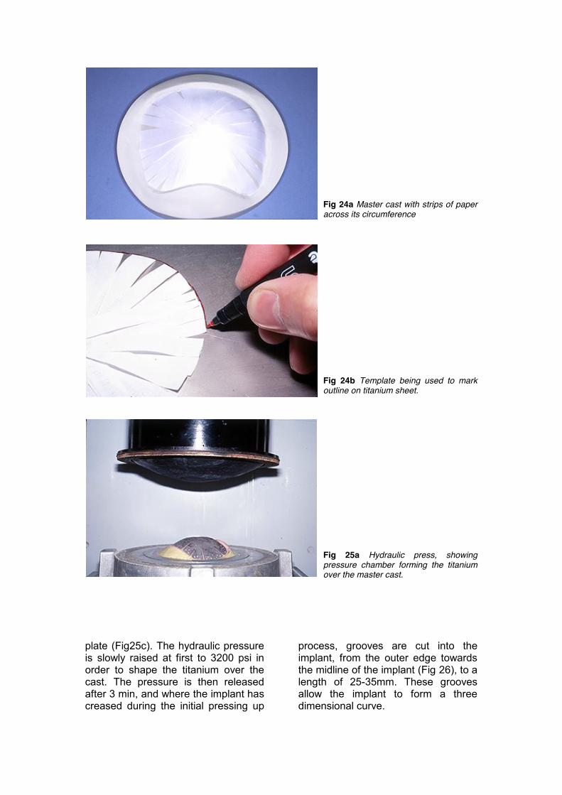
Fig 24a Master cast with strips of paper across its circumference
Fig 24b Template being used to mark outline on titanium sheet.
Fig 25a Hydraulic press, showing pressure chamber forming the titanium over the master cast.
plate (Fig25c). The hydraulic pressure is slowly raised at first to 3200 psi in order to shape the titanium over the cast. The pressure is then released after 3 min, and where the implant has creased during the initial pressing up
process, grooves are cut into the implant, from the outer edge towards the midline of the implant (Fig 26), to a length of 25-35mm. These grooves allow the implant to form a three dimensional curve.
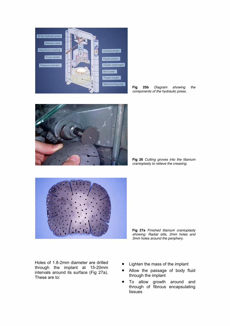
Fig 25b Diagram showing the components of the hydraulic press.
Fig 26 Cutting groves into the titanium cranioplasty to relieve the creasing.
Fig 27a Finished titanium cranioplasty showing: Radial slits, 2mm holes and 3mm holes around the periphery.
Holes of 1.8-2mm diameter are drilled through the implant at 15-20mm intervals around its surface (Fig 27a). These are to:
x Lighten the mass of the implant x Allow the passage of body fluid
through the implant x To allow growth around and
through of fibrous encapsulating tissues
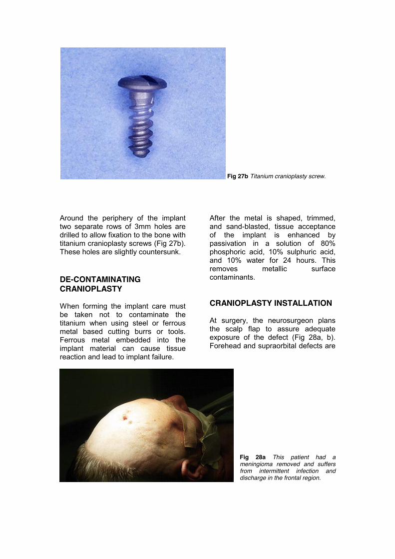
Fig 27b Titanium cranioplasty screw. Around the periphery of the implant two separate rows of 3mm holes are drilled to allow fixation to the bone with titanium cranioplasty screws (Fig 27b). These holes are slightly countersunk. DE-CONTAMINATING CRANIOPLASTY When forming the implant care must be taken not to contaminate the titanium when using steel or ferrous metal based cutting burrs or tools. Ferrous metal embedded into the implant material can cause tissue reaction and lead to implant failure.
After the metal is shaped, trimmed, and sand-blasted, tissue acceptance of the implant is enhanced by passivation in a solution of 80% phosphoric acid, 10% sulphuric acid, and 10% water for 24 hours. This removes metallic surface contaminants. CRANIOPLASTY INSTALLATION At surgery, the neurosurgeon plans the scalp flap to assure adequate exposure of the defect (Fig 28a, b). Forehead and supraorbital defects are
Fig 28a This patient had a meningioma removed and suffers from intermittent infection and discharge in the frontal region.
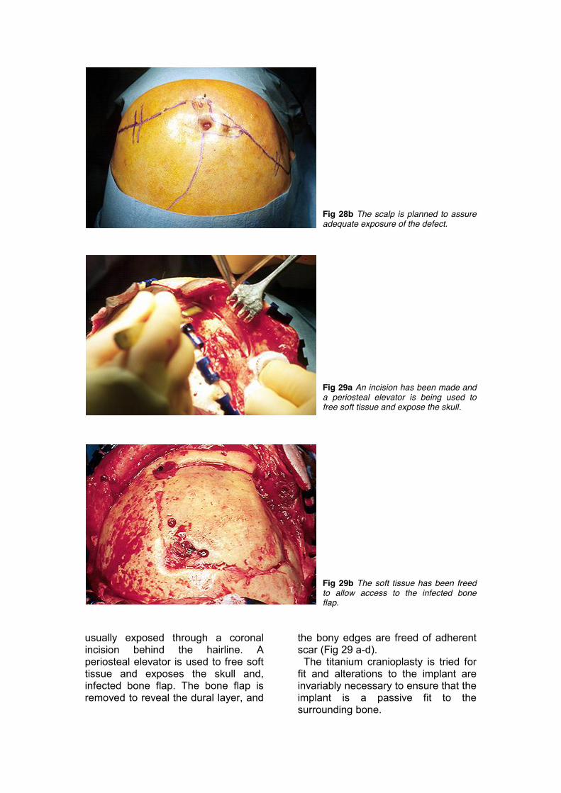
Fig 28b The scalp is planned to assure adequate exposure of the defect.
Fig 29a An incision has been made and a periosteal elevator is being used to free soft tissue and expose the skull.
Fig 29b The soft tissue has been freed to allow access to the infected bone flap.
usually exposed through a coronal incision behind the hairline. A periosteal elevator is used to free soft tissue and exposes the skull and, infected bone flap. The bone flap is removed to reveal the dural layer, and
the bony edges are freed of adherent scar (Fig 29 a-d). The titanium cranioplasty is tried for fit and alterations to the implant are invariably necessary to ensure that the implant is a passive fit to the surrounding bone.
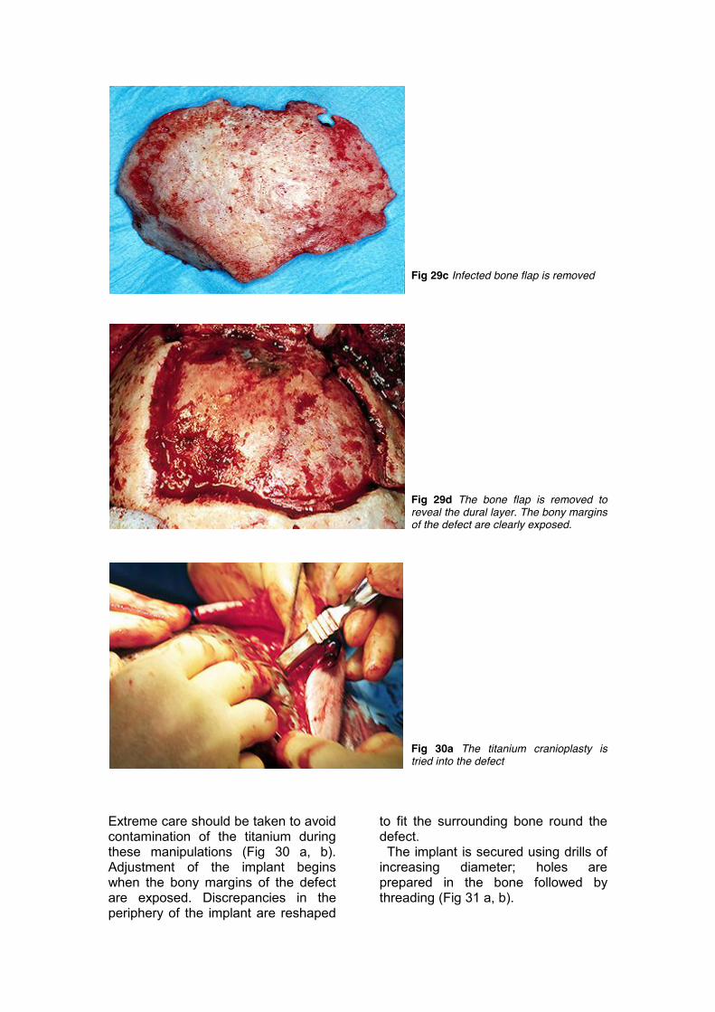
Fig 29c Infected bone flap is removed
Fig 29d The bone flap is removed to reveal the dural layer. The bony margins of the defect are clearly exposed.
Fig 30a The titanium cranioplasty is tried into the defect
Extreme care should be taken to avoid contamination of the titanium during these manipulations (Fig 30 a, b). Adjustment of the implant begins when the bony margins of the defect are exposed. Discrepancies in the periphery of the implant are reshaped
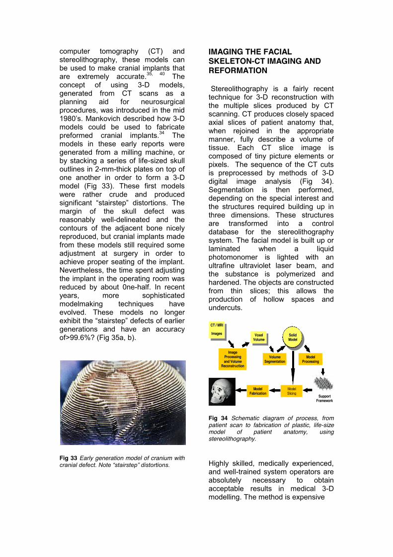
to fit the surrounding bone round the defect. The implant is secured using drills of increasing diameter; holes are prepared in the bone followed by threading (Fig 31 a, b).
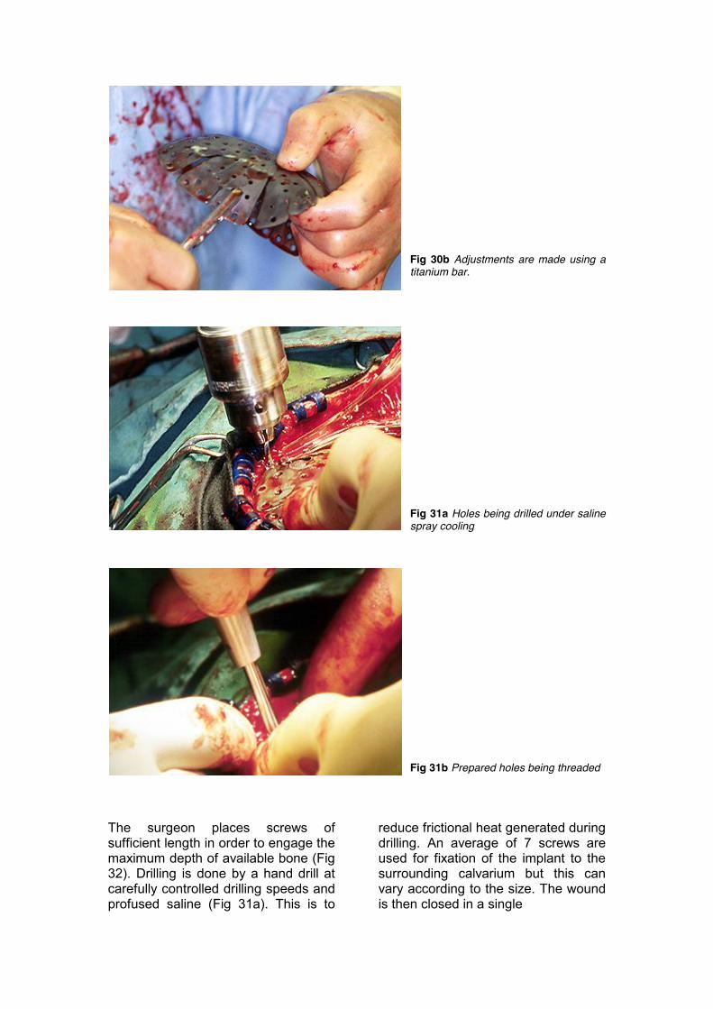
Fig 30b Adjustments are made using a titanium bar.
Fig 31a Holes being drilled under saline spray cooling
Fig 31b Prepared holes being threaded The surgeon places screws of sufficient length in order to engage the maximum depth of available bone (Fig 32). Drilling is done by a hand drill at carefully controlled drilling speeds and profused saline (Fig 31a). This is to
reduce frictional heat generated during drilling. An average of 7 screws are used for fixation of the implant to the surrounding calvarium but this can vary according to the size. The wound is then closed in a single
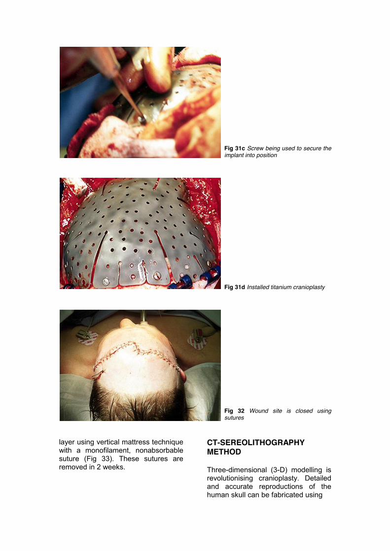
Fig 31c Screw being used to secure the implant into position
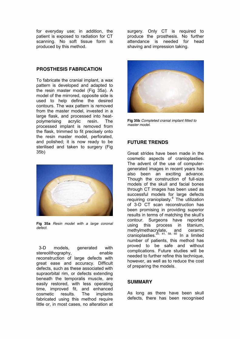
Fig 31d Installed titanium cranioplasty
Fig 32 Wound site is closed using sutures
layer using vertical mattress technique with a monofilament, nonabsorbable suture (Fig 33). These sutures are removed in 2 weeks.
CT-SEREOLITHOGRAPHY METHOD Three-dimensional (3-D) modelling is revolutionising cranioplasty. Detailed and accurate reproductions of the human skull can be fabricated using
computer tomography (CT) and stereolithography, these models can be used to make cranial implants that are extremely accurate.35, 40 The concept of using 3-D models, generated from CT scans as a planning aid for neurosurgical procedures, was introduced in the mid 1980’s. Mankovich described how 3-D models could be used to fabricate preformed cranial implants.34 The models in these early reports were generated from a milling machine, or by stacking a series of life-sized skull outlines in 2-mm-thick plates on top of one another in order to form a 3-D model (Fig 33). These first models were rather crude and produced significant “stairstep” distortions. The margin of the skull defect was reasonably well-delineated and the contours of the adjacent bone nicely reproduced, but cranial implants made from these models still required some adjustment at surgery in order to achieve proper seating of the implant. Nevertheless, the time spent adjusting the implant in the operating room was reduced by about 0ne-half. In recent years, more sophisticated modelmaking techniques have evolved. These models no longer exhibit the “stairstep” defects of earlier generations and have an accuracy of>99.6%? (Fig 35a, b).
Fig 33 Early generation model of cranium with cranial defect. Note “stairstep” distortions.
IMAGING THE FACIAL SKELETON-CT IMAGING AND REFORMATION Stereolithography is a fairly recent technique for 3-D reconstruction with the multiple slices produced by CT scanning. CT produces closely spaced axial slices of patient anatomy that, when rejoined in the appropriate manner, fully describe a volume of tissue. Each CT slice image is composed of tiny picture elements or pixels. The sequence of the CT cuts is preprocessed by methods of 3-D digital image analysis (Fig 34). Segmentation is then performed, depending on the special interest and the structures required building up in three dimensions. These structures are transformed into a control database for the stereolithography system. The facial model is built up or laminated when a liquid photomonomer is lighted with an ultrafine ultraviolet laser beam, and the substance is polymerized and hardened. The objects are constructed from thin slices; this allows the production of hollow spaces and undercuts.
Fig 34 Schematic diagram of process, from patient scan to fabrication of plastic, life-size model of patient anatomy, using stereolithography. Highly skilled, medically experienced, and well-trained system operators are absolutely necessary to obtain acceptable results in medical 3-D modelling. The method is expensive
CT / MRI
Images
ImageProcessingand Volume
Reconstruction
VoxelVolume
VolumeSegmentation
S
ModelProcessing
ModelSlicing
ModelFabrication
SolidModel
SupportFramework
for everyday use; in addition, the patient is exposed to radiation for CT scanning. No soft tissue form is produced by this method. PROSTHESIS FABRICATION To fabricate the cranial implant, a wax pattern is developed and adapted to the resin master model (Fig 35a). A model of the mirrored, opposite side is used to help define the desired contours. The wax pattern is removed from the master model, invested in a large flask, and processed into heat-polymerising acrylic resin. The processed implant is removed from the flask, trimmed to fit precisely onto the resin master model, perforated, and polished; it is now ready to be sterilised and taken to surgery (Fig 35b)
Fig 35a Resin model with a large coronal defect. 3-D models, generated with stereolithography, enable reconstruction of large defects with great ease and accuracy. Difficult defects, such as these associated with supraorbital rim, or defects extending beneath the temporalis muscle, are easily restored, with less operating time, improved fit, and enhanced cosmetic results. The implants fabricated using this method require little or, in most cases, no alteration at
surgery. Only CT is required to produce the prosthesis. No further attendance is needed for head shaving and impression taking.
Fig 35b Completed cranial implant fitted to master model. FUTURE TRENDS Great strides have been made in the cosmetic aspects of cranioplasties. The advent of the use of computer-generated images in recent years has also been an exciting advance. Though the construction of full-size models of the skull and facial bones through CT images has been used as successful models for large defects requiring cranioplasty.6 The utilization of 3-D CT scan reconstruction has been promising in providing superior results in terms of matching the skull’s contour. Surgeons have reported using this process in titanium, methylmethacrylate, and ceramic cranioplasties.20, 41, 58, 60 In a limited number of patients, this method has proved to be safe and without complications. Future studies will be needed to further refine this technique, however, as well as to reduce the cost of preparing the models. SUMMARY As long as there have been skull defects, there has been recognised
need to cover them in some way. Cranioplasty is the surgical correction of skull defects. The two major purposes of performing a cranioplasty are to protect the brain and to provide reasonable cosmesis. The two physical requirements of the implant are strength and malleability. Originally, foreign materials such as precious metals were used. Autogenous bone grafts have also achieved successful results. Over the past quarter-century, the popularisation of acrylics and radiolucent metals has favoured them over bone because of their ease of use, the absence of need to harvest donor bone, and, particularly, bone’s tendency to resorb or scar. Yet foreign materials can cause excessive inflammation, producing a synovial membrane at the interface between the host bone and cranioplasty construct, increasing the risk of infection. Currently, hydroxyapatite-based ceramics, which may induce bone growth into the implant, are increasingly being used. Future applications will include antibiotic-impregnated implants and computer-generated models to improve the precision of cranioplasty fit and cosmesis.

REFERENCES 1. Alesch F, Bauer R: Polyacryl prosthesis for cranioplasty: Their production in silicon rubber casts. Acta Neurochir 77:68-71, 1985 2. Asenjo A: Neurosurgical Techniques. Springfield, Charles C Thomas, 1963. 3. Asimacopoulos TJ, Papadakins N, Mark VH: Anew method of cranioplasty. J Neurosurg 47:790, 1977 4. Beck C: Cranioplastic operations. JAMA 23:893-899, 1894 5. Belloste A, Verduc L: The Hospital Surgeon. London, D. Midwinter, 1732. 6. Blake GB, MacFarlane MR, Hinton JW: Titanium in reconstructive surgery of the skull and face. Br J Plast Surg 43:528-535, 1990. 7. Booth JA, Curtis BF: Report of a case of tumour of the left frontal lobe of the cerebrum: Operation Recovery. Ann Surg 17:128-139, 1893. 8. Brown RC: The repair of skull defects. Med J Aust 20:409-411, 1917. 9. Busch E, Bing J, Hansen EH: Gelatine and polyethylene film as dura substitutes and polyethylene plates as bone subsititute in skull defects. Acta Chir Scand 97:410-416, 1949 10. Campbell E, Meirowsky A, Hyde G:Studies on the use of metals in surgery. Ann Surg 144:472-479, 1941. 11. Cooper PR, Schlechter GB, Jacobs GB, Rubin RC, Witte RL: A Pre-Formed Methyl Methacrylate Cranioplasty. Neurol Surg 8:219, 1977. 12. Courtemanche AD, Thompson GB: Silastic cranioplasty following craniofacial injuries. Plast Recon Surg 41:165-170, 1968 13. Courville CB: Cranioplasty in prehistoric times. Bull Los Ang Neurol Soc 24:1-8, 1959. 14. Delashaw JB, Persing JA: Cranial defects and their repair. In Youmans JR (ed): Neurological Surgery: A Comprehensive Reference Guide to the Diagnosis and management of Neurosurgical Problems, ed 3. Philadelphia, WB Saunders, 2290-2304, 1990.
15. Farnum EJ: Traumatic epilepsy: Insert of hard-rubber plate in skull. Chicago Med Times 30:45-48, 1897 16. Fletcher Am, Purnaveja S, Amin WM, Ritchie GM, Moradians S, and Wood AW: The level of residual monomer in self-curing denture base materials. J Dent Res 62:118, 1983. 17. Fodstadt H, Ekstedt J, Friden H: CSF hydrodynamic studies before and after cranioplasty. Acta Neurochir 28:514, 1979. 18. Foustanos AP, Anagnostopoulos D, Kotsianos G, and Rapidis AD: Cranioplasty: A Review of 10 cases. J. Maxillofac Surg 11:83, 1983. 19. Fulcher OH: Tantalum as a metallic implant to repair cranial defects. JAMA 121:931-933, 1943. 20. Gordon DS, Blair GAS. Titanium cranioplasty. BMJ 2:478, 1974. 21. Grant FC, Norcross NC. Repair of cranial defects by cranioplasty. Ann Surg 110:488-512, 1939. 22. Haeseker B: Mr. Job van Meekeren (1611-1666) and surgery of the head. Plast Recon Surg 82:539-546, 1988. 23. Hammon WM, Kempe LG: Methyl methacrylate cranioplasty. ACTA Neurochir. 25:69, 1971. 24. Hansson HA, Albrektsson T, Branemark P-I. Structural aspects of the interface between tissue and titanium implants. J Pros Dent 50:108-13, 1983. 25. Hockley AD, Goldin MJC, Wake, JI et al: Skull repair in children. Pediatr Neurosurg 16:271-275, 1990-91. 26. Huggett R, Brooks SC, and Bates JF: The effect of different curing cycles on levels of residual monomer in acrylic resin denture base materials. QuintDent Technol 8:365, 1984. 27. Ingraham FD, Alexander E, Matson DD: Polyethylene, a new synthetic plastic for use in surgery: Experimental applications in neurosurgery. JAMA 135:82-87, 1947. 28. Jackson I, Hoffmann GT: Depressed comminuted fracture of plastic cranioplasty. Neurosurg J 13:116-117, 1956.

29. Joffe JM, McDermott PJ, Linney AD, et al: Computer-generated titanium cranioplasty: Report of a new technique for repairing skull defects. Br J Neurosurg 6:343-350, 1992 30. Kane EO: Sheet mica plate for brain covering. Railway Surg J 19:476-477, 1917 31. Kasemo B. Biocompatability of titanium surface science aspects. J Pros Dent 49:832-7. 32. Kraaber S. Thulin H, and Nielsen E: Skin Sensitivy to denture base materials in burning mouth syndrome. Contact Dermatitis 5:90, 1979. 33. Lamb DJ, Ellis B, and Priestly, D: The effects of processing variables on levels of residual monomer in autopolymerizing dental acrylic resin. Dent J 11:80, 1983. 34. Linder I, Albreklsson T, Branemark P, et al: Electron microscopic analysis of the bone-titanium interface. Acta Orthop 54:45-52, 1983. 35. Mankovich N, Curtis D, Kagawa T, et al.: Comparison of computer-based fabrication of alloplastic cranial implants with conventional techniques. J Prosthet Dent. 55:606, 1986. 36. Mankovich NJ, Samson D, Pratt W, et al.: Surgical planning using three-dimensional imaging and computer modelling. Otolaryng Clin NA. 27:875, 1994 37. Mankovich NJ, Yue A, Ammirati M, et al.: Solid models for CT/MR image display accuracy and utility in surgical planning (p. 2). In: Medical Imaging V: Image Capture, Formatting, and Display. San Jose, CA, 1992. 38. Mayfield FH, Levitch LA: Repair of cranial defects with tantalum. Am J Surg 67:319-332, 1945. 39. Meekeren JJ: Observationes Medico-Chirugicae. Amsterdam, Ex Officina Henrici & Vidnae Theodori Boom, 1682. 40. Munroe AR: The operation of cartilage-cranioplasty. Can Med Assoc J 14:47-49, 1924 41. Ney KW: The repair of cranial defects with celluloid. Am J Surg 44:394-399, 1939. 42. Nishimura R, Gagnon F, Roumanas E, Mankovich N: Alloplastic craniofacial implants fabricated from computer tomographic-scan generated casts. In: Proceedings of First International Congress on Maxillofacial Prosthetics. Zlotolow I, Esposito S, Beumer J, eds. New York, 1995.
43. Ono I, Gunji H, Kaneko F, et al: Treatment of extensive cranial bone defects using computer designed hydroxyapatite ceramic and perioseal flaps. Plast Reconstr Surg 92:819-830, 1993. 44. Piggot S: A trepanned skull of the Beaker Period from Dorset and the practice of trepanning in prehistoric Europe. Proc Prehist Soc East Anglia 6:112-132, 1940. 45. Prolo DJ, Oklund SA: The use of bone grafts and alloplastic materials in cranioplasty. Clin Orthop 268:270-278, 1991. 46. Pudenz RH: The repair of cranial defects with tantalum: An experimental study. JAMA 121:478-481, 1943. 47. Ranu H: The thermal properties of human cortical bone: An in-vitro study. Eng in Med 16:175-176, 1987. 48. Prolo DJ. Cranial defects and cranioplasty. Neurosurgery, vol I. New York: McGraw Hill, 1985: 1647-56. 49. Reeves DL: Cranioplasty. Springfield, Charles C Thomas, 1950. 50. Rogers SL: Primitive Surgery: Skills Before Science. Springfield, Charles C Thomas, 1985. 51. Scott M, Wycis HT, Murtagh F: Long-term evaluation of stainless steel cranioplasty. Surg Gyn Obst. 115:453, 1962. 52. Semlitsch M: Mechanical properties of selected implant metals used for artificial hip joints. In Ducheyne P, Hastings GW: Metal and Ceramic Biomaterials, vol II. Boca Raton, FL, CRC Press 1993, 1-21. 53. Sessions RB, Wolf SK, Moiel RH, and Cheer WR: Wire mesh foundation for methyl methacrylate cranioplasty. Laryngoscope 84:1020-30, 1974. 54. Shaw RC, Thering HR: Reconstruction of cranial defects. Clinics Plast Surg. 2:539, 1975. 55. Simpson DS: Titanium in cranioplasty. J Neurosurg 22-292-293, 1965. 56.Spence WT: Form fitting cranioplasty. J Neurosurg. 11:219, 1954. 57. Tabadder K, LaMorgese J: Complications of large cranial defect: case report. J Neurosurg. 44:506, 1976. 58. Timmons RL: Cranial defects and their repair. In Youmans J: Neurological Surgery: A Comprehensive Guide to the Diagnosis and management of Neurosurgical Problems, ed 2. Philadelphia, WB Saunders, 1982, 2228-2250.

59. Van Gool AV: Preformed polymethylmethacrylate cranial restorations: Report of 45 cases. J Maxillofac Surg 13:2-8. 60. Van Puttern MC, Yamada S: Alloplastic cranial implants made from computer tomographic scan generated casts. J Prosthet Dent 68:103-108, 1992 61. Venable CS, Stuck WG, Beach A: The effects on bone of the presence of metals: Based upon electrolysis. Ann Surg 105:917-938, 1992 62. Waite PD, Morawetz RB, Zeiger HE: Reconstruction of cranial defects with porous hydroxylapatite blocks. Neurosurgery 25:214-217, 1989. 63. Walker AE: A History of Neurological Surgery. Baltimore, Williams & Wilkins, 1951, pp 216-247 64. Williams DF. Titanium as a metal for implantation. J Med Eng Technol 101:195-202, 1977. 65. Williams DF. Titanium as a metal for implantation. J Med Eng Technol 101:266-70, 1977. 66. Woodhall B, Spurling RG: Tantalum cranioplasty for war wounds of the skull. Ann Surg 121:649-671, 1945. 67. Woodroffe HL: The reparation of cranial defects by means of cartilaginous grafts. Br J Surg 5:42-52, 1917. 68. Woolf JI, Walker AE: Crainoplasty: Collective review. Int Abs Surg 81:1-23, 1945.