Embed Size (px)
Citation preview
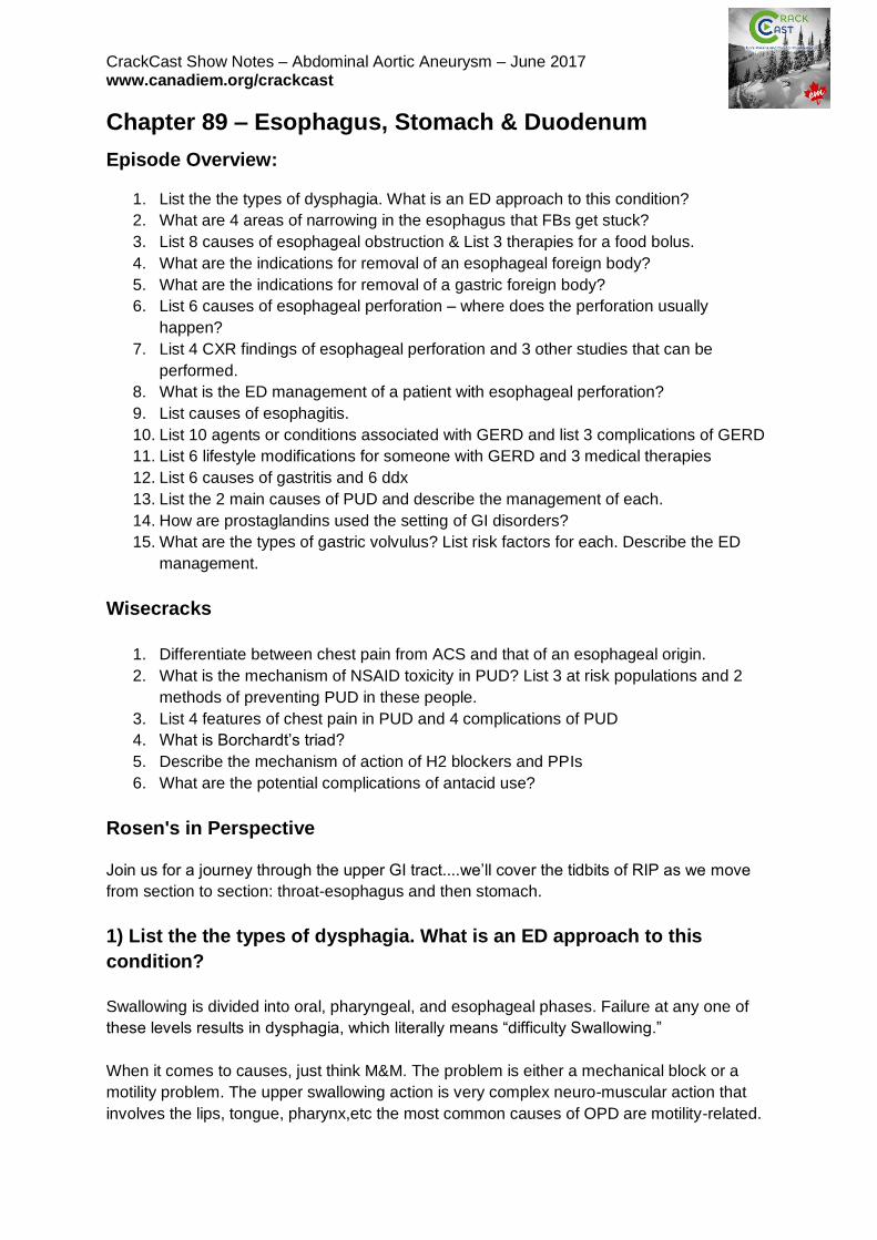
CrackCast Show Notes – Abdominal Aortic Aneurysm – June 2017 www.canadiem.org/crackcast
Chapter 89 – Esophagus, Stomach & Duodenum
Episode Overview:
1. List the the types of dysphagia. What is an ED approach to this condition?
2. What are 4 areas of narrowing in the esophagus that FBs get stuck?
3. List 8 causes of esophageal obstruction & List 3 therapies for a food bolus.
4. What are the indications for removal of an esophageal foreign body?
5. What are the indications for removal of a gastric foreign body?
6. List 6 causes of esophageal perforation – where does the perforation usually
happen?
7. List 4 CXR findings of esophageal perforation and 3 other studies that can be
performed.
8. What is the ED management of a patient with esophageal perforation?
9. List causes of esophagitis.
10. List 10 agents or conditions associated with GERD and list 3 complications of GERD
11. List 6 lifestyle modifications for someone with GERD and 3 medical therapies
12. List 6 causes of gastritis and 6 ddx
13. List the 2 main causes of PUD and describe the management of each.
14. How are prostaglandins used the setting of GI disorders?
15. What are the types of gastric volvulus? List risk factors for each. Describe the ED
management.
Wisecracks
1. Differentiate between chest pain from ACS and that of an esophageal origin.
2. What is the mechanism of NSAID toxicity in PUD? List 3 at risk populations and 2
methods of preventing PUD in these people.
3. List 4 features of chest pain in PUD and 4 complications of PUD
4. What is Borchardt’s triad?
5. Describe the mechanism of action of H2 blockers and PPIs
6. What are the potential complications of antacid use?
Rosen's in Perspective
Join us for a journey through the upper GI tract....we’ll cover the tidbits of RIP as we move
from section to section: throat-esophagus and then stomach.
1) List the the types of dysphagia. What is an ED approach to this
condition?
Swallowing is divided into oral, pharyngeal, and esophageal phases. Failure at any one of
these levels results in dysphagia, which literally means “difficulty Swallowing.”
When it comes to causes, just think M&M. The problem is either a mechanical block or a
motility problem. The upper swallowing action is very complex neuro-muscular action that
involves the lips, tongue, pharynx,etc the most common causes of OPD are motility-related.

CrackCast Show Notes – Abdominal Aortic Aneurysm – June 2017 www.canadiem.org/crackcast
a. List 4 causes of oropharyngeal dysphagia: “immediate or transfer
dysphagia”
i. Neuromuscular - swallowing muscles don’t work
1. Due to a cerebrovascular accident - #1 cause!
2. Degenerative aging:
a. Alzheimer's, ALS, Diabetic neuropathy, muscular
dystrophy, Parkinson's, Brain tumour
3. Immunologic - poly/dermatomyositis / MS / Myasthenia
Gravis / scleroderma
4. Infectious - botulism, diphtheria, polioMyelitis, rabies,
Sydenham’s chorea, tetanus
5. Metabolic - thyrotoxicosis, lead poisoning, Mg deficiency
ii. Obstructive
1. Dysphagia lusoria
b. List 6 causes of esophageal dysphagia. “Delayed / gets stuck”
i. Again think M&M
1. Mechanical
a. Intrinsic issue: strictures, webs, rings, tumours,
i. EsophagitIs, ***foreign bodies***
b. Extrinsic compression: osteopHytes, ***mediastinal
masses***, aortic aneurysm, thyroid goitre
2. Motility
a. Intrinsic: Achalasia, diffuse esophageal spasm,
hypertensive LES, scleroderma, CREST syndrome,
nutcracker esophagus
b. Extrinsic:
i. Gastric volvulus
ii. Alcoholism, diabetes, GERD,
Key History points to ask:
● Immediate vs. Delayed swallowing difficulty?
● Solids, liquids, or both?
● Intermittent or progressive symptoms?
● Associated symptoms, GI history, family history?
Key physical exam:
● HEENT
● Observed swallowing attempt
○ People with neuromuscular disease can’t swallow while prone (they depend
on gravity to do the work!)
ii. Oropharyngeal - OPD
1. Hx:
a. “Can’t begin / initiate a swallow; sputter and gag;
choke; drool; regurgitate through the nose”
CrackCast Show Notes – Abdominal Aortic Aneurysm – June 2017 www.canadiem.org/crackcast
b. **repeated swallowing attempts**
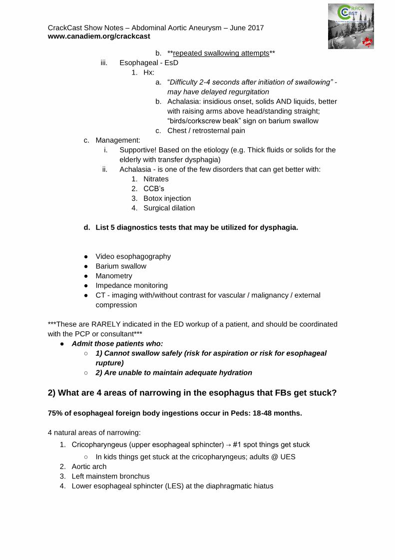
iii. Esophageal - EsD
1. Hx:
a. “Difficulty 2-4 seconds after initiation of swallowing” -
may have delayed regurgitation
b. Achalasia: insidious onset, solids AND liquids, better
with raising arms above head/standing straight;
“birds/corkscrew beak” sign on barium swallow
c. Chest / retrosternal pain
c. Management:
i. Supportive! Based on the etiology (e.g. Thick fluids or solids for the
elderly with transfer dysphagia)
ii. Achalasia - is one of the few disorders that can get better with:
1. Nitrates
2. CCB’s
3. Botox injection
4. Surgical dilation
d. List 5 diagnostics tests that may be utilized for dysphagia.
● Video esophagography
● Barium swallow
● Manometry
● Impedance monitoring
● CT - imaging with/without contrast for vascular / malignancy / external
compression
***These are RARELY indicated in the ED workup of a patient, and should be coordinated
with the PCP or consultant***
● Admit those patients who:
○ 1) Cannot swallow safely (risk for aspiration or risk for esophageal
rupture)
○ 2) Are unable to maintain adequate hydration
2) What are 4 areas of narrowing in the esophagus that FBs get stuck?
75% of esophageal foreign body ingestions occur in Peds: 18-48 months.
4 natural areas of narrowing:
1. Cricopharyngeus (upper esophageal sphincter) → #1 spot things get stuck
○ In kids things get stuck at the cricopharyngeus; adults @ UES
2. Aortic arch
3. Left mainstem bronchus
4. Lower esophageal sphincter (LES) at the diaphragmatic hiatus

CrackCast Show Notes – Abdominal Aortic Aneurysm – June 2017 www.canadiem.org/crackcast
3) List 8 causes of esophageal obstruction & list 3 therapies for a food
bolus.
Clinical presentation:
● Dysphagia, odynophagia, neck/chest pain / epigastric pain
● Complete obstruction = unable to swallow secretions (spit basin); choking; stridor;
cough
○ Cafe’ coronary: sudden cyanosis and collapse after a piece of swallowed food
lodges in the airway leading to hypoxic arrest/syncope
○ Steakhouse syndrome: large piece of food (steak) causes complete
obstruction of the distal esophagus. (fish bones RARELY obstruct the
esophagus)
● Partial obstruction = usually the case in children, who present atypically:
○ Fever, wheezing, stridor, rhonchi, poor feeding
Causes of esophageal obstruction:
1. Mechanical obstruction:
a. Internal
i. Foreign body
1. Adults - meat/bones; dentures; pen caps, etc.
2. Kids - coins/batteries
ii. Strictures / rings (Schatzki’s ring) / webs
iii. Eosinophilic esophagitis
iv. Malignancy
b. External factors
i. Large Left atrium / ventricle
ii. Goiter
iii. Mediastinal tumour
2. Motor factors (remember than the distal 2/3rds are innervated by involuntary
smooth muscle)
a. Achalasia
b. Infectious (botulism, tetanus, etc.)
Food bolus therapies: (from Ep 60)
● Glucagon (0.5 to 2 mg) IV (Glucagon receptor agonist) – Theoretical action is
lowering the smooth muscle tone at the lower esophageal sphincter without
inhibiting normal esophageal peristalsis. Data is unsupporting of its use in
modern treatment, and it is NO longer recommended.
● Benzodiazepine (GABA agonist): Anxiolytic +/- improves effect of Glucagon.
Probably safe, but not shown to be better than placebo.
● Nitroglycerin (Nitric oxide which is a potent vasodilator) thought to work to relax
lower esophageal sphincter tone
● Nifedipine (Calcium Channel Blocker) – thought to work to relax lower
esophageal sphincter tone
● Enzymatic degradation w/ proteolytic enzyme papain or coca-cola – ineffective
and has fallen out of favour
CrackCast Show Notes – Abdominal Aortic Aneurysm – June 2017 www.canadiem.org/crackcast In summary; Do not use:
○ Glucagon
○ Meat tenderizer (papain)
○ Effervescent agents
4) What are the indications for removal of an esophageal foreign body?
We covered some of this previously: https://canadiem.org/crackcast-e060-foreign-bodies/
● First you have to diagnose the foreign body:
○ AP and lateral radiographs of: neck soft tissues/chest/abdomen (as
applicable)
○ Nasopharyngoscopy → laryngoscopy → endoscopy
○ Contrast swallow studies - are high risk for aspiration / perforation
○ CT - more sensitive for that elusive fish bone/chicken bone or to look for signs
of inflammation/free air.
INDICATIONS FOR REMOVAL: (endoscopy, foley, Magill, etc).
● Button batteries (can cause severe tissue damage in 2 hrs!)
● Large objects (e.g. dentures, tooth brush)
● Sharp objects
● Coins lodged in the proximal esophagus
● Complete esophageal obstructions (preventing liquid past)
○ Incomplete obstructions must be removed in 24 hrs
Three mechanisms by which button batteries cause damage:
● Leakage of alkaline electrolyte
● Pressure necrosis
● Generation of electrical current - causing electrolysis of tissue fluids
5) What are the indications for removal of a gastric foreign body?
● Most can be observed conservatively, however if they have any of these features
they may need to be removed:
○ Longer than 5 cm
○ Wider than 2.5 cm (rarely leave the duodenum)
○ Sharp and pointed objects (high risk of intestinal perforation)
○ Objects that remain in the stomach for more than 3-4 weeks
○ Objects that stay in the same intestinal location for 1 week
CrackCast Show Notes – Abdominal Aortic Aneurysm – June 2017 www.canadiem.org/crackcast
6) List 6 causes of esophageal perforation – where does the perforation
usually happen?
● The esophagus has two layers: inner circular layer of muscle & the outer longitudinal
layer of muscle
● Clinical presentation: odynophagia, fever, decreased PO intake, chest pain, dyspnea,
hoarseness, subcutaneous air
● Causes:
○ Food stuffs:
■ Chicken / fish / animal bones
■ Large unchewed pieces (e.g. steak)
○ Button batteries
○ Coins
○ Forceful valsalva:
■ Vomiting
■ Childbirth
■ Coughing / heavy lifting
○ Iatrogenic:
■ Post EGD
■ Post NG placement
■ Post endotracheal intubation
○ Caustic substance ingestion
○ Severe esophagitis
○ Blunt / penetrating trauma
○ Carcinoma
90% of spontaneous perforations happen in the distal esophagus; other sites:
pharyngoesophageal junction.
Iatrogenic injuries happen at the esophagogastric junction.
7) List 4 CXR findings of esophageal perforation and 3 other studies that
can be performed.
CXR:
● Pneumothorax
● Subcutaneous air / emphysema
● Hydropneumothorax
● Pneumomediastinum
● Pleural effusions
● Pulmonary infiltrates
● Wide mediastinum
CrackCast Show Notes – Abdominal Aortic Aneurysm – June 2017 www.canadiem.org/crackcast Other studies:
● Abdominal radiography
● Gastrograffin swallow (water soluble agent)
● CT chest
● Flexible EGD - 100% sensitivity (although has a risk of enlarging a minimal
transmural opening due to insufflation of air)
8) What is the ED management of a patient with esophageal perforation?
Mackler’s triad: subcutaneous emphysema, chest pain, vomiting = esophageal perforation
Misdiagnosis of people with esophageal perforation as ACS/PE/Aortic dissection/perforated
ulcer/pancreatitis/pneumothorax/lung abscess - due to chest and abdominal pain!
Management:
● Broad spectrum abx
● Admission
● NPO
9) List causes of esophagitis.
Symptoms: dysphagia, odynophagia, chest pain, bleeding → hemorrhage → perforation!
● GERD (#1 cause)
● Eosinophilic esophagitis
○ Unknown etiology; typically in non-responders to PPI’s and kids with atopy.
Don’t respond to high dose PPIs, may need steroids inhalers.
● Infection
○ Usually an immunocompromised (steroid use, ETOH, immunosuppression, or
HIV seroconversion syndrome) person developing Candida (but can also be
CMV/HSV). Usually have odynophagia not improved with antacids.
● Foreign body / toxic ingestion
○ Aka. Pill esophagitis - from large pills sticking to the esophageal wall. Can
lead to erosions/ulcer/strictures.
● Post-radiation
10) List 10 agents or conditions associated with GERD and list 3
complications of GERD
Some physiologic GER occurs in everyone, when it becomes symptomatic then it is “GERD”.
Conditions associated with GERD: (ie. anything that causes the LES to relax of become
overwhelmed) - this comes from Box 79-2
● Decreased LES pressure
○ Meds:
■ CCB, nitrates, anticholinergics, albuterol, benzos,
○ Fatty meals / Chocolate / Caffeine / Ethanol / Peppermint / nicotine
CrackCast Show Notes – Abdominal Aortic Aneurysm – June 2017 www.canadiem.org/crackcast
○ Pregnancy
○ Estrogen / progesterone
● Increased gastric emptying time / Increased intra-abd. Pressure
○ Anticholinergic drugs
○ Pregnancy
○ Obesity
○ Coughing / bending /
○ Supine position
○ Gastroparesis / neuromuscular disease leading to gastric outlet obstruction
● Decreased esophageal motility
○ Achalasia
○ DM
○ Scleroderma
GEeRD = “gastric emptying; esophageal relaxation drugs” mnemonic
Complications of GERD
1. Esophagitis
○ Progressing to Barrett’s metaplasia, erosion, ulcerations, scarring
○ Stricture formation due to persistent scarring and inflammation
2. Reflux induced asthma
○ Microaspiration of
3. Adenocarcinoma of the esophagus
4. Esophageal perforation → mediastinitis
11) List 6 lifestyle modifications for someone with GERD and 3 medical
therapies
Lifestyle mods:
● Avoidance of high risk foods (caffeine, chocolate, alcohol, fatty, spicy foods)
● Weight loss*
● Smoking cessation
● Elevation of the head of bed, and avoidance of lying down after meals*
● Exercise
● Alcohol cessation
Therapies:
● Acid neutralization
○ Milk of magnesia
○ TUMS
○ Pepto Bismol - bismuth salts
● Decrease acid production (most effective)
○ PPIs
■ Pantoprazole 20 - 40 mg PO - daily or BID before meals for 4-8 wks.
○ H2 blockers

CrackCast Show Notes – Abdominal Aortic Aneurysm – June 2017 www.canadiem.org/crackcast
■ Ranitidine 150 mg PO
● Strengthening LES / motility amplification
○ Maxeran 10 mg
● Mucosal protection
○ Sucralfate 1 g - q 6 hrs (safe in pregnancy!)
○ Ideally given 30-60 mins before meals.
12) List 6 causes of gastritis and 6 DDx
True gastritis is a histology diagnosis.
Typically, we use the term clinically. Usually mild-moderate burning pain or bloating
epigastrically. Few diagnostic tests are needed (but you need to exclude other things on the
ddx!)
Causes:
1. Acute Helicobacter pylori infection
2. Acute phlegmonous gastritis (rare, but lifethreatening!)
3. Shock states
4. Lymphoma
5. Adenocarcinoma
6. NSAID use
7. ETOH abuse / use
8. Drugs:
a. 5FU, bisphosphonates, iron
9. Smoking
10. Bile / pancreatic secretions
11. Stress
12. Glucocorticoids
DDx:
● PUD
● Pancreatitis
● Mesenteric ischemia
● Biliary disease
● ACS - angina
● Pericarditis
● SBO

CrackCast Show Notes – Abdominal Aortic Aneurysm – June 2017 www.canadiem.org/crackcast
13) List the 2 main causes of PUD and describe the management of
each.
1. H. Pylori infection - #1 cause
○ 70% of duodenal/gastric ulcers thought to be caused by this!
○ Oral-oral route/fecal-oral route/iatrogenic
○ Up to 40% of the population is infected with this bacteria (but only 5-
10% of people infected seem to get ulcers)
○ Testing: serum antibody test, stool antigen testing, breath test,
mucosal bx.
○ Management:
i. Triple therapy:
1. Clarithromycin 500 mg po BID for 10 days
2. Amoxicillin 1 g bid PO for 10 days
3. A PPI for 4-8 weeks.
4. (USE metronidazole to replace #1,2 for penicillin
allergy)
ii. Some ppl need quadruple therapy-
bismuth/metronidazole/tetracycline/PPI
iii. Commercial combination packs exist (Prevpac)
iv. Use adjunct agents for GERD/gastritis
2. NSAID associated (chronic)
○ Due to suppressed gastric prostaglandin synthesis
○ Varying risks for ulceration based on NSAID type and co-use of
glucocorticosteroids, anticoagulants
○ Management:
i. Stop NSAID use
ii. PPI for 4-8 weeks
iii. Adjunct agents for GERD/gastritis
3. < 1% of PUD is caused by Zollinger-Ellison syndrome
4. Systemic illness (especially in infants and children)
Adjuncts:
● Prostaglandins (misoprostol)
● Sucralfate
● Bismuth salts
● Stop all GERD / gastritis causing foods/lifestyle choices/meds
Reasons to referral to GI:
● Age >55 yrs with:
○ New onset
■ Dyspepsia
■ Progressive weight loss
■ Persistent vomiting
■ Iron deficiency anemia
■ Epigastric mass
CrackCast Show Notes – Abdominal Aortic Aneurysm – June 2017 www.canadiem.org/crackcast
14) How are prostaglandins used the setting of GI disorders?
Prostaglandins protect the gastric mucosa! (Inhibit acid secretion, increase
mucous/bicarbonate/stimulate blood flow).
Misoprostol (an analogue of prostaglandin) - used for high risk patients - to prevent
progression of NSAID-induced ulcers.
15) What are the types of gastric volvulus? List risk factors for each.
Describe the ED management.
This is a life-threatening closed-loop obstruction of the stomach! Because the stomach is
only fixed a two points - it can easily twist on itself.
Defined by a few different ways:
1. Cause: primary vs. secondary
a. Primary:
i. subdiaphragmatic - stabilizing ligaments are too lax.
b. Secondary:
i. Supradiaphragmatic - due to diaphragmatic defects (hiatal hernia,
gastric ulcer/cancer, diaphragm paralysis, and other).
2. Anatomy: axis of rotation
a. Twisting on its long axis (organoaxial)
b. Twisting on its short axis (mesenteroaxial)
3. Onset: acute vs. chronic
Look for the triad: severe epigastric pain and distension; vomiting with violent retching;
inability to pass an NG tube.
In children: look for sudden life-threatening events with apnea/cyanosis/respiratory distress!
Risk factors:
● Age 40-50 yrs
● Paraesophageal hernia
● **20% of cases occur in infants due to a congenital diaphragmatic defect (secondary)
Management:
● This disease can result in gastric ischemia, perforation and death.
Goal is REDUCTION!
● Attempt NG tube placement
● May go for attempted endoscopic reduction vs. immediate surgery! (call both!)
CrackCast Show Notes – Abdominal Aortic Aneurysm – June 2017 www.canadiem.org/crackcast
Wisecracks
1) Differentiate between chest pain from ACS and that of an esophageal
origin.
Esophageal --overlapping symptoms/signs--
ACS
● prolonged, non exertional pain that:
○ Interrupts sleep / lying down
○ Is related to meals
○ Relief with antacids
○ Regurgitation ○ DysphaGia ○ Worse with
swallowing
substernal chest pain Crushing, squeezing pain
Radiation to neck, shoulder, arm
Response to nitroglycerin
● exertional symptoms ● Substernal
dyscomfort ● Dyspnea ● (Atypical anginal
symptoms may mislead us!)
These are clues, NOT slam-dunk rules! Think worst-first!
Once a workup is “negative” for the killers - then consider a GI source of the symptoms!
(Gastritis, esophagitis, PUD, biliary tract disease).
2) What is the mechanism of NSAID toxicity in PUD? List 3 at risk
populations and 2 methods of preventing PUD in these people.
See above!
● Prostaglandin inhibition due to COX inhibition!
● NSAID users, age > 60, hx of PUD, steroid/anticoag use
● Change to lower risk NSAID / stop use
● Concurrent PPI / acid suppressants.
3) List 4 features of chest pain in PUD and 4 complications of PUD
● Burning , gnawing epigastric pain
● Fullness, nausea, early satiety, bloating
● Onset of pain 2-5 hrs post meal, OR worse around 2 am in the morning (when acid
production is at its highest!)
● Relief with eating (pain eating relief)
CrackCast Show Notes – Abdominal Aortic Aneurysm – June 2017 www.canadiem.org/crackcast (VomitiNG is atypical!!) - so think of other things: gastric volvulus, outlet obstruction, SBO,
pancreatitis, biliary disease.
● Complications
○ Hemorrhage
○ Perforation (duodenal ulcers)
○ Penetration (e.g. posteriorly into the retroperitoneum)
○ Gastric outlet obstruction
4) What is Borchardt’s triad?
“This condition may present as an acute abdominal emergency or a chronic cause of upper
abdominal discomfort. Borchardt's triad is believed to be diagnostic for acute gastric
volvulus and consists of unproductive retching, epigastric pain and distention, and
the inability to pass a nasogastric tube. Carter et al suggest three additional findings that
may be very suggestive of gastric volvulus: minimal abdominal findings when the stomach is
in the thorax; a gas-filled viscus in the lower chest or upper abdomen on chest radiograph,
especially when associated with a paraesophageal hernia; and obstruction at the site of the
volvulus shown by upper gastrointestinal (GI) series. Mortality from acute gastric volvulus
has been reported to be as high as 30–50%. As a result, quick recognition and prompt
surgical correction remain key in therapy for acute gastric volvulus.”
Copied from: https://www.ncbi.nlm.nih.gov/pmc/articles/PMC3072542/
5) Describe the mechanism of action of H2 blockers and PPIs
Histamine - is the primary stimulus for gastric acid secretion. H2 blockers attach onto the H2
receptor of the parietal cell and prevent the release of hydrochloric acid.
The parietal cell’s proton pump (H, K, ATP-ase) that produces HCl is irreversibly blocked by
PPI’s. Very effective - production of acid can be reduced up to 95%. Require regular use to
achieve therapeutic benefit - (if a patient is taking drugs on a PRN basis a H2 blocker would
be a better choice).
These therapies are novel - because ulcers can’t develop in the absence of acid!
6) What are the potential complications of antacid use?
● Magnesium containing antacids:
○ Can cause diarrhea
● Calcium containing antacids: watch for milk-alkali syndrome
○ Acid rebound
○ Constipation
○ Hypercalcemia
○ Alkalosis
○ Renal insufficiency

CrackCast Show Notes – Abdominal Aortic Aneurysm – June 2017 www.canadiem.org/crackcast
● Aluminum containing antacids:
○ Constipation
○ Electrolyte abnormalities
● Decrease Absorption of: (Reduce this by taking them 1-3 hrs post meals)
○ Warfarin
○ Digoxin
○ Antibiotics,
○ Anticonvulsants