Embed Size (px)
Citation preview

Grand Round July 29, 2010
Amy Kulak,MD PGY3

Case Presentation
CC: “increasing size of left eye HPI:46 y.o Female c/o of left eye
“swelling” and associated with burning and tearing x 4 days.
Patient was brought to ER by family because of an episode of syncope with AMS, weakness and abdominal pain.
Interpersonal and communication skills, patient care

Case Presentation PMH: HIV+ x 12 years, recently started on
HAART,?CD4 unknown by patient but last confirmed as <200
Hx of +PPD, treated with INH Psychiatric disorder POH: None ALL: NKDA Sochx: ex-smoker Famhx: no glc or blindness Meds: Truvada, seroquel, zolpidem, Dronabinol
Interpersonal and communication skills, patient care

EXTERNAL PHOTO

Case Presentation
Nvasc: 20/25, 20/30 (+2.00)
P: 4-2mm, no APD CVF: full ou EOM: Full OD, OS: -1 limitation medial
and -1/2 lateral gaze, -1/2 up and downgaze Patient denies diplopia or
pain with eye movement
Color: Full ou Hertels: 122: 19,24 Tpen: 14, 18 @ 7pm
+resistance to retropulsion
Interpersonal and communication skills, patient care
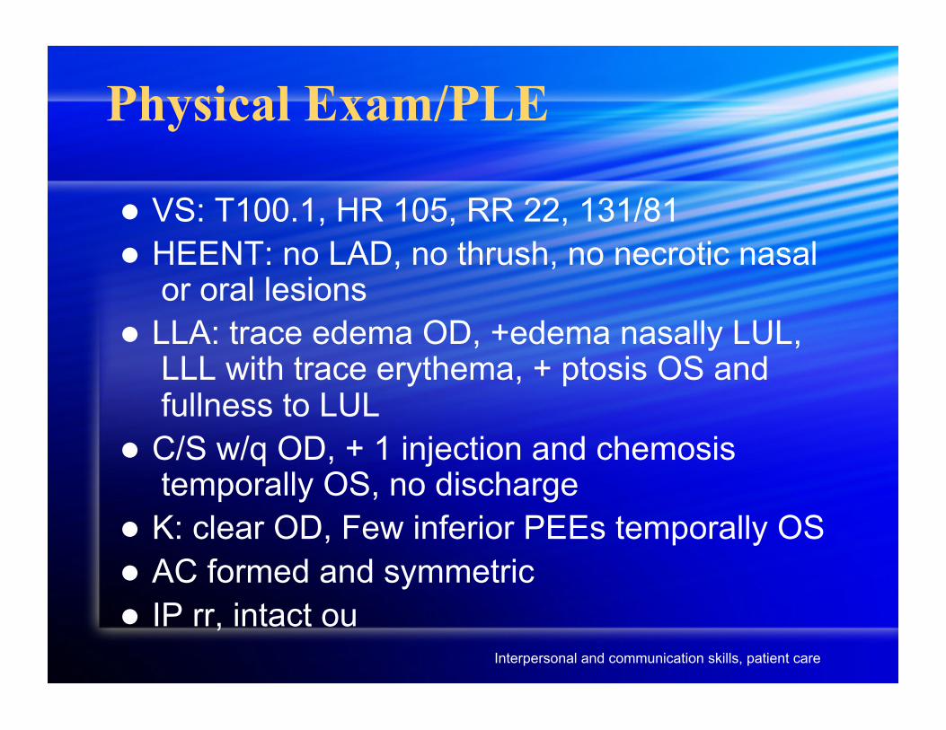
Physical Exam/PLE
VS: T100.1, HR 105, RR 22, 131/81 HEENT: no LAD, no thrush, no necrotic nasal
or oral lesions LLA: trace edema OD, +edema nasally LUL,
LLL with trace erythema, + ptosis OS and fullness to LUL
C/S w/q OD, + 1 injection and chemosis temporally OS, no discharge
K: clear OD, Few inferior PEEs temporally OS AC formed and symmetric IP rr, intact ou
Interpersonal and communication skills, patient care

DFE
V: clear ou D: s/p ou, c/d 0.4 ou M: flat ou V; wnl OD, Mildly dilated OS P: WNL od, no heme, holes, tears or
chorioretinal folds ou
patient care

DDX for Unilateral Proptosis:

DDX unilateral proptosis Endocrine/Inflammatory
*Thyroid eye disease Orbital pseudotumor
Neoplastic: *orbital metastasis *lymphoma optic glioma meningioma hemangioma Hemangioblastoma orbital rhabdomyosarcoma leukemia
Misc: Pseudoproptosis contralateral enophthalmos
Infectous Orbital cellulitis *Orbital abscess +Mucormycosis
Trauma retrobulbar hematoma
Vascular: Carotid art-cavernous sinus fistula cavernous sinus thrombosis
Granulomatous Wegners Histiocytosis Hand-Schullar Christian syndrome
Congenital: orbital dermoid cavernous lymphangioma

More history…
On ROS patient reports constitutional symptoms including weakness, night sweats and weight loss of 15lbs/3 weeks and loss of appetite
Patient also reports BRBPR over last few months which she attributed to hemorroids
Interpersonal and communication skills, patient care

What are our next steps???
Laboratory workup: cbc, cmp, coags, TFT, CD4 /Tcell subset, viral load, blood cultures x 2, amylase, lipase, hemoccult
Imaging: MRI or CT orbits, CT abd/pelvis/chest
Medical Knowledge, patient care

Labs:
WBC: 2.9 (L) H/H: 8(↓13)/25.5 Plat:249 Na: 135, K:4.6, cl:107, bun:12, Cr: 0.6 Amylase: 118, Lipase: 391 Hemoccult + stool TFT; normal
Medical Knowledge, patient care

Imaging

What next???

HOSPITAL COURSE Patient was transfused 3 units of PRBC for anemia Patient was scheduled for an MRI but unfortunately
refused test CT scan of abd/pelvis
revealed several scattered low attenuation lesions in liver (some>3cm), an 18mm lesion in head of pancreas, and L hilar kidney mass, and retroperitoneal lymphadenopathy. Patient also had thickening of folds of stomach wall.
Patient had mild SOB, TTE performed and revealed moderate size pericardial effusion w/o evidence of tamponade.CT surgery consulted- no intervention
Patient was scheduled for EGD/Colonoscopy by GI service for possible GI bleed
Patient was scheduled by our service for L orbitotomy/ biopsy of orbital mass.
Medical Knowledge, patient care

Surgical Pathology
Orbital tissue: High grade Malignant B-cell lymphoma
EGD: revealed several friable masses in body, antrum of stomach extending into duodenal bulb. Surgical path also consistent with high grade malignant B cell lymphoma
Immunohistochemistry : +CD10, Bcl-6, CD20, and Ki-67, Neg: Bcl-2, CD 3
Final dx: Burkitt Lymphoma vs Intermediate Diffuse Large B-cell Lymphoma
care
Professionalism, Medical Knowledge, patient care

Orbital Lymphoma and AIDS

Ocular manifestations
There is a long list of ocular manifestations associated with HIV that affect eyelids, orbit, adnexa, anterior and posterior segments
Orbital involvement, with HIV is seen less commonly 1) infectious: Aspergillus, Mucor, Toxoplasma gondii, and Pneumocystis carinii. HIV infected Children may present with recurrent episodes of orbital/peri-orbital cellulitis. 2) orbital lymphoma
Medical Knowledge, patient care

Background The association between Non Hodgkin
lymphoma and AIDS has been well recognized. However, orbital involvement is rare
HIV -associated lymphoma was first incorporated in the US CDC definition as ands AIDS defining illness in 1985
Before HAART, accounted for 3-4% of all AIDS defining illness (reported to CDC)
In the HAART era, steady 1.6% Without effective ART, CDC estimated that
5-10% of all HIV-infected individuals will have lymphoma as either an initial or subsequent AIDS defining illness
Medical Knowledge, practice based learning

Background…
Approximately 95% of HIV associated lymphomas are B cell origin ( CD 19, CD20 +) and Non-Hodgkin’s
1991 review(2,500 cases of HIV-1-associated lymphomas revealed that approximately 80% arose in the periphery 20% arose in the central nervous system (CNS)
This distribution of lymphomas remains the same in the era of highly active antiretroviral therapy era
Systems based practice, practice based learning

Non Hodgkin lymphoma:
NHL is a malignant neoplasm derived from a clonal proliferation of B or T Lymphocytes
There are more than 40 different subtypes, which can arise in extranodal tissue and in lymph nodes
In general, orbital lymphomas account for ~2% of all NHL
Fourth most common malignancy among male and females
Systems based practice, practice based learning
NHL orbital lymphoma
REAL ( Revised European American Lymphoma) the 4 most common orbital lymphomas in the general population:
1)MALT( Mucosa-Associated Lymphoid Tissue) 40-60%
2)CLL -Chronic Lymphocytic Lymphoma 3)Follicular center lymphomas 4)High grade lymphomas (Mantle, Diffuse large
cell, Burkitt, B-ALL)
Medical knowledge, systems based practice

Pathogenesis
AIDS related lymphoma is thought to arise as a consequence of long-term stimulation and proliferation of lymphocytes due to HIV virus itself
The reactivation of prior EBV infection secondary to immunosuppression likely plays a role.
Activation of c-myc and bcl oncogene is probably involved in the pathogenesis
( C-MYC translocaton 8:14 in Burkitt’s)
Medical knowledge

Clinical presentation of orbital lymphoma Gradual, painless palpable mass in orbit or
eyelid (In the high grade , HIV associated lymphomas, onset may be more rapid)
Proptosis Eyelid or periorbital swelling Ptosis Excessive tearing Diplopia Limitations of EOM Blurry vision +APD
Patient care, professionalism, interpersonal and communication skills

Clinical features of HIV associated lymphoma and Epidemiology Cummulative risk for lymphoma is 3-8% in
AIDS patients 1-3% of AIDS patients with NHL will have
orbital Rapid and aggressive course
which may be interpreted initially as infectious or inflammatory
Much higher incidence of extranodal involvement in HIV infected patients with NHL approx 78%-98%(Reifler et al1994)
As compared to approx 50% (Hatef 2007) GI tract most common extranodal site
Patient care, professionalism, interpersonal and communication skills

Imaging: CT ORBITs : *classically will reveal “puttylike”
molding of the tumor to normal structures.
*Bone erosion or infiltration is rare except sometimes in high grade malignant lymphomas. *Up to 50% orbital lymphoproliferative lesions arise from lacrimal fossa
In AIDs associated: -Indentation and/or globe displacement. -Bony destruction -extension into paranasal sinus
Diagnosis
Open biopsy is preferred
Patient care, medical knowledge

Histopathologic features of AIDS associated lymphomas 62-81% AIDS patients have high grade NHL Approximately 75-80% of these lymphomas
are classified histologically as large-cell lymphomas and the remaining, 20-25% as Burkitt (or small-cell)
Compared to general population: Burkitts makes up <10 % of intermediate and high grade (remember most NHL are low grade)
Patient care, professionalism, interpersonal and communication skills

Burkitt’s Lymphoma “starry sky” appearance Neoplastic Bcells
(purple) with macrophages (white
Sm to med size lymphocytes
Mitotic figures Round to oval vesicular
nuclei 1-3 prominent nucleoli
Patient care, medical knowedge
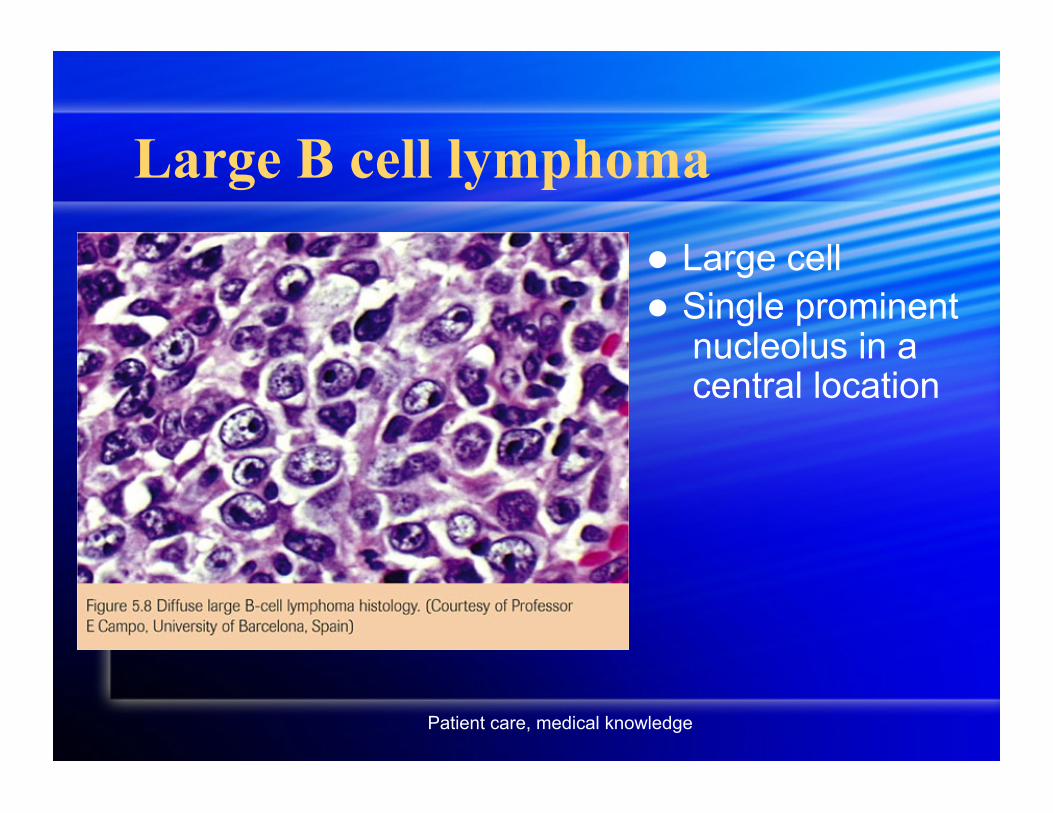
Large B cell lymphoma Large cell Single prominent
nucleolus in a central location
Patient care, medical knowledge
Treatment Treatment depends on many factors, type of
lymphoma, morphologic and pathologic features, systemic involvement, CD 4 count, Most importantly, the patient’s and family’s wishes
Prognosis is generally poor Initial attempts to treat High grade
lymphomas in HIV patients aggressively resulted in many patients succumbing to fatal relapses, opportunistic infections, and had a decreased quality of life
Generally, modified form of chemotherapy with lower dose +/- radiation maybe offered
HAART therapy is usually started after chemo/radx
Patient care, professionalism, interpersonal and communication skills
Back To Our Patient… Patient was counseled on her diagnosis and
prognosis and oncology was consulted in house.
Patient was discharged a few days later after her condition stabilized. Repeat TTE showed no increased size in pericardial effusion. Her H/H remained stable and patient did not require further transfusion.
Unfortunately, oncology follow-up notes have not been made available in the patient’s medical record from SUNY downstate as of yet.
Patient missed her follow-up with ophthalmology
Interpersonal and communication skills, patient care

Reflective Practice
Even in the “HAART era” we still encounter numerous patients with ophthalmic manifestations associated with HIV
To consider HIV associated lymphoma when presented with HIV positive patient with unilateral proptosis, regardless of CD4 count
I feel this patient was managed appropriately and in a timely manner.

Core Competencies Patient Care- compassion and appropriate management
was displayed Interpersonal and communication skills: explained to
patient and family members in non-medical terminology our recommendations.
Medical knowledge: reviewed clinical and scientific features of orbital lymphoma and sought appropriate expertise from other services, such as oncology and radiology.
Professionalism: this case stressed the importance of a multidisciplinary approach. Informed consent properly obtained prior to procedures.
Practice based learning: used evidence based medicine to manage patient care
Systems based practice: appropriate tests and diagnostic studies were selected based on necessity to appropriately manage patient

References BSCS: section 7 Orbits, Eyelids and Lacrimal System 2009-2010. AAO
D. Reifler MD FACS, M. Warznarski, PhD, Warren. Blount MD, D. Graham MD, K.Mill MD . Orbital Lymphoma Associated with Acquired Immune Deficiency Syndrome (AIDS) SURVEY OF OPHTHALMOLOGY VOLUME 38(4). JANUARY-FEBRUARY 1994
FM Rahhal, Daniel Rosberger, MH Heinemann. Aggressive orbital lymphoma in AIDS. British J of Ophthalmology 1994; 78(319-321)
Lene Sjo,Eisabeth Rifkiaer, Jan Prause, Jorgen Petersen, Jakob Madsen, Niels Pedersen, Steffen Heegaard. Increasing Incidence of Ophthalmic Lymphoma in Denmark from 1980 to 2005. IOVS August 2008. Vol 49 (8).
Elham Hatef, MD, Dianna Roberts, PHD Peater Mclaughlin, MD, Barbara Pro, MD; Bita Esmaeli, MD. Prevalence and Nature of Systemic Involvement and Stage at Initial Examination in Patients with Orbital and Ocular Adnexal Lymphoma. Arch. Ophthalmol Dec 2007. Vol 125(12).
R.I. Aviva,*, K. Miszkielb PICTORIAL REVIEW Orbital imaging: Part 2. Intraorbital pathology The National Institute for Neurology and Neurosurgery and Moorfields Eye Hospital, London, UK Clinical Radiology (2005) 60,
288-307 "