Embed Size (px)
Citation preview
Hearing Research 73 (lYY4) 190-104 ELSEVI ER
Covariation of binaural, concurrently-measured spontaneous otoacoustic emissions
M.J. Penner a,*, S.E. Brauth a, Pawel J. Jastreboff ’
’ Psychology Department, University of Maryland, College Park, Maryland 20742, USA; h Uniczrsity of Marylund School of Mdicine. Baltimore. h4myland 21201, USA
(Received 3 July 1993; Revision received 22 September 1993: Accepted 26 October 1993)
Abstract
Simultaneous recordings of binaural spontaneous otoacoustic emissions (SOAEs) were made for 2 female subjects. Fol SOAEs below about 3.6 kHz measured within a testing session, the frequencies of nearby monaural and binaural SOAEs tended to move in tandem, whereas widely separated SOAEs did not. Across many testing sessions spanning a menstrual cycle, all monaural and binaural SOAE frequencies shifted in tandem. Possible mechanisms consistent with these results are discussed.
Key words: Binaural SOAEs; Efferent system; Middle-ear muscles
1. Introduction
Spontaneous otoacoustic emissions (SOAEs), sounds which may be recorded when a sensitive miniature microphone is inserted into the ear canal, are thought to result from active biomechanical elements in the cochlea (Bialek and Wit, 1984; Kemp, 1979; Kim, 1986). Although some instability in SOAEs might be expected from alterations in cochlear properties (Keefe et al., 1990; Long et al., 1991; Talmadge et al., 1991; Zurek, 19811, the frequency and amplitude of SOAEs may additionally be influenced both by efferent activity (Mott et al., 1989) and by cardiovascular changes which may be linked to circadian (Bell, 1992; Wit, 1985) and, for premenopausal women, menstrual (Bell, 1992; Hag- gerty, 1990; Penner and Glotzbach, 1994) rhythms in the frequency of an SOAE.
Instabilities in one cochlea may be observed with a single microphone (Zurek, 1981). Although Whitehead (1991) has studied changes in SOAEs when a single microphone is moved from one ear to the other, the effects of the efferent system and cardiovascular changes might best be revealed by monitoring both ears simultaneously.
* Corresponding author.
0378-5955/94/$07X10 0 1994 Elsevier Science B.V. All rights reserved
SD1 0378-5955(93)EO180-J
In an attempt to explore the factors affecting SOAE variability, binaural SOAEs were simultaneously moni- tored (1) during one testing session and (2) during a menstrual cycle. To the extent that binaurally uncorre- lated alterations in cochlear processes mediate SOAE generation, the frequencies and amplitudes of binaural SOAEs would not be expected to change in tandem. On the other hand, if efferent activity, cardiovascular changes, or binaurally correlated peripheral changes influenced the emission generator, then the frequen- cies and amplitudes of binaural SOAEs would be ex- pected to shift in tandem.
2. Materials and methods
2. I. Subjects
Two female subjects were recruited for measure- ments of SOAEs made during a menstrual cycle. Each subject had clinically normal hearing (within 25 dB of ANSI 1970). Both subjects had been previously tested (Penner et al., 19931, and so were known to have binaural SOAEs.
2.2. Protocol
Subjects were seated in a double-walled sound-proof booth. An ER 10B microphone was placed in the left
and an ER 10 low-noise microphone was placed in the
right ear. The subject touched any key on a keyboard
interfaced to a computer to begin the simultaneous digitizations of both ear’s outputs. The lights on the keyboard were illuminated during the recording to inform the subject that data collection was in progress. After each digitization, the system paused and waited for the subject to touch another key to initiate the next recording.
For both subjects, testing constituted an initial ses- sion in which data for thirty binaural spectra were
collected with the digitizations spaced in as close tem-
poral proximity as possible, approximately, one spectra per minute. In addition, the SOAEs were measured once daily during a menstrual cycle and immediately preceding and following it. To avoid changes due to circadian rhythms, subjects were tested at approxi- mately the same time each day. For Subject 1, mea- surements were made between 4 and 5 p.m. For Sub- ject 2. measurements were made between 1 and 2 p.m. For the within-session measures, the SOAEs were
monitored for about 30 min during which time the data for 30 spectra were collected.
2.3. Mrasurirzg SOAEs
Details of the measurement procedure have been presented elsewhere (Penner et al., 1993). Briefly, the signal from the ear was amplified by 80 dB and served as input to a 16-bit analog-to-digital converter (Data Translation model 2823). Data were sampled at a 40- kHz rate for 0.82 s for each FFT, resulting in a fre- quency resolution of 1.22 Hz. Each spectrum was based on the average of 32 Fast Fourier Transforms (FFTs), so that the data for each spectrum were collected in 26.2 s. The data were stored on the Syquest cartridges for later analyses.
3. Results
3. I. The SOAEs
We wished to explore covariation in pairs of binau-
ral SOAE frequencies as a function of their frequency separation. Therefore, our subjects were selected be- cause they had binaural SOAEs. However, the fre- quency separation of extant SOAEs was not manipu- lated by the experimenter, so our conclusions are nec- essarily limited by the existing SOAEs. In addition, only independent SOAEs were studied: neither linked SOAEs, nor SOAEs produced by the nonlinear inter- action of independent SOAEs (Burns et al., 1984; Penner and Glotzbach, 1993) were investigated be- cause fluctuation in such SOAEs is not independent.
Because the fluctuations of pairs of SOAEs within
and between-sessions were to be compared, only those
SOAEs which typically appeared could be considered. For the within-session data. the SOAEs were required to appear on at least 5 spectra so that covariation of the SOAEs could subsequently be studied. For the within-session data for Subject 1, 10 SOAEs met this
criterion. Six SOAEs in the right (at nominal frequen- cies of 2.139, 2.567, 3.386, 3.827, 5.385, and 5.969 kHz1, and four SOAEs in the left (at nominal frequencies of 1.339, 1.774, 3.676, and 4.136 kHz1 ear were tracked.
For the within-session data, for Subject 2. nine SOAEs met this criterion. Four SOAEs in the right (at nominal frequencies of 1.258, 1.698, 2.481, and 3.562 kHz). and five SOAEs in the left (at nominal frequencies of
1.151, 1.549, 1.726, 2.077 and 2.617 kHz1 car were tracked.
For the between-session data, the SOAEs were re- quired to appear on half of the days tested. For the between-session data. for Subject 1, four SOAEs in the
right (at nominal frequencies of 2.13Y. 3.386, 3.827, 5.385 kHz1, and three SOAEs in the left (at nominal
frequencies of 1.774, 3.676, and 4.136 kHz1 ear were tracked. For Subject 2, four SOAEs in the right (at
nominal frequencies of 1.258, 1.698, 2.481, and 3.562 kHz1, and four SOAEs in the left (at nominal frequen- cies of 1.549, 1.726. 2.617 and 3.Y27 kHz1 ear were tracked.
3.2. Comparison of within- und hctween-session data:
monaural
In order to explore the hypothesis that local changes in peripheral processes affected nearby SOAEs simi- larly, covariation of all possible pairs of simultaneously measured SOAEs were examined. For each spectrum, the frequency of one SOAE served as one value of the
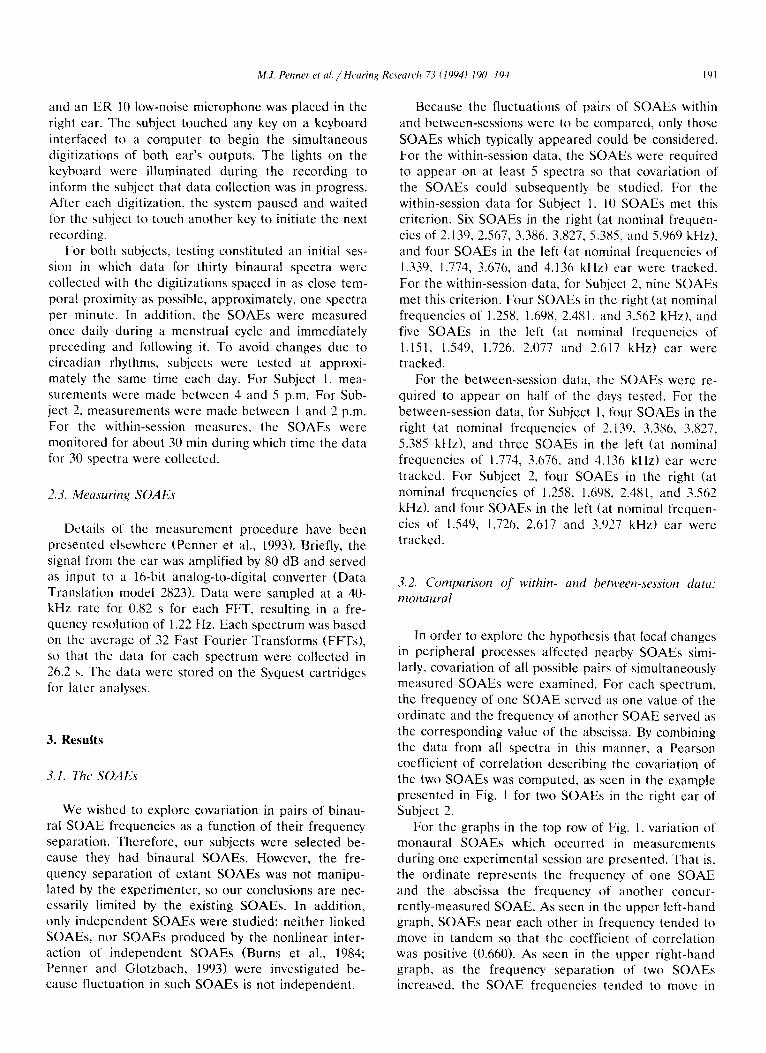
ordinate and the frequency of another SOAE served as the corresponding value of the abscissa. By combining the data from all spectra in this manner. a Pearson coefficient of correlation describing the covariation of the two SOAEs was computed. as seen in the example presented in Fig. 1 for two SOAEs in the right ear of Subject 2.
For the graphs in the top row of Fig. 1, variation of monaural SOAEs which occurred in measurements during one experimental session arc presented. That is, the ordinate represents the frequency of one SOAE and the abscissa the frequency of another concur- rently-measured SOAE. As seen in the upper left-hand graph, SOAEs near each other in frequency tended to move in tandem so that the coefficient of correlation was positive (0.660). As seen in the upper right-hand graph. as the frequency separation of two SOAEs increased, the SOAE frequencies tended to move in
142
1.700 1.705 1710 3.555 3.565
FREQUENCY (ktiz)
Fig. 1. Covariation of pairs of monaural SOAJZ frequencies for Subject 2. Within-session covariation is presented in the top row (with numbers indicating the number of spectra in which the SOAE frequency on the ordinate was measured at the same time as the SOAE frequency on the abscissa). Between-session covariation is presented in the bottom row (circles) for data spanning a menstrual cycle.
opposite directions so that the coefficient of correla- tion was negative ( - 0.586).
For the graphs in the bottom row of Fig. 1, variation of SOAEs which occurred during the course of the menstrual cycle are presented. For the monthly data, the frequencies of all the SOAEs tended to move in tandem as will be seen in Fig. 3, so that the coefficient of correlation was positive nearly independent of the frequency separation of the SOAJ% (for the graph on the right side of the bottom row of Fig. 1, r = 0.330; for the graph on the left, I = 0.385).
3.3. Comparison of within- and between-session data: binaural
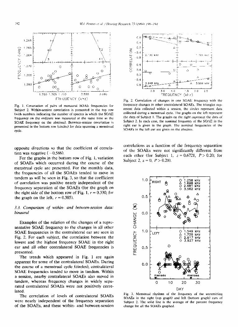
Examples of the relation of the changes of a repre- sentative SOAE frequency to the changes in all other SOAE frequencies in the contralateral ear are seen in Fig. 2. For each subject, the correlation between the lowest and the highest frequency SOAE in the right ear and all other contralateral SOAE frequencies is presented.
The trends which appeared in Fig. 1 are again apparent for some of the contralateral SOAEs. During the course of a menstrual cycle, (circles), contralateral SOAE frequencies tended to move in tandem. Within a session, nearby contralateral SOAEs also moved in tandem, whereas frequency changes in widely sepa- rated contralateral SOAEs were not positively corre- lated.
The correlation of levels of contralateral SOAEs were nearly independent of the frequency separation of the SOAEs, and these within- and between-session
0.0
z -0.4 ., F 2.130 kHz ’ 265 kYr i Q -0.8 i i
-0.4 - A
5.948 kHz __ &/.A---
a 3.559 kHz -0.8 w
A --
1 , I T I I I ;
2.0 3.0 4.0 1.5 20 25
FREQUENCY (kHz)
Fig. 2. Correlation of changes in one SOAE frequency with the frequency changes in other contralateral SOAEs. The triangles rep- resent data collected within a session, the circles represent data collected during a menstrual cycle. The graphs on the left represent the data of Subject 1. The graphs on the right represent the data of Subject 2. In each case, the nominal frequency of the SOAE in the right ear is given in the graph. The nominal frequencies of the SOAEs in the left ear are given on the abscissa.
correlations as a function of the frequency separation of the SOAEs were not significantly different from each other (for Subject 1, z = 0.6721, P > 0.20; for Subject 2, z = 0, P > 0.20).
8 0 A 11.258 1.698 kl& kHz 0 2.481 kHz 0 3.562 kHz
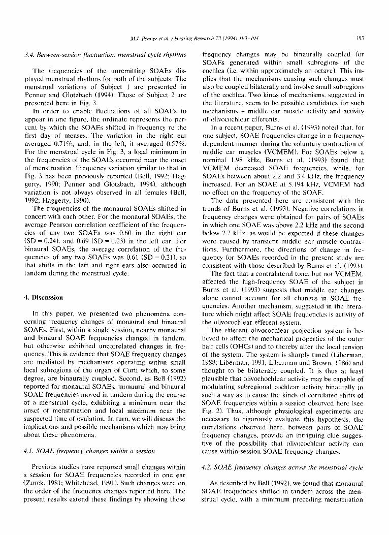
DAY Fig. 3. Menstrual rhythms of the frequency of the unremitting SOAEs in the right (top graph) and left (bottom graph) ears of Subject 2. The solid line is the average of the percent frequency change for all the SOAEs graphed.
M.J. Penner et al. / Hearmg Research 7.3 (19941 I YO- 194 103
3.4. Between-session fluctuation: menstrual cycle rhythms
The frequencies of the unremitting SOAEs dis- played menstrual rhythms for both of the subjects. The menstrual variations of Subject 1 are presented in Penner and Glotzbach (1994). Those of Subject 2 are
presented here in Fig. 3. In order to enable fluctuations of all SOAEs to
appear in one figure, the ordinate represents the per- cent by which the SOAEs shifted in frequency re the first day of menses. The variation in the right ear
averaged O.71%, and, in the left, it averaged 0.57%. For the menstrual cycle in Fig. 3, a local minimum in the frequencies of the SOAEs occurred near the onset of menstruation. Frequency variation similar to that in
Fig. 3 has been previously reported (Bell, 1992; Hag- gerty, 1990; Penner and Glotzbach, 1994), although variation is not always observed in all females (Bell,
1992; Haggerty, 1990). The frequencies of the monaural SOAEs shifted in
concert with each other. For the monaural SOAEs, the
average Pearson correlation coefficient of the frequcn- ties of any two SOAEs was 0.60 in the right ear (SD = 0.24), and 0.69 (SD = 0.23) in the left ear. For binaural SOAEs, the average correlation of the fre- quencies of any two SOAEs was 0.61 (SD = 0.21), so that shifts in the left and right ears also occurred in tandem during the menstrual cycle.
4. Discussion
In this paper, we presented two phenomena con- cerning frequency changes of monaural and binaural SOAEs. First, within a single session, nearby monaural and binaural SOAE frequencies changed in tandem, but otherwise exhibited uncorrelated changes in fre- quency. This is evidence that SOAE frequency changes are mediated by mechanisms operating within small local subregions of the organ of Corti which, to some degree, are binaurally coupled. Second, as Bell (1992) reported for monaural SOAEs, monaural and binaural SOAE frequencies moved in tandem during the course of a menstrual cycle, exhibiting a minimum near the onset of menstruation and local maximum near the suspected time of ovulation. In turn, we will discuss the implications and possible mechanisms which may bring about these phenomena.
4.1. SOAE frequency changes within a session
Previous studies have reported small changes within a session for SOAE frequencies recorded in one ear (Zurek, 1981; Whitehead, 1991). Such changes were on the order of the frequency changes reported here. The present results extend these findings by showing these
frequency changes may be binaurally coupled for SOAEs generated within small subregions of the
cochlea (i.e. within approximately an octave). This im- plies that the mechanisms causing such changes must also be coupled bilaterally and involve small subregions of the cochlea. Two kinds of mechanisms, suggested in the literature, seem to be possible candidates for such mechanisms - middle ear muscle activity and activity
of olivocochlear efferents. In a recent paper, Burns et al. (1993) noted that, for
one subject, SOAE frequencies change in a frequency-
dependent manner during the voluntary contraction of middle ear muscles (VCMEM). For SOAEs below a nominal 1.98 kHz, Burns et al. (1903) found that
VCMEM decreased SOAE frequencies. while, for SOAEs between about 2.2 and 3.4 kHz, the frequency increased. For an SOAE at 5.194 kHz, VCMEM had
no effect on the frequency of the SOAE. The data presented here are consistent with the
trends of Burns et al. (1993). Negative correlations in
frequency changes were obtained for pairs of SOAEs in which one SOAE was above 2.2 kHz and the second below 2.2 kHz, as would be expected if these changes were caused by transient middle ear muscle contrac- tions. Furthermore, the directions of change in fre- quency for SOAEs recorded in the present study are consistent with those described by Burns et al. (1993).
The fact that a contralateral tone, but not VCMEM, affected the high-frequency SOAE of the subject in Burns et al. (1993) suggests that middle ear changes alone cannot account for all changes in SOAE fre- quencies. Another mechanism, suggested in the litera- ture which might affect SOAE frequencies is activity of the olivocochlear efferent system.
The efferent olivocochlear projection system is be- lieved to affect the mechanical properties of the outer
hair cells (OHCs) and to thereby alter the local tension of the system. The system is sharply tuned (Liberman, 1988; Liberman, 1991; Liberman and Brown, 1986) and thought to be bilaterally coupled. It is thus at least plausible that olivochochlear activity may be capable of modulating subregional cochlear activity binaurally in such a way as to cause the kinds of correlated shifts of SOAE frequencies within a session observed here (see Fig. 2). Thus, although physiological experiments are necessary to rigorously evaluate this hypothesis, the correlations observed here, between pairs of SOAE frequency changes. provide an intriguing clue sugges- tive of the possibility that olivocochlear activity can cause within-session SOAE frequency changes.
4.2. SOAE frequency changes across the menstrual cycle
As described by Bell (1992), we found that monaural SOAE frequencies shifted in tandem across the men- strual cycle, with a minimum preceding menstruation
and a local maximum occurring near the time of ovula- tion. Binaural SOAE frequencies changed in the same manner, thus indicating that the mechanism(s) underly- ing these SOAE frequency changes are also bilaterally coupled. This is consistent with Bell’s (1992) suggestion that SOAE frequency changes across the menstrual cycle are related to cardiovascular changes involving cerebrospinal fluid (CSF) pressure. Because SOAEs tend to move in tandem throughout the menstrual cycle, these changes seem quite different from those occurring within sessions which, as described above, seem to be caused by mechanisms operating within subregions of the cochlea. Given the magnitude of the effects (see Figs. 2 and 3), CSF pressure may have a larger effect on SOAEs than within-session factors such as changes in middle-ear pressure.
In summary, our data suggest the existence of cou- pling between SOAE generator mechanisms locally, within the organ of Corti, as well as binaurally. Possi- ble explanations include changes in middle ear muscle activity as well as coordinated bilateral activity in the olivocochlear efferent bundles. More than one mecha- nism may well be involved and mechanisms may inter- act. The existence of between-session changes across the menstrual cycle in premenopausal women suggest that other metabolic factors, probably affecting CSF pressure, may interact as well. The exciting possibility is that future studies of SOAE frequency changes, including studies of the physiological mediators, might prove useful in suggesting and testing hypotheses about how cochlear transduction mechanisms interact within the organ of Corti.
5. Acknowledgements
This research was supported by a grant from the National Institutes of Health (NIDCD 1ROl DCOO068) and by the University of Maryland at College Park.
6. References
American National Standards Institute (1970) American national
standard specifications for audiometers (ANSI 53.6-1969). New York: ANSI.
Bell, A. (1992) Circadian and menstrual rhythms in frequency varia-
Bialek, W.S. and Wit, H.P. (lYX4) Quantum itmlt\ [<I ~I\~I~I;I~~!I
stability: theory and experiments on acoustic cmiAon\ l’rom tlx
human ear. Phya. Lett. A 104. I73- 17%
Burns. EM.. Harrison, WA. Bulen. J.C. and Keel’<. D.H. t lW1) Voluntary contraction of middle car muscles: Effects trn input
impcdancc. energy reflectance and spontaneous otoacoustic enlis
sions. Hear. Res. 67. 117- 127.
Burns, E.M., Strickland. E.A.. Tubis. A. and Jonc~. K. (lYX4) Inlrl-
actions among spontaneous otoacoustic emissions. 1. Distortion
products and linked emissions. Hear. Res. If), 771-278.
Haggerty. H.S. (1990) Spontaneous otoacoustic emissions: Evidcncr
for a monthly rhythm of frequency variation in women. Assoc.
Res. Otolaryngol. Abstr. 268. page 233.
Keefe. D.H.. Burns. E.M.. Ling. R.. and Laden, B. (1090) Chaotic
dynamics of otoacoustic emissions. In: P. Dallas, C.D. Geisler.
J.W. Matthews. M. Ruggero and C.R. Steele (Eds.). The Mechatl-
its and Biophysics of Hearing, pp. 194-201. Springer Verlag,
New York.
Kemp. D.T. (1979) Evidence of mechanical nonlinearity and fre-
quency selective wave amplification in the cochlea. Arch. Oto-
Rhino-Laryngol. 224, 37-45.
Kim, D.O. (1986) Active nonlinear cochlear biomechanics and the
role of outer-hair-cell subsystem in the mammalian auditory
system. Hear. Res. 22, 105-l 14.
Liberman, M.C. (1988) Response properties of cochlear afferent
neurons: Monaural vs. binaural stimulation. J. Neurophysiol. 60.
1779%17’)s.
Liberman, M.C. (1991) The olivocochlear efferent bundle and the
susceptibility of the inner ear to acoustic injury. J. Neurophysiol.
65, 123-132.
Liberman, M.C. and Brown, M.C. (1986) Physiology and anatomy of
single olivocochlear neurons in the cat. Hear. Res. 24, 17-36.
Long, G.R.. Tubis. A., Jones, K.L. (1991) Modelling synchronization
and suppression of spontaneous otoacoustic emissions using Van
der Pol oscillators: Effect of aspirin administration. J. Acoust.
Sot. Am. X9, 1201-1212.
Mott, J.B., Norton, S.J., Neely, S.T. and Warr, W.B. (IYXY) Changes
in spontaneous otoacoustic emissions produced by acoustic stimu-
lation of the contralateral ear. Hear. Res. 38, 229-242.
Penner, M.J. and Glotzbach, L. (19941 Covariation of tinnitus pitch
and the associated emission: a case study. Arch. Otolaryngol. -
Head and Neck Surg. (in press).
Penner, M.J., Glotzbach. L. and Huang, T. (lYY3) Spontaneous
otoacoustic emissions: measurement and results. Hear. Res. 6X.
229-231.
Talmadge, CL., Tubis, A. and Wit H.P. (1991) Are spontaneoua
otoacoustic emissions generated by self-sustained cochlear oscil-
lators? J. Acoust. Sot. Am. 89. 2391-2399.
Whitehead, M.L. (1991) Slow variations of the amplitude and fre-
quency of spontaneous otoacoustic emissions. Hear. Res. 53.
269-280.
Wit, H.P. (1985) Diurnal cycle for spontaneous oto-acoustic emission
frequency. Hear. Res. 18, 197-199. Zurek, P.M. (1981) Spontaneous narrowband acoustic signals emit-
ted from human ears. J. Acoust. Sot. Am. 69, 514-523.