Embed Size (px)
Citation preview

School Nurses Guide to Otoacoustic Emissions
(OAEs)
www.maico-diagnostics.com
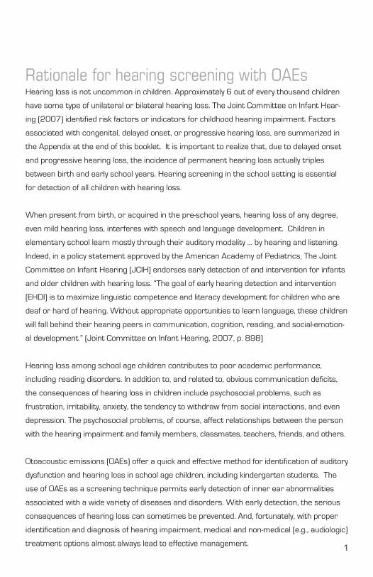
Rationale for hearing screening with OAEsHearing loss is not uncommon in children. Approximately 6 out of every thousand children
have some type of unilateral or bilateral hearing loss. The Joint Committee on Infant Hear-
ing (2007) identified risk factors or indicators for childhood hearing impairment. Factors
associated with congenital, delayed onset, or progressive hearing loss, are summarized in
the Appendix at the end of this booklet. It is important to realize that, due to delayed onset
and progressive hearing loss, the incidence of permanent hearing loss actually triples
between birth and early school years. Hearing screening in the school setting is essential
for detection of all children with hearing loss.
When present from birth, or acquired in the pre-school years, hearing loss of any degree,
even mild hearing loss, interferes with speech and language development. Children in
elementary school learn mostly through their auditory modality … by hearing and listening.
Indeed, in a policy statement approved by the American Academy of Pediatrics, The Joint
Committee on Infant Hearing (JCIH) endorses early detection of and intervention for infants
and older children with hearing loss. “The goal of early hearing detection and intervention
(EHDI) is to maximize linguistic competence and literacy development for children who are
deaf or hard of hearing. Without appropriate opportunities to learn language, these children
will fall behind their hearing peers in communication, cognition, reading, and social-emotion-
al development.” (Joint Committee on Infant Hearing, 2007, p. 898)
Hearing loss among school age children contributes to poor academic performance,
including reading disorders. In addition to, and related to, obvious communication deficits,
the consequences of hearing loss in children include psychosocial problems, such as
frustration, irritability, anxiety, the tendency to withdraw from social interactions, and even
depression. The psychosocial problems, of course, affect relationships between the person
with the hearing impairment and family members, classmates, teachers, friends, and others.
Otoacoustic emissions (OAEs) offer a quick and effective method for identification of auditory
dysfunction and hearing loss in school age children, including kindergarten students. The
use of OAEs as a screening technique permits early detection of inner ear abnormalities
associated with a wide variety of diseases and disorders. With early detection, the serious
consequences of hearing loss can sometimes be prevented. And, fortunately, with proper
identification and diagnosis of hearing impairment, medical and non-medical (e.g., audiologic)
treatment options almost always lead to effective management. 1
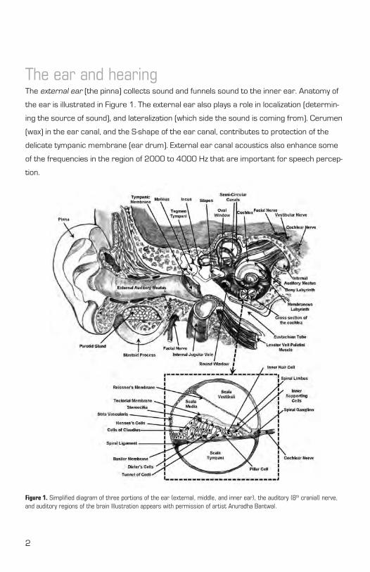
The ear and hearingThe external ear (the pinna) collects sound and funnels sound to the inner ear. Anatomy of
the ear is illustrated in Figure 1. The external ear also plays a role in localization (determin-
ing the source of sound), and lateralization (which side the sound is coming from). Cerumen
(wax) in the ear canal, and the S-shape of the ear canal, contributes to protection of the
delicate tympanic membrane (ear drum). External ear canal acoustics also enhance some
of the frequencies in the region of 2000 to 4000 Hz that are important for speech percep-
tion.
2
Figure 1. Simplified diagram of three portions of the ear (external, middle, and inner ear), the auditory (8th cranial) nerve, and auditory regions of the brain Illustration appears with permission of artist Anuradha Bantwal.
The middle ear consists of the tympanic membrane and the ossicles (malleus, incus, and
stapes). Sound waves reaching the tympanic membrane are amplified by the middle ear
system, providing an increase in sound intensity of almost 30 dB. Mechanical energy from
sound waves is converted to electrical signals by specialized hair cells located within the
inner ear (the cochlea). The term “hair cells” is used because there are extending from the
top of each cell hundreds of thin hair-like protein-based cilia. There are about 15,000 hair
cells in the human ear. One third of the hair cells, the inner hair cells located medially in
the cochlea (see Figure 1), communicate (synapse) with auditory (8th cranial nerve) fibers.
Activation of the inner hair cells leads to firing of auditory nerve fibers and stimulation of
auditory regions of the central nervous system (also shown in Figure 1). The remaining two-
thirds of the hair cells located more laterally within the cochlea, referred to as outer hair
cells, are capable of motility (movement). Upon activation, metabolism within the outer hair
cells increases dramatically, and the outer hair cells rapidly elongate (during hyper-polariza-
tion) and become shorter (during depolarization). Changes in outer hair cell length generate
energy within the cochlea that contributes to hearing sensitivity and the ability to distinguish
small differences in the frequencies of sounds. Outer hair cell movement also produces
otoacoustic emissions, as reviewed briefly in the next section.
At this point, it’s important keep in mind that although the ear is clearly important in hear-
ing, we really hear with our brain. High level auditory processing, including speech percep-
tion, occurs within a complex network of central nervous system pathways and centers
(nuclei) containing millions of neurons. Clinically, hearing evaluation is not complete unless it
includes procedures for evaluating how the brain processes relatively sophisticated sounds,
such as speech. Audiologists regularly perform such procedures in hearing assessment.
Audiologic tests used to evaluate function of the ear, such as otoacoustic emissions (OAEs),
are very important in the diagnosis of hearing loss. However, OAEs alone are not a test of
hearing.
3
What are OAEs and how are they recorded?Otoacoustic emissions (OAEs) are sounds measured in the external ear canal that reflect
movement of the outer hair cells in the cochlea. Energy produced by outer hair cell motility
serves as an amplifier within the cochlea, contributing to better hearing. Indeed, normal out-
er hair cells are essential for perfectly normal auditory function. OAEs are produced by the
energy from outer hair cell motility that makes its way outward from the cochlea through
the middle ear, vibrating the tympanic membrane, and propagating into the external ear
canal. Although the amplification produced by outer hair cell movement within the cochlea
may be as high as 50 dB, residual energy reaching the ear canal …otoacoustic emissions …
is normally in the range of 0 to 15 dB.
Two types of OAEs may be measured clinically with FDA-approved devices. Transient evoked
OAEs (TEOAEs) are elicited with very brief (transient) sounds, such as clicks or tone bursts,
presented at an intensity level of 80 dB SPL. TEOAEs reflecting cochlear (outer hair cell)
activity are generally recorded over the frequency range of 500 to about 4000 Hz. Distor-
tion product OAEs (DPOAEs) are elicited with sets of two pure tone frequencies, abbreviated
f2 and f
1, that are closely spaced and presented simultaneously at moderate intensity levels,
such as (respectively) 55 and 65 dB SPL. DPOAEs can be recorded across a frequency re-
gion of 500 to 8,000 Hz and sometimes even higher frequencies. Mechanisms and clinical
applications of OAEs are described in recent textbooks (cited at the end of the booklet) and
in thousands of peer reviewed journal articles. An Internet search for OAE literature can
easily be performed via the National Library of Medicine website (www.nlm.nih.gov, Health
Care Professionals).
OAEs are non-invasive and technically simple to record. In school age children, hearing
screening with OAEs usually requires less than two minutes for both ears. Screening time
for one ear is often less than 30 seconds! Sedation is not indicated for OAE measurement,
even in children. No behavioral response is required for participating in the testing, so the
procedure is not affected by a child’s motivation, attention, or cognitive status. The tech-
nique for hearing screening with OAEs will now be summarized.
Briefly, a soft disposable probe tip is gently inserted into the outer portion of the external
ear canal (Figure 2). An airtight seal between the probe tip and the ear canal isn’t neces-
4
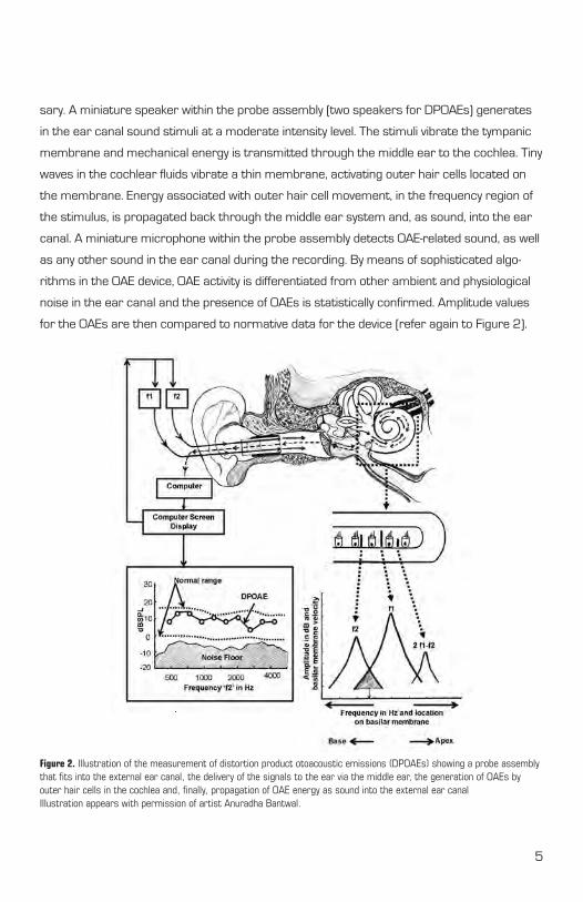
sary. A miniature speaker within the probe assembly (two speakers for DPOAEs) generates
in the ear canal sound stimuli at a moderate intensity level. The stimuli vibrate the tympanic
membrane and mechanical energy is transmitted through the middle ear to the cochlea. Tiny
waves in the cochlear fluids vibrate a thin membrane, activating outer hair cells located on
the membrane. Energy associated with outer hair cell movement, in the frequency region of
the stimulus, is propagated back through the middle ear system and, as sound, into the ear
canal. A miniature microphone within the probe assembly detects OAE-related sound, as well
as any other sound in the ear canal during the recording. By means of sophisticated algo-
rithms in the OAE device, OAE activity is differentiated from other ambient and physiological
noise in the ear canal and the presence of OAEs is statistically confirmed. Amplitude values
for the OAEs are then compared to normative data for the device (refer again to Figure 2).
Figure 2. Illustration of the measurement of distortion product otoacoustic emissions (DPOAEs) showing a probe assembly that fits into the external ear canal, the delivery of the signals to the ear via the middle ear, the generation of OAEs by outer hair cells in the cochlea and, finally, propagation of OAE energy as sound into the external ear canalIllustration appears with permission of artist Anuradha Bantwal.
5
Analysis and interpretation of OAEsModern OAE devices typically include software for automated data analysis in hearing
screening, including algorithms for calculation of amplitude values, noise floor levels, and for
statistical confirmation the OAEs are present or absent. Visual inspection of OAE data with
manual analysis is almost always an option, and particularly important for diagnostic appli-
cation of OAEs. There are three general steps in the analysis of OAE findings. The first step
is to verify adequate measurement conditions. Specifically, noise levels must be sufficiently
low (usually less than – 10 dB SPL) to permit confident detection of OAE activity and the
stimulus intensity levels in the ear canal should be close to the desired (target) levels. OAE
devices invariably perform a quick calibration of stimulus intensity levels prior to data collec-
tion. The next step in data analysis is to determine whether reliable (repeatable) OAEs are
recorded, that is, whether OAE amplitude exceeds the noise level by 6 dB or more at the
test frequency. Finally, when the difference between OAE amplitude and noise floor ≥ 6 dB
SPL, findings are analyzed with respect to an appropriate normal region for OAE amplitude.
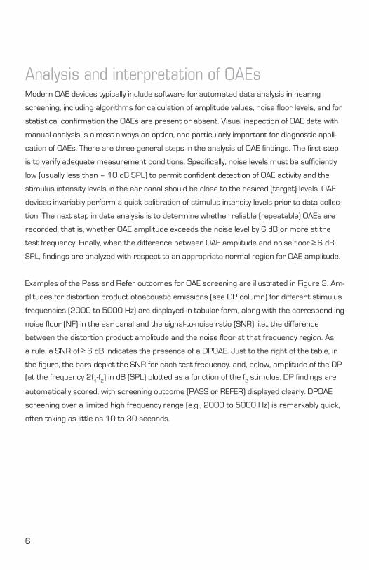
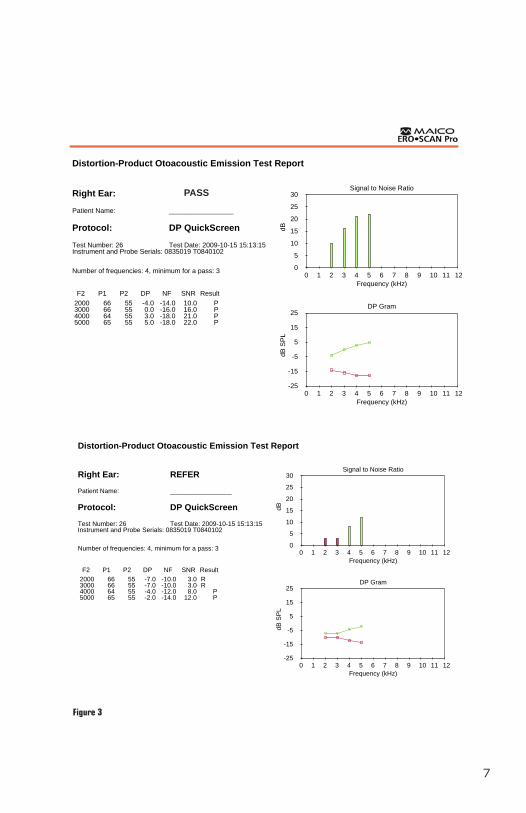
Examples of the Pass and Refer outcomes for OAE screening are illustrated in Figure 3. Am-
plitudes for distortion product otoacoustic emissions (see DP column) for different stimulus
frequencies (2000 to 5000 Hz) are displayed in tabular form, along with the correspond-ing
noise floor (NF) in the ear canal and the signal-to-noise ratio (SNR), i.e., the difference
between the distortion product amplitude and the noise floor at that frequency region. As
a rule, a SNR of ≥ 6 dB indicates the presence of a DPOAE. Just to the right of the table, in
the figure, the bars depict the SNR for each test frequency. and, below, amplitude of the DP
(at the frequency 2f1-f
2) in dB (SPL) plotted as a function of the f
2 stimulus. DP findings are
automatically scored, with screening outcome (PASS or REFER) displayed clearly. DPOAE
screening over a limited high frequency range (e.g., 2000 to 5000 Hz) is remarkably quick,
often taking as little as 10 to 30 seconds.
6
7
Figure 3
Distortion-Product Otoacoustic Emission Test Report
Right Ear: REFER
Patient Name: _________________
Protocol: DP QuickScreen
Test Number: 26 Test Date: 2009-10-15 15:13:15Instrument and Probe Serials: 0835019 T0840102
Number of frequencies: 4, minimum for a pass: 3
F2 P1 P2 DP NF SNR Result 2000 66 55 -4.0 -14.0 10.0 P 3000 66 55 0.0 -16.0 16.0 P 4000 64 55 3.0 -18.0 21.0 P 5000 65 55 5.0 -18.0 22.0 P
0 1 2 3 4 5 6 7 8 9 10 11 12Frequency (kHz)
0
5
10
15
20
25
30
dB
Signal to Noise Ratio
0 1 2 3 4 5 6 7 8 9 10 11 12Frequency (kHz)
-25
-15
-5
5
15
25
dB S
PL
DP Gram
Etymotic Research Inc. - Copyright 2009
PASS
Distortion-Product Otoacoustic Emission Test Report
Right Ear: REFER
Patient Name: _________________
Protocol: DP QuickScreen
Test Number: 26 Test Date: 2009-10-15 15:13:15Instrument and Probe Serials: 0835019 T0840102
Number of frequencies: 4, minimum for a pass: 3
F2 P1 P2 DP NF SNR Result 2000 66 55 -7.0 -10.0 3.0 R 3000 66 55 -7.0 -10.0 3.0 R 4000 64 55 -4.0 -12.0 8.0 P 5000 65 55 -2.0 -14.0 12.0 P
0 1 2 3 4 5 6 7 8 9 10 11 12Frequency (kHz)
0
5
10
15
20
25
30
dB
Signal to Noise Ratio
0 1 2 3 4 5 6 7 8 9 10 11 12Frequency (kHz)
-25
-15
-5
5
15
25
dB S
PL
DP Gram
Etymotic Research Inc. - Copyright 2009
Why are OAEs valuable in children?OAEs are rapidly gaining popularity as a technique for hearing screening of school age and
pre-school children. One important reason is simplicity. OAE screening outcome is generally
described as either “Pass” or “Refer.” A pass outcome is reported when OAEs are present
(≥ 6 dB above the noise floor) for the majority of test frequencies. Although the presence
of OAEs does not always indicate normal hearing sensitivity, a pass outcome in most cases
rules out a serious degree of hearing loss. A refer OAE screening outcome should be viewed
as a clear risk factor for hearing loss that could affect communication and school perfor-
mance. Children who yield a reliable refer outcome for OAE screening should be referred
for diagnostic hearing assessment by an audiologist, and possible audiological or medical
management. The literature contains hundreds of peer reviewed scientific papers reporting
evidence in support of OAE measurement in children.
OAE are widely applied in pediatric populations for a variety of reasons. As already noted,
OAEs are an index of outer hair cell activity. Because of their dependence on normal cell
metabolism, OAEs are exquisitely sensitive to even subtle outer hair cell dysfunction. Almost
all insults to the cochlea first affect the outer hair cells. Therefore, assuming normal middle
ear function, OAE abnormalities provide early and compelling evidence of cochlear (outer
hair cell) dysfunction. Additional clinical advantages of OAE are:
• Brief test time: Usually less than a minute per ear
• Relatively simple technique: Little training is required
• Can be recorded without a special sound-treated test room
• Objective: Unaffected by attention, cognition, cooperation
• Independent of age: OAEs can be recorded easily from pre-school and
kindergarten children, and even newborn infants
• Ear specific: Separate test results for each ear
• Frequency specific: Information for many individual frequencies
8
OAEs in School Age ChildrenHearing screening of school age children is a long-standing convention for detection of
hearing loss that will interfere with academic performance. In the past, a pure tone hearing
screening approach was the only option for screening hearing of pre-school and school age
children. Pure tone hearing screening utilizing an audiometer is associated with multiple
practical problems, particularly in pre-school and young school age (e.g., kindergarten) chil-
dren. Unacceptably high refer rates for pure tone hearing screening, up to 70% in the pre-
school population (e.g., Hall & Swanepoel, 2010), may result from a combination of factors,
such as inexperienced or poorly trained screening personnel, excessive levels of ambient
noise in the test environment, and listener variables common in young children (e.g., atten-
tion, cognition, motivation) that sometimes preclude a valid screening outcome.
Within recent years, clinical studies have documented the value and many advantages of
OAEs as a hearing screening technique in pediatric populations, including school age chil-
dren. OAEs are now the technique of choice for hearing screening of pre-school and school
age children, given the proven effectiveness and feasibility of OAEs as a hearing screening
technique in newborn infants (Hall & Swanepoel, 2010). Overall refer rates (either or both
ears) are lower for distortion product OAEs (12.5%) than for pure tone audiometry (17%),
reducing the number of children who require rescreening or medical referral. OAEs are very
sensitive to both middle ear and inner ear disorders, yet not affected by the child’s ability to
understand instructions or to attend to sounds.
9
Pulling it all togetherOAEs are a quick, non-invasive, sensitive, and objective procedure for detecting in the school
setting hearing loss secondary to middle ear or inner ear (cochlear) auditory dysfunction. In
other words, OAEs are a handy and proven technique for identifying children at risk for hear-
ing impairment. Despite the many clinical advantages and applications of OAE measure-
ment, it’s important to remember that OAEs are not a test of hearing. OAEs may be absent
in children with normal hearing sensitivity who have residual minor middle ear disorders.
Conversely, OAEs may be present, even with amplitudes entirely within normal limits, in chil-
dren with rarely encountered inner hair cell dysfunction or retrocochlear auditory pathology.
Selected ReferencesDhar S & Hall JW III. (2010). Otoacoustic Emissions: Principles, Procedures, and Protocols.
San Diego: Plural Publishing
Hall JW III. (2000). Handbook of Otoacoustic Emissions. San Diego: Singular Publishing Company
Hall JW III & Swanepoel, D. (2010). Objective Assessment of Hearing. San Diego: Plural Publishing
National Center for Hearing Assessment and Management (NCHAM). (2006). Early identi-
fication of hearing loss: Conducting periodic otoacoustic emissions (OAE) hearing screening
with infants and toddlers during well-child visits. For more information, contact NCHAM at
Utah State University, Logan UT 84322. Available online at: www.infanthearing.org or www.
hearandnow.org/periodicscreening
Year 2007 Position Statement: Principles and Guidelines for Early Hearing Detection and
Intervention Programs. Joint Committee on Infant Hearing Pediatrics, 120, pp. 898-921
NOTE: Anyone with Internet access can quickly perform a literature review on the topic
of otoacoustic emissions at the National Library of Medicine website (www.nlm.nih.gov,
Health Care Professionals). A search will produce abstracts of thousands of articles
containing the word “otoacoustic emissions.” A more refined search can be performed
with combinations of terms, such as “otoacoustic emissions” and “dementia.” Articles of
interest can then be requested via email of the author designated for correspondence.
10
CreditsJames W. Hall III, Ph.D. contributed to the preparation of this booklet. Dr. Hall earned
his Masters degree from Northwestern University and his Ph.D. in Audiology from Baylor
College of Medicine. He is the author of over 150 journal articles and book chapters, plus
10 textbooks including the Handbook of Otoacoustic Emissions and the recently published
Otoacoustic Emissions: Principles, Procedures, and Protocols. Dr. Hall is Clinical Professor in
the Department of Communicative Disorders at the University of Florida where he maintains
a clinical practice, teaches doctoral level students, and conducts externally funded
research.
Kathryn Sutherland, Marketing Manager, served as production coordinator for this booklet.
Anuradha Bantwal provided the artwork appearing in Figure 1 and Figure 2 of this
booklet. Ms. Bantwal is an Audiologist and Speech-Language Pathologist working in India.
Additional ResourcesAmerican Academy of Audiology. www.audiology.org
American Academy of Pediatrics. www.aap.org
Better Hearing Institute. www.betterhearing.org
National Center for Hearing Assessment and Management (NCHAM).
www.infanthearing.org
Otoacoustic Emissions Portal Zone. www.otoemissions.org
11
Appendix: Evidenced-based risk indicators that are associated with hearing loss in
childhood, including permanent congenital, delayed onset, or progressive hearing loss,
according to the Joint Committee on Infant Hearing.*
• Caregiver concern regarding hearing, speech, language, or developmental delay
• Family history of permanent childhood hearing loss
• Neonatal intensive care of more than 5 days or any of the following regardless
of length of stay: ECMO, assisted ventilation, exposure to ototoxic medications
(gentimycin and tobramycin) or loop diuretics (furosemide/Lasix), and
hyperbilirubinemia that requires exchange transfusion
• In utero infections, such as CMV, herpes, rubella, syphilis, and toxoplasmosis
• Craniofacial anomalies, including those that involve the pinna, ear canal, ear tags,
ear pits, and temporal bone anomalies
• Physical findings, such as white forelock, that are associated with a syndrome
known to include a sensorineural or permanent conductive hearing loss
• Syndromes associated with hearing loss or progressive or late-onset hearing loss,
such as neurofibromatosis, osteopetrosis, and Usher syndrome other frequently
identified syndromes include Waardenburg, Alport, Pendred, and Jervell and
Lange-Nielson
• Neurodegenerative disorders, such as Hunter syndrome, or sensory motor
neuropathies, such as Friedreich ataxia and Charcot-Marie-Tooth syndrome
• Culture-positive postnatal infections associated with sensorineural hearing loss,
including confirmed bacterial and viral (especially herpes viruses and varicella)
meningitis
• Head trauma, especially basal skull/temporal bone fracture that requires
hospitalization
• Chemotherapy
______________________________________________________________________
* Source: Joint Committee on Infant Hearing. (2007). Year 2007 Position Statement:
Principles and Guidelines for Early Hearing Detection and Intervention Programs.
Pediatrics, 120, pp. 898-921
12

Notes
13
EXCELLENCE IN HEARING SCREENING SINCE 1937MAICO DIAGNOSTICS • 10393 West 70th Street, Eden Prairie, MN 55344
toll free 888-941-4201 • fax 952-903-4100 • www.maico-diagnostics.com
©M
aico
Dia
gnos
tics,
200
9
08/16
OA
E G
uide
- S
choo
l Nur
ses


![A comparative study of evoked otoacoustic emissions in ...SOAEs - We also examined spontaneous otoacoustic emissions, which had been previously reported in this gecko species [5]](https://img.pdfslide.us/doc/110x75/60806f7e0c731c1c4f6b0c15/a-comparative-study-of-evoked-otoacoustic-emissions-in-soaes-we-also-examined.jpg)