Embed Size (px)
Citation preview

1
Corso di Immunologia
A.A. 2011-12
Immunoglobuline
Struttura e Funzione

2
Definitions
• Immunoglobulins (Ig) - Glycoprotein molecules which are produced by
plasma cells in response to an immunogen and which function as
antibodies. The immunoglobulins derive their name from the finding that
when antibody-containing serum is place in an electrical field the
antibodies, migrated with the globular proteins.
Keywords
Immunoglobulin, Valence, Heavy chain, Light chain, Variable region,
Constant region, Hinge region, Domain, Hypervariable region,
Framework region, Groups & subgroups, Fab & Fc, F(ab')2, Type &
subtype, Class & subclass, Opsonin, J chain, Secretory component

3
Immunoglobulins: Structure and Function
• Definition: Glycoprotein molecules that are produced by plasma cells in response to an immunogen and which function as antibodies.
Immune serum
Ag adsorbed serum
+ -albumin
globulins
Mobility
Am
ount
of p
rote
in
α1 α2 γβ
4
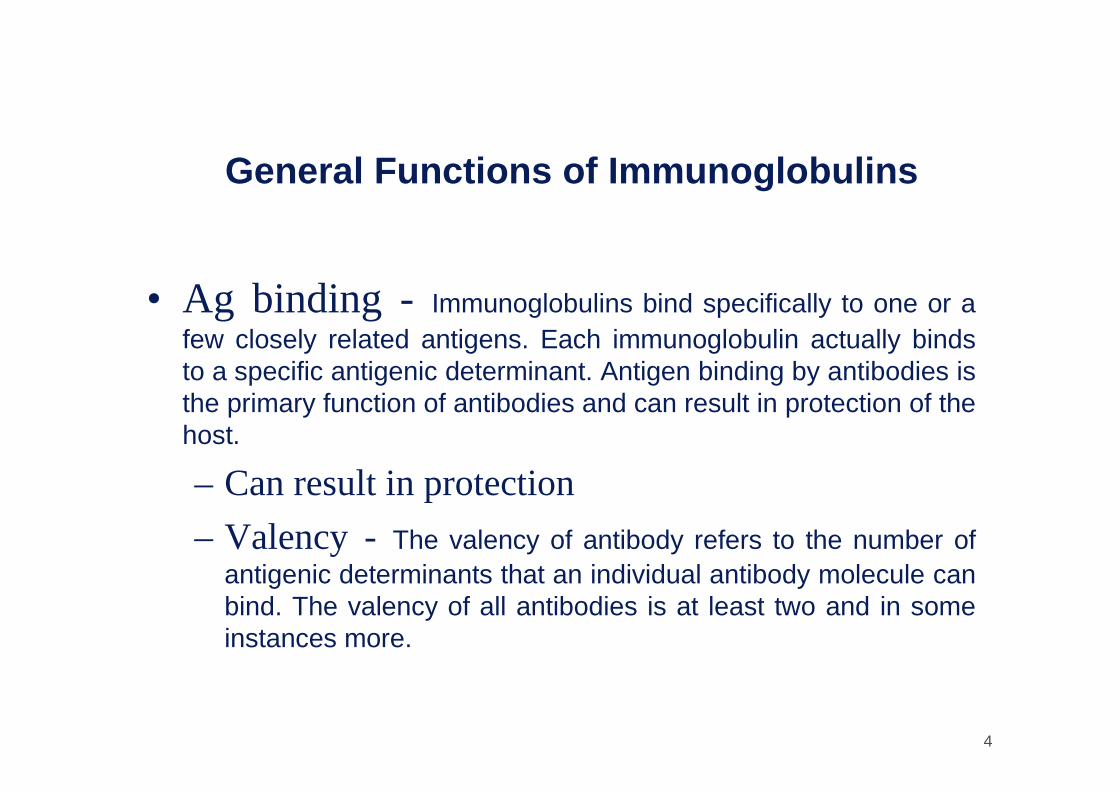
General Functions of Immunoglobulins
• Ag binding - Immunoglobulins bind specifically to one or a few closely related antigens. Each immunoglobulin actually bindsto a specific antigenic determinant. Antigen binding by antibodies isthe primary function of antibodies and can result in protection of the host.
– Can result in protection
– Valency - The valency of antibody refers to the number of antigenic determinants that an individual antibody molecule can bind. The valency of all antibodies is at least two and in some instances more.

5
General Functions of Immunoglobulins
• Effector functions -(Usually require Ag binding) -Oftenthe binding of an antibody to an antigen has no direct biological effect. Rather, the significant biological effects are a consequence of secondary "effector functions" of antibodies. The immunoglobulinsmediate a variety of these effector functions. Usually the ability to carryout a particular effector function requires that the antibody bind to itsantigen. Not every immunoglobulin will mediate all effector functions.
– Fixation of complement -lysis of cells, release of biologically active molecules.
– Binding to various cells -phagocytic cells, lymphocytes, platelets, mast cells, and basophils have receptors that bindimmunoglobulins and the binding can activate the cells to performsome function. Some immunoglobulins also bind to receptors on placental trophoblasts. The binding results in transfer of the immunoglobulin across the placenta and the transferred maternalantibodies provide immunity to the fetus and newborn.

6
Alcune Funzioni Anticorpali

7
Basic Immunoglobulin Structure
Although different immunoglobulins can differ structurally they all are built from the same basic unit.
• Immunoglobulins - heterogeneous
• Myeloma proteins - homogeneous immunoglobulins
CH1
VL
CL
VH
CH2 CH3
Hinge Region
Carbohydrate
Disulfide bond
Dominio Immunoglobulinico:
2 foglietti betastabilizzati da un
legame disolfurointracatena ripiegati.
8
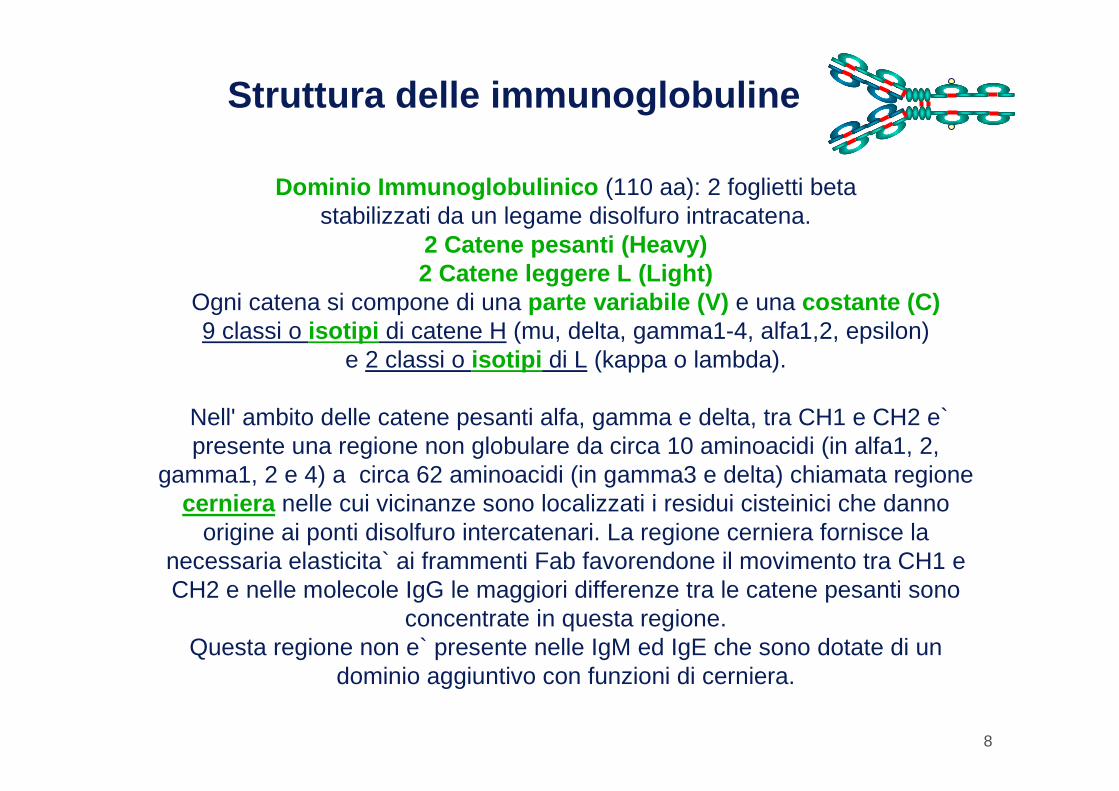
Struttura delle immunoglobuline
Dominio Immunoglobulinico (110 aa): 2 foglietti beta stabilizzati da un legame disolfuro intracatena.
2 Catene pesanti (Heavy)2 Catene leggere L (Light)
Ogni catena si compone di una parte variabile (V) e una costante (C)9 classi o isotipi di catene H (mu, delta, gamma1-4, alfa1,2, epsilon)
e 2 classi o isotipi di L (kappa o lambda).
Nell' ambito delle catene pesanti alfa, gamma e delta, tra CH1 e CH2 e` presente una regione non globulare da circa 10 aminoacidi (in alfa1, 2,
gamma1, 2 e 4) a circa 62 aminoacidi (in gamma3 e delta) chiamata regione cerniera nelle cui vicinanze sono localizzati i residui cisteinici che danno
origine ai ponti disolfuro intercatenari. La regione cerniera fornisce la necessaria elasticita` ai frammenti Fab favorendone il movimento tra CH1 e CH2 e nelle molecole IgG le maggiori differenze tra le catene pesanti sono
concentrate in questa regione.Questa regione non e` presente nelle IgM ed IgE che sono dotate di un
dominio aggiuntivo con funzioni di cerniera.

9
Immunoglobulin Structure (1)
• Heavy & Light Chains -All immunoglobulins have a fourchain structure as their basic unit. They are composed of two identicallight chains (23Kd) and two identical heavy chains (50-70Kd)
CH1
VL
CL
VH
CH2 CH3
Hinge Region
Carbohydrate
Disulfide bond

10
Immunoglobulin Structure (2)• Disulfide bonds
– Inter-chain - The heavy and light chains and the two heavychains are held together by inter-chain disulfide bonds and by non-covalent interactions. The number of interchain disulfide bonds variesamong different immunoglobulin molecules.
– Intra-chain - Within each of the polypeptide chains there are also intra-chain disulfide bonds.
CH1
VL
CL
VH
CH2 CH3
Hinge Region
Carbohydrate
Disulfide bond

11
Immunoglobulin Structure
• Variable & Constant Regions - After the amino acid sequences of many different heavy chains and light chains werecompared, it became clear that both the heavy and light chain could bedivided into two regions based on variability in the amino acid sequences. – 1. Light Chain - VL (110 aa) and CL (110 aa)
– 2. Heavy Chain - VH (110 aa) and CH (330-440 aa)
CH1
VL
CL
VH
CH2 CH3
Hinge Region
Carbohydrate
Disulfide bond

12
Immunoglobulin Structure
• Hinge Region - The region at which the arms of the antibody molecule forms a Y is called the hinge region because there is some flexibility in the molecule at this point.
CH1
VL
CL
VH
CH2 CH3
Hinge Region
Carbohydrate
Disulfide bond
13
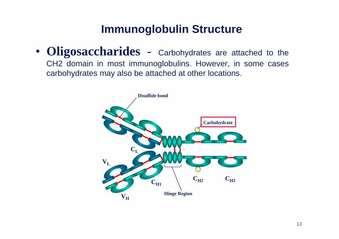
Immunoglobulin Structure
• Oligosaccharides - Carbohydrates are attached to the CH2 domain in most immunoglobulins. However, in some casescarbohydrates may also be attached at other locations.
CH1
VL
CL
VH
CH2 CH3
Hinge Region
Carbohydrate
Disulfide bond
14
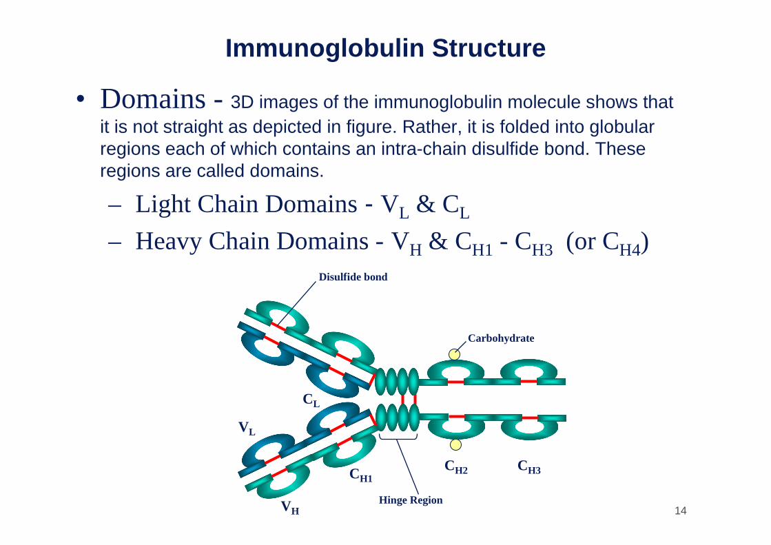
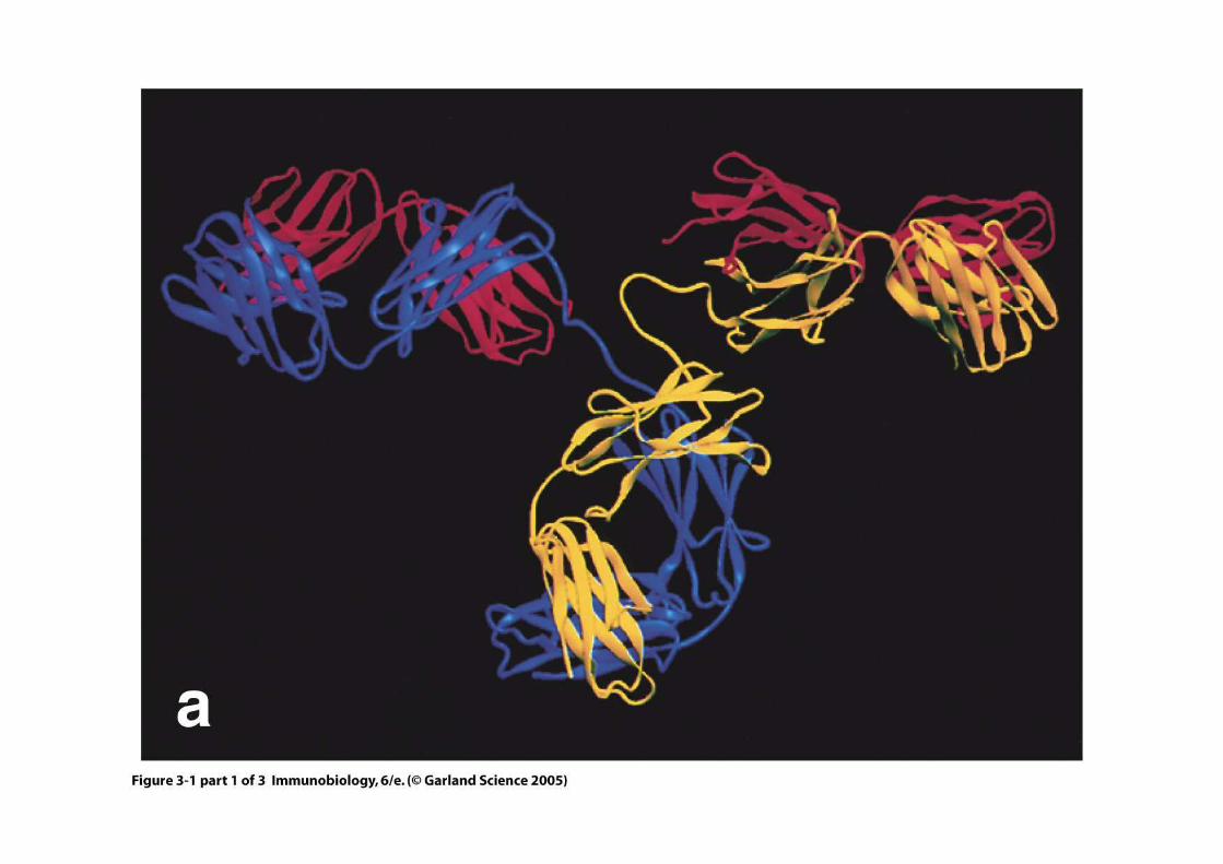
Immunoglobulin Structure
• Domains -3D images of the immunoglobulin molecule shows thatit is not straight as depicted in figure. Rather, it is folded into globularregions each of which contains an intra-chain disulfide bond. Theseregions are called domains.
– Light Chain Domains- VL & CL
– Heavy Chain Domains - VH & CH1 - CH3 (or CH4)
CH1
VL
CL
VH
CH2 CH3
Hinge Region
Carbohydrate
Disulfide bond

15
Dominio Immunoglobulinico:
2 foglietti betastabilizzati da un
legame disolfurointracatena ripiegati.
16
Figure 3-1 part 1 of 3

17
Figure 3-1 part 2 of 3

18
Figure 3-1 part 3 of 3

19
Figure 3-2

20
Complessi antigene-anticorpo
EM

21
Structure of the Variable Region• Hypervariable (HVR ) or complimentarity determining
regions (CDR) - Comparisons of the amino acid sequences of the variable regions of Igs show that most of the variability resides in threeregions are called the hypervariable regions or the complementaritydetermining regions as illustrated in figure. Antibodies with differentspecificities (i.e. different combining sites) have different CDR's whileantibodies of the exact same specificity have identical CDR's (i.e. CDR --> Ab Combing site). CDR's are found in both the H and the L chains.
HVR3
FR1 FR2 FR3 FR4
HVR1HVR2
Va
riabi
lity
Inde
x
25 7550 100Amino acid residue
150
100
50
0

22
Structure of the Variable RegionHVR3
FR1 FR2 FR3 FR4
HVR1HVR2
Va
riabi
lity
Inde
x
25 7550 100Amino acid residue
150
100
50
0
• Framework regions (FR) - The regions between the CDR's in the variable region are called the framework regions. Based on similarities and differences in the framework regions the immunoglobulin heavy and light chain variable regions can be dividedinto groups and subgroups. These represent the products of differentvariable region genes.
23
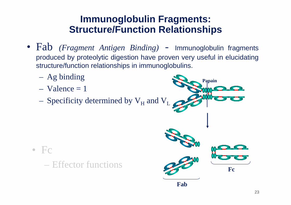
Immunoglobulin Fragments: Structure/Function Relationships
• Fab (Fragment Antigen Binding) - Immunoglobulin fragmentsproduced by proteolytic digestion have proven very useful in elucidatingstructure/function relationships in immunoglobulins.
– Ag binding
– Valence = 1
– Specificity determined by VH and VL
Papain
Fc
Fab
• Fc– Effector functions
24
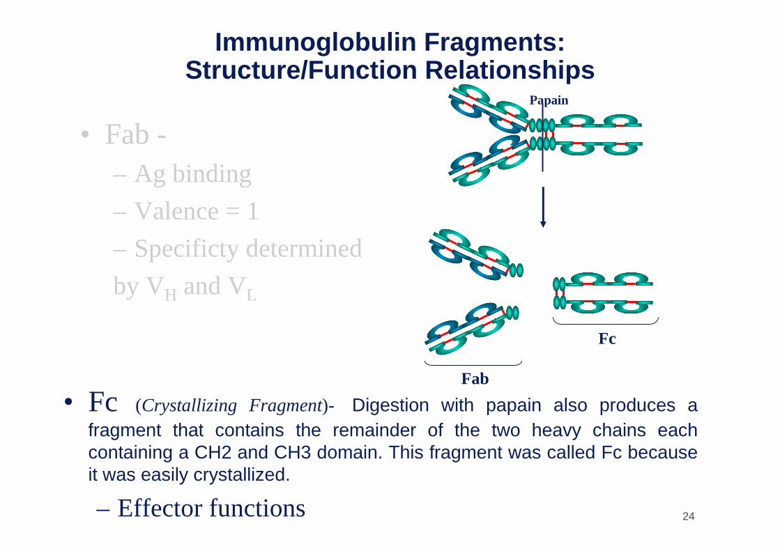
Immunoglobulin Fragments: Structure/Function Relationships
• Fab -– Ag binding
– Valence = 1
– Specificty determined
by VH and VL
Papain
Fc
Fab
• Fc (Crystallizing Fragment)- Digestion with papain also produces a fragment that contains the remainder of the two heavy chains eachcontaining a CH2 and CH3 domain. This fragment was called Fc becauseit was easily crystallized.
– Effector functions

25
Immunoglobulin Fragments: Structure/Function Relationships
• Fab
– Ag binding - These fragments were called the Fab fragments
because they contained the antigen binding sites of the antibody.
Each Fab fragment is monovalent whereas the original molecule was
divalent. The combining site of the antibody is created by both VH
and VL. An antibody is able to bind a particular antigenic determinant
because it has a particular combination of VH and VL. Different
combinations of a VH and VL result in antibodies that can bind a
different antigenic determinants.

26
Immunoglobulin Fragments: Structure/Function Relationships
Papain
Fc
Fab
• Fc– Effector functions -The effector
functions of immunoglobulins are mediated by this part of the molecule.
Different functions are mediated by the different domains in this fragment.
Normally the ability of an antibody to carryout an effector function requires the priorbinding of an antigen. However, there are exceptions to this rule.
27
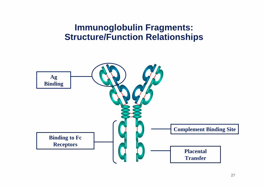
Immunoglobulin Fragments: Structure/Function Relationships
Ag Binding
Complement Binding Site
Placental Transfer
Binding to FcReceptors
28
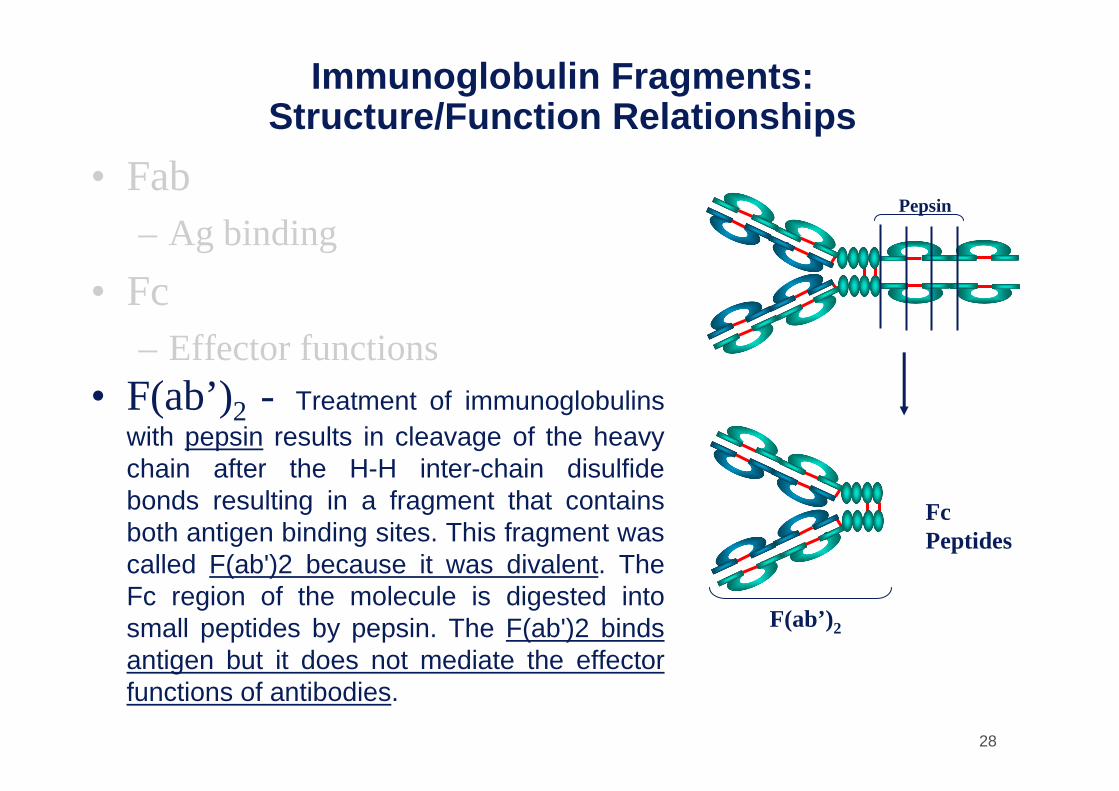
Immunoglobulin Fragments: Structure/Function Relationships
• Fab– Ag binding
• Fc– Effector functions
• F(ab’)2 - Treatment of immunoglobulinswith pepsin results in cleavage of the heavychain after the H-H inter-chain disulfidebonds resulting in a fragment that containsboth antigen binding sites. This fragment wascalled F(ab')2 because it was divalent. The Fc region of the molecule is digested intosmall peptides by pepsin. The F(ab')2 bindsantigen but it does not mediate the effectorfunctions of antibodies.
Pepsin
FcPeptides
F(ab’)2
29
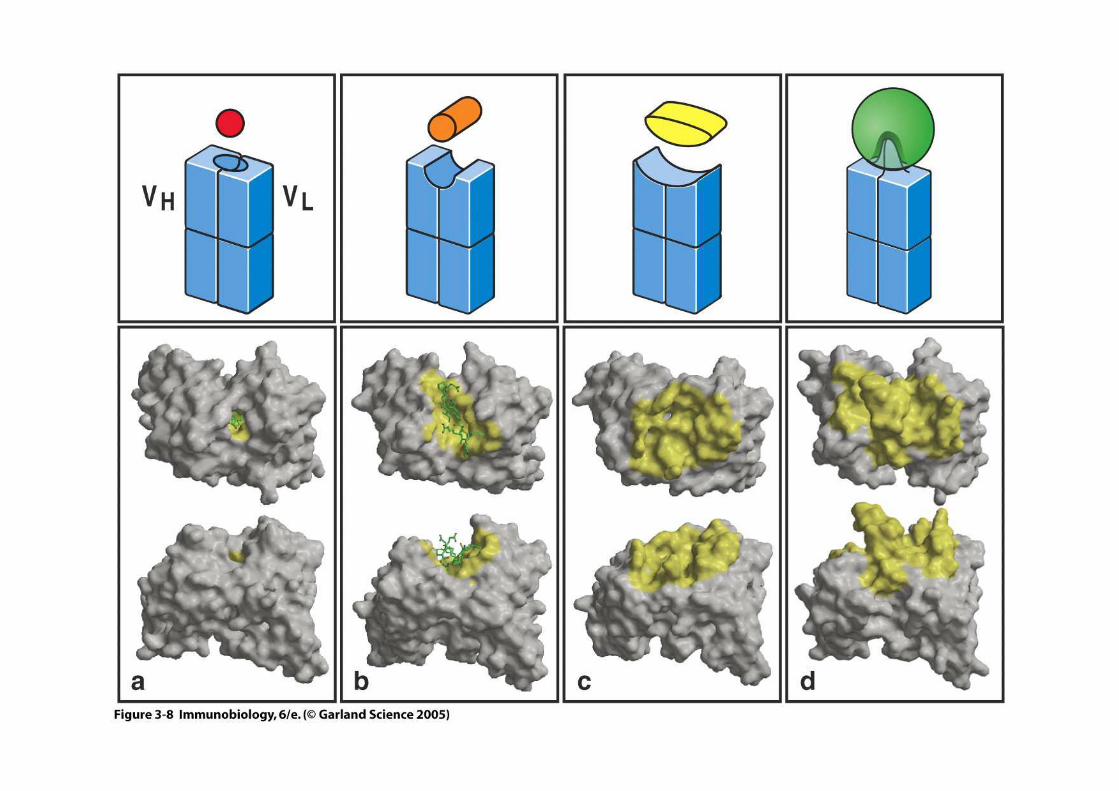
Figure 3-8
30
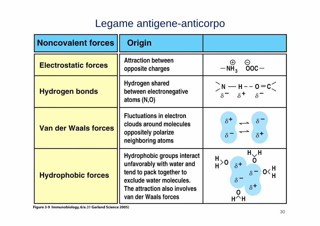
Legame antigene-anticorpo
31
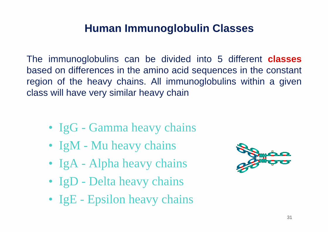
Human Immunoglobulin Classes
• IgG - Gamma heavy chains
• IgM - Mu heavy chains
• IgA - Alpha heavy chains
• IgD - Delta heavy chains
• IgE - Epsilon heavy chains
The immunoglobulins can be divided into 5 different classesbased on differences in the amino acid sequences in the constantregion of the heavy chains. All immunoglobulins within a givenclass will have very similar heavy chain

32
Human Immunoglobulin Subclasses
• IgG Subclasses– IgG1 - Gamma 1 heavy chains
– IgG2 - Gamma 2 heavy chains
– IgG3 - Gamma 3 heavy chains
– IgG4 - Gamma 4 heavy chains
• IgA subclasses– IgA1 - Alpha 1 heavy chains
– IgA2 - Alpha 2 heavy chains
The classes of immunoglobulins can de divided into subclasses based on small differences in the amino acid sequences in the constant region of the heavy chains. All immunoglobulins within a subclass will have very similarheavy chain constant region amino acid sequences. Again these differencesare most commonly detected by serological means.

33
Human ImmunoglobulinLight Chain Types
• Kappa
• Lambda
Immunoglobulins can also be classified by the type of light chain that theyhave. Light chain types are based on differences in the amino acid sequence in the constant region of the light chain. These differences are detected by serological means.

34
Human ImmunoglobulinLight Chain Subtypes
• Lambda light chains– Lambda 1
– Lambda 2
– Lambda 3
– Lambda 4
The light chains can also be divided into subtypes based on differencesin the amino acid sequences in the constant region of the light chain.
35
Immunoglobulins
• Nomenclature -Immunoglobulins are named based on the class, or subclass of the heavy chain and type or subtype of light chain. Unless it is stated precisely you are to assume that allsubclass, types and subtypes are present. IgG means that allsubclasses and types are present.
– IgM (kappa)
– IgA1(lambda 2)
– IgG
• Heterogeneity -Immunoglobulins considered as a populationof molecules are normally very heterogeneous because they are composed of different classes and subclasses each of which hasdifferent types and subtypes of light chains. In addition, differentimmunoglobulin molecules can have different antigen bindingproperties because of different VH and VL regions.
36
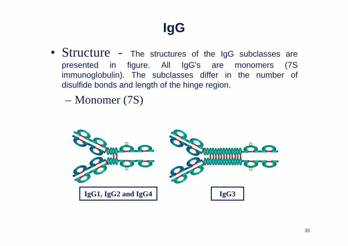
IgG
• Structure - The structures of the IgG subclasses are presented in figure. All IgG's are monomers (7S immunoglobulin). The subclasses differ in the number of disulfide bonds and length of the hinge region.
– Monomer (7S)
IgG1, IgG2 and IgG4 IgG3

37
IgG
• Structure• Properties -Most versatile immunoglobulin because it is
capable of carrying out all of the functions of immunoglobulinmolecules.
– Major serum Ig– Major Ig in extravascular spaces– Placental transfer – Does not require Ag
binding (IgG2 not well)– Fixes complement (except IgG4)– Binds to Fc receptors (IgG2, IgG4 do not bind)
• Phagocytes - opsonization• K cells - ADCC

38
IgG
• Structure• Properties
– Major serum Ig - 75% of serum Ig is IgG– Major Ig in extravascular spaces– Placental transfer – Does not require Ag
binding (IgG2)– Fixes complement (IgG4)– Binds to Fc receptors (IgG2, IgG4)
• Phagocytes - opsonization• K cells - ADCC
39
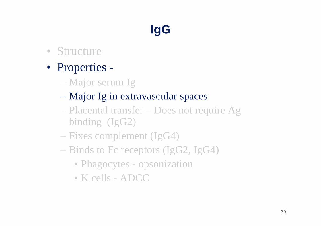
IgG
• Structure• Properties -
– Major serum Ig– Major Ig in extravascular spaces– Placental transfer – Does not require Ag
binding (IgG2)– Fixes complement (IgG4)– Binds to Fc receptors (IgG2, IgG4)
• Phagocytes - opsonization• K cells - ADCC
40
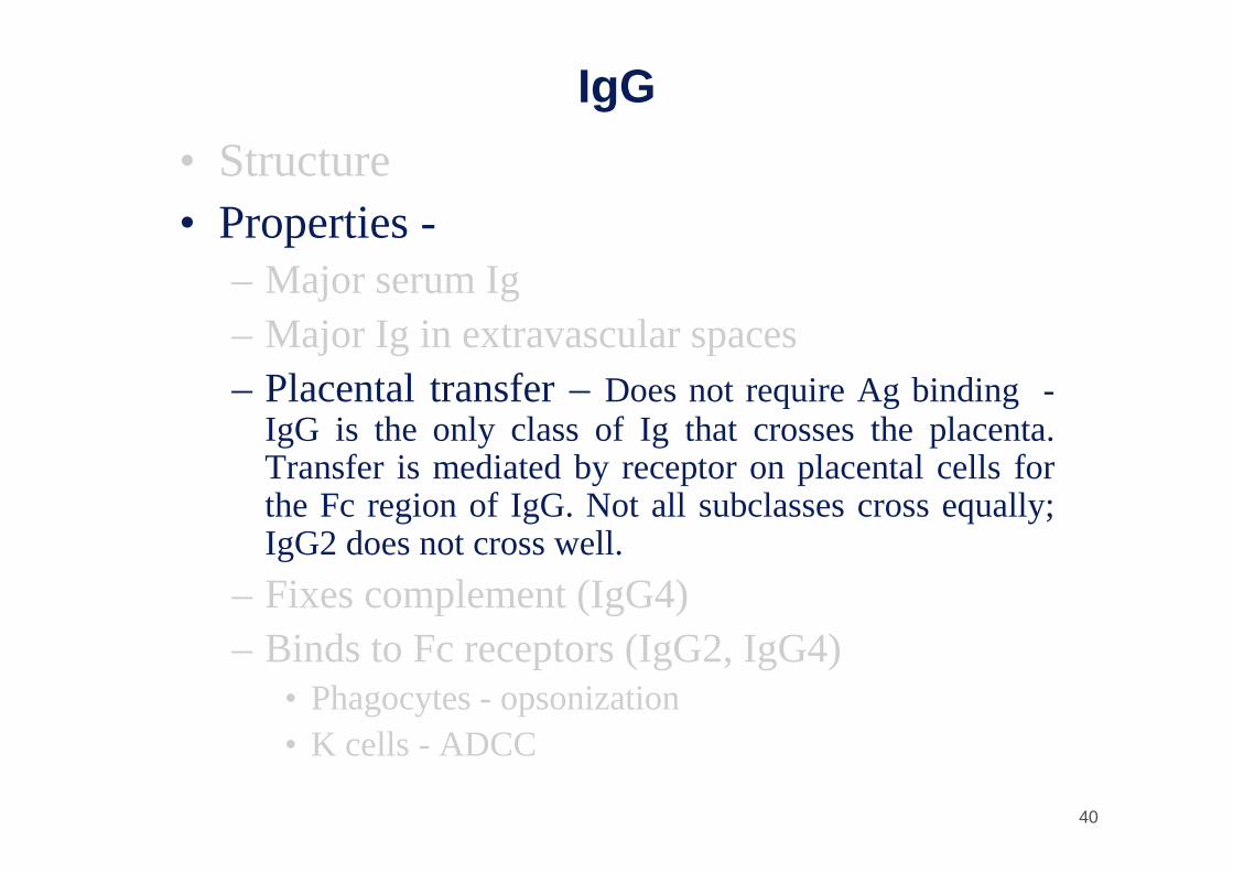
IgG
• Structure• Properties -
– Major serum Ig– Major Ig in extravascular spaces– Placental transfer –Does not require Ag binding -
IgG is the only class of Ig that crosses the placenta. Transfer is mediated by receptor on placental cells forthe Fc region of IgG. Not all subclasses cross equally; IgG2 does not cross well.
– Fixes complement (IgG4)– Binds to Fc receptors (IgG2, IgG4)
• Phagocytes - opsonization• K cells - ADCC

41
IgG
• Structure• Properties
– Major serum Ig– Major Ig in extravascular spaces– Placental transfer – Does not require Ag
binding (IgG2)– Fixes complement - Not all subclasses fix
equally well; IgG4 does not fix complement– Binds to Fc receptors (IgG2, IgG4)
• Phagocytes - opsonization• K cells - ADCC

42
IgG• Structure• Properties
– Major serum Ig– Major Ig in extravascular spaces– Placental transfer – Does not require Ag binding
(IgG2)– Fixes complement - (IgG4)– Binds to Fc receptors -Macrophages, monocytes, PMN's and
some lymphocytes have Fc receptors for the Fc region of IgG. Not allsubclasses bind equally well; IgG2 and IgG4 do not bind to Fc receptors. A consequence of binding to the Fc receptors on PMN's, monocytes and macrophages is that the cell can now internalize the antigen better. The antibody has prepared the antigen for eating by the phagocytic cells. The term opsonin is used to describe substances that enhance phagocytosis. IgG is a good opsonin. Binding of IgG to Fc receptors on other types of cellsresults in the activation of other functions.
• Phagocytes - opsonization• K cells - ADCC

43
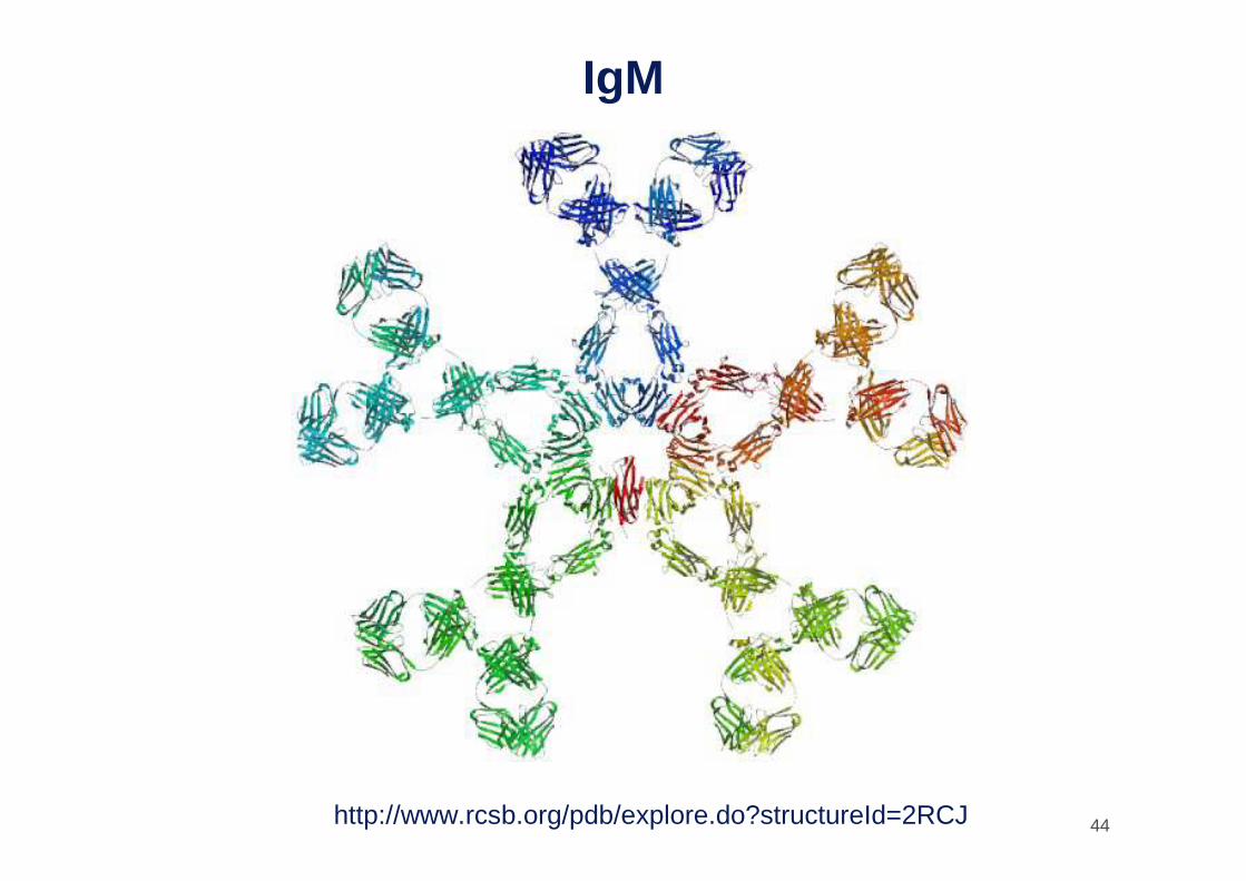
IgM• Structure -IgM normally
exists as a pentamer (19S immunoglobulin) but it can also exist as a monomer. In the pentameric form all heavychains are identical and alllight chains are identical.Thus, the valence istheoretically 10. IgM has anextra domain on the ì chain(CH4) and it has anotherprotein covalently bound via a S-S bond called the J chain. This chain functions in polymerization of the moleculeinto a pentamer.
– Pentamer (19S)
– Extra domain (CH4)
– J chain
Cµµµµ4
J Chain
44
IgM
http://www.rcsb.org/pdb/explore.do?structureId=2RCJ

45
IgM
• Structure
• Properties– 3rd highest serum Ig -IgM is the 3rd most common serum Ig.
– First Ig made by fetus and B cells -IgM is the first Ig to bemade by the fetus and the first Ig to be made by a virgin B cellswhen it is stimulated by antigen.
– Fixes complement -As a consequence of its pentamericstructure, IgM is a good complement fixing Ig. Thus, IgM antibodiesare very efficient in leading to the lysis of microrganisms.

46
IgM• Structure
• Properties– 3rd highest serum Ig
– First Ig made by fetus and B cells
– Fixes complementTail Piece– Agglutinating Ig - As a consequence of its
structure, IgM is also a good agglutinating Ig. Thus, IgM antibodies are very good in clumpingmicroorganisms for eventual elimination from the body.
– Binds to Fc receptors -IgM binds to some cells via Fc receptors.
– B cell surface Ig

47
IgM• Structure
• Properties– 3rd highest serum Ig– First Ig made by fetus
and B cells– Fixes complement
Tail Piece
– Agglutinating Ig
– Binds to Fc receptors
– B cell surface Ig -Surface IgM exists as a monomer and lacks J chain but it has anextra 20 amino acids at the C-terminal end to anchor it into the membrane (see figure). Cell surfaceIgM functions as a receptor for antigen on B cells. Surface IgM is noncovalently associated with twoadditional proteins in the membrane of the B cell called Ig-αααα and Ig-ββββ as indicated in the next figure. These additional proteins act as signal transducing molecules since the cytoplasmic tail of the Ig molecule itself is too short to transduce a signal. Contact between surface immunoglobulin and anantigen is required before a signal can be transduced by the Ig-α and Ig-β chains. In the case of T-independent antigens (LPS batterico, capsule polisaccaridiche, flagelli batterici), contact between the antigen and surface immunoglobulin is sufficient to activate B cells to differentiate into antibody secreting plasma cells. However, for T-dependent antigens, a second signal provided by helper T cellsis required before B cells are activated.

48
B Cell Antigen Receptor (BcR)
Ig-αIg-β Ig-βIg-α
Vi sono due forme di molecole immunoglobuliniche: di membrana (recettori Ig) e secretorie (immunoglobuline circolanti). Queste due forme si differenziano per l'estremita` COOH terminale: la forma secretoria possiede un peptide terminale altamente idrofilo chiamato Ts (secretory tail), mentre la forma di membrana termina con la sequenza idrofoba Tm (membrane tail) che ne permette l'ancoraggio alla membrana

49
IgA
• Structure– Serum - monomer -Serum IgA is a monomer but IgA found in
secretions is a dimer as presented in figure. When IgA exits as a dimer, a J chain is associated with it.
– Secretions (sIgA)• Dimer (11S)
• J chain
• Secretory component
J ChainSecretory Piece

50
IgA• Properties• IgA is the 2nd most common serum Ig.• IgA is the major class of Ig in secretions - tears, saliva, colostrum,
mucus. Since it is found in secretions secretory IgA is important in local(mucosal) immunity.
• Normally IgA does not fix complement, unless aggregated.• IgA can binding to some cells - PMN's and some lymphocytes.
J ChainSecretory Piece
51
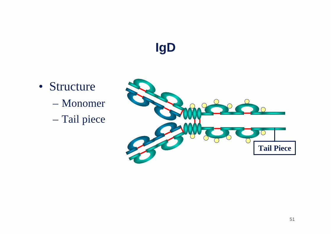
IgD
• Structure– Monomer
– Tail piece
Tail Piece

52
IgD
• Structure
• Properties– 4th highest serum Ig -IgD is found in low levels in
serum; its role in serum uncertain.– B cell surface Ig -IgD is primarily found on B cell
surfaces where it functions as a receptor for antigen. IgD on the surface of B cells has extra amino acids at C-terminalend for anchoring to the membrane. It also associates withthe Ig-αααα and Ig-ββββ chains.
– Does not bind complement

53
IgE
• Structure -IgE exists as a monomer and has an extra domain in the constant region.
– Monomer
– Extra domain (CH4)
CH4
54
IgE
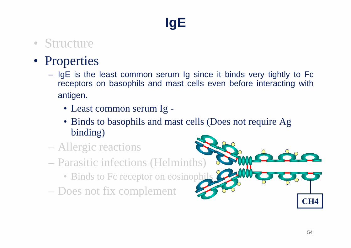
• Structure• Properties
– IgE is the least common serum Ig since it binds very tightly to Fcreceptors on basophils and mast cells even before interacting withantigen.
• Least common serum Ig -• Binds to basophils and mast cells (Does not require Ag
binding)
– Allergic reactions– Parasitic infections (Helminths)
• Binds to Fc receptor on eosinophils
– Does not fix complementCH4

55
IgE• Structure• Properties
– Least common serum Ig– Binds to basophils and mast cells (Does not require Ag
binding)– Allergic reactions - Involved in allergic reactions - As a
consequence of its binding to basophils an mast cells, IgE is involvedin allergic reactions. Binding of the allergen to the IgE on the cellsresults in the release of various pharmacological mediators that resultin allergic symptoms.
– Parasitic infections (Helminths)• Binds to Fc receptor on eosinophils
– Does not fix complement
CH4
56
IgE
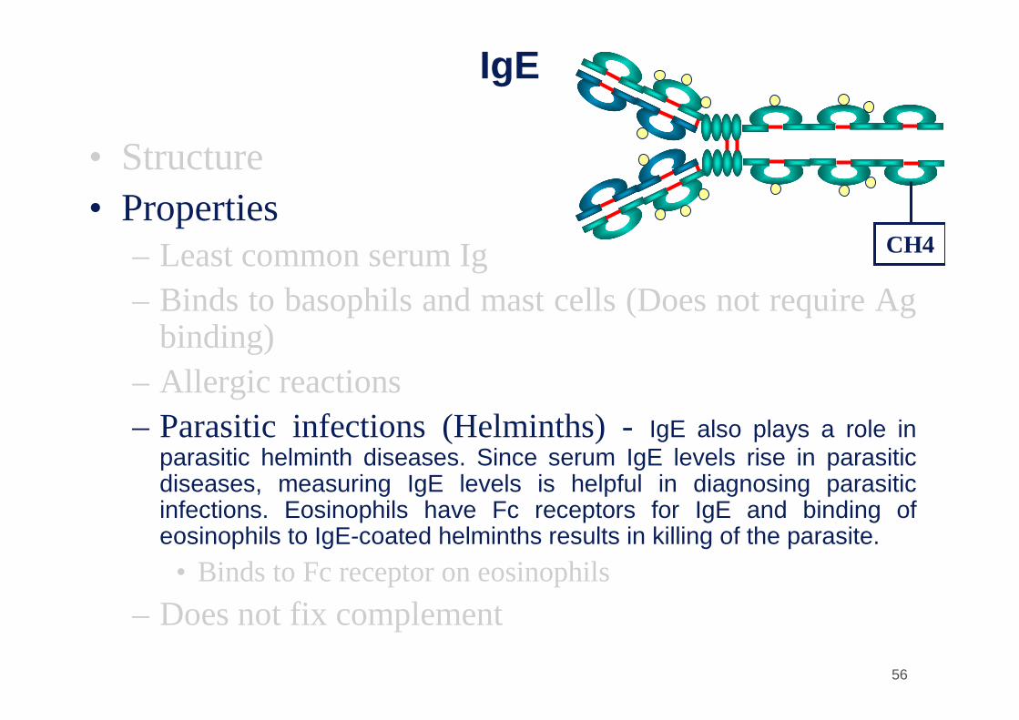
• Structure• Properties
– Least common serum Ig– Binds to basophils and mast cells (Does not require Ag
binding)– Allergic reactions– Parasitic infections (Helminths) -IgE also plays a role in
parasitic helminth diseases. Since serum IgE levels rise in parasiticdiseases, measuring IgE levels is helpful in diagnosing parasiticinfections. Eosinophils have Fc receptors for IgE and binding of eosinophils to IgE-coated helminths results in killing of the parasite.
• Binds to Fc receptor on eosinophils
– Does not fix complement
CH4
57
IgE
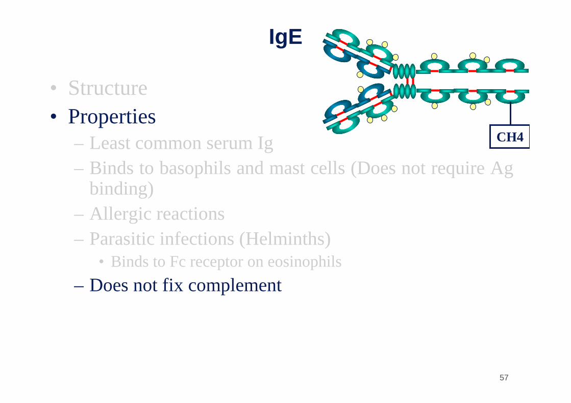
• Structure• Properties
– Least common serum Ig– Binds to basophils and mast cells (Does not require Ag
binding)– Allergic reactions– Parasitic infections (Helminths)
• Binds to Fc receptor on eosinophils
– Does not fix complement
CH4