Embed Size (px)
Citation preview
MEDICAL SCHOOL
DEPARTMENT OF PHSYIOLOGY
MSc: MOLECULAR AND APPLIED PHYSIOLOGY
CORRELATION BETWEEN SEMINAL PLASMA LEVES OF GROWTH HORMONE, INSULIN-LIKE GROWTH FACTOR-1 AND MALE INFERTILITY
MAZIOTIS EVANGELOS
RN: 201500345
SUPERVISING COMMITTEE:
SIMOPOULOU MARIA, Assistant Professor, Physiology, Medical School
KOUTSILIERIS MICHAEL, Professor, Head of Physiology Department, Medical
School
VAIOPOULOS GEORGIOS, Honored Professor, Physiology, Medical School
ATHENS, 2017
CONTENTS
ACKNOWLEDGEMENTS ............................................................................................................. 3
ABSTRACT .................................................................................................................................. 4
INTRODUCTION ......................................................................................................................... 5
Spermatogenesis ................................................................................................................... 5
Biology of semen ................................................................................................................... 6
Male Infertility ....................................................................................................................... 7
SEMEN ANALYSIS DIAGNOSIS ................................................................................................ 9
Approach and management of male infertility within the scope of Assisted Reproduction
Technologies (ART) .............................................................................................................. 10
Growth Hormone ................................................................................................................ 11
IGF-1 .................................................................................................................................... 13
Association between Male Infertility, IGF-1 and GH ........................................................... 14
MATERIALS AND METHODS .................................................................................................... 16
Sample Collection ................................................................................................................ 16
Seminogram ........................................................................................................................ 16
DNA Fragmentation Test ..................................................................................................... 17
ELISA .................................................................................................................................... 17
Sandwich ELISA ................................................................................................................ 18
Competitive ELISA............................................................................................................ 18
Statistical analysis ................................................................................................................ 19
RESULTS ................................................................................................................................... 20
DISCUSSION ............................................................................................................................. 28
REFERENCES ............................................................................................................................ 32

ACKNOWLEDGEMENTS
I would like to express my gratitude to my supervisor, Assistant Professor Mara
Simopoulou, for her continuous help and support, and the faith she showed in me
entrusting me with this project. I would also like to thank Assistant Professor
Anastasios Philippou for selflessly offering his counseling and lab assistance during
this project. Moreover I would like to thank Genesis Clinic for their cooperation and
especially Dr. Ioannis Nitsos without whom the completion of this project would be
almost impossible. Furthermore I would like to express my gratitude to Professor
Michael Koutsilieris and Honored Professor George Vaiopoulos for accepting to
elaborate my thesis. Finally I would like to thank my colleagues Vagelis Zevolis and
Stella Tassou for their lab assistance at all times.
ABSTRACT
Infertility is a common problem affecting a considerable percentage of the male
population worldwide and has been a research topic for many decades. Growth
Hormone and Insulin-like Growth Factor 1 have been proposed as factors that may
have a pivotal role in male infertility due to their anabolic effect. The aim of this
study is to investigate a possible correlation between seminal plasma levels of GH and
IGF-1 and sperm parameters that may exert an impact on fertility. The study took
place between May 2017 and September 2017 with the participation of 50 patients. A
marginally non-significant statistic difference (p=0.063) in GH levels has been found
between asthenospermic group and normal group. A statistically significant
correlation (p<0.05) has been found between levels of GH and IGF-1 in the group
with asthenospermia and at least one more abnormal parameter. The above correlation
has been found also in patients with low sperm concentration, vitality, volume and
abnormal morphology. According to our knowledge this is a novel finding and further
studies are required in order to clarify the biological significance of this correlation.
ΣΥΝΟΨΗ
Η ππνγνληκόηεηα είλαη έλα ζύλεζεο πξόβιεκα πνπ αθνξά έλα ζεκαληηθό πνζνζηό
ηνπ παγθόζκηνπ αλδξηθνύ πιεζπζκνύ θαη απνηειεί εξεπλεηηθό ηνκέα εδώ θαη
αξθεηέο δεθαεηίεο. Η απμεηηθή νξκόλε θαη ν Insulin-like Growth Factor-1 έρνπλ
πξνηαζεί ωο παξάγνληεο πνπ δηαδξακαηίδνπλ θαζνξηζηηθό ξόιν ζηελ αλδξηθή
ππνγνληκόηεηα εμαηηίαο ηεο αλαβνιηθήο δξάζεο ηνπο. Ο ζθνπόο ηεο παξνύζαο
κειέηεο είλαη ε δηεξεύλεζε πηζαλήο ζπζρέηηζεο κεηαμύ ηωλ επηπέδωλ GH θαη IGF-1
ηνπ ζπεξκαηηθνύ πιάζκαηνο θαη ηωλ ζπεξκαηηθώλ παξακέηξεωλ πνπ πηζαλώο
επεξεάδνπλ ηελ αλδξηθή ππνγνληκόηεηα. H κειέηε πξαγκαηνπνηήζεθε κεηαμύ Μαΐνπ
2017 θαη Σεπηεκβξίνπ 2017 κε ηε ζπκκεηνρή 50 αζζελώλ. Μηα νξηαθά κε ζεκαληηθή
ζηαηηζηηθή δηαθνξά (ξ=0.063) ζηα επίπεδα ηεο GH βξέζεθε αλάκεζα ζην
θπζηνινγηθό θαη ην αζζελνζπεξκηθό γθξνππ. Μηα ζηαζηηζηηθά ζεκαληηθή ζπζρέηηζε
(ξ<0.05) βξέζεθε αλάκεζα ζηα επίπεδα ηεο GH θαη ηνπ IGF-1 ζην γθξνππ κε
αζζελνζπεξκία θαη ηνπιάρηζηνλ κηα αθόκε κε θπζηνινγηθή ζπεξκαηηθή παξάκεηξν.
Η παξαπάλω ζπζρέηηζε βξέζεθε θαη ζηνπο αζζελείο κε ρακειή ζπγθέληξωζε
ζπεξκαηνδωαξίωλ, ρακειή δωηηθόηεηα ζπεξκαηνδωαξίωλ θαη ρακειό όγθν
ζπέξκαηνο. Σύκθωλα κε ηελ γλώζε καο πξόθεηηαη γηα έλα θαηλνηόκν εύξεκα πνπ
ρξεηάδεηαη πεξαηηέξω έξεπλα πξνθεηκέλνπ λα δηαζαθεληζηεί ε βηνινγηθή ζεκαζία ηεο
ζπζρέηηζεο απηήο.
INTRODUCTION
Spermatogenesis Spermatogenesis is the multi-step process that begins with the first mitotic division of
germ cells in order to become mature spermatozoa. During embryogenesis the
primordial germ cells migrate into the gonad and become immature germ cells named
spermatogonia. Spermatogonia have 46 chromosomes. After puberty and almost
throughout life these cells divide mitotically.
Some of the spermatogonia enter into their first meiotic division and become primary
spermatocytes. At the prophase of this meiotic division the chromosomes undergo
cross-over and as a result the primary spermatocytes become tetraploid cells (46 sets
of chromosomes). Completing this meiotic division, the daughter cells become
secondary spermatocytes, which have 23 sets of chromosomes (22 autosomal and a
duplicated X or Y chromosomes). Undergoing another meiosis the secondary
spermatocytes become spermatids which are haploid cells, having one of each
autosomal chromosomes and a X or Y chromosome. Spermatids form the inner layer
of the epithelium and are found in rather discrete aggregates as the cells derived from
a single spermatogonium tend to remain together and differentiate synchronously
[Boron, 2012]
For the production of mature spermatozoa by spermatids another process is required,
the spermiogenesis. Spermatids are located adjacent to the lumen of the seminiferous
tubules during the early stages of spermiogenesis and are surrounded by processes of
Sertoli cell cytoplasm. The Sertoli cells are large, polyhedral cells extending from the
basement membrane toward the lumen of the seminiferous tubule. Tight junctions
connect the adjacent Sertoli cells to forming a blood-testis barrier providing a
protective environment for developing germ cells. In addition, gap junctions between
the Sertoli cells and developing spermatozoa may represent a mechanism for
transferring material between these two types of cells. Release of the spermatozoa
from the Sertoli cell has been called spermiation [Guyton & Hall, 2011].
During spermiation spermatozoa are immotile and are passively moved from the
Sertoli cells through the seminiferous tubules to the rete testes and to epididymis.
Spermatozoa are transferred to epididymis with assistance by ciliary action of the
luminal epithelium and contractility of the smooth muscle elements of the efferent
duct wall. [Vanders, 2001]
Sperm are stored in the epididymis, where they undergo a process of maturation
before they are capable of progressive motility and fertilization. Spermatozoa released
at ejaculation are fully motile and capable of fertilization providing they undergo
changes during their journey to the salpinx, whereas spermatozoa obtained directly
from the testis are functionally immature insofar as they cannot penetrate an ovum.
However, these immature spermatozoa can fertilize if they are injected into an ovum
employing the technique of Intracytoplasmic Sperm Injection ICSI [Vanderzwalmen
et al., 1995]. During maturation in the epididymis, spermatozoa undergo changes in
motility, metabolism, and morphology. Spermatozoa derived from the head of the
epididymis are often unable to fertilize ova, whereas larger proportions of
spermatozoa captured from the body are fertile. Spermatozoa obtained from the tail of
the epididymis, or from the vas deferens, are almost always capable of fertilization.
[Boron, 2012]
The epididymis empties into the vas deferens, which is responsible for the movement
of sperm along the tract. The vas deferens contains well-developed muscle layers that
facilitate sperm movement. The vas deferens passes through the inguinal canal,
traverses the ureter, and continues medially to the posterior and inferior aspect of the
urinary bladder, where it is joined by the duct arising from the seminal vesicle;
together, they form the ejaculatory duct.
Almost 74 days are needed for spermatogonia to become mature spermatozoa and
each stage of the process has a specific duration. The rate of spermatogenesis is
constant and cannot be accelerated by hormones such as gonadotropins or androgens.
Germ cells must comply with a specific timeline in their differentiation. If the
environment is unfavorable and makes it impossible for them to continue their
differentiation at the normal rate they degenerate. The production rate of spermatozoa
is the highest for a man in his 20‟s and drops progressively throughout life. In men
older than 50 years old a significant decrease in the percentage of morphologically
normal and motile spermatozoa is found. [Chrysikopoulos, 2001]
Biology of semen Only 10% of the volume of semen is sperm cells. According to World Health
Organisation the normal concentration of sperm cells is greater than 15 million/mL,
and the typical ejaculate volume is greater than 1.5 mL. The typical ejaculate content
varies between 150 and 600 million spermatozoa.
Aside from the sperm cells, the remainder of the semen (i.e., 90%) is seminal plasma,
the extracellular fluid of semen. Very little seminal plasma accompanies the
spermatozoa as they move through the testes and epididymis. The seminal plasma
originates primarily from the accessory glands (the seminal vesicles, prostate gland,
and the bulbourethral glands). The seminal vesicles contribute around 70% of the
volume of semen. Aside from the sperm, the remaining 20% represents epididymal
fluids, as well as secretions of the prostate gland and bulbourethral glands.
Seminal plasma contains a plethora of sugars and ions. Fructose and citric acid are
contributed to the seminal plasma by the accessory glands, and their concentrations
vary with the volume of semen ejaculated. Ascorbic acid and traces of B vitamins are
also found in human seminal plasma.
Seminal plasma also contains low molecular weight polypeptides and proteins. Sperm
functionality is relative to these proteins, which have a major role in sperm
interactions with the various environments along the female genital tract towards the
oocyte vestments. Specific peptides and proteins act as signals for the female immune
system to modulate sperm rejection or tolerance, perhaps even influencing the relative
intrinsic fertility of the male and/or couple by attaining a status of maternal tolerance
towards embryo and placental development [Rodriguez-Martinez et al., 2011].
Male Infertility Infertility is defined as the inability of a couple to achieve pregnancy after at least one
year of unprotected intercourse (WHO). Infertility is a common problem and
according to recent researche about 15% of couples are infertile worldwide. Around
50% of total infertility is attributed to male factor infertility alone or combined with
female infertility. Another study supports that infertility affects almost 7% of the total
male population [Lotti & Maggi, 2015]. These numbers may be even higher because
male infertility is not well reported due to social reasons especially in the developing
countries or the patriarchal societies.
According to European Urology Association almost 70% of male infertility is a result
of urogenital abnormalities, malignancies, urogenital track infections, increased
scrotal temperature, endocrine disturbances, genetic abnormalities and immunological
factors. In the other 30% no associated factor can be found although the semen
analysis is abnormal. This is called idiopathic infertility. A part of idiopathic
infertility is suspected to be caused by epigenetic disorders or life style habits.
Causes
Urogenital abnormalities may be congenital or acquired and refer to cryptorchidism,
hypospadias (dislocation of the urethra), congenital absence of vas deference (may be
a symptom of cystic fibrosis), obstructions, testicular torsion and orchitis (a result of
either viral or bacterial infection).
Cryptorchidism is the absence of one or both testes from the scrotum. It is one of the
most common congenital defects of the male reproductive system. It affects
approximately 2-4% of new-born boys and may have an impact at the health of the
male adult. Testosterone and Insuline-like factor 3 (INSL3) are major regulators of
the testicular descent. The cause of the abnormal descent may be genetic, including
mutations on the INSL3 gene, chromosomal alterations or polymorphisms [Foresta et
al., 2008]. Environmental reasons may be another cause of cryptorchidism including
maternal lifestyle habits like increased alcohol consumption [Daamgard et al., 2007]
and smoking [Jensen et al., 2007]. Other factors like pre-mature birth and low body
weight of the infant is found to increase the risk of cryptorchidism.
Increased scrotal temperature may be a result of varicocele. Varicocele is the
enlargement of the veins in the scrotum. It affects almost 15% of all adult males and
is amongst the most common causes of male infertility. Varicocele has an adverse
effect on spermatogenesis not only due to increased temperature but also causes
increased intratesticular pressure, hypoxia, reflux of toxic metabolites from the
adrenal glands and hormonal abnormalities [Kantartzi et al., 2007]. Scrotal
hyperthermia may also be caused by febrile illness or lifestyle habits (such as laptop
use [Sheynkin et al., 2005]).
Urogenital track infections are accounted for almost 15% of male infertility. The
infections may affect different locations of the urogenital track, which may be the
testes, the epididymis and the accessory male glands [Pellati et al., 2008]. The
spermatozoa may be affected during the different stages of their development
resulting in reduced vitality and reduced sperm count.
Endocrine disturbances may be the cause of infertility in a small percentage of men.
Usually they are a result of a dysfunction in hypothalamic-pituitary-testicular axis and
on rare occasions they are a result of thyroid or adrenal disorders. The causes of the
endocrine disturbances may be genetic or acquired. Acquired causes include tumors,
inflammation or injury of the hypothalamus or the pituitary.
Genetic disorders may act indirectly (as described above) or directly towards male
infertility. Genetic syndromes that include an abnormal chromosomal number are a
cause of infertility. Reciprocal translocations, ring chromosomal abnormalities,
Robertsonian translocations, inversions and reciprocal translocations are associated
with male infertility. Mutations affecting the Y chromosome are possible causes of
male infertility such as the AZF deletions [Shamsi et al., 2011]
Immune Infertility is an autoimmune disease caused by anti-sperm antibodies.
Abnormalities of the blood-testis barrier may cause the production of anti-sperm
antibodies. The anti-sperm antibodies may inhibit sperm functions that are important
for fertilization mostly affecting zona and oolemma binding [Bohring & Krause,
2003].
According to recent research another cause of infertility may be epigenetic disorders.
Epigenetic changes occur in many stages during spermatogenesis. These changes
include DNA methylation and phosphorylation, hyperacytelation, histone degradation,
ubiquitylation and sumoylation [Dada et al., 2012]. miRNAs also play an important
role in the fertility as there are more than 300 differentially regulated miRNAs when
comparing fertile and azoospermic men. [Gunes et al., 2016].
Lifestyle habits or work conditions may also have an impact in male fertility.
Gasoline, lead and zync fumes have been found to have a negative result in male
fertility. Smoking, obesity and drugs may also be a cause of infertility [Sharma et al.,
2013].
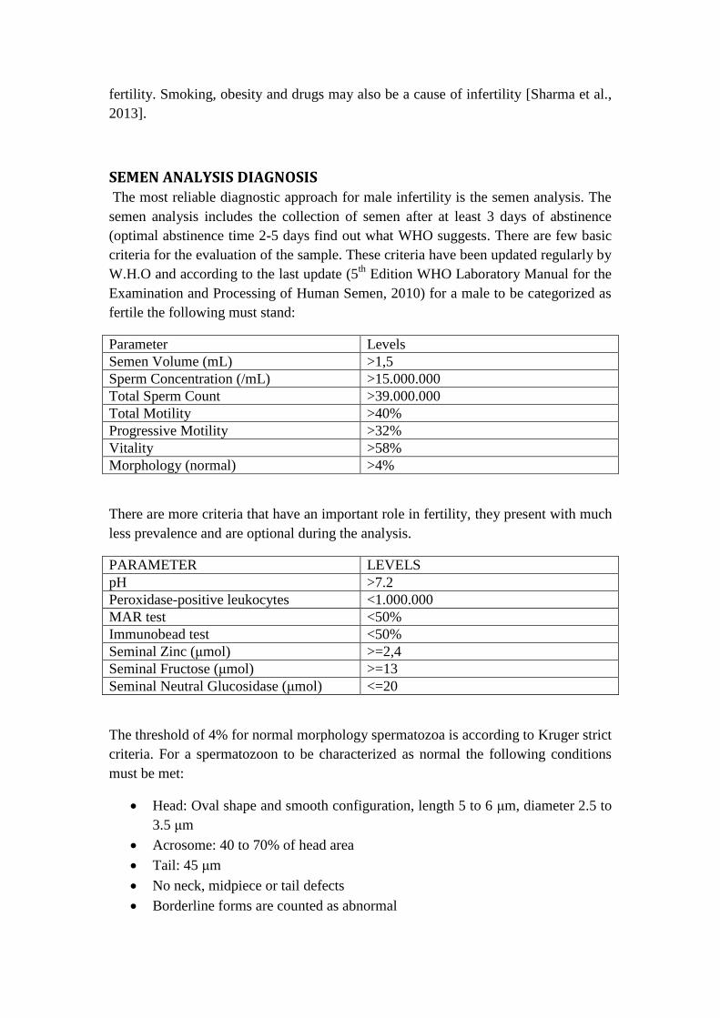
SEMEN ANALYSIS DIAGNOSIS The most reliable diagnostic approach for male infertility is the semen analysis. The
semen analysis includes the collection of semen after at least 3 days of abstinence
(optimal abstinence time 2-5 days find out what WHO suggests. There are few basic
criteria for the evaluation of the sample. These criteria have been updated regularly by
W.H.O and according to the last update (5th
Edition WHO Laboratory Manual for the
Examination and Processing of Human Semen, 2010) for a male to be categorized as
fertile the following must stand:
Parameter Levels
Semen Volume (mL) >1,5
Sperm Concentration (/mL) >15.000.000
Total Sperm Count >39.000.000
Total Motility >40%
Progressive Motility >32%
Vitality >58%
Morphology (normal) >4%
There are more criteria that have an important role in fertility, they present with much
less prevalence and are optional during the analysis.
PARAMETER LEVELS
pH >7.2
Peroxidase-positive leukocytes <1.000.000
MAR test <50%
Immunobead test <50%
Seminal Zinc (κmol) >=2,4
Seminal Fructose (κmol) >=13
Seminal Neutral Glucosidase (κmol) <=20
The threshold of 4% for normal morphology spermatozoa is according to Kruger strict
criteria. For a spermatozoon to be characterized as normal the following conditions
must be met:
Head: Oval shape and smooth configuration, length 5 to 6 κm, diameter 2.5 to
3.5 κm
Acrosome: 40 to 70% of head area
Tail: 45 κm
No neck, midpiece or tail defects
Borderline forms are counted as abnormal
[Kruger et al., 1986 & 1988]
Approach and management of male infertility within the scope of
Assisted Reproduction Technologies (ART) Male infertility is a dysfunction with complex etiology so there are few treatment
options. Drug administration, surgical corrections and Assisted Reproductive
Techniques are some of them. Since a large portion of infertility is caused by
idiopathic reasons, or other treatments may not have the expected results, ART are
commonly performed.
Intrauterine Insemination (IUI) is an assisted reproductive technique (ART) in which
sperm is transferred to a woman‟s uterine cavity in order to achieve fertilization. This
procedure has a long history of usage, mostly used in animals during the early years.
The protocol has changed over the course of time and after the discovery of modern
laboratory techniques in order to achieve higher success rates. After collection of the
semen, the sample is processed and washed in the laboratory, separating the sperm
from the other proteins and cells of the seminal fluid and only the highly motile
spermatozoa are transferred to the uterine cavity using a catheter. IUI is commonly
used as the first line of treatment managing an infertile couple with aetiology that
does not include male factor. However it may also be employed when mildly low
sperm count or motility is detected. IUI has a success rate of approximately 10% per
cycle (Soria et al., 2012), which is much less than other ARTs but is less invasive and
less costly.
The first successful IVF took place in 1978 and created many new opportunities for
couples suffering with infertility [Steptoe and Edwards, 1978] During the first years
of IVF practice success rates were relatively low. IVF procedure includes
gonadotropin medication treatment for women in order to cause controlled ovarian
stimulation resulting to production of multiple follicles of the ovaries. After the
follicles have reached a certain degree of development, other medications are
administered (usually chorionic gonadotropin) which trigger oocyte release. The
oocytes are retrieved using ultrasound guided transvaginal paracentesis and aspiration
of follicular fluid.
After collection of both the oocytes and the semen, selection procedures occur for
both samples. The oocyte selection is based mainly on the assessment of the cumulus
oocyte complex morphology. Further research has lead to the proposal and
enrichment of assessment criteria of selection such as on intrinistic (mitochondrial
status, glucose-6-phosphate dehydrogenase 1 activity) and extrinistic factors
(apoptosis of follicular cells or the levels of transforming of growth factor beta
superfamily in follicular fluid or serum) [Wang et al., 2007] or morphological
parameters (zona pellucida imaging) obtained by light or polarized light microscopy.
During sperm selection motile spermatozoa separation are separated from the semen,
mucus and other immotile or non-viable spermatozoa, using a technique called sperm
washing. This is performed by density gradient centrifugation or a direct swim up
technique. [Volpes et al., 2016]
The spermatozoa and the oocyte are incubated together in a culture media at a rate of
about 75000:1 (day 0). The incubation time preferred for better outcome is 1-4 hours
[Zhang et al., 2013]. If fertilization occurs two nuclei will be visible at the fertilized
oocyte following 16-18 hours post incubation(day 1). The fertilized oocyte then is
transferred to the appropriate growth medium. Embryos grow in culture until cleavage
stage (days 2-4) or until blastocyst stage (days 5-6). During the cleavage (except day
4) or blastocyst stage embryos are available either for embryo transfer or
cryopreservation. The embryo qualified for embryo transfer is evaluated using mainly
a morphological scoring system. Morphological parameters are acquired through
microscopy at certain times or time-lapse microscopy. Embryos could also be
evaluated through Preimplantation Genetic Diagnosis (PGD). Embryos may be
biopsied for PGD during 3 stages (polar body {preconception Diagnosis}, cleavage
stage and blastocyst stage). [Geraedts & De Wert, 2009]
In case of severe male infertility Intracytoplasmic Sperm Injection (ICSI) is preferred
instead of the standard IVF. First human birth using ICSI was reported in 1992
[Palermo et al., 1992] . A complete absence of spermatozoa occurs in 10-15% of
infertile men. This condition is named azoospermia and is categorized into obstructive
and non-obstructive. In case of obstructive azoospermia sperm retrieval occurs with
Percutaneous or Microscopic Epididymal Sperm Aspirations (PESA/MESA). The
non-obstructed condition requires a more inasive procedure called Testicular Sperm
Extraction (TESE) and micro-TESE. [Esteves, 2013]
The procedure requires the deposition of a single spermatozoon into the cytoplasm of
an oocyte at MII stage of metaphasis. The oocyte is held with the aid of a holding
pipette while the injection pipette penetrates the zona pellucida and the single
spermatozoon is released into the cytoplasm [Palermo et al., 1992]. In order to avoid
damage to the meiotic spindle (that may lead to genetic abnormalities) the polar body
is rotated at the 11o clock position during sperm injection. ICSI is usually performed
at 4 hours after oocyte retrieval [Simopoulou et al., 2016]. The fertilized oocyte is
then transferred to another medium similar to the classic IVF process.
Growth Hormone Growth hormone is secreted by somatotrophs in the anterior pituitary, is a part of the
somatotropin prolactin family of hormones and plays a major role in growth
regulation. GH is coded by a single gene (GH1), related with 4 other genes, and is
located on chromosome 17 where all 5 genes are interspersed in the same
transcriptional orientation. Those genes share a remarkable degree of sequence
identity and form a gene cluster, the growth hormone locus, which is thought to have
evolved from continuous duplications. Alternative splicing of the mRNA coded by
those genes results to different isomorphs of the 5 growth hormones. The GH1 is
expressed in the pituitary but not in the placental tissue in contrast to the 4 other genes
of the growth hormone locus.
Like other hormones GH is synthesized as a larger prehormone. The prehormone is
processed by the endoplasmic reticulum and the Golgi system, resulting to the
removal of several small peptides. The predominant form of GH is a 22-kDA
polypeptide with two sulfhydryl bonds.
Growth hormone secretion is regulated by growth hormone–releasing hormone and
somatostatin and ghrelin. GH is secreted in pulses, throughout the day mostly during
the first hours of sleep. The coordination of GH secretion by the somatotrophs during
a secretory pulse presumably occurs in response to both positive and negative
hypothalamic control signals.
Growth Hormone Releasing Hormone Small diameter neurons in the arcuate nucleus
of the hypothalamus secrete GH-releasing hormone (GHRH), a 43 amino acid peptide
that reaches the somatotrophs in the anterior pituitary. This neuropeptide promotes
GH secretion by the somatotrophs. GHRH is made principally in the hypothalamus,
but it can also be found in neuroectodermal tissue outside the Central Nervous
System.
The hypothalamus also synthesizes Somatostatin, a 14 amino acid neuropeptide.
Somatostatin is produced in the periventricular region of the hypothalamus and is
secreted into the hypophyseal portal blood. It is a potent inhibitor of GH secretion.
Somatostatin is also produced elsewhere in the brain and in selected tissues outside
the Central Nervous System. The primary regulation of GH secretion is stimulatory,
because sectioning the pituitary stalk, thereby interrupting the portal blood flow from
the hypothalamus to the pituitary, leads to a decline in GH secretion. It also appears
that the pulses of GH secretion are entrained by the pulsatile secretion of GHRH.
Ghrelin is a 28 amino acids hormone. It is released by distinct endocrine cells within
the mucosal layer of the stomach in response to fasting. Endocrine cells of the
gastrointestinal tract also produce ghrelin, although the highest ghrelin concentrations
are found in the fundus of the stomach. The arcuate nucleus of the hypothalamus also
makes small amounts of ghrelin. Infusion of ghrelin either into the bloodstream or
into the cerebral ventricles markedly increases growth hormone secretion.
GH has anabolic, lipolytic and antinatriuretic action. GH deficiency in children results
to a reduced growth rate according to growth charts. Children with reduced GH levels
may suffer from pituitary dwarfism. GH deficiency in adults may result to reduced
lean body mass, reduced extracellular water and higher fat mass respectively [Carroll
et al., 1998]. It may also result to psychological problems (depression, anxiety, lack of
well-being), muscular system dysfunctions (fatigue, lack of strength, fibromyalgia
syndrome and neuromuscular dysfunction), cardiovascular abnormalities (impaired
cardiac function, increased low-density lipoprotein, prothrombotic state, accelerated
atherogenesis), decreased insulin sensitivity and decreased sweating and
thermoregulation [Gupta, 2011]. Growth hormone has been associated with fertility
as GH is required for timing the onset of puberty and the induction of sexual
maturation both in males and females. Moreover GH modulates gonadotropin
secretion and regulates the growth and actions of secondary sexual tissues, including
the activation of uterus in females and the prostate and seminal vesicles in males [Hull
& Harvey, 2014].
Excessive GH secretion during childhood is the cause of gigantism development. An
excess of GH after puberty results in the clinical syndrome of acromegaly. This
condition is characterized by the growth of bone and many other somatic tissues,
including skin, muscle, heart, liver, and the gastrointestinal tract. The lengthening of
long bones is not part of the syndrome because the epiphyseal growth plates close at
the end of puberty. Thus, acromegaly causes progressive thickening of bones and soft
tissues of the head, hands, feet, and other parts of the body. If untreated, these somatic
changes cause significant morbidity and shorten life as a result of joint deformity,
hypertension, pulmonary insufficiency, and heart failure.
IGF-1 Insulin-like Growth Factor 1 (IGF-1) is similar to insulin in function and structure and is a
member of a family of proteins involved in mediating growth and development. The
encoded protein is processed from a precursor, bound by a specific receptor, and secreted.
The gene coding this protein is the IGF1 gene and is located at the q arm of chromosome 12
at position 23.2. The human IGF-1 gene consists of six exons, including two leader exons, and
has two promoters. IGF-1 precursor protein includes different isoforms (IGF-1Ea, IGF-1Eb,
IGF-1Ec or MGF) which undergo post-translational modification [Philippou et al., 2007]. IGF-
1 is a small peptide containing 70 amino acids with a molecular weight of 7649 Da.
The production of IGF-1 is induced by GH in liver and its paracrine production is also
regulated by GH. IGF-1 acts as a suppressor of GH secretion. An increase in the circulating
concentration of IGF- 1 suppresses GH secretion through both direct and indirect
mechanisms. IGF-1 exerts a direct action on the pituitary to suppress GH secretion by the
somatotrophs. IGF-1 also appears to suppress GHRH release in the hypothalamus and to
increase somatostatin secretion.
IGF-1 is a hormone similar in molecular structure to insulin. It plays an important role in
childhood growth and continues to have anabolic effects in adults. IGF-1 is produced
throughout life. The highest rates of IGF-1 production occur during the pubertal growth
spurt.
Its primary action is mediated by binding to its specific receptor, the insulin-like growth
factor 1 receptor (IGF1R), which is present on many cell types in many tissues. Binding to the
IGF1R, a receptor tyrosine kinase, initiates intracellular signaling. IGF-1 binds to at least two
cell surface receptors: the IGF-1 receptor (IGF1R), and the insulin receptor. The IGF1R binds
IGF-1 at significantly higher affinity than the insulin receptor. Like the insulin receptor, the
IGF1R receptor is a tyrosine kinase receptor.
IGF-1 binds with Insulin-like Growth Factor Binding Proteins (IGFBPs) with high affinity.
IGFBPs block IGF-1 ability to activate IGF1R. IGF affinity for IGFBPs can be decreased with
limited proteolysis. IGFBPs may bind to specific IGFBP receptors (IGFBPR) and then
internalize into the cell or transmit inhibitory signal from cell surface [Baxter, 2014].
IGF-1 deficiency has been linked with growth retardation. Laron syndrome is an example of
IGF-1 deficiency in children and results to dwarfism, retarded skeletal maturation and organ
growth, osteopenia and muscle underdevelopment. IGF-1 deficiency has been associated
with an number of other conditions, like chronic liver disease, cardiovascular diseases
including reduced heart contractibility, angiotensin II sensitivity and altered expression of
genes coding proteins required for proper cardiac structure and function [Gonzalez-Guerra
et al., 2017]. Metabolic syndrome, neurodegenerative diseases, musculoskeletal disorders,
renal diseases and catabolic syndrome are also linked with IGF-1 deficiency [Puche et al.,
2012].
Furthermore IGF-1 deficiency is associated with intrauterine growth restriction implying the
major role of IGF-1 in normal fetal and placental growth and differentiation. Intrauterine
growth restriction (IUGR) is the second most frequent cause of perinatal morbidity and
mortality, defined as the inability to achieve the expected weight for gestational age.
[Martin-Estal et al., 2016].
In testicular tissues the production of IGF-1 is independent of GH [Richards et al., 2002] and
is localized in spermatogenic and Leydig cells [Yoon et al., 2011]. The stimulation of IGF-1 in
reproductive tissues and cells is yet a controversial issue. IGF-1 in human males is thought to
be stimulated by gonadotropins [Grizard, 1994] and more specifically FSH and testosterone
[Itoh et al., 1994]. Similar findings exist in fish [Sambroni et al., 2013]. In granulosa cells of
mice females IGF-1 seems to regulate FSH [Zhou et al., 1997]. Gonadotropin axis hormone
stimulates the somatotropic hormone axis to produce pubertal growth spurt [Rogol, 2004].
IGF-1 and FSH act synergistically in regulating FSHR [Minegishi et al., 2000] AKT and
steroidogenic genes [Zhou et al., 2013] in rat granulosa cells. IGF-1 regulates FOXO1 which
downregulates production of gonadotropins [Skarra et al., 2015].
Association between Male Infertility, IGF-1 and GH There is wide controversy about the roles of Growth Hormone and Insulin-like
Growth Factor 1 in male fertility. Although both these hormones regulate human
growth and secondary sexual tissues, it is yet uncertain if they have a direct effect on
fertility.
GH has been identified as a potential treatment for infertility. Research has shown that
GH improves semen volume, count and motility [Kalra et al., 2008]. GH improved
motility, but not sperm count, in asthenozoospermic and oligozoospermic men, with
reported pregnancies in the asthenozoospermic group but not in the oligozoospermic
group [Ovesen et al., 1996]. Recombinant-hGH was found to increase sperm
concentration and motility in half of the male subjects with idiopathic
oligozoospermia [Radicioni et al., 1994].
On the other hand GH treatment did not increase sperm count on men with severe
idiopathic oligozoospermia [Lee et al., 1995]. Moreover male Growth Hormone
Receptor knock-out dwarf chickens had no difference in fertility in comparison with
normal ones [Zheng et al., 2007]. Male rats with GHR-ko abolished only part of their
fertility (14 out of 19 were fertile in comparison to 21 out of 22 in the control group)
[Chandrashekar et al., 1999].
IGF-1 deficiency is found to have an effect on sperm count [Colombo et al., 1999]. A
more recent study found correlation between IGF-1 serum levels and low sperm
concentration but not with seminal plasma levels of IGF-1 [Lee et al., 2016]. IGF-1
receptor was absent from seminal plasma from patients with a history of failed
fertilization, though IGF1R was present both in fertile and infertile men [Sanchez-
Luego, 2005]. IGF-1 levels in seminal plasma from patients with varicocele (common
cause of male infertility) was significantly different from that following
varicocelectomy and from that of control group, though IGF-1 was not correlated with
semen quality [Naderi et al., 2015]. Transgenic mice overexpressing IGFBP-1 showed
and alteration of spermatogenesis resulting in lower production and quality of sperm
[Froment et al., 2004]. However, a research in stallions on the other hand showed no
direct association between IGF-1 and infertility or subfertility [Hess & Rosser, 2001].
MATERIALS AND METHODS
Sample Collection The study was carried out between March 2017 and September 2017. Sample
collection and sperm analysis was performed at Genesis Athens Clinic. Samples were
then transferred to Laboratory of Physiology at Medical School of National and
Kapodistrian University of Athens. Participants were between 18 and 55 years old.
All patients were asked to sign the consent form in order to participate in this study.
The consent form and the study were approved by Greek Authority for Personal Data
Protection. The samples were collected either in a private room adjacent to the
laboratory or transferred to the laboratory within 30 minutes following abstinence of 2
to 5 days.). Sperm analysis was performed at the same day of collection and the
samples were then centrifuged twice, once at 2830 rpm (1000g) for 10 minutes and
once at 6560rpm (4000g) for 20 minutes. Samples were then stored at -80℃ until
ELISA tests were performed.
Seminogram Seminogram is a diagnostic test used for the assessment of male fertility. Sample is
collected in a specially designed room near the laboratory if possible or has to be
transferred to the laboratory within 30 min following collection. The parameters
evaluated are seminal volume, sperm count, sperm concentration, total motility,
progressive motility, morphology, pH and liquefaction.
Sperm count, concentration, total motility, progressive motility and morphology are
evaluated using a Mackler counting chamber under a microscope. The counting
cambers is constructed from two pieces of optically flat glass, the upper layer serves
as a cover glass, with a 1 sq.mm fine grid in the center subdivided into 100 squares of
0.1 x 0.1 mm each.
5 κL of the sample are transferred to the counting chamber. Spermatozoa counted in
10 squares represent the sperm concentration of the sample in millions. Non-motile
sperm are counted within an area of nine or sixteen squares in the center of the grid.
Moving sperms are then counted and graded. The procedure is repeated in several
areas.
Grade a: These are the strongest and swim fast in a straight line.
Grade b: These also move forward but tend to travel in a curved or crooked motion.
Grade c: These have non-progressive motility because they do not move forward
despite the fact that they move their tails.
Grade d: These are immotile and fail to move at all.
Grade a and b spermatozoa represent the progressive motility percentage. Total
motility is grade a, b and c.
The same method with motility evaluation is used for morphology evaluation but
under a light microscope under higher magnification.
According to the results of the seminogram, if abnormal findings occur, one of the
following conditions may be diagnosed:
Condition Description
Oligospermia Decreased Number of Spermatozoa
Asthenospermia Decreased Motile Spermatozoa
(either progressive motility or total motility)
Teratospermia Decreased amount of morpohologically
normal Spermatozoa
Azoospermia Absence of spermatozoa but presence of
semen
The above conditions may be combined. I.e. oligoasthenospermia means that the
patient has both decreased motility and decreased number of spermatozoa.
In case of abnormal findings a second seminogram following 75 days is suggested for
improved reliability.
DNA Fragmentation Test Sperm DNA fragmentation test evaluates the integrity of sperm DNA which is
essential for the accurate transmission of genetic information [Agarwal et al., 2003].
DNA Fragmentation rate has been negatively correlated with sperm morphology
[Keshteli et al., 2016] and sperm concentration [Irvine et al., 2000]. DNA
Fragmentation is also correlated with possible miscarriages [Robinson et al., 2012].
Sperm DNA fragmentation is evaluated via many possible techniques. The most
commonly employed laboratory techniques are: single cell gel electrophoresis
(COMET assay), Sperm Chromatin Structure Assay (SCSA), In Situ Nick Translation
(NT: Nick Translation), Sperm Chromatin Dispersion Test (SCD) and Terminal
Uridine Nick-End Labelling (TUNEL assay).
ELISA ELISA (enzyme-linked immunosorbent assay) is a plate-based assay technique
designed for detecting and quantifying substances such as peptides, proteins,
antibodies and hormones. ELISA is being used both for diagnostic and research
purposes. As an analytic biochemistry assay, ELISA involves detection of an analyte
(the specific substance whose presence is being quantitatively analyzed) in a liquid
sample by a method that continues to use liquid reagents during the analysis. ELISA
is performed in specific plates containing 96 wells used for standards, controls and
samples. The main detection system for ELISA is colorimetric, using a
spectrophotometer, but chemiluminescent and fluorescent detection systems have
been developed. There are 4 different types of ELISA: direct, indirect, sandwich and
competitive ELISA.
For the assessment of GH levels protocol of sandwich ELISA was used (R&D hGH
ELISA kit) and for IGF-1 levels protocol of competitive ELISA was used (Origene
IGF-1 ELISA kit).
Sandwich ELISA
The sandwich ELISA quantifies antigens between two layers of antibodies. The
capture antibody, which is highly specific for the antigen, is attached to a solid
surface. The antigen is then added, followed by addition of a second antibody, the
detection antibody. The detection antibody binds the antigen at a different epitope
than the capture antibody. As a result, the antigen is „sandwiched‟ between the two
antibodies. The antibody binding affinity for the antigen is usually the main
determinant of immunoassay sensitivity. As the antigen concentration increases, the
amount of detection antibody increases, leading to a higher measured response. The
standard curve of a sandwich-binding assay has a positive slope. To quantify the
extent of binding, different reporters can be used. These reporters can be directly
attached to the detection antibody or to a secondary antibody which binds the
detection antibody. The substrate for the enzyme is added to the reaction that forms a
colorimetric readout as the detection signal. The signal generated is proportional to
the amount of target antigen present in the sample. The antibody linked reporter used
to measure the binding event determines the detection mode
Competitive ELISA
The central event of competitive ELISA is a competitive binding process executed by
original antigen (sample antigen) and add-in antigen. The procedures of competitive
ELISA are different in some respects compared with Indirect ELISA, Sandwich
ELISA and Direct ELISA.
Primary unlabeled antibody is incubated with sample antigen. Antibody-antigen
complexes are then added to 96-well plates which are pre-coated with the same
antigen. Unbound antibody is removed by washing the plate. The more antigen in the
sample, the less antibody will be able to bind to the antigen in the well. The secondary
antibody that is specific to the primary antibody and conjugated with an enzyme is
added. A substrate is added, and remaining enzymes elicit a chromogenic or
fluorescent signal.
Statistical analysis Normality of the distribution in the three groups (normal, asthenospermic and
asthenospermic plus at least one more abnormal condition) was tested using Shapiro-
Wilks test. Only normal group presented normal distributions. Results of Shapiro-
Wilks test were confirmed using Qualitive-Quantitative plots (Q-Q plots). Since not
all distributions were normal Wilcoxon rank-sum test (Mann-Whitney U) was
preferred instead of Pearson t-test. “Descriptive statistics” was used in order to
present the results of the seminograms and the levels of IGF-1 and GH. Spearman‟s
Correlation Coefficient was used to establish statistical dependence between the
rankings of two dependents (sperm parameters, levels of GH and IGF-1).
Statistical analysis was performed using R programming language via RStudio
development environment interpreter adding the necessary library for Q-Q plots
(fBasics). A confidence level of 95% (p<0.05) was used in order to establish
statistical significance. 0.1>p>0.05 is widely considered as statistical indication since
statistical indication in small samples (such as the sample used in this study) could
potentially become statistical significance in larger samples.
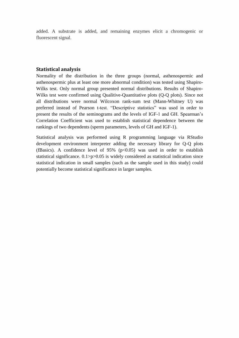
RESULTS Participants were aged between 27 and 55 years with a mean age of 42.3 years, and
had at least 3 days of abstinence (optimal abstinence days 2-5). It is important to
mention that 90% of our sample had abnormal progressive motility (45 out of 50).
Only 5 samples were classified without abnormal parameters in their seminogram.
Out of the 50 patients, 16 requested only for a DNA fragmentation test, so there are
no data of vitality or morphology based on straining (although 2 samples were
checked for morphology as well). The mean values and standard deviations of sperm
parameters and GH and IGF-1 levels in seminal plasma are presented in table 1.
Table 1: Mean values of sperm parameters and mean levels of IGF-1 and GH in
seminal plasma
N Mean Standard Deviation
Sperm Concentration 50 26 * 10^6 30*10^6
Progressive Motility 50 12.96% 11.68%
Total Motility 50 22.87% 17.75%
Vitality 34 36.91% 32.75%
Morphology 36 9.02% 8.01%
DNA Index 16 0.32 0.12
Volume 50 2.9 1.6
Seminal GH 50 31.28 30.16
Seminal IGF-1 50 40.16 16.86
Table 2: Normal and abnormal findings in each parameter
Sperm Parameters Normal Abnormal
Sperm Concentration 22 28
Progressive Motility 5 45
Total Motility 12 38
Vitality 10 24
Morphology 22 14
DNA Index 8 8
Volume 43 7
More detailed results are depicted in the histograms below:
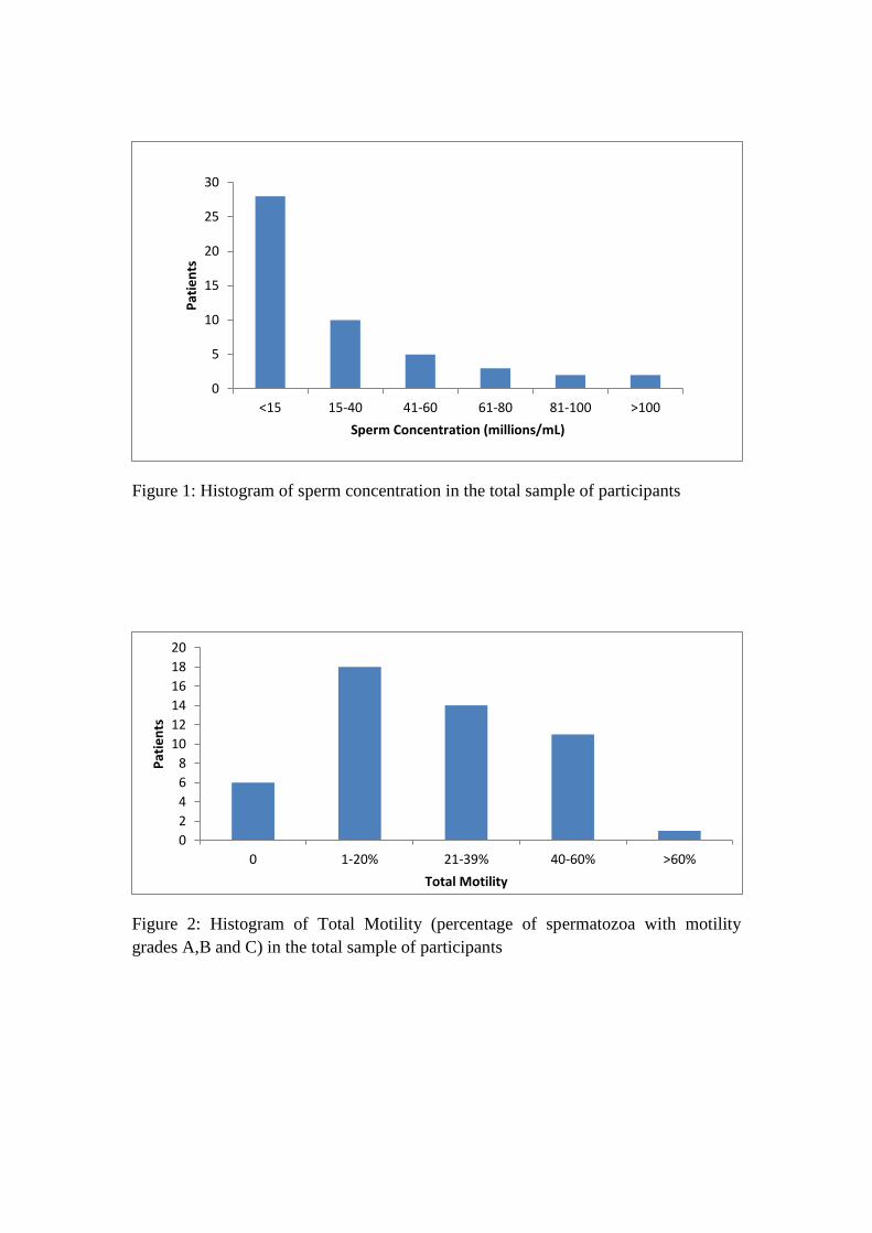
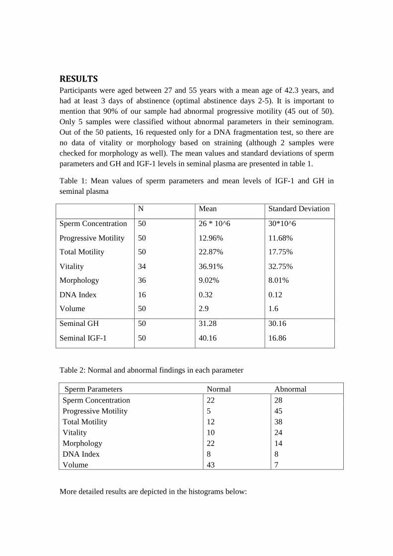
Figure 1: Histogram of sperm concentration in the total sample of participants
Figure 2: Histogram of Total Motility (percentage of spermatozoa with motility
grades A,B and C) in the total sample of participants
0
5
10
15
20
25
30
<15 15-40 41-60 61-80 81-100 >100
Pat
ien
ts
Sperm Concentration (millions/mL)
0
2
4
6
8
10
12
14
16
18
20
0 1-20% 21-39% 40-60% >60%
Pat
ien
ts
Total Motility
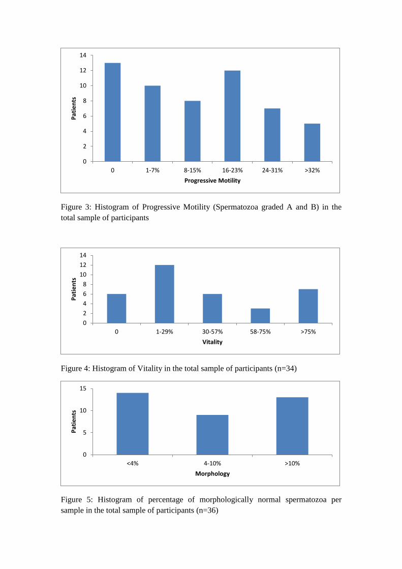
Figure 3: Histogram of Progressive Motility (Spermatozoa graded A and B) in the
total sample of participants
Figure 4: Histogram of Vitality in the total sample of participants (n=34)
Figure 5: Histogram of percentage of morphologically normal spermatozoa per
sample in the total sample of participants (n=36)
0
2
4
6
8
10
12
14
0 1-7% 8-15% 16-23% 24-31% >32%
Pat
ien
ts
Progressive Motility
0
2
4
6
8
10
12
14
0 1-29% 30-57% 58-75% >75%
Pat
ien
ts
Vitality
0
5
10
15
<4% 4-10% >10%
Pat
ien
ts
Morphology
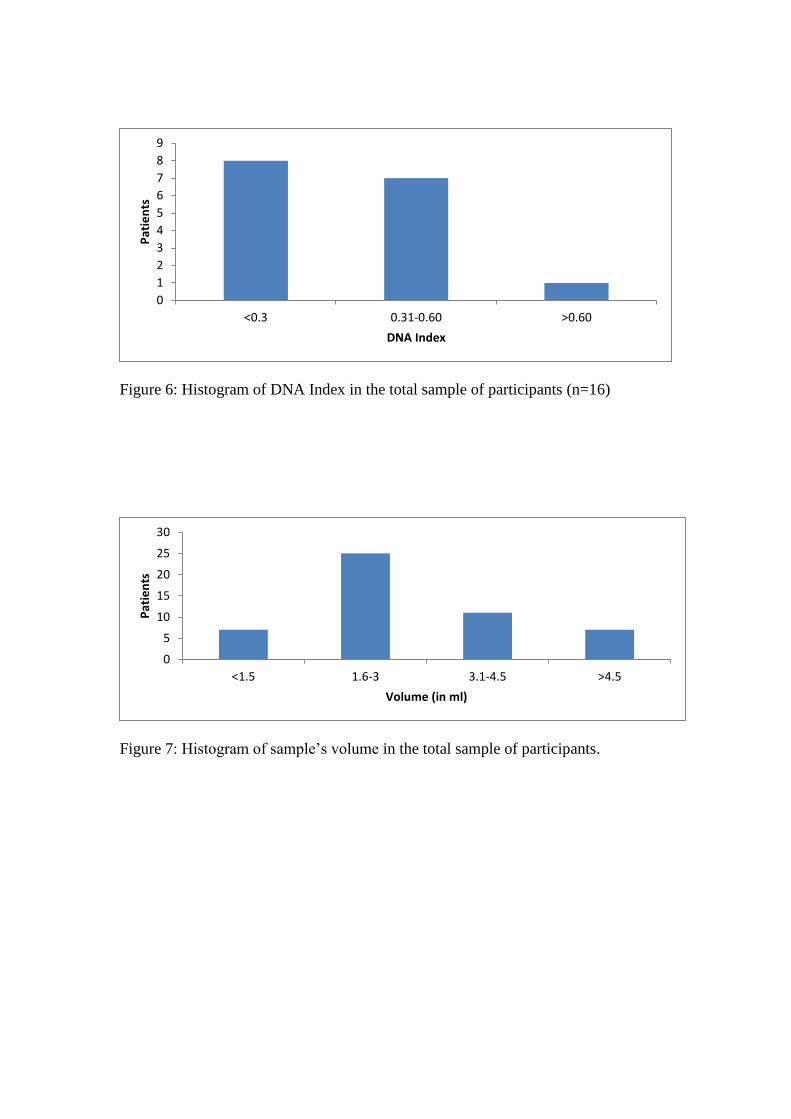
Figure 6: Histogram of DNA Index in the total sample of participants (n=16)
Figure 7: Histogram of sample‟s volume in the total sample of participants.
0
1
2
3
4
5
6
7
8
9
<0.3 0.31-0.60 >0.60
Pat
ien
ts
DNA Index
0
5
10
15
20
25
30
<1.5 1.6-3 3.1-4.5 >4.5
Pat
ien
ts
Volume (in ml)
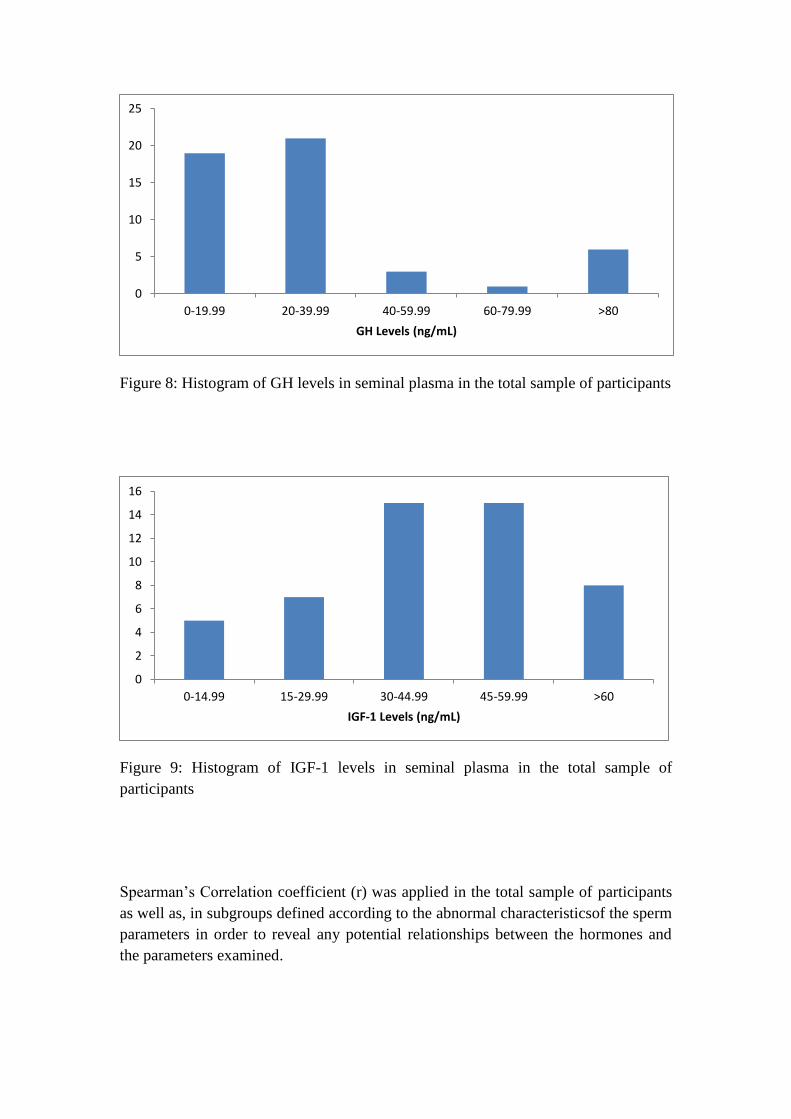
Figure 8: Histogram of GH levels in seminal plasma in the total sample of participants
Figure 9: Histogram of IGF-1 levels in seminal plasma in the total sample of
participants
Spearman‟s Correlation coefficient (r) was applied in the total sample of participants
as well as, in subgroups defined according to the abnormal characteristicsof the sperm
parameters in order to reveal any potential relationships between the hormones and
the parameters examined.
0
5
10
15
20
25
0-19.99 20-39.99 40-59.99 60-79.99 >80
GH Levels (ng/mL)
0
2
4
6
8
10
12
14
16
0-14.99 15-29.99 30-44.99 45-59.99 >60
IGF-1 Levels (ng/mL)
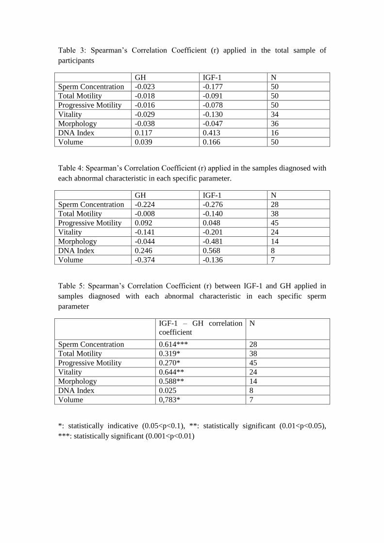
Table 3: Spearman‟s Correlation Coefficient (r) applied in the total sample of
participants
GH IGF-1 N
Sperm Concentration -0.023 -0.177 50
Total Motility -0.018 -0.091 50
Progressive Motility -0.016 -0.078 50
Vitality -0.029 -0.130 34
Morphology -0.038 -0.047 36
DNA Index 0.117 0.413 16
Volume 0.039 0.166 50
Table 4: Spearman‟s Correlation Coefficient (r) applied in the samples diagnosed with
each abnormal characteristic in each specific parameter.
GH IGF-1 N
Sperm Concentration -0.224 -0.276 28
Total Motility -0.008 -0.140 38
Progressive Motility 0.092 0.048 45
Vitality -0.141 -0.201 24
Morphology -0.044 -0.481 14
DNA Index 0.246 0.568 8
Volume -0.374 -0.136 7
Table 5: Spearman‟s Correlation Coefficient (r) between IGF-1 and GH applied in
samples diagnosed with each abnormal characteristic in each specific sperm
parameter
IGF-1 – GH correlation
coefficient
N
Sperm Concentration 0.614*** 28
Total Motility 0.319* 38
Progressive Motility 0.270* 45
Vitality 0.644** 24
Morphology 0.588** 14
DNA Index 0.025 8
Volume 0,783* 7
*: statistically indicative (0.05<p<0.1), **: statistically significant (0.01<p<0.05),
***: statistically significant (0.001<p<0.01)
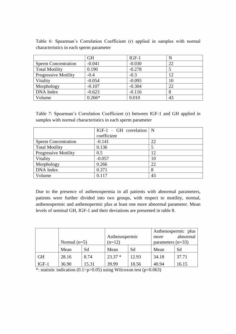
Table 6: Spearman‟s Correlation Coefficient (r) applied in samples with normal
characteristics in each sperm parameter
GH IGF-1 N
Sperm Concentration -0.041 -0.030 22
Total Motility 0.190 -0.278 5
Progressive Motility -0.4 -0.3 12
Vitality -0.054 -0.095 10
Morphology -0.107 -0.304 22
DNA Index -0.623 -0.116 8
Volume 0.266* 0.010 43
Table 7: Spearman‟s Correlation Coefficient (r) between IGF-1 and GH applied in
samples with normal characteristics in each sperm parameter
IGF-1 – GH correlation
coefficient
N
Sperm Concentration -0.141 22
Total Motility 0.136 5
Progressive Motility 0.5 12
Vitality -0.057 10
Morphology 0.266 22
DNA Index 0.371 8
Volume 0.117 43
Due to the presence of asthenospermia in all patients with abnormal parameters,
patients were further divided into two groups, with respect to motility, normal,
asthenospermic and asthenopermic plus at least one more abnormal parameter. Mean
levels of seminal GH, IGF-1 and their deviations are presented in table 8.
Normal (n=5)
Asthenospermic
(n=12)
Asthenospermic plus
more abnormal
parameters (n=33)
Mean Sd Mean Sd Mean Sd
GH 28.16 8.74 23.37 * 12.93 34.18 37.71
IGF-1 36.90 15.31 39.99 18.56 40.94 16.15
*: statistic indication (0.1>p>0.05) using Wilcoxon test (p=0.063)

Table 9: Spearman‟s Correlation Coefficient (r) between IGF-1 and GH in each group
IGF-1 and GH correlation
Normal (n=5)
Asthenospermic
(n=12)
Asthenospermia plus at least on
more abnormal finding (n=33)
Coefficient 0.5 -0.133 0.366**
**: statistically significant (0.01>p>0.05).
DISCUSSION
Infertility is a common disorder affecting 7% of the male population worldwide [Lotti
& Maggi, 2015]. It is a multi-factor disorder that has been the topic of many studies.
Common aims of those studies were to either investigate possible new biomarkers
and/or causes of infertility, , or to test new therapeutic treatments. The purpose of the
present study was to investigate possible associations between GH, IGF-1 and male
infertility.
In this study, a marginally non-significant difference between asthenospermic (group
B) and normal group was revealed, while group C (i.e., asthenospermia plus at least
one more abnormal finding) presented no statistically significant difference in
comparison to any other group. The lack of statistical significance in the differences
between normal group and group C may be due to the fact that 4 samples in the latter
group showed extremely high levels of GH, increasing the inter-individual variability.
Thus, it could be speculated that this variability might have been smoothened if the
total number of participants was larger and potentially might reveal significant
differences between the normal group and group C.
The correlation found between GH and sperm motility is in accordance with the
literature. Specifically, GH has been shown to improve motility, but not sperm
concentration, in asthenospermic and oligospermic men, with pregnancies reported in
the asthenospermic group but not in the oligospermic group [Ovesen et al., 1996]. In
addition, treatment with recombinant-hGH was found to increase motility in half of
the male subjects with idiopathic oligospermia [Radicioni et al., 1994].
In addition, GH treatment has been found to raise seminal volume in oligospermic
patients [Kalra et al., 2008] but to our knowledge no study has been conducted for
oligo-astheno-teratozoospermic patients. In our study, a correlation between GH and
seminal volume in samples with normal-ranged volume marginally failed to reach
statistical significance, while such correlation was not established neither in patients
with low seminal volume nor at aggregation of both groups. The reason for that may
be the fact that all patients with low seminal volume had abnormalities in all other
sperm parameters examined.
GH has been proposed as a novel therapy for both male and female infertility with
promising results [Magon et al., 2011]. Especially in females, GH has been used to
enhance ovarian stimulation in poor responders during IVF/ICSI cycles.
Administration of GH increases the number of MII oocytes retrieved and the number
of embryos obtained [Yu et al., 2015]. Moreover, administration of low-dose GH
along with standard gonadotropin protocol improves embryo quality and pregnancy
rates in poor responders [Lattes et al., 2015]. Ovarian stimulation associated with GH
has been proposed for poor responders with history of assisted reproduction failures
[Hazout et al., 2009]. Co-administration of GH and aspirin, along with the GnRH
antagonist protocol, has been found to increase the rates of retrieved oocytes, promote
the maturation of oocytes and improve the fertilization rate in IVF [Guan et al., 2007].
However, other studies have reported controversial results.GH co-administration
along with GnRH antagonist protocol did not increase pregnancy rates, although it did
increase the number of retrieved oocytes [Eftekhar et al., 2013; Kucuk et al., 2008].
Another study contradicted the above-mentioned results, concluding that GH
supplements improved pregnancy and live birth rates but did not improve oocyte
retrieval [de Ziegler et al., 2011].
According to the evidences of the present study and the literature reports, GH plays a
role in the physiology of reproduction. However, further research is required in order
to investigate which subgroups of infertile men and women may be benefited by its
use as therapeutic intervention, and at which dose it will be beneficial. Nevertheless,
reduced fertility was found in transgenic mice overexpressing GH [Bartke et al.,
1999]. More specifically, in Bartke‟s study [1999], both GH-resistant (GHR-KO) and
GH-overexpressing mice had reduced fertility, interestingly however it was concluded
that GHR-KO mice subfertility was mild in comparison to IGF-1-KO. Thus, overall
the study suggested that IGF-1 production in reproductive tissues is independent of
GH. This is in agreement with the findings of other research groups that concluded
that IGF-1 production in testicular tissues is stimulated by gonadotropins [Grizard,
1994] and more specifically by FSH and testosterone [Itoh et al., 1994].
In our study, though, a strong positive correlation was found between IGF-1 and GH
in patients with low sperm concentration, with low vitality and abnormal morphology
spermatozoa, as well as a marginally significant correlation in patients with low
progressive, total motility and low volume. Such correlations do not exist in the total
sample of participants or in healthy groups for each of the above aspects. The
indicative correlation between GH and IGF-1 in patients with low motility
(progressive and or total) may be a false positive indication, since all our patients
suffered from asthenospermia. A further analysis after the subdivision of samples in
normal, asthenospermic and asthenospermic plus at least one more abnormality
(group C) showed asignificant correlation between GH and IGF-1 in patients with
asthenospermia and at least one more abnormal condition, however there was not
statistically significant correlation between GH and IGF-1 in asthenospermic patients.
(p<0.05).
To the best of our knowledge, there are no studies correlating GH and IGF-1 in
seminal plasma or male reproductive tissues in general. The fact that this correlation
occurred only in subfertile patients requires further research, in order to investigate
and delineate a possible biological significance of this relationship and to
characterize the nature and mechanisms of this association. This could be a novel
research topic and its conclusions might contribute to the explanation of the
controversial results reported in the literature and, subsequently, to the better
understanding of male infertility.
Seminal IGF-1 levels showed no statistically significant association with any of the
sperm parameters, which is in accordance with another recent study [Lee et al., 2016].
However, in that study there were statistically significant associations between serum
IGF-1 levels and male infertility. Another study in stallions showed no direct
association between IGF-1 and infertility or subfertility [Hess & Rosser, 2001]. These
results contradicted the findings of a previous study that found statistically significant
differences in seminal IGF-1 levels in patients with low sperm concentration
compared with normal subjects [Colombo et al., 1999]. The reason of these
conflicting results could be the role of IGF binding proteins (IGFBPs) in the
regulation of the IGF-1 bioavailability, or the absence of IGF-1 receptor [Sanchez-
Luengo et al., 2005]. IGF-1 levels in seminal plasma derived from patients with
varicocele (common cause of male infertility) were significantly different compared
with that following varicocelectomy and that of control group, though IGF-1 levels
were not correlated with semen quality [Naderi et al., 2015]. Transgenic mice
overexpressing IGFBP-1 showed alterations in spermatogenesis, resulting in lower
production and quality of sperm [Froment et al., 2004].
IGF-1 has been investigated in the scope of female infertility, embryo growth and
Assisted Reproduction Techniques (ART) as well. IGF-1 and IGFBP-3 serum levels
increased during ovarian stimulation in females with polycystic ovaries syndrome,
undergoing GnRH antagonist protocols without the supplement of GH.. Patients
whose IGF-1 levels decreased from day 3 to day of hCG had a higher number of
oocytes retrieved, but less mature oocytes than patients whose IGF-1 levels were
elevated. Moreover, an increase in IGFBP-3 levels during stimulation was correlated
with a higher number of pregnancies [Dragisic et al., 2005]. Levels of IGF-1 in
follicular fluid were found to be directly correlated with embryo quality and
pregnancy outcome [Mehta et al., 2013]. This is in agreement with a recent study that
concluded that follicular fluid level of IGF-1 may have a predictive value in IVF
outcome, since IGF-1 levels were positively correlated with higher pregnancy rates
[Faraj et al., 2017].
IGF-1 deficiency is associated with intrauterine growth restriction, implying the major
role of IGF-1 in normal fetal and placental growth and differentiation. Intrauterine
growth restriction (IUGR) is the second most frequent cause of perinatal morbidity
and mortality, defined as the inability to achieve the expected weight for gestational
age. [Martin-Estal et al., 2016]. IGF-1 levels were higher to women with a live birth
in comparison to women with spontaneous abortion, ectopic pregnancy or them who
were not pregnant. That study concluded that maternal serum levels of IGF-1 and
IGFBP-1 were positively associated with the likelihood of a live birth, whereas levels
of IGF-2 were negatively correlated with live birth [Ramer et al., 2015].
To conclude, IGF-1 and GH appear to have a pivotal role both in the physiology of
reproduction and in assisted reproduction. In our study, a possible association
between GH and asthenospermia was revealed. IGF-1 seminal levels had no
significant association with the sperm parameters examined. The novel finding of this
study is the significant correlation between IGF-1 and GH in patients with
asthenospermia and at least one more abnormal sperm parameter. This finding
requires further investigation in order to establish a possible biological significance,
the functionality of this association and possibly a better understanding of male
infertility.
REFERENCES 1. Agarwal, A., & Said, T. M. (2003). Role of sperm chromatin abnormalities
and DNA damage in male infertility. Hum Reprod Update, 9(4), 331-345.
2. Bartke, A., Chandrashekar, V., Turyn, D., Steger, R. W., Debeljuk, L.,
Winters, T. A., . . . Kopchick, J. J. (1999). Effects of growth hormone
overexpression and growth hormone resistance on neuroendocrine and
reproductive functions in transgenic and knock-out mice. Proc Soc Exp Biol
Med, 222(2), 113-123.
3. Boron, Walter F.Boulpaep, Emile L. (Eds.) (2009) Medical physiology :a
cellular and molecular approach Philadelphia, PA : Saunders/Elsevier
4. Bohring, C., & Krause, W. (2003). Characterization of spermatozoa surface
antigens by antisperm antibodies and its influence on acrosomal exocytosis.
Am J Reprod Immunol, 50(5), 411-419.
5. Carroll, P. V., Christ, E. R., Bengtsson, B. A., Carlsson, L., Christiansen, J. S.,
Clemmons, D., . . . Thorne, M. (1998). Growth hormone deficiency in
adulthood and the effects of growth hormone replacement: a review. Growth
Hormone Research Society Scientific Committee. J Clin Endocrinol Metab,
83(2), 382-395. doi:10.1210/jcem.83.2.4594
6. Chandrashekar, V., Bartke, A., Coschigano, K. T., & Kopchick, J. J. (1999).
Pituitary and testicular function in growth hormone receptor gene knockout
mice. Endocrinology, 140(3), 1082-1088. doi:10.1210/endo.140.3.6557
7. Chrysikopoulos Α. (2002). Υπνγνληκόηεηα - ζηείξωζε, Ιαηξηθέο Εθδόζεηο Π.
Χ. Παζραιίδεο
8. Colombo, J. B., & Naz, R. K. (1999). Modulation of insulin-like growth
factor-1 in the seminal plasma of infertile men. J Androl, 20(1), 118-125.
9. Dada, R., Kumar, M., Jesudasan, R., Fernández, J. L., Gosálvez, J., &
Agarwal, A. (2012). Epigenetics and its role in male infertility. J Assist
Reprod Genet, 29(3), 213-223. doi:10.1007/s10815-012-9715-0
10. Damgaard, I. N., Jensen, T. K., Petersen, J. H., Skakkebaek, N. E., Toppari, J.,
& Main, K. M. (2007). Cryptorchidism and maternal alcohol consumption
during pregnancy. Environ Health Perspect, 115(2), 272-277.
doi:10.1289/ehp.9608
11. de Ziegler, D., Streuli, I., Meldrum, D. R., & Chapron, C. (2011). The value of
growth hormone supplements in ART for poor ovarian responders. Fertil
Steril, 96(5), 1069-1076. doi:10.1016/j.fertnstert.2011.09.049
12. Dragisic, K. G., Davis, O. K., Fasouliotis, S. J., & Rosenwaks, Z. (2005). Use
of a luteal estradiol patch and a gonadotropin-releasing hormone antagonist
suppression protocol before gonadotropin stimulation for in vitro fertilization
in poor responders. Fertil Steril, 84(4), 1023-1026.
doi:10.1016/j.fertnstert.2005.04.031
13. Eftekhar, M., Aflatoonian, A., Mohammadian, F., & Eftekhar, T. (2013).
Adjuvant growth hormone therapy in antagonist protocol in poor responders
undergoing assisted reproductive technology. Arch Gynecol Obstet, 287(5),
1017-1021. doi:10.1007/s00404-012-2655-1
14. Esteves, S. C. (2013). Microdissection testicular sperm extraction (micro-
TESE) as a sperm acquisition method for men with nonobstructive
azoospermia seeking fertility: operative and laboratory aspects. Int Braz J
Urol, 39(3), 440; discussion 441.
15. Faraj, N., Alhalabi, M., & Al- Quobaili, F. (2017). Predictive value of
follicular fluid insulin like growth factor-1 in IVF outcome of normo-
ovulatory women. Middle East Fertility Society Journal, 22(2), 101-104.
doi:https://doi.org/10.1016/j.mefs.2017.01.002
16. Foresta, C., Zuccarello, D., Garolla, A., & Ferlin, A. (2008). Role of
hormones, genes, and environment in human cryptorchidism. Endocr Rev,
29(5), 560-580. doi:10.1210/er.2007-0042
17. Froment, P., Staub, C., Hembert, S., Pisselet, C., Magistrini, M., Delaleu, B., .
. . Monget, P. (2004). Reproductive abnormalities in human insulin-like
growth factor-binding protein-1 transgenic male mice. Endocrinology, 145(4),
2080-2091. doi:10.1210/en.2003-0956
18. Geraedts, J. P., & De Wert, G. M. (2009). Preimplantation genetic diagnosis.
Clin Genet, 76(4), 315-325. doi:10.1111/j.1399-0004.2009.01273.x
19. González-Guerra, J. L., Castilla-Cortazar, I., Aguirre, G. A., Muñoz, Ú.,
Martín-Estal, I., Ávila-Gallego, E., . . . García-Villalón, Á. (2017). Partial
IGF-1 deficiency is sufficient to reduce heart contractibility, angiotensin II
sensibility, and alter gene expression of structural and functional cardiac
proteins. PLoS One, 12(8), e0181760. doi:10.1371/journal.pone.0181760
20. Grizard, G. (1994). [IGF(s) and testicular functions. Secretion and action of
IGF-1 on Leydig cells]. Contracept Fertil Sex, 22(9), 551-555.
21. Guan, Q., Ma, H. G., Wang, Y. Y., & Zhang, F. (2007). [Effects of co-
administration of growth hormone(GH) and aspirin to women during in vitro
fertilization and embryo transfer (IVF-ET) cycles]. Zhonghua Nan Ke Xue,
13(9), 798-800.
22. Gunes, S., Arslan, M. A., Hekim, G. N. T., & Asci, R. (2016). The role of
epigenetics in idiopathic male infertility. J Assist Reprod Genet, 33(5), 553-
569. doi:10.1007/s10815-016-0682-8
23. Gupta, V. (2011). Adult growth hormone deficiency. Indian J Endocrinol
Metab, 15 Suppl 3, S197-202. doi:10.4103/2230-8210.84865
24. Hall, J. E., & Guyton, A. C. (2011). Guyton and Hall textbook of medical
physiology. Philadelphia, PA: Saunders Elsevier
25. Hasanzadeh Keshteli, S., Farsi, M. M., & Khafri, S. (2016). Should We
Perform Semen Analysis, DNA Fragmentation, and Hypo-osmotic Swelling
Tests together? Int J Mol Cell Med, 5(4), 246-254.
26. Hazout, A., Junca, A., Ménézo, Y., Demouzon, J., & Cohen-Bacrie, P. (2009).
Effect of growth hormone on oocyte competence in patients with multiple IVF
failures. Reprod Biomed Online, 18(5), 664-670.
27. Hess, M. F., & Roser, J. F. (2001). The effects of age, season and fertility
status on plasma and intratesticular insulin-like growth factor I concentration
in stallions. Theriogenology, 56(5), 723-733.
28. Hull, K. L., & Harvey, S. (2014). Growth hormone and reproduction: a review
of endocrine and autocrine/paracrine interactions. Int J Endocrinol, 2014,
234014. doi:10.1155/2014/234014
29. Irvine, D. S., Twigg, J. P., Gordon, E. L., Fulton, N., Milne, P. A., & Aitken,
R. J. (2000). DNA integrity in human spermatozoa: relationships with semen
quality. J Androl, 21(1), 33-44.
30. Itoh, N., Nanbu, A., Tachiki, H., Akagashi, K., Nitta, T., Mikuma, N., . . .
Kumamoto, Y. (1994). Restoration of testicular transferrin, insulin-like growth
factor-1 (IGF-1), and spermatogenesis by exogenously administered purified
FSH and testosterone in medically hypophysectomized rats. Arch Androl,
33(3), 169-177.
31. Jensen, M. S., Bonde, J. P., & Olsen, J. (2007). Prenatal alcohol exposure and
cryptorchidism. Acta Paediatr, 96(11), 1681-1685. doi:10.1111/j.1651-
2227.2007.00506.x
32. Jensen, M. S., Toft, G., Thulstrup, A. M., Bonde, J. P., & Olsen, J. (2007).
Cryptorchidism according to maternal gestational smoking. Epidemiology,
18(2), 220-225. doi:10.1097/01.ede.0000254061.90686.9f
33. Kantartzi, P. D., Goulis, C. D., Goulis, G. D., & Papadimas, I. (2007). Male
infertility and varicocele: myths and reality. Hippokratia, 11(3), 99-104.
34. Kruger, T. F., Acosta, A. A., Simmons, K. F., Swanson, R. J., Matta, J. F., &
Oehninger, S. (1988). Predictive value of abnormal sperm morphology in in
vitro fertilization. Fertil Steril, 49(1), 112-117.
35. Kruger, T. F., Menkveld, R., Stander, F. S., Lombard, C. J., Van der Merwe, J.
P., van Zyl, J. A., & Smith, K. (1986). Sperm morphologic features as a
prognostic factor in in vitro fertilization. Fertil Steril, 46(6), 1118-1123.
36. Kucuk, T., Kozinoglu, H., & Kaba, A. (2008). Growth hormone co-treatment
within a GnRH agonist long protocol in patients with poor ovarian response: a
prospective, randomized, clinical trial. J Assist Reprod Genet, 25(4), 123-127.
doi:10.1007/s10815-008-9212-7
37. Lattes, K., Brassesco, M., Gomez, M., & Checa, M. A. (2015). Low-dose
growth hormone supplementation increases clinical pregnancy rate in poor
responders undergoing in vitro fertilisation. Gynecol Endocrinol, 31(7), 565-
568. doi:10.3109/09513590.2015.1025378
38. Lee, H. S., Park, Y. S., Lee, J. S., & Seo, J. T. (2016). Serum and seminal
plasma insulin-like growth factor-1 in male infertility. Clin Exp Reprod Med,
43(2), 97-101. doi:10.5653/cerm.2016.43.2.97
39. Lee, K. O., Ng, S. C., Lee, P. S., Bongso, A. T., Taylor, E. A., Lin, T. K., &
Ratnam, S. S. (1995). Effect of growth hormone therapy in men with severe
idiopathic oligozoospermia. Eur J Endocrinol, 132(2), 159-162.
40. Lotti, F., Corona, G., Vitale, P., Maseroli, E., Rossi, M., Fino, M. G., &
Maggi, M. (2015). Current smoking is associated with lower seminal vesicles
and ejaculate volume, despite higher testosterone levels, in male subjects of
infertile couples. Hum Reprod, 30(3), 590-602. doi:10.1093/humrep/deu347
41. Lotti, F., & Maggi, M. (2015). Ultrasound of the male genital tract in relation
to male reproductive health. Hum Reprod Update, 21(1), 56-83.
doi:10.1093/humupd/dmu042
42. Magon, N., Agrawal, S., Malik, S., & Babu, K. M. (2011). Growth hormone in
the management of female infertility. Indian J Endocrinol Metab, 15 Suppl 3,
S246-247. doi:10.4103/2230-8210.84876
43. Magon, N., Singh, S., Saxena, A., & Sahay, R. (2011). Growth hormone in
male infertility. Indian J Endocrinol Metab, 15 Suppl 3, S248-249.
doi:10.4103/2230-8210.84877
44. Martín-Estal, I., de la Garza, R. G., & Castilla-Cortázar, I. (2016). Intrauterine
Growth Retardation (IUGR) as a Novel Condition of Insulin-Like Growth
Factor-1 (IGF-1) Deficiency. Rev Physiol Biochem Pharmacol, 170, 1-35.
doi:10.1007/112_2015_5001
45. Mehta, B. N., Chimote, N. M., Chimote, M. N., Chimote, N. N., & Nath, N.
M. (2013). Follicular fluid insulin like growth factor-1 (FF IGF-1) is a
biochemical marker of embryo quality and implantation rates in in vitro
fertilization cycles. J Hum Reprod Sci, 6(2), 140-146. doi:10.4103/0974-
1208.117171
46. Naderi, G., Mohseni Rad, H., Tabassomi, F., & Latif, A. (2015). Seminal
insulin-like growth factor-I may be involved in the pathophysiology of
infertility among patients with clinical varicocele. Hum Fertil (Camb), 18(2),
92-95. doi:10.3109/14647273.2014.965759
47. Ovesen, P., Jørgensen, J. O., Ingerslev, J., Ho, K. K., Orskov, H., &
Christiansen, J. S. (1996). Growth hormone treatment of subfertile males.
Fertil Steril, 66(2), 292-298.
48. Ovesen, P., Jørgensen, J. O., Kjaer, T., Ho, K. K., Orskov, H., & Christiansen,
J. S. (1996). Impaired growth hormone secretion and increased growth
hormone-binding protein levels in subfertile males. Fertil Steril, 65(1), 165-
169.
49. Palermo, G., Joris, H., Devroey, P., & Van Steirteghem, A. C. (1992).
Pregnancies after intracytoplasmic injection of single spermatozoon into an
oocyte. Lancet, 340(8810), 17-18.
50. Pellati, D., Mylonakis, I., Bertoloni, G., Fiore, C., Andrisani, A., Ambrosini,
G., & Armanini, D. (2008). Genital tract infections and infertility. Eur J
Obstet Gynecol Reprod Biol, 140(1), 3-11. doi:10.1016/j.ejogrb.2008.03.009
51. Philippou, A., Maridaki, M., Halapas, A., & Koutsilieris, M. (2007). The role
of the insulin-like growth factor 1 (IGF-1) in skeletal muscle physiology. In
Vivo, 21(1), 45-54.
52. Puche, J. E., & Castilla-Cortázar, I. (2012). Human conditions of insulin-like
growth factor-I (IGF-I) deficiency. J Transl Med, 10, 224. doi:10.1186/1479-
5876-10-224
53. Radicioni, A., Paris, E., Dondero, F., Bonifacio, V., & Isidori, A. (1994).
Recombinant-growth hormone (rec-hGH) therapy in infertile men with
idiopathic oligozoospermia. Acta Eur Fertil, 25(5), 311-317.
54. Ramer, I., Kanninen, T. T., Sisti, G., Witkin, S. S., & Spandorfer, S. D.
(2015). Association of in vitro fertilization outcome with circulating insulin-
like growth factor components prior to cycle initiation. Am J Obstet Gynecol,
213(3), 356.e351-356. doi:10.1016/j.ajog.2015.04.026
55. Rodríguez-Martínez, H., Kvist, U., Ernerudh, J., Sanz, L., & Calvete, J. J.
(2011). Seminal plasma proteins: what role do they play? Am J Reprod
Immunol, 66 Suppl 1, 11-22. doi:10.1111/j.1600-0897.2011.01033.x
56. Rogol, A. D. (2004). Gender and hormonal regulation of growth. J Pediatr
Endocrinol Metab, 17 Suppl 4, 1259-1265.
57. Sambroni, E., Rolland, A. D., Lareyre, J. J., & Le Gac, F. (2013). FSH and LH
have common and distinct effects on gene expression in rainbow trout testis. J
Mol Endocrinol, 50(1), 1-18. doi:10.1530/JME-12-0197
58. Shamsi, M. B., Kumar, K., & Dada, R. (2011). Genetic and epigenetic factors:
Role in male infertility. Indian J Urol, 27(1), 110-120. doi:10.4103/0970-
1591.78436
59. Sharma, R., Biedenharn, K. R., Fedor, J. M., & Agarwal, A. (2013). Lifestyle
factors and reproductive health: taking control of your fertility. Reprod Biol
Endocrinol, 11, 66. doi:10.1186/1477-7827-11-66
60. Sheynkin, Y., Jung, M., Yoo, P., Schulsinger, D., & Komaroff, E. (2005).
Increase in scrotal temperature in laptop computer users. Hum Reprod, 20(2),
452-455. doi:10.1093/humrep/deh616
61. Simopoulou, M., Giannelou, P., Bakas, P., Gkoles, L., Kalampokas, T.,
Pantos, K., & Koutsilieris, M. (2016). Making ICSI Safer and More Effective:
A Review of the Human Oocyte and ICSI Practice. In Vivo, 30(4), 387-400.
62. Simopoulou, M., Gkoles, L., Bakas, P., Giannelou, P., Kalampokas, T.,
Pantos, K., & Koutsilieris, M. (2016). Improving ICSI: A review from the
spermatozoon perspective. Syst Biol Reprod Med, 62(6), 359-371.
doi:10.1080/19396368.2016.1229365
63. Skarra, D. V., & Thackray, V. G. (2015). FOXO1 is regulated by insulin and
IGF1 in pituitary gonadotropes. Mol Cell Endocrinol, 405, 14-24.
doi:10.1016/j.mce.2015.02.006
64. Soria, M., Pradillo, G., García, J., Ramón, P., Castillo, A., Jordana, C., &
Paricio, P. (2012). Pregnancy predictors after intrauterine insemination:
analysis of 3012 cycles in 1201 couples. J Reprod Infertil, 13(3), 158-166.
65. Steptoe, P. C., & Edwards, R. G. (1978). Birth after the reimplantation of a
human embryo. Lancet, 2(8085), 366.
66. Sánchez-Luengo, S., Fernández, P. J., & Romeu, A. (2005). Insulin growth
factors may be implicated in human sperm capacitation. Fertil Steril, 83(4),
1064-1066. doi:10.1016/j.fertnstert.2004.12.003
67. Vander A.J. ,Sherman J.H, Luciano D.S. (2001). Human Physiology: The
mechanisms of body function PA: Mac-Graw Hill
68. Volpes, A., Sammartano, F., Rizzari, S., Gullo, S., Marino, A., & Allegra, A.
(2016). The pellet swim-up is the best technique for sperm preparation during
in vitro fertilization procedures. J Assist Reprod Genet, 33(6), 765-770.
doi:10.1007/s10815-016-0696-2
69. Wang, Q., & Sun, Q. Y. (2007). Evaluation of oocyte quality: morphological,
cellular and molecular predictors. Reprod Fertil Dev, 19(1), 1-12.
70. Yoon, M. J., Berger, T., & Roser, J. F. (2011). Localization of insulin-like
growth factor-I (IGF-I) and IGF-I receptor (IGF-IR) in equine testes. Reprod
Domest Anim, 46(2), 221-228. doi:10.1111/j.1439-0531.2010.01643.x
71. Yu, X., Ruan, J., He, L. P., Hu, W., Xu, Q., Tang, J., . . . Peng, Y. F. (2015).
Efficacy of growth hormone supplementation with gonadotrophins in vitro
fertilization for poor ovarian responders: an updated meta-analysis. Int J Clin
Exp Med, 8(4), 4954-4967.
72. Zhang, X. D., Liu, J. X., Liu, W. W., Gao, Y., Han, W., Xiong, S., . . . Huang,
G. N. (2013). Time of insemination culture and outcomes of in vitro
fertilization: a systematic review and meta-analysis. Hum Reprod Update,
19(6), 685-695. doi:10.1093/humupd/dmt036


![Caderno Seminal Digital - Universidade Do Estado Do Rio De ...1]Cad.Seminal_N19... · Caderno Seminal Digital 3 Caderno Seminal Digital Ano 19, nº 19, V. 19 (Jan-Jun/2013) – ISSN](https://img.pdfslide.us/doc/110x75/5f57a10ef6f45308c6546f90/caderno-seminal-digital-universidade-do-estado-do-rio-de-1cadseminaln19.jpg)


![Temperature-dependent seminal recovery in the …...seminal reserves) and their regeneration rate [15]. Therefore, an important factor involved in seminal depletion may be the time](https://img.pdfslide.us/doc/110x75/5ebcebf3778b5b4567241695/temperature-dependent-seminal-recovery-in-the-seminal-reserves-and-their-regeneration.jpg)