Embed Size (px)
Citation preview
Cornealendothelium:developmentalstrategies forregeneration
J Zavala1, GR Lopez Jaime2,
CA Rodrıguez Barrientos1 and J Valdez-Garcia1 ;3
Abstract
The main treatment available for restoration of
the corneal endothelium is keratoplasty. This
procedure is faced with several difficulties,
including the shortage of donor tissue, post-
surgical complications associated with the use
of drugs to prevent immune rejection, and a
significant increase in the occurrence of glau-
coma. Recently, surgical procedures such as
Descemet’s stripping endothelial keratoplasty
have focused on the transplant of corneal
endothelium, yielding better visual results but
still facing the need for donor tissue. The
emergent strategies in the field of cell biology
and tissue cultivation of corneal endothelial
cells aim at the production of transplantable
endothelial cell sheets. Cell therapy focuses on
the culture of corneal endothelial cells
retrieved from the donor, in the donor’s
cornea, followed by transplantation into the
recipient. Recently, research has focused on
overcoming the challenge of harvesting
human corneal endothelial cells and the
generation of new biomembranes to be used
as cell scaffolds in surgical procedures. The
use of corneal endothelial precursors from the
peripheral cornea has also demonstrated to be
effective and represents a valuable tool for
reducing the risk of rejection in allogeneic
transplants. Several animal model reports also
support the use of adult stem cells as therapy
for corneal diseases. Current results represent
important progresses in the development of
new strategies based on alternative sources of
tissue for the treatment of corneal endothe-
liopathies. Different databases were used to
search literature: PubMed, Google Books,
MD Consult, Google Scholar, Gene Cards, and
NCBI Books. The main search terms used
were: ‘cornea AND embryology AND tran-
scription factors’, ‘human endothelial kerato-
plasty AND risk factors’, ‘(cornea OR corneal)
AND (endothelium OR endothelial) AND cell
culture’, ‘mesenchymal stem cells AND cell
therapy’, ‘mesenchymal stem cells AND
cornea’, and ‘stem cells AND (cornea OR
corneal) AND (endothelial OR endothelium)’.
Eye (2013) 27, 579–588; doi:10.1038/eye.2013.15;
published online 8 March 2013
Keywords: corneal endothelium; tissue
engineer; stems cells
Introduction
The cornea is a transparent avascular tissue that
in conjunction with the sclera forms the outer
portion of the eye. It is a connective tissue that
acts as the primary barrier against infection and
mechanical damage to the internal structures of
the eye. Along with the tear film on the ocular
surface, it accounts for more than two-thirds of
the total refractive power of the eye. It is
organized into three cell layers: epithelium,
stroma, and endothelium, and two interfaces:
Bowman’s layer and Descemet’s membrane.1
The epithelium provides a biodefense system on
the anterior surface of the eye, helps to keep the
corneal surface optically smooth, and provides a
barrier to external biological agents and
chemical damage. Bowman’s layer serves as an
interface between the epithelium and the stroma
and consists of randomly arranged collagen
fibers and proteoglycan types I and III. The
stroma constitutes about 90% of the thickness of
the cornea and is composed of extracellular
matrix, keratinocytes, and nerve fibers. It
provides structural strength, shape, stability,
and transparency to the cornea. The
endothelium is a thin monolayer of polygonal
cells covering the posterior surface of
Descemet’s membrane and is in contact with the
1Ophthalmology ResearchChair, Tecnologico deMonterrey, School ofMedicine and HealthSciences, Monterrey, Mexico
2Medical and Surgical RetinaResidency ProgramDepartment, Universidad deGuadalajara Instituto deOftalmologıa y CienciasVisuales, Guadalajara,Mexico
3Instituto de Oftalmologıa yCiencias Visuales - TecSalud, Monterrey, Mexico
Correspondence:J Valdez-Garcia,Tecnologico de Monterrey,School of Medicine andHealth Sciences, 3000Morones Prieto AvenueCol. Los DoctoresMonterrey, Monterrey64710 Mexico. Tel/Fax:+518188882066;E-mail: [email protected]
Received: 12 September2012Accepted in revised form:25 January 2013Published online: 8 March2013
RE
VIE
W
Eye (2013) 27, 579–588& 2013 Macmillan Publishers Limited All rights reserved 0950-222X/13
www.nature.com/eye
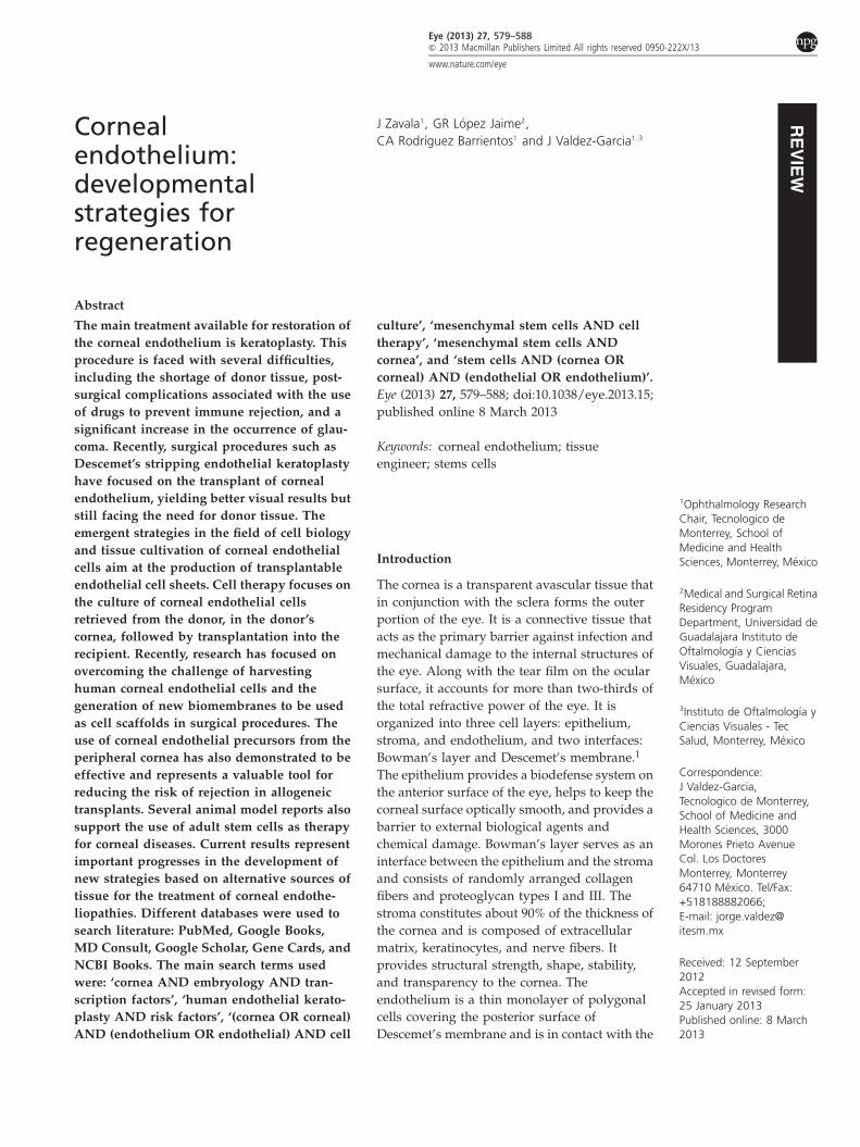
aqueous humor (Figure 1). The main function is to
regulate the hydration state through an active ATP and
biocarbonate-dependent pump; thereby providing
transparency to the cornea, which allows the eye to
perform its visual functions.2 It is also an important
system for the passage of nutrients and waste removal
through simple diffusion, facilitated diffusion, and
active transport mechanisms.3
The corneal endothelium is the cell layer with the
lowest mitotic activity.4 Given the importance of its
function, damage to the endothelium is potentially more
serious than that to the other corneal layers and can
result in cell loss and irreversible damage to the
endothelial cytoskeleton, that ultimately affecting visual
function.5 The main treatment for this condition is
corneal transplant. Nevertheless, given the difficulty
obtaining donor tissue, the development of novel
strategies has focused on the use of cultured corneal
endothelial cells, corneal endothelial stem cells, and stem
cells of extra-ocular origin. In this article, we describe the
corneal endothelium’s embryology and physiology, the
main conditions that affects it and the cell therapies
currently under development.
Embryology
The embryological origins of the major structures of the
eye are diverse. The central part of the cornea, including
the endothelium, is derived from neural crest cells.
The retina and the epithelial layers of the iris and ciliary
body are derived from the anterior neural plate, the lens
from surface ectoderm, and the corneal epithelium
from epidermal ectoderm.6,7
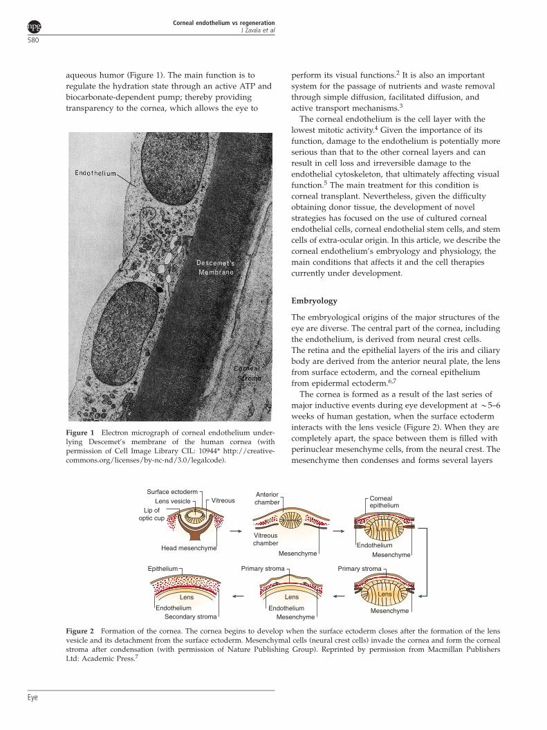
The cornea is formed as a result of the last series of
major inductive events during eye development at B5–6
weeks of human gestation, when the surface ectoderm
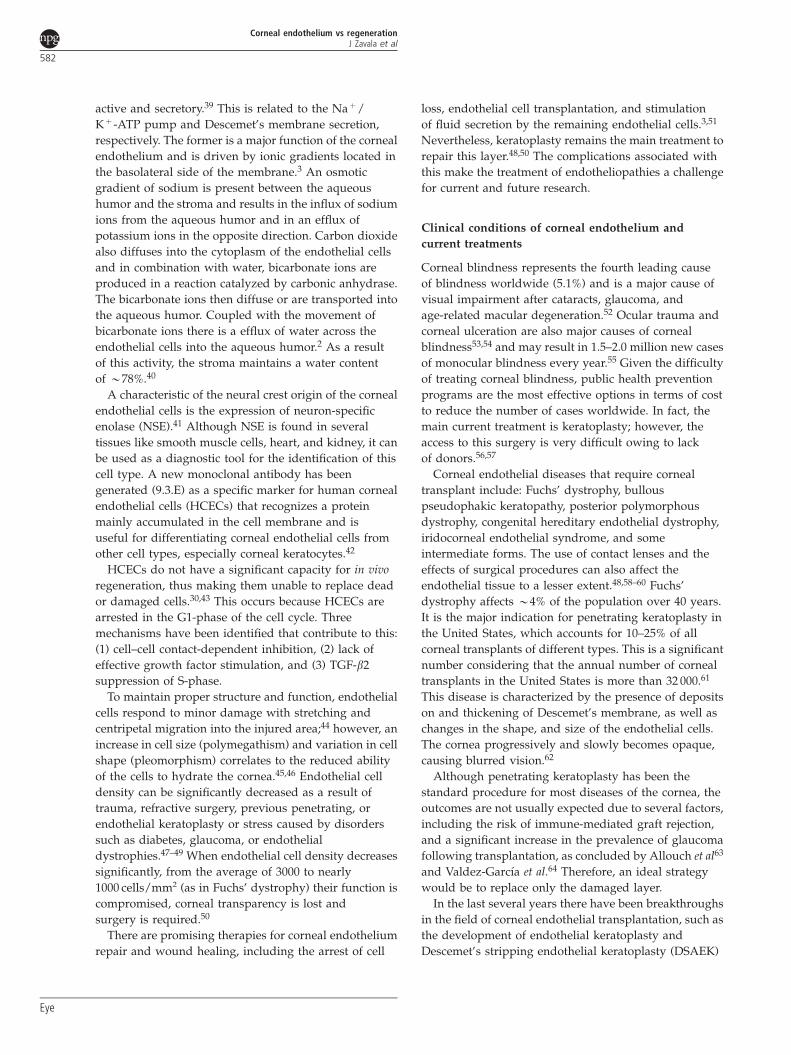
interacts with the lens vesicle (Figure 2). When they are
completely apart, the space between them is filled with
perinuclear mesenchyme cells, from the neural crest. The
mesenchyme then condenses and forms several layers
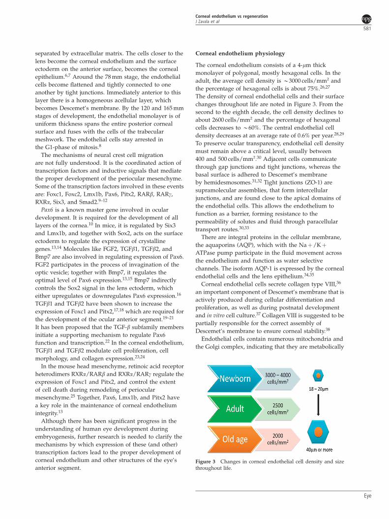
Figure 1 Electron micrograph of corneal endothelium under-lying Descemet’s membrane of the human cornea (withpermission of Cell Image Library CIL: 10944* http://creative-commons.org/licenses/by-nc-nd/3.0/legalcode).
Lip ofoptic cup
Head mesenchyme
Vitreous
Mesenchyme
Vitreouschamber
Anteriorchamber
Mesenchyme
Endothelium
Cornealepithelium
Mesenchyme
Primary stroma
EndotheliumMesenchyme
Primary stroma
Lens
Endothelium
Epithelium
Lens
Secondary stroma
Lens vesicle
Lens
Lens
Surface ectoderm
Figure 2 Formation of the cornea. The cornea begins to develop when the surface ectoderm closes after the formation of the lensvesicle and its detachment from the surface ectoderm. Mesenchymal cells (neural crest cells) invade the cornea and form the cornealstroma after condensation (with permission of Nature Publishing Group). Reprinted by permission from Macmillan PublishersLtd: Academic Press.7
Corneal endothelium vs regenerationJ Zavala et al
580
Eye
separated by extracellular matrix. The cells closer to the
lens become the corneal endothelium and the surface
ectoderm on the anterior surface, becomes the corneal
epithelium.6,7 Around the 78 mm stage, the endothelial
cells become flattened and tightly connected to one
another by tight junctions. Immediately anterior to this
layer there is a homogeneous acellular layer, which
becomes Descemet’s membrane. By the 120 and 165 mm
stages of development, the endothelial monolayer is of
uniform thickness spans the entire posterior corneal
surface and fuses with the cells of the trabecular
meshwork. The endothelial cells stay arrested in
the G1-phase of mitosis.8
The mechanisms of neural crest cell migration
are not fully understood. It is the coordinated action of
transcription factors and inductive signals that mediate
the proper development of the periocular mesenchyme.
Some of the transcription factors involved in these events
are: Foxc1, Foxc2, Lmx1b, Pax6, Pitx2, RARb, RARg,RXRa, Six3, and Smad2.9–12
Pax6 is a known master gene involved in ocular
development. It is required for the development of all
layers of the cornea.10 In mice, it is regulated by Six3
and Lmx1b, and together with Sox2, acts on the surface
ectoderm to regulate the expression of crystalline
genes.13,14 Molecules like FGF2, TGFb1, TGFb2, and
Bmp7 are also involved in regulating expression of Pax6.
FGF2 participates in the process of invagination of the
optic vesicle; together with Bmp7, it regulates the
optimal level of Pax6 expression.13,15 Bmp7 indirectly
controls the Sox2 signal in the lens ectoderm, which
either upregulates or downregulates Pax6 expression.16
TGFb1 and TGFb2 have been shown to increase the
expression of Foxc1 and Pitx2,17,18 which are required for
the development of the ocular anterior segment.19–21
It has been proposed that the TGF-b subfamily members
initiate a supporting mechanism to regulate Pax6
function and transcription.22 In the corneal endothelium,
TGFb1 and TGFb2 modulate cell proliferation, cell
morphology, and collagen expression.23,24
In the mouse head mesenchyme, retinoic acid receptor
heterodimers RXRa/RARb and RXRa/RARg regulate the
expression of Foxc1 and Pitx2, and control the extent
of cell death during remodeling of periocular
mesenchyme.25 Together, Pax6, Lmx1b, and Pitx2 have
a key role in the maintenance of corneal endothelium
integrity.13
Although there has been significant progress in the
understanding of human eye development during
embryogenesis, further research is needed to clarify the
mechanisms by which expression of these (and other)
transcription factors lead to the proper development of
corneal endothelium and other structures of the eye’s
anterior segment.
Corneal endothelium physiology
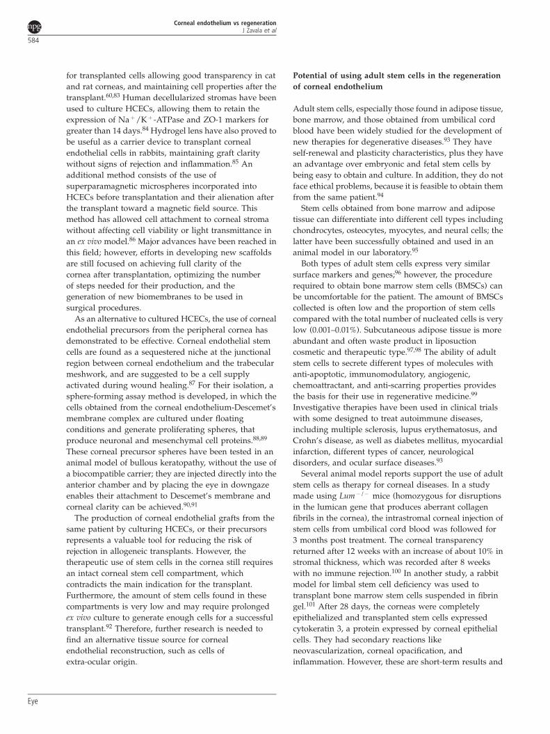
The corneal endothelium consists of a 4-mm thick
monolayer of polygonal, mostly hexagonal cells. In the
adult, the average cell density is B3000 cells/mm2 and
the percentage of hexagonal cells is about 75%.26,27
The density of corneal endothelial cells and their surface
changes throughout life are noted in Figure 3. From the
second to the eighth decade, the cell density declines to
about 2600 cells/mm2 and the percentage of hexagonal
cells decreases to B60%. The central endothelial cell
density decreases at an average rate of 0.6% per year.28,29
To preserve ocular transparency, endothelial cell density
must remain above a critical level, usually between
400 and 500 cells/mm2.30 Adjacent cells communicate
through gap junctions and tight junctions, whereas the
basal surface is adhered to Descemet’s membrane
by hemidesmosomes.31,32 Tight junctions (ZO-1) are
supramolecular assemblies, that form intercellular
junctions, and are found close to the apical domains of
the endothelial cells. This allows the endothelium to
function as a barrier, forming resistance to the
permeability of solutes and fluid through paracellular
transport routes.30,33
There are integral proteins in the cellular membrane,
the aquaporins (AQP), which with the Naþ/KþATPase pump participate in the fluid movement across
the endothelium and function as water selective
channels. The isoform AQP-1 is expressed by the corneal
endothelial cells and the lens epithelium.34,35
Corneal endothelial cells secrete collagen type VIII,36
an important component of Descemet’s membrane that is
actively produced during cellular differentiation and
proliferation, as well as during postnatal development
and in vitro cell culture.37 Collagen VIII is suggested to be
partially responsible for the correct assembly of
Descemet’s membrane to ensure corneal stability.38
Endothelial cells contain numerous mitochondria and
the Golgi complex, indicating that they are metabolically
Figure 3 Changes in corneal endothelial cell density and sizethroughout life.
Corneal endothelium vs regenerationJ Zavala et al
581
Eye
active and secretory.39 This is related to the Naþ/
Kþ -ATP pump and Descemet’s membrane secretion,
respectively. The former is a major function of the corneal
endothelium and is driven by ionic gradients located in
the basolateral side of the membrane.3 An osmotic
gradient of sodium is present between the aqueous
humor and the stroma and results in the influx of sodium
ions from the aqueous humor and in an efflux of
potassium ions in the opposite direction. Carbon dioxide
also diffuses into the cytoplasm of the endothelial cells
and in combination with water, bicarbonate ions are
produced in a reaction catalyzed by carbonic anhydrase.
The bicarbonate ions then diffuse or are transported into
the aqueous humor. Coupled with the movement of
bicarbonate ions there is a efflux of water across the
endothelial cells into the aqueous humor.2 As a result
of this activity, the stroma maintains a water content
of B78%.40
A characteristic of the neural crest origin of the corneal
endothelial cells is the expression of neuron-specific
enolase (NSE).41 Although NSE is found in several
tissues like smooth muscle cells, heart, and kidney, it can
be used as a diagnostic tool for the identification of this
cell type. A new monoclonal antibody has been
generated (9.3.E) as a specific marker for human corneal
endothelial cells (HCECs) that recognizes a protein
mainly accumulated in the cell membrane and is
useful for differentiating corneal endothelial cells from
other cell types, especially corneal keratocytes.42
HCECs do not have a significant capacity for in vivo
regeneration, thus making them unable to replace dead
or damaged cells.30,43 This occurs because HCECs are
arrested in the G1-phase of the cell cycle. Three
mechanisms have been identified that contribute to this:
(1) cell–cell contact-dependent inhibition, (2) lack of
effective growth factor stimulation, and (3) TGF-b2
suppression of S-phase.
To maintain proper structure and function, endothelial
cells respond to minor damage with stretching and
centripetal migration into the injured area;44 however, an
increase in cell size (polymegathism) and variation in cell
shape (pleomorphism) correlates to the reduced ability
of the cells to hydrate the cornea.45,46 Endothelial cell
density can be significantly decreased as a result of
trauma, refractive surgery, previous penetrating, or
endothelial keratoplasty or stress caused by disorders
such as diabetes, glaucoma, or endothelial
dystrophies.47–49 When endothelial cell density decreases
significantly, from the average of 3000 to nearly
1000 cells/mm2 (as in Fuchs’ dystrophy) their function is
compromised, corneal transparency is lost and
surgery is required.50
There are promising therapies for corneal endothelium
repair and wound healing, including the arrest of cell
loss, endothelial cell transplantation, and stimulation
of fluid secretion by the remaining endothelial cells.3,51
Nevertheless, keratoplasty remains the main treatment to
repair this layer.48,50 The complications associated with
this make the treatment of endotheliopathies a challenge
for current and future research.
Clinical conditions of corneal endothelium and
current treatments
Corneal blindness represents the fourth leading cause
of blindness worldwide (5.1%) and is a major cause of
visual impairment after cataracts, glaucoma, and
age-related macular degeneration.52 Ocular trauma and
corneal ulceration are also major causes of corneal
blindness53,54 and may result in 1.5–2.0 million new cases
of monocular blindness every year.55 Given the difficulty
of treating corneal blindness, public health prevention
programs are the most effective options in terms of cost
to reduce the number of cases worldwide. In fact, the
main current treatment is keratoplasty; however, the
access to this surgery is very difficult owing to lack
of donors.56,57
Corneal endothelial diseases that require corneal
transplant include: Fuchs’ dystrophy, bullous
pseudophakic keratopathy, posterior polymorphous
dystrophy, congenital hereditary endothelial dystrophy,
iridocorneal endothelial syndrome, and some
intermediate forms. The use of contact lenses and the
effects of surgical procedures can also affect the
endothelial tissue to a lesser extent.48,58–60 Fuchs’
dystrophy affects B4% of the population over 40 years.
It is the major indication for penetrating keratoplasty in
the United States, which accounts for 10–25% of all
corneal transplants of different types. This is a significant
number considering that the annual number of corneal
transplants in the United States is more than 32 000.61
This disease is characterized by the presence of deposits
on and thickening of Descemet’s membrane, as well as
changes in the shape, and size of the endothelial cells.
The cornea progressively and slowly becomes opaque,
causing blurred vision.62
Although penetrating keratoplasty has been the
standard procedure for most diseases of the cornea, the
outcomes are not usually expected due to several factors,
including the risk of immune-mediated graft rejection,
and a significant increase in the prevalence of glaucoma
following transplantation, as concluded by Allouch et al63
and Valdez-Garcıa et al.64 Therefore, an ideal strategy
would be to replace only the damaged layer.
In the last several years there have been breakthroughs
in the field of corneal endothelial transplantation, such as
the development of endothelial keratoplasty and
Descemet’s stripping endothelial keratoplasty (DSAEK)
Corneal endothelium vs regenerationJ Zavala et al
582
Eye

techniques. In 2005, only 4.5% of the donor corneas were
used for endothelial keratoplasty. By 2007, this number
increased to 50%.51 Nevertheless, the main problem with
endothelial keratoplasty is a postoperative cell loss
comparable to or higher than that observed with
penetrating keratoplasty.65 It has been documented that
endothelial cell density decreases B49% 24 months post-
surgery and that cell loss can be higher in patients with
previous glaucoma surgery. Neither donor age nor initial
cell density proved to have significant influence on
endothelial cell loss. The most promising strategy by
which postoperative cell loss can be reduced effectively
is the strict and adequate lowering of intraocular
pressure.66 Techniques such as Descemet’s membrane
endothelial keratoplasty and Descemet’s membrane
automated endothelial keratoplasty were developed,
offering better visual results, with less scarring and less
optical stromal aberrations. These procedures opened the
possibility to replace the corneal endothelium with
endothelium reconstructed by bioengineering.67
However, clinically available procedures with artificial
corneas have limitations such as inflammation of the
retroprosthetic membrane and development of
glaucoma, and are reserved for high-risk patients.68,69
The limited availability of donor corneas and the
current issues in surgical procedures require the
development of new methods in the field of tissue
engineering in order to improve corneal endothelial cell
survival and increase corneal endothelial cell density.
The emergent strategies in the field of cell biology and
tissue cultivation of corneal endothelial cells aim at the
production of transplantable endothelial cell sheets.
Cell therapy
Currently, cell therapy is aimed at reducing the problem
of the lack of donor tissue. To repair the corneal
endothelium, cell therapy focuses on the culture of
corneal endothelial cells retrieved from the donor, in the
donor’s cornea, followed by transplantation into the
recipient. Recent reports have demonstrated that corneal
endothelial cells possess the ability to undergo mitosis in
culture using these methods. HCEC ex vivo models are
able to overcome the G1-phase and complete the cell
cycle; this occurs after the release of cell–cell junctions
and in the presence of appropriate growth factors. In fact,
it has been proven that endothelial cells from both central
and peripheral areas of the cornea proliferate in vitro70
and that they can be cultured from young and adult
donors, obtaining similar numbers of cells when
specific growth factors are used.71
To successfully engineer human corneal endothelium
from a small number of cells, the processes of isolation,
preservation and expansion are critical. Recently,
research has focused on overcoming the challenge of
harvesting HCECs. It is known that the main factors that
influence the mitotic capacity of HCECs in vitro are: the
method of culture, the nature of the growth factors
contained in the medium, and the viability of the donor
cornea. The latter is influenced by age, cell density, donor
death-to-preservation time, preservation period, overall
health of the donor, and the specific cause of death.72
The common methodology to isolate endothelial cells
involves: (1) the retrieval of the corneal endothelium,
(2) dissociation of cell junctions in paired Descemet’s
membrane/endothelial layer, and (3) culture in proper
media. Based on the method used to dissociate the cell
junctions, the procedures have been classified as
enzymatic and non-enzymatic. The first is based on the
use of enzymes such as collagenase, trypsin, or
dispase.73,74 This technique has the tendency of leading
to cellular degradation due to the incubation time
required to detach cells from the matrix, and because it
also allows for the dissociation of the collagen matrix in
which the keratocytes are located. Additionally, it
often results in contamination from the stromal cells.
To overcome this problem, magnetic cell separation
improves HCECs yield, allowing for a high separation
efficacy.75 The non-enzymatic method is based on the use
of ethylenediamine tetraacetic acid (EDTA) to release
cell–cell junctions at the same time as it promotes cell
division upon exposure to mitogens.30,70,72,76 In this
process, EDTA can also cause cell damage and
decrease cell yield.77 There is a combined method
that uses collagenase II to generate preservable HCEC
aggregates and a brief treatment with trypsin/EDTA
leading to a high proliferation rate with less cell
damage.78
The addition of several different growth factors in
the culture media has been used to promote HCEC
expansion. The use of insulin and basic fibroblastic
growth factor (bFGF) have been shown to promote
mitosis in cells from peripheral cornea but not in the
central zone.79 In another study, nerve growth factor
(NGF) along with bovine pituitary extract and epidermal
growth factor (EGF) supported the expansion of cells
from both central and peripheral areas of the cornea.72 In
addition, a recent study concluded that a culture media
that combines EGF, insulin, transferrin, bFGF, NGF, and
pituitary extract promotes proliferative capacity up to
the third passage.73
In order to replicate the monolayer of the corneal
endothelium, there have been developments to maintain
cell morphology, density, and function. Materials like
collagen, amniotic membrane, and biodegradable
polymers have successfully been used in culture media
and animal models.80–82 Porcine corneal matrix and
Descemet’s membrane have also been used as scaffolds
Corneal endothelium vs regenerationJ Zavala et al
583
Eye
for transplanted cells allowing good transparency in cat
and rat corneas, and maintaining cell properties after the
transplant.60,83 Human decellularized stromas have been
used to culture HCECs, allowing them to retain the
expression of Naþ/Kþ -ATPase and ZO-1 markers for
greater than 14 days.84 Hydrogel lens have also proved to
be useful as a carrier device to transplant corneal
endothelial cells in rabbits, maintaining graft clarity
without signs of rejection and inflammation.85 An
additional method consists of the use of
superparamagnetic microspheres incorporated into
HCECs before transplantation and their alienation after
the transplant toward a magnetic field source. This
method has allowed cell attachment to corneal stroma
without affecting cell viability or light transmittance in
an ex vivo model.86 Major advances have been reached in
this field; however, efforts in developing new scaffolds
are still focused on achieving full clarity of the
cornea after transplantation, optimizing the number
of steps needed for their production, and the
generation of new biomembranes to be used in
surgical procedures.
As an alternative to cultured HCECs, the use of corneal
endothelial precursors from the peripheral cornea has
demonstrated to be effective. Corneal endothelial stem
cells are found as a sequestered niche at the junctional
region between corneal endothelium and the trabecular
meshwork, and are suggested to be a cell supply
activated during wound healing.87 For their isolation, a
sphere-forming assay method is developed, in which the
cells obtained from the corneal endothelium-Descemet’s
membrane complex are cultured under floating
conditions and generate proliferating spheres, that
produce neuronal and mesenchymal cell proteins.88,89
These corneal precursor spheres have been tested in an
animal model of bullous keratopathy, without the use of
a biocompatible carrier; they are injected directly into the
anterior chamber and by placing the eye in downgaze
enables their attachment to Descemet’s membrane and
corneal clarity can be achieved.90,91
The production of corneal endothelial grafts from the
same patient by culturing HCECs, or their precursors
represents a valuable tool for reducing the risk of
rejection in allogeneic transplants. However, the
therapeutic use of stem cells in the cornea still requires
an intact corneal stem cell compartment, which
contradicts the main indication for the transplant.
Furthermore, the amount of stem cells found in these
compartments is very low and may require prolonged
ex vivo culture to generate enough cells for a successful
transplant.92 Therefore, further research is needed to
find an alternative tissue source for corneal
endothelial reconstruction, such as cells of
extra-ocular origin.
Potential of using adult stem cells in the regeneration
of corneal endothelium
Adult stem cells, especially those found in adipose tissue,
bone marrow, and those obtained from umbilical cord
blood have been widely studied for the development of
new therapies for degenerative diseases.93 They have
self-renewal and plasticity characteristics, plus they have
an advantage over embryonic and fetal stem cells by
being easy to obtain and culture. In addition, they do not
face ethical problems, because it is feasible to obtain them
from the same patient.94
Stem cells obtained from bone marrow and adipose
tissue can differentiate into different cell types including
chondrocytes, osteocytes, myocytes, and neural cells; the
latter have been successfully obtained and used in an
animal model in our laboratory.95
Both types of adult stem cells express very similar
surface markers and genes;96 however, the procedure
required to obtain bone marrow stem cells (BMSCs) can
be uncomfortable for the patient. The amount of BMSCs
collected is often low and the proportion of stem cells
compared with the total number of nucleated cells is very
low (0.001–0.01%). Subcutaneous adipose tissue is more
abundant and often waste product in liposuction
cosmetic and therapeutic type.97,98 The ability of adult
stem cells to secrete different types of molecules with
anti-apoptotic, immunomodulatory, angiogenic,
chemoattractant, and anti-scarring properties provides
the basis for their use in regenerative medicine.99
Investigative therapies have been used in clinical trials
with some designed to treat autoimmune diseases,
including multiple sclerosis, lupus erythematosus, and
Crohn’s disease, as well as diabetes mellitus, myocardial
infarction, different types of cancer, neurological
disorders, and ocular surface diseases.93
Several animal model reports support the use of adult
stem cells as therapy for corneal diseases. In a study
made using Lum� /� mice (homozygous for disruptions
in the lumican gene that produces aberrant collagen
fibrils in the cornea), the intrastromal corneal injection of
stem cells from umbilical cord blood was followed for
3 months post treatment. The corneal transparency
returned after 12 weeks with an increase of about 10% in
stromal thickness, which was recorded after 8 weeks
with no immune rejection.100 In another study, a rabbit
model for limbal stem cell deficiency was used to
transplant bone marrow stem cells suspended in fibrin
gel.101 After 28 days, the corneas were completely
epithelialized and transplanted stem cells expressed
cytokeratin 3, a protein expressed by corneal epithelial
cells. They had secondary reactions like
neovascularization, corneal opacification, and
inflammation. However, these are short-term results and
Corneal endothelium vs regenerationJ Zavala et al
584
Eye
overall, the study provides evidence that bone marrow
stem cells can be used for the treatment of corneal
disorders. As an alternative, differentiated adult stem
cells into corneal endothelial cells have demonstrated
efficacy in the corneal clarity restoration in animal
models. In our experience, differentiated cells are more
likely to promote healing processes as compared with the
effect of only adult stem cells (unpublished data). In
2010, Du et al102 demonstrated that stem cells isolated
from human adipose tissue can differentiate into corneal
keratocytes. After 3 weeks in pellet culture with enriched
medium, stem cells adopted the keratocyte phenotype
and expressed keratocan and keratan sulfate. In this
study, the addition of bovine corneal extract to the
culture medium did not enhance the levels of expressed
keratocan, which suggest that differentiation relies more
on the three dimensional culture environments than on
molecular supplementation. A different study,
demonstrated the ability of endothelial progenitor
cells from bone marrow to differentiate into corneal
endothelial cells.60 In this experiment, the co-culture of
corneal endothelial cells with endothelial bone marrow
precursors for 10 days produced endothelial-like cells
and the expression of AQP-1, tight junctions, and NSE.
Moreover, differentiated cells were transplanted using
porcine corneal acellular matrix as carrier into cat’s
corneas with stripped endothelium, returning
corneal transparency after 28 days with little edema.
Recently, we reported a preliminary
over-representation analysis of the main difference in the
gene expression pattern between adipose mesenchymal
stem cells and corneal endothelial cells to set a baseline
for in vitro differentiation assays.103 In this study, we
identified 195 highly different expressed genes and their
related pathways, protein interactions, growth factors,
and biological processes. These data provide a valuable
tool for designing a more appropriate induction media.
Among the animal models used for adult stem cell
transplantation into corneal endothelium, White New
Zealand rabbit has demonstrated to be advantageous
given its resemblance with human corneal endothelium.
In a previous study, we demonstrated that rabbits older
than 6 months possess limited replicative ability to
restore corneal endothelium, making it a suitable
model of the human cornea in endothelial wound
healing studies.104
In order to be used as therapy for the regeneration of
the corneal endothelium, bioengineered tissue needs to
overcome the limits of obtaining functional cells in
enough quantities for transplantation, development of
better techniques of tissue engineering to grow an ex vivo
endothelial cell sheet, and improve the mechanisms to
slow the loss of endothelial cells following
transplantation. Current results represent important
progresses in the development of new strategies based
on alternative sources of tissue for the treatment of
corneal endotheliopathies.
Conclusion
The lack of donor tissue for corneal transplants makes
endothelium regeneration a challenge for researchers.
The recently discovered ability of corneal endothelial
cells to proliferate in vitro has opened the possibility
of regenerating the corneal endothelium through
bioengineering.
Currently, research is aimed at identifying optimal
conditions for the isolation and culture of corneal
endothelial cells and the optimal biomaterials use as
scaffolds in transplantation. Differentiation assays
should be supported by generic expression studies in
order to ensure specific cell functionality. The advances
of animal models show promising results, allowing for
recovery of cornea transparency almost entirely.
Moreover, mesenchymal stem cells obtained from
umbilical cord blood and bone marrow have shown an
ability to regenerate the endothelium in animal models.
Recently, it was demonstrated that in adipose tissue stem
cells are found in quantities greater than those found in
bone marrow, providing a more accessible source of cells.
Furthermore, the discovery of progenitor cells in the
periphery of the cornea has also shown potential for their
use in healing the endothelium. Thus, bioengineered
corneal endothelium using cells from the same patient
represent a potential new treatment to restore visual
acuity in patients with critical reduced endothelial
corneal density. This new treatment would eliminate the
main problems of corneal transplantation: lack of donors,
the possibility of immune reaction after the surgery, and
post-surgical complications such as infection and
development of glaucoma.
Conflict of interest
The authors declare no conflict of interest.
References
1 DelMonte DW, Kim T. Anatomy and physiology of thecornea. J Cataract Refract Surg 2011; 37(3): 588–598.
2 Krachmer H, Manis J, Holland J. Cornea: Fundamentals,Diagnosis and Management. 2nd edn. Elsevier Mosby:Beijing, China, 2005.
3 Bonanno JA. Identity and regulation of ion transportmechanisms in the corneal endothelium. Prog Retin Eye Res2003; 22(1): 69–94.
4 Joyce NC, Harris DL, Mello DM. Mechanisms of mitoticinhibition in corneal endothelium: contact inhibition andTGF-beta2. Invest Ophthalmol Vis Sci 2002; 43(7): 2152–2159.
Corneal endothelium vs regenerationJ Zavala et al
585
Eye
5 Mehta D, Malik AB. Signaling mechanisms regulating
endothelial permeability. Physiol Rev 2006; 86(1): 279–367.6 Tuft SJ, Coster DJ. The corneal endothelium. Eye 1990;
4(Pt 3): 389–424.7 Graw J. Eye development. In: Koopman Peter (ed).
Current Topics in Developmental Biology Vol. 90.
Academic Press: San Diego, CA, USA, 2010, pp 343–386.8 Fargo A, McDermott L, Soong K. Corneal anatomy,
physiology, and wound healing. In: Yanoff M, Jay Duker
(eds). Ophthalmology. 3rd edn. Mosby Elsevier: China, 2004,
pp 203.9 Bennett JL, Zeiler SR, Jones KR. Patterned expression of
BDNF and NT-3 in the retina and anterior segment of the
developing mammalian eye. Invest Ophthalmol Vis Sci 1999;
40(12): 2996–3005.10 Collinson JM, Quinn JC, Hill RE, West JD. The roles of Pax6
in the cornea, retina, and olfactory epithelium of the
developing mouse embryo. Dev Biol 2003; 255(2): 303–312.11 Chen J, Wong-Chong J, SundarRaj N. FGF-2- and TGF-�1-
induced downregulation of lumican and keratocan in
activated corneal keratocytes by JNK signaling pathway.
Invest Ophthalmol Vis Sci 2011; 52(12): 8957–8964.12 Lee JG, Kay EP. NF-KB is the transcription factor for FGF-2
that causes endothelial mesenchymal transformation in
cornea. Invest Ophthalmol Vis Sci 2012; 53(3): 1530–1538.13 Lang RA. Pathways regulating lens induction in the
mouse. Int J Dev Biol 2004; 48(8-9): 783–791.14 Liu W, Lagutin OV, Mende M, Streit A, Oliver G. Six3
activation of Pax6 expression is essential for mammalian
lens induction and specification. EMBO J 2006; 25(22):
5383–5395.15 Gotoh N, Laks S, Nakashima M, Lax I, Schlessinger J.
FRS2 family docking proteins with overlapping roles in
activation of MAP kinase have distinct spatial-temporal
patterns of expression of their transcripts. FEBS Lett 2004;
564(1-2): 14–18.16 Wawersik S, Purcell P, Rauchman M, Dudley A,
Robertson E, Maas R. BMP7 acts in murine lens placode
development. Dev Biol 1999; 207(1): 176–188.17 Ittner LM, Wurdak H, Schwerdtfeger K, Kunz T, Ille F,
Leveen P et al. Compound developmental eye disorders
following inactivation of TGFbeta signaling in neural-crest
stem cells. J Biol 2005; 4(3): 1.18 Nelms B, Labosky P. Transcriptional Control of Neural Crest
Development. Morgan & Claypool Life Sciences: San Rafael,
CA, USA, 2010.19 Cvekl A, Tamm ER. Anterior eye development and ocular
mesenchyme: new insights from mouse models and
human diseases. Bioessays 2004; 26(4): 374–386.20 Matt N, Ghyselinck NB, Pellerin I, Dupe V. Impairing
retinoic acid signalling in the neural crest cells is sufficient
to alter entire eye morphogenesis. Dev Biol 2008; 320(1):
140–148.21 Gage PJ, Zacharias AL. Signaling "cross-talk" is integrated
by transcription factors in the development of the anterior
segment in the eye. Dev Dyn 2009; 238(9): 2149–2162.22 Grocott T, Johnson S, Bailey AP. Neural crest cells organize
the eye via TGF-� and canonical Wnt signalling. NatCommun 2011; 2: 265.
23 Hassell JR, Birk DE. The molecular basis of corneal
transparency. Exp Eye Res 2010; 91(3): 326–335.24 Whikehart D. Corneal endothelium: Overview. In: Dartt
DarleneA (eds). Encyclopedia of the eye. Academic Press:
Oxford, UK, 2010, pp 424–434.
25 Matt N, Dupe V, Garnier JM, Dennefeld C, Chambon P,
Mark M et al. Retinoic acid-dependent eye morphogenesis
is orchestrated by neural crest cells. Development 2005;
132(21): 4789–4800.26 Graue-Weichers L, Valdez-Garcıa J, Ramırez-Luquın T,
Claros A. Densidad celular endotelial. Estudio en
poblacion general de la Ciudad de Mexico. Rev Mex
Oftalmol 1989; 63(3): 91–95.27 Worner CH, Olguın A, Ruız-Garcıa JL, Garzon-Jimenez N.
Cell pattern in adult human corneal endothelium.
PLoS One 2011; 6(5): e19483.28 Bourne WM, Nelson LR, Hodge DO. Central corneal
endothelial cell changes over a ten-year period. Invest
Ophthalmol Vis Sci 1997; 38(3): 779–782.29 Yee RW, Matsuda M, Schultz RO, Edelhauser HF. Changes
in the normal corneal endothelial cellular pattern as a
function of age. Curr Eye Res 1985; 4(6): 671–678.30 Joyce NC. Proliferative capacity of corneal endothelial
cells. Exp Eye Res 2012; 95(1): 16–23.31 Maurice DM. The location of the fluid pump in the cornea.
J Physiol 1972; 221(1): 43–54.32 Fischbarg J, Maurice DM. An update on corneal hydration
control. Exp Eye Res 2004; 78(3): 537–541.33 Srinivas SP. Dynamic regulation of barrier integrity of the
corneal endothelium. Optom Vis Sci 2010; 87(4): E239–E254.34 Hamann S, Zeuthen T, La Cour M, Nagelhus N, Ottersen
O, Agre P et al. Aquaporins in complex tissues: distribution
of aquaporins 1-5 in human and rat eye. Am J Physiol 1998;
274(5 pt 1): C1332–C1345.35 Verkman AS. Role of aquaporin water channels in eye
function. Exp Eye Res 2003; 76(2): 137–143.36 Biswas S, Munier FL, Yardely J, Hart-Holden N, Perveen R,
Cousin P et al. Missense mutations in COL8A2, the gene
encoding the alpha2 chain of type VIII collagen, cause two
forms of corneal endothelial dystrophy. Hum Mol Genet
2001; 10(21): 2415–2423.37 Kabosova A, Azar DT, Bannikov GA, Campbell K,
Durbeej M, Ghohestani R et al. Compositional differences
between infant and adult human corneal basement
membranes. Invest Ophthalmol Vis Sci 2007; 48(11):
4989–4999.38 Puk O, Dalke C, Calzada-Wack J, Ahmad N, Klaften M,
Wagner S et al. Reduced corneal thickness and enlarged
anterior chamber in a novel ColVIIIa2G257D mutant
mouse. Invest Ophthalmol Vis Sci 2009; 50(12): 5653–5661.39 Waring GO, Bourne WM, Edelhauser HF, Kenyon KR.
The corneal endothelium. Normal and pathologic structure
and function. Ophthalmology 1982; 89(6): 531–590.40 Geroski DH, Matsuda M, Yee RW, Edelhauser HF. Pump
function of the human corneal endothelium. Effects of age
and cornea guttata. Ophthalmology 1985; 92(6): 759–763.41 Bohnke M, Vogelberg K, Engelmann K. Detection of
neurone-specific enolase in long-term cultures of human
corneal endothelium. Graefes Arch Clin Exp Ophthalmol
1998; 236(7): 522–526.42 Engelmann K, Bednarz J, Schafer HJ, Friedl P. Isolation and
characterization of a mouse monoclonal antibody against
human corneal endothelial cells. Exp Eye Res 2001; 73(1):
9–16.43 Yokoi T, Seko Y, Yokoi T, Makino H, HAtou S,
Yamada N et al. Establishment of functioning human
corneal endothelial cell line with high growth potential.
PloS One 2012; 7(1): e29677.
Corneal endothelium vs regenerationJ Zavala et al
586
Eye

44 Sheng H, Bullimore MA. Factors affecting corneal
endothelial morphology. Cornea 2007; 26(5): 520–525.45 Polse KA, Brand RJ, Cohen SR, Guillon M. Hypoxic effects
on corneal morphology and function. Invest Ophthalmol VisSci 1990; 31(8): 1542–1554.
46 Corkidi G, Marquez J, Usisima R, Toledo R, Valdez J,
Graue E. Automated in vivo and online morphometry of
human corneal endothelium. Med Biol Eng Comput 1993;
31: 432–437.47 Inoue K, Kato S, Inoue Y, Amano S, Oshika T. The corneal
endothelium and thickness in type II diabetes mellitus. JpnJ Ophthalmol 2002; 46(1): 65–69.
48 Bourne WM. Biology of the corneal endothelium in health
and disease. Eye 2003; 17(8): 912–918.49 Hatou S, Yamada M, Akune Y, Mochizuki H, Shiraishi A,
Joko T et al. Role of insulin in regulation of Naþ -/Kþ -
dependent ATPase activity and pump function in corneal
endothelial cells. Invest. Ophthalmol Vis Sci 2010; 51(8):
3935–3942.50 Eghrari AO, Gottsch JD. Fuchs’ corneal dystrophy.
Expert Rev Ophthalmol 2010; 5(2): 147–159.51 Rose L, Kelliher C, Jun AS. Endothelial keratoplasty:
historical perspectives, current techniques, future
directions. Can J Ophthalmol. 2009; 44(4): 401–405.52 Resnikoff S, Pascolini D, Etya’ale D. Global data on visual
impairment in the year 2002. Bull World Health Organ 2004;
82(11): 844–851.53 Trevino E, Duran F, Valdez J. IOVS 1993, Vol 34, ARVO
Abstract, Program 1176 1993.54 Valdez J, Trevino E, Duran F. IOVS 1993, Vol 34, ARVO
Abstract, Program 2037 1993.55 Whitcher JP, Srinivasan M, Upadhyay MP. Corneal
blindness: a global perspective. Bull World Health Organ2001; 79(3): 214–221.
56 Vajpayee RB, Sharma N, Jhanji V, Titiyal JS, Tandon R.
One donor cornea for 3 recipients: a new concept for
corneal transplantation surgery. Arch Ophthalmol 2007;
125(4): 552–554.57 Lawlor M, Kerridge I. Anything but the eyes: culture,
identity, and the selective refusal of corneal donation.
Transplantation 2011; 92(11): 1188–1190.58 Valdez-Garcıa JE, Pauli A, Madrid-Valero G,
Grawe-Weichers E. Analisis morfometrico automatizado
del ojo contralateral en queratopatıa bulosa pseudofaquica.
Rev Mex Oftalmol 2000; 74(6): 267–270.59 Valdez-Garcıa JE, Graue-Weichers E, Marquez L,
Rodrıguez-Valdes C. Hallazgos morfometricos endoteliales
en cirugıa de catarata. Estudio comparativo extracpasular
vs. intracapsular. Rev Mex Oftalmol 2000; 74(4): 169–172.60 Shao C, Fu Y, Lu W, Fan X. Bone marrow-derived
endothelial progenitor cells: a promising therapeutic
alternative for corneal endothelial dysfunction. CellsTissues Organs 2011; 193(4): 253–263.
61 Klintworth GK. Corneal dystrophies. Orphanet J Rare Dis2009; 4: 7.
62 Biswell R. Cornea. In: Riordan-Eva P, John P (eds). Vaughan& Asbury’s General Ophtalmology. McGraw Hill: USA, 2004,
pp 1–29.63 Allouch C, Borderie V, Touzeau O, Scheer S, Nordmann J,
Laroche L. Incidence and factors influencing glaucoma
after penetrating keratoplasty. J Fr Ophtalmol 2003; 26(6):
553–561.64 Valdez-Garcıa JE, Morales-Lozano J, Gonzalez-Gonzalez A,
Madero-Frech A, Quintanilla-Dieck J. Resultados del
transplante de cornea en pacientes con queratopatıa
bulosa. Rev Mex Oftalmol 2005; 79(3): 242–244.65 Engelmann K, Valtink M, Lindemann D, Nitschke M.
Transplantation of corneal endothelium–chances and
challenges. Klin Monbl Augenheilkd 2001; 228(8): 712–723.66 Bertelmann E, Pleyer U, Rieck P. Risk factors for
endothelial cell loss post-keratoplasty. Acta Ophthalmol
Scand 2006; 84(6): 766–770.67 Proulx S, Brunette I. Methods being developed for
preparation, delivery and transplantation of a
tissue-engineered corneal endothelium. Exp Eye Res 2011;
95(1): 68–75.68 Duan D, Klenkler BJ, Sheardown H. Progress in the
development of a corneal replacement: keratoprostheses
and tissue-engineered corneas. Expert Rev Med Devices
2006; 3(1): 59–72.69 Carlsson DJ, Li F, Shimmura S, Griffith M. Bioengineered
corneas: how close are we? Curr Opin Ophthalmol 2003;
14(4): 192–197.70 Mimura T, Joyce NC. Replication competence and
senescence in central and peripheral human corneal
endothelium. Invest Ophthalmol Vis Sci 2006; 47(4):
1387–1396.71 Joyce NC, Zhu CC. Human corneal endothelial cell
proliferation: potential for use in regenerative medicine.
Cornea 2004; 23(8 Suppl): S8–S19.72 Konomi K, Zhu C, Harris D, Joyce NC. Comparison of the
proliferative capacity of human corneal endothelial cells
from the central and peripheral areas. Invest Ophthalmol Vis
Sci 2005; 46(11): 4086–4091.73 Peh GS, Toh KP, Wu FY, Tan DT, Mehta JS. Cultivation of
human corneal endothelial cells isolated from paired
donor corneas. PloS One 2011; 6(12): e28310.74 Zhu C, Rawe I, Joyce NC. Differential protein expression in
human corneal endothelial cells cultured from young and
older donors. Mol Vis 2008; 14: 1805–1814.75 Peh GS, Lee MX, Wu FY, Toh K, Balehosur D, Mehta JS.
Optimization of human corneal endothelial cells for
culture: the removal of corneal stromal fibroblast
contamination using magnetic cell separation. Int J
Biomater 2012; 12: 601302.76 Senoo T, Obara Y, Joyce NC. EDTA: a promoter of
proliferation in human corneal endothelium. Invest
Ophthalmol Vis Sci 2000; 41(10): 2930–2935.77 Chen KH, Azar D, Joyce NC. Transplantation of adult
human corneal endothelium ex vivo: a morphologic study.
Cornea 2001; 20(7): 731–737.78 Li W, Sabater AL, Chen YT, Hayashida Y, Chen SY,
He H et al. A novel method of isolation, preservation,
and expansion of human corneal endothelial cells. Invest
Ophthalmol Vis Sci 2007; 48(2): 614–620.79 Bednarz J, Rodokanaki-von Schrenck A, Engelmann K.
Different characteristics of endothelial cells from central
and peripheral human cornea in primary culture and after
subculture. In Vitro Cell Dev Biol Anim 1998; 34(2): 149–153.80 Mimura T, Yamagami S, Yokoo S, Hayashida Y, Chen S,
He H et al. Cultured human corneal endothelial cell
transplantation with a collagen sheet in a rabbit model.
Invest. Ophthalmol Vis Sci 2004; 45(9): 2992–2997.81 Ishino Y, Sano Y, Nakamura T, Connon CJ, Rigby H,
Fullwood NJ et al. Amniotic membrane as a carrier for
cultivated human corneal endothelial cell transplantation.
Invest Ophthalmol Vis Sci 2004; 45(3): 800–806.
Corneal endothelium vs regenerationJ Zavala et al
587
Eye

82 Hadlock T, Singh S, Vacanti JP, McLaughlin BJ. Ocular cellmonolayers cultured on biodegradable substrates. TissueEng 1999; 5(3): 187–196.
83 Schwartzkopff J, Bredow L, Mahlenbrey S, Boehringer D,Reinhard T. Regeneration of corneal endotheliumfollowing complete endothelial cell loss in rat keratoplasty.Mol Vis 2010; 16: 2368–2375.
84 Choi JS, Williams JK, Greven M, Walter KA, Laber PW,Khang G et al. Bioengineering endothelialized neo-corneasusing donor-derived corneal endothelial cells anddecellularized corneal stroma. Biomaterials 2010; 31(26):6738–6745.
85 Mohay J, Wood TO, McLaughlin BJ. Long-term evaluationof corneal endothelial cell transplantation. Trans AmOphthalmol Soc 1997; 95: 131–148.
86 Patel SV, Bachman LA, Hann CR, Bahler CK, Fautsch MP.Human corneal endothelial cell transplantation in a humanex vivo model. Invest Ophthalmol Vis Sci 2009; 50(5):2123–2131.
87 Whikehart DR, Parikh CH, Vaughn AV, Mishler K,Edelhauser HF. Evidence suggesting the existence of stemcells for the human corneal endothelium. Mol Vis 2005; 11:816–824.
88 Yokoo S, Yamagami S, Yanagi Y, Uchida S, Mimura T, UsuiT et al. Human corneal endothelial cell precursors isolatedby sphere-forming assay. Invest. Ophthalmol Vis Sci 2005;46(5): 1626–1631.
89 Yamagami S, Mimura T, Yokoo S, Takato T, Amano S.Isolation of human corneal endothelial cell precursors andconstruction of cell sheets by precursors. Cornea 2006; 25:S90–S92.
90 Mimura T, Yokoo S, Araie M, Amano S, Yamagami S.Treatment of rabbit bullous keratopathy with precursorsderived from cultured human corneal endothelium. InvestOphthalmol Vis Sci 2005; 46(10): 3637–3644.
91 Mimura T, Yamagami S, Usui T, Seiichi, Honda N,Amano S. Necessary prone position time for humancorneal endothelial precursor transplantation in a rabbitendothelial deficiency model. Curr Eye Res 2007; 32(7-8):617–623.
92 De Miguel MP, Alio JL, Arnalich-Montiel F,Fuentes-Julian S, de Benito-Llopies L, Amparo F et al.Cornea and ocular surface treatment. Curr Stem Cell ResTher 2010; 5(2): 195–204.
93 Lodi D, Iannitti T, Palmieri B. Stem cells in clinical practice:applications and warnings. J Exp Clin Cancer Res 2011; 30: 9.
94 Abdallah BM, Kassem M. The use of mesenchymal(skeletal) stem cells for treatment of degenerative diseases:current status and future perspectives. J Cell Physiol 2009;218(1): 9–12.
95 Martınez HR, Zavala-Arcos J, Moreno-Cuevas J,Gutierrez-Alcala J, Gonzalez-Garza MT. XXXIV ReunionAnual de la Academia Mexicana de Neurologıa.Rev Mex Neurosci 2010; 11(5): 380–437.
96 Lee RH, Kim B, Choi I, Kim H, Choi H, Suh K et al.Characterization and expression analysis of mesenchymalstem cells from human bone marrow and adipose tissue.Cell Physiol Biochem 2004; 14(4-6): 311–324.
97 Liras A. Future research and therapeutic applications ofhuman stem cells: general, regulatory, and bioethicalaspects. J Transl Med 2010; 8: 131.
98 Zuk PA. The adipose derived stem cell: looking back andlooking ahead. Mol Biol Cell 2010; 21(11): 1783–1787.
99 Meirelles Lda S, Nardi NB. Methodology, biology andclinical applications of mesenchymal stem cells. FrontBiosci 2009; 14: 4281–4298.
100 Liu H, Zhang J, Liu CY, Wang I, Sieber M, Chang J et al.Cell therapy of congenital corneal diseases with umbilicalmesenchymal stem cells: lumican null mice. PLoS One2010; 5(5): e10707.
101 Gu S, Xing C, Han J, Tso MO, Hong J. Differentiation ofrabbit bone marrow mesenchymal stem cells into cornealepithelial cells in vivo and ex vivo. Mol Vis 2009; 15: 99–107.
102 Du Y, Roh DS, Funderburgh ML, Mann M, Marra K,Rubin J et al. Adipose-derived stem cells differentiate tokeratocytes in vitro. Mol Vis 2010; 16: 2680–2689.
103 Valdez J, Zavala J, Trevino V, Martinez E. IOVS 2012,ARVO E-Abstract, Program 6008, 2012.
104 Valdez JE, Oak SS, Laing RA. IOVS 1994, Vol 35, ARVOAbstract, Program 1604 1994.
Corneal endothelium vs regenerationJ Zavala et al
588
Eye