Embed Size (px)
Citation preview

7/22/2019 Coral Diseases
http://slidepdf.com/reader/full/coral-diseases 1/20
i
Supervised by: Sally Lim & Ian Baker
Muhammad Erdi Lazuardi
2nd
project of Deep 2, English language course
2012
CORAL DISEASES

7/22/2019 Coral Diseases
http://slidepdf.com/reader/full/coral-diseases 2/20
ii
CORAL DISEASES
2nd
project of Deep 2, English language course
James Cook University – Pathway College
Written by:
Muhammad Erdi Lazuardi
All cover pictures by:
Muhammad Erdi Lazuardi
Supervised by:
Sally Lim
Ian Baker
January, 2012

7/22/2019 Coral Diseases
http://slidepdf.com/reader/full/coral-diseases 3/20
iii
Contents
1. Introduction ......................................................................................2
1.1 Background ................................................................................................................. 2
1.2 Objectives .................................................................................................................... 3
2. Coral diseases ...................................................................................4
2.1 What is disease? .......................................................................................................... 4
2.2 The description of coral lesions in the field ................................................................ 5
2.2.1 Known causes of lesions by animals and the environment.................................. 6
2.2.2 Lesion type of diseases ........................................................................................ 8
2.2.2.1 Tissue loss..................................................................................................... 8
2.2.2.2 Growth anomalies ....................................................................................... 10
2.2.2.3 Tissue discoloration .................................................................................... 10
3. Monitoring coral diseases ..............................................................11
4. How to deal with coral diseases based on scientific research .......12
5. Conclusion .....................................................................................14
Acknowledgements .............................................................................15
References ...........................................................................................16
7/22/2019 Coral Diseases
http://slidepdf.com/reader/full/coral-diseases 4/20
iv
Figures
Figure 1. Map of coral reefs worldwide. .................................................................................... 2
Figure 2. A decision tree identifying coral lesions based on visual identification in the field. . 5
Figure 3. Fish bites on massive coral ( Porites sp.). ................................................................... 6
Figure 4. Drupella gastropods bite on branching coral ( Acropora sp.). .................................... 6
Figure 5. Tube formers living inside coral................................................................................. 7
Figure 6. Algae grows rapidly on a wide coral’s colony. .......................................................... 7
Figure 7. Crown of Thorn predation on coral. Photo: author .................................................... 7
Figure 8. Sedimentation on coral. Photo: author. ...................................................................... 8
Figure 9. a. Black-band disease on massive coral; b. White-band disease on stag-horn coral;
c. Red-band disease on gorgonian. ............................................................................. 8
Figure 10. Red-band disease on massive coral (Faviidae). ........................................................ 9
Figure 11. Growth anomaly on massive coral ( Porites sp.). ................................................... 10
Figure 12. a. Tissue discoloration on massive coral ( Montipora sp.). b. Tissue discoloration
on sub-massive coral (Goniopora sp.). Possibly causes by thermal bleaching.
Photo: author. .......................................................................................................... 10
Figure 13. Coral disease monitoring activities. Photo: author. ................................................ 11
Figure 14. White pox disease on Acropora palmata in Caribbean reefs. ................................ 12

7/22/2019 Coral Diseases
http://slidepdf.com/reader/full/coral-diseases 5/20
2
Coral Diseases
1. Introduction
1.1 Background
Many scientists say coral reefs are comparable to tropical rainforests underwater due to
their abundance of species and great ecological complexity. They are like an oasis in the
middle of the desert due to the primary energy they produce, and the benefits they provide for
other creatures living around them as well as for humankind.
Coral reefs are typical of the tropical ecosystem. Nybakken (1988) says coral reefs are
massive deposits of calcium carbonate mainly from coral, the organisms of phylum Cnidaria,
class Anthozoa and the order of Scleractinia, and other marine organisms also contribute to
the creation of the reefs that produce calcium material.
Corals themselves are a kind of invertebrate animal. Mainly, they live in tropical waters
due to the sunlight and warm sea temperature stability they need. They need sunlight and
stable temperatures due to algae (named Zooxanthellae) that live inside their body as
symbiosis mutualism. Zooxanthellae needs photosynthesis to produce nutrition and contribute
to coral’s nutrition, while coral provides a
haven for zooxanthellae.
Coral is classified a “hard coral” or “soft
coral” related to the calcareous skeleton they
build. The majority of coral grows in
colonies, creates life-form structures such as
massive, sub-massive, branching, tabulate, digit like (digitate), and encrusting. Some species
of corals have solitaire forms, mainly as ‘mushroom’ life-form.
Figure 1. Map of coral reefs worldwide.

7/22/2019 Coral Diseases
http://slidepdf.com/reader/full/coral-diseases 6/20
3
Coral Diseases
There are many benefits of coral reefs. Firstly, in terms of physical benefits, coral reefs
could prevent erosion of coastal area from waves. Secondly, biological benefits are that they
provide feeding and nursery grounds, and habitats to other organisms such as fish, crustacean
and mollusc. Thirdly, as far as economic benefits are also are concerned, coral reefs provide
economic resources such as fish and medicines’ substances. Fourthly, there are also esthetic
benefits as coral reefs provide picturesque scenery and enable for marine tourism.
However, despite their importance, reefs are still negatively affected by what Raymundo,
Couch & Harvell (2008) refer to as “the big four” human activities that threaten their
sustainability: climate change, land-and marine-based pollution, habitat degradation, and
over-fishing. They also state:
“One phenomenon which has recently gained the attention of coral reef
scientists and managers is disease. Diseases affecting corals have
increased in both frequency and severity within the last three decades and
caused major community shifts on reefs. Yet we are only beginning to
understand enough about drivers of disease outbreaks to consider
management actions”.
1.2 Objectives
This report was written for students and coral reef observers, governments, managers and
stakeholders of marine protected areas who are interested in coral diseases. This article
explains what is currently known about coral reefs diseases, identification and causes, and
furthermore states about coral diseases monitoring. It also provides solutions for how to deal
with coral diseases based on scientific research.

7/22/2019 Coral Diseases
http://slidepdf.com/reader/full/coral-diseases 7/20
4
Coral Diseases
2. Coral diseases
Regarding the words ‘coral diseases’, normally, scientists clarify coral diseases in terms
of the diseases that infect the colony of hard corals. Although the diseases also infect some
organisms such as Gorgonian (sea fan) and soft coral on coral reefs, most diseases are found
on hard coral.
2.1 What is disease?
Raymundo et al. (2008) use the terms disease to mean “any impairment to health
resulting in physiological dysfunction”. They also say disease engages a relation between a
host, an agent, and the environment.
There are several diseases in coral reefs as Raymundo et al. (2008) mention. First,
infectious biotic diseases, caused by a microbial agent, for example bacterium, fungus or
virus, broadened between host organisms and harmfully affect the host’s health. Second,
abiotic diseases; they do not associate a microbial agent but weaken health. Possibly, those
caused straightforwardly by sedimentation, stress, temperature, toxic chemicals, nutrient
disparity and UV radiation, as environmental agents. Lastly, noninfectious biotic diseases;
they does not spread among organisms, although they may be effected by a microbial agent.
Thus, it is not effected by microbial directly, but a toxin taken out from microbes destroys the
host.
In a simple way, disease is a lack of health normally shown by the emergence of an
injury (a morphologic deformity). Raymundo et al. (2008) mention injury as “lesion”.

7/22/2019 Coral Diseases
http://slidepdf.com/reader/full/coral-diseases 8/20
5
Coral Diseases
2.2 The description of coral lesions in the field
This aim in this section is not to divide diseases based on causes, because more
information is needed from laboratory observations, but to divide coral diseases based on
visual coral lesions and early assessment.
According to Raymundo et al. (2008), there is a “decision tree” designed for “field-based
assessment of diseases” describing coral lesions and relevant to wide reefs.
One of the most important things is coral identification. Besides the ability to dive,
observers must have the ability in coral identification. It is a visual identification in coral
genus as well as species if possible.
Figure 2. A decision tree identifying coral lesions based on visual identification in the field.
3b. Yes record:
1. Lesions present 2. Host affected 3. Scene investigation:
known cause?
3a. No record:4. Lesion type
Tissue loss
Growth anomaly
Tissue discoloration
/ overlying
pigmented material
Fish bites/ skeletal damage
Gastropod bites
Galls, tube formers
Algal abrasion/ overgrowth
Crown of thorn predation
Sediment damage
7/22/2019 Coral Diseases
http://slidepdf.com/reader/full/coral-diseases 9/20
6
Coral Diseases
Figure 4. Drupella gastropods biteon branching coral
( Acropora sp.).
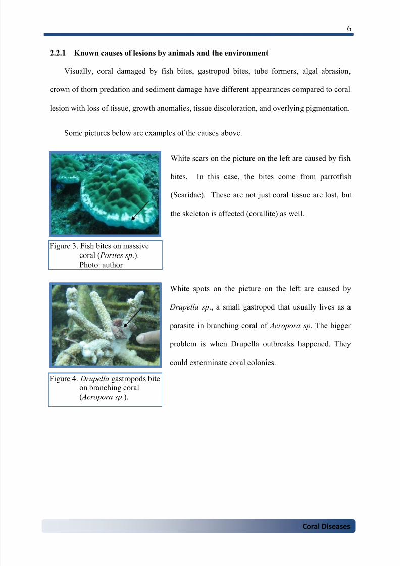
2.2.1 Known causes of lesions by animals and the environment
Visually, coral damaged by fish bites, gastropod bites, tube formers, algal abrasion,
crown of thorn predation and sediment damage have different appearances compared to coral
lesion with loss of tissue, growth anomalies, tissue discoloration, and overlying pigmentation.
Some pictures below are examples of the causes above.
White scars on the picture on the left are caused by fish
bites. In this case, the bites come from parrotfish
(Scaridae). These are not just coral tissue are lost, but
the skeleton is affected (corallite) as well.
White spots on the picture on the left are caused by
Drupella sp., a small gastropod that usually lives as a
parasite in branching coral of Acropora sp. The bigger
problem is when Drupella outbreaks happened. They
could exterminate coral colonies.
Figure 3. Fish bites on massive
coral ( Porites sp.).
Photo: author

7/22/2019 Coral Diseases
http://slidepdf.com/reader/full/coral-diseases 10/20
7
Coral Diseases
Figure 5. Tube formers living
inside coral.
Photo: author.
Figure 6. Algae grows rapidly on
a wide coral’s colony.
White spots on the picture on the left are caused by tube
formers that live from inside coral to the coral’s surface.
Although it is harmful, coral colony could continue
survive.
Algae grows rapidly covering a wide coral’s colony. This
happens when sedimentation or run off from land enters
the waters more than usual. Thus, corals could suffer from
these competing algae.
The branching coral’s colony seems to be bleached caused
by Crown of Thorns starfish (COT/ Acantharter planci)
predation. There are not only coral tissues that are lost,
but all coral polyps leaving only dead coral with clean
white corallite. COTs could be more dangerous when
outbreaks of their population. They could destroy colony
of corals in a huge area.
Figure 7. Crown of Thorn
predation on coral.
Photo: author

7/22/2019 Coral Diseases
http://slidepdf.com/reader/full/coral-diseases 11/20
8
Coral Diseases
Figure 9. a. Black-band disease on massive coral; b. White-band disease on stag-horn coral;
c. Red-band disease on gorgonian.
Sedimentation prevents coral from breathing properly
because their polyps are covered by sedimentation.
Moreover, sedimentation blocks the sunlight so that
zooxanthellae could not perform photosynthesis. As a
result, the coral becoming stressed and releases mucus
until coral dies, if sedimentation continues.
Figures 3 – 8 describe known causes from other animals and the environment. The
affect could be local such as fish and Drupella bites, but the other could be wider such as
crown of thorn predation and sedimentation.
2.2.2 Lesion type of diseases
2.2.2.1 Tissue loss
Tissue is a collective of cells in an organism that contains related structure and function
(biology-online.org, 2008). In terms of coral, tissue is a structure that covers the corallite
(coral skeleton) consisting of a body and tentacles to what we call a polyp. When disease
attaches, it causes loss of tissue and leaves only the corallite.
Some pictures below show diseases with tissue loss.
Figure 8. Sedimentation on coral.
Photo: author.
b ca

7/22/2019 Coral Diseases
http://slidepdf.com/reader/full/coral-diseases 12/20
9
Coral Diseases
Black-band disease is distinguished by a black line or crescent-formed band, dividing
healthy tissue and white dead coral. The disease is largely caused by a cyanobacteria
combining with sulfide-oxidizing bacteria and sulfur-reducing bacteria. Black-band disease
was discovered for the first time on the reefs of Belize and Florida in 1972 (coris.noaa.gov,
2010).
The cause of white-band disease is unidentified so far. Although strange microbe is
founded, scientists still could not identify this microorganism (coris.noaa.gov, 2010). White-
band disease appearance is similar with black-band disease except the color, which is white.
It is also separates healthy tissue and white dead coral. Normally it appears in coral branching
of Acropora sp.
Red-band disease is characterized by a red line separating healthy tissue and dead loss
tissue. As in figure 9.c., it happens not only on hard coral, but gorgonian as well. It appears
by a slender band of filamentous cyanobacteria moving slowly across the coral surface as
well as executing living tissue (coris.noaa.gov, 2010).
The picture on the left shows different types of red-
band disease in the filaments of cyanobacteria which
stretch round in a net-like manner leaving dead coral in
the middle.
Figure 10. Red-band disease on
massive coral
(Faviidae).
Photo: author.

7/22/2019 Coral Diseases
http://slidepdf.com/reader/full/coral-diseases 13/20
10
Coral Diseases
Figure 11. Growth anomaly on
massive coral ( Porites
sp.).
2.2.2.2 Growth anomalies
As in the introduction, corals mostly grow as massive, sub-massive, branching,
tabulate, digit-like (digitate), and encrusting forms colony. In reefs, sometimes we find an
abnormal growth in a normal coral colony life-form. The abnormality could have bigger or
smaller skeletal components. The cause and the dangers
are unknown. On the other hand, corallite could be
totally missing, and the growth anomaly leaves a white
plaque over the surface of coral colony (Raymundo et al.
2008).
2.2.2.3 Tissue discoloration
Tissue discoloration is the change of colour in the coral colony with colours such as
pink, purple and pale. It is considered to be a reaction of the coral to various stressors such as
coral life-competition, boring fauna, thermal bleaching, and abrasion from algae. (Raymundo
et al. 2008).
Figure 12. a. Tissue discoloration on massive coral ( Montipora sp.).
b. Tissue discoloration on sub-massive coral (Goniopora
sp.). Possibly causes by thermal bleaching. Photo: author.
a b
7/22/2019 Coral Diseases
http://slidepdf.com/reader/full/coral-diseases 14/20
11
Coral Diseases
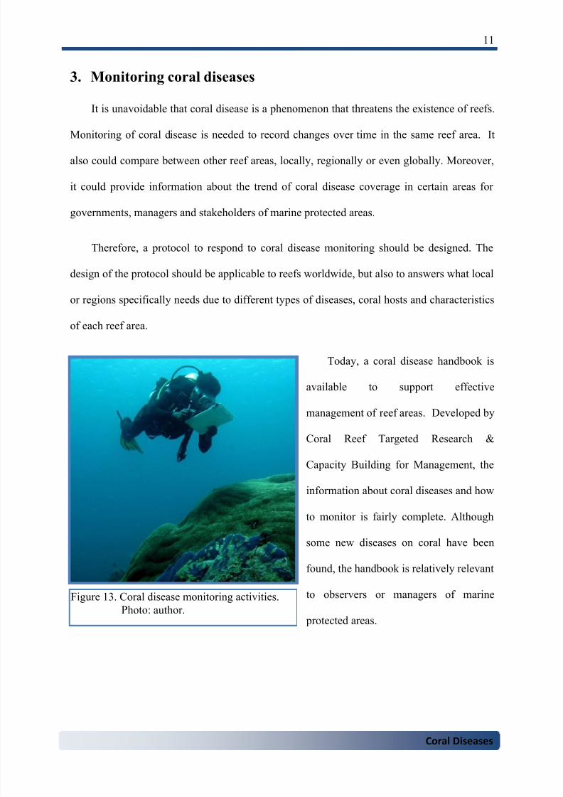
3. Monitoring coral diseases
It is unavoidable that coral disease is a phenomenon that threatens the existence of reefs.
Monitoring of coral disease is needed to record changes over time in the same reef area. It
also could compare between other reef areas, locally, regionally or even globally. Moreover,
it could provide information about the trend of coral disease coverage in certain areas for
governments, managers and stakeholders of marine protected areas.
Therefore, a protocol to respond to coral disease monitoring should be designed. The
design of the protocol should be applicable to reefs worldwide, but also to answers what local
or regions specifically needs due to different types of diseases, coral hosts and characteristics
of each reef area.
Today, a coral disease handbook is
available to support effective
management of reef areas. Developed by
Coral Reef Targeted Research &
Capacity Building for Management, the
information about coral diseases and how
to monitor is fairly complete. Although
some new diseases on coral have been
found, the handbook is relatively relevant
to observers or managers of marine
protected areas.
Figure 13. Coral disease monitoring activities.
Photo: author.
7/22/2019 Coral Diseases
http://slidepdf.com/reader/full/coral-diseases 15/20
12
Coral Diseases
4. How to deal with coral diseases based on scientific research
The sea water provides microbial diversity. It remains unknown how many of these are
pathogens or prospectively so. Moreover, microbial diversity is not just supported by sea
water but linked to what happens on land. In addition, imperfect knowledge of coral, their
diseases and the factors that influence coral health, their resistance and their resilience are our
challenges in developing effective management (Raymundo et al., 2008).
Some evidence of threats to coral damage are known. For examples, outbreaks of crown
of thorns are caused by the extinction of their natural predator and nutrient run-off increased
by human use of coastal areas. Moreover, fast growing development of coastal areas brings
sediment into the reefs. (www.reef.crc.org.au, n.d). These cause coral damage and so coral
dies in huge areas.
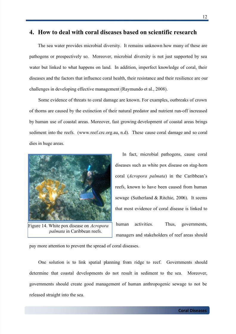
In fact, microbial pathogens, cause coral
diseases such as white pox disease on stag-horn
coral ( Acropora palmata) in the Caribbean’s
reefs, known to have been caused from human
sewage (Sutherland & Ritchie, 2006). It seems
that most evidence of coral disease is linked to
human activities. Thus, governments,
managers and stakeholders of reef areas should
pay more attention to prevent the spread of coral diseases.
One solution is to link spatial planning from ridge to reef. Governments should
determine that coastal developments do not result in sediment to the sea. Moreover,
governments should create good management of human anthropogenic sewage to not be
released straight into the sea.
Figure 14. White pox disease on Acropora
palmata in Caribbean reefs.

7/22/2019 Coral Diseases
http://slidepdf.com/reader/full/coral-diseases 16/20
13
Coral Diseases
The other solution is to create as many reefs or marine protected areas as possible to
preserve coral reefs areas and take the benefits of them sustainably. Additionally,
governments or managers of marine protected areas could work together to prevents threats
to coral reefs.
Finally, all stakeholders must be involved in management of marine protected areas,
especially local communities who take benefits directly from their reefs.

7/22/2019 Coral Diseases
http://slidepdf.com/reader/full/coral-diseases 17/20
14
Coral Diseases
5. Conclusion
Coral disease is a phenomenon that threatens the existence of reefs today. There are at
least three kind of diseases based on causes in coral lesion: infectious biotic disease, abiotic
disease, and non- infectious biotic disease. On the other hand, there are some lesions and
damage to corals caused by other animals such as parrotfish and environmental aspects such
as sedimentation and changes of sea temperature.
Coral disease monitoring is to record changes over time in the same reef area as well as a
tool to compare between other reef areas, locally, regionally or globally. It is important to
give this information to governments, managers and stakeholders of marine protected areas as
an input in management of reef area.
Finally, incomplete knowledge of coral, coral diseases and the factors that influence
coral’s health, their resistance and their resilience are our challenges in developing effective
management of reef areas.

7/22/2019 Coral Diseases
http://slidepdf.com/reader/full/coral-diseases 18/20
15
Coral Diseases
Acknowledgements
Praise to Allah almighty.
I have been indebted in the preparation of this report to my teacher, my supervisor, Sally
Lim and Ian Baker of James Cook University – Pathway College, who have been improving
my English as well as my academic writing with their patience and kindness. I am very
grateful to all of JCU-Pathway College staffs who really helpful and nice. Everything is so
well-organized here.
To Dr. Agni Boedhihartono and Prof. Jeffrey Sayer, who encouraged me to continue
study in James Cook University. It is an 8 year of waiting, and in one more step I am going to
be a student of James Cook University, Australia. Thus, it is an honour.
I am very gratefully thanks to my parents, my brother and my sisters, who have been
providing thoughtful supports.
To Ditta, my wife and Aldebaran my son who always accompany, you are my joy, my
happiness, my reasons, my life.
And in the end the love you take is equal to the love you make (The Beatles).

7/22/2019 Coral Diseases
http://slidepdf.com/reader/full/coral-diseases 19/20
16
Coral Diseases
References
Algae overgrowth [image]. (2006). Retrieved January 25, 2012 from
http://www.coralmorphologic.com
Biology online. (2008). Dictionary. Retrieved January 2, 2012 from
http://www.biology-online.org/dictionary/Tissue
Black-band disease [image]. (2010). Retrieved January 25, 2012, from
http://coris.noaa.gov/about/diseases/
Coral Reef Map [image]. (2008). Retrieved January 20, 2012 from
http://www.coralreefinfo.com
Growth anomalies [image]. (2005). Retrieved January 26, 2012 from
http://archives.starbulletin.com/2005/12/25/news/story03.html
Nybakken, J.W. (1988). Marine biology: An ecological approach (M. Eidman, Ed.). Jakarta:
PT. Gramedia.
Raymundo, L.J., Couch, C.S. & Harvell, C.D. (Eds). (2008). Coral disease handbook.
Guidelines for assessment, monitoring & management. Melbourne, Australia: Coral
Reef Targeted Research and Capacity Building for Management Program.
Reef Research Center. (n.d). What causes crown of thorn starfish outbreaks?. Retrieved
January 26, 2012, from
http://www.reef.crc.org.au/discover/plantsanimals/cots/cotstheory.html
Red-band disease [image]. (2008). Retrieved January 25, 2012 from
http://www.coralreefinfo.com/coralglossary/glossary_r.htm
Sutherland, K.P & Ritchie, K.B. (2006). White pox disease of the Caribbean elkhorn coral,
Acropora palmata. Retrieved January 26, 2012, from
http://isurus.mote.org/Keys/microbiology/chapter16_patterson-sutherland_ritchie.pdf
White-band disease [image]. (2010). Retrieved January 25, 2012, from
http://coris.noaa.gov/about/diseases/

7/22/2019 Coral Diseases
http://slidepdf.com/reader/full/coral-diseases 20/20
17
Coral Diseases
White pox disease [image]. (2007). Retrieved January 25, 2012, from
http://soundwaves.usgs.gov