Embed Size (px)
Citation preview
Copyright © 2010 Pearson Education, Inc.
C h a p t e r
16
The Digestive System
PowerPoint® Lecture Slides prepared by Jason LaPres
Lone Star College - North Harris
Copyright © 2010 Pearson Education, Inc.
Copyright © 2010 Pearson Education, Inc.
Introduction to the Digestive System
• Acquires nutrients from environment
• Anabolism
– Uses raw materials to synthesize essential
compounds
• Catabolism
– Decomposes substances to provide energy cells need
to function
Copyright © 2010 Pearson Education, Inc.
Digestive Tract
• Digestive tract also called gastrointestinal (GI) tract or alimentary canal– Is a muscular tube– Extends from oral cavity to anus:
• Passes through pharynx, esophagus, stomach, and small and large intestines
Copyright © 2010 Pearson Education, Inc.
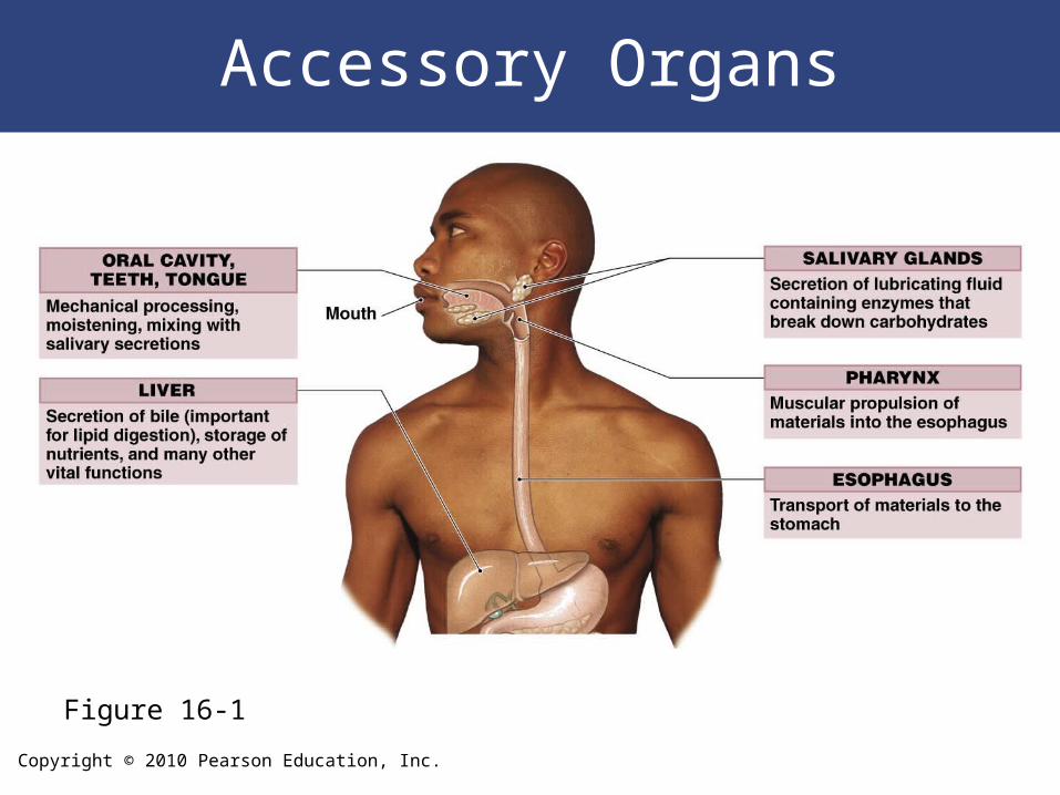
Accessory Organs
Figure 16-1
Copyright © 2010 Pearson Education, Inc.
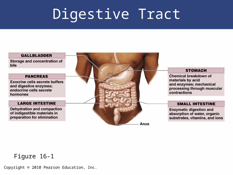
Digestive Tract
Figure 16-1
Copyright © 2010 Pearson Education, Inc.
Functions of the Digestive System
1. Ingestion – Occurs when materials enter digestive tract via the mouth
2. Mechanical processing – Crushing and shearing– Makes materials easier to propel along digestive tract
3. Digestion – The chemical breakdown of food into small organic
fragments for absorption by digestive epithelium
Copyright © 2010 Pearson Education, Inc.
Functions of the Digestive System
4. Secretion– Is the release of water, acids, enzymes, buffers, and salts
– By epithelium of digestive tract
– By glandular organs
5. Absorption – Movement of organic substrates, electrolytes, vitamins,
and water
– Across digestive epithelium
– Into interstitial fluid of digestive tract
6. Excretion – Removal of waste products from body fluids
Copyright © 2010 Pearson Education, Inc.
Digestive Tract
• Lining of the digestive tract protects surrounding tissues against– Corrosive effects of digestive acids and enzymes– Mechanical stresses, such as abrasion– Bacteria either ingested with food or that reside in
digestive tract
Copyright © 2010 Pearson Education, Inc.
Histological Organization of the Digestive Tract
• Major layers of the digestive tract– Mucosa– Submucosa– Muscularis externa– Serosa
Copyright © 2010 Pearson Education, Inc.
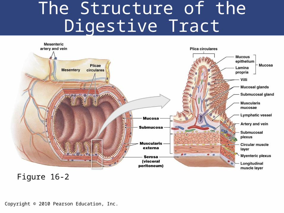
The Structure of the Digestive Tract
Figure 16-2
Copyright © 2010 Pearson Education, Inc.
Histological Organization of the Digestive Tract
• The Mucosa– Is the inner lining of the digestive tract– Is a mucous membrane consisting of:
• Epithelium, moistened by glandular secretions• Lamina propria of areolar tissue
Copyright © 2010 Pearson Education, Inc.
Histological Organization of the Digestive Tract
• The Digestive Epithelium
– Mucosal epithelium is simple or stratified:
• Depending on location, function, and stresses:
– oral cavity, pharynx, and esophagus:
» mechanical stresses
» lined by stratified squamous epithelium
– stomach, small intestine, and most of large intestine:
» absorption
» simple columnar epithelium with mucous (goblet) cells
Copyright © 2010 Pearson Education, Inc.
Histological Organization of the Digestive Tract
• The Submucosa– Is a layer of dense, irregular connective tissue– Surrounds muscularis mucosae– Has large blood vessels and lymphatic
vessels– May contain exocrine glands:
• Secrete buffers and enzymes into digestive tract
Copyright © 2010 Pearson Education, Inc.
Digestive Tract
• The Serosa– Serous membrane covering muscularis externa– Visceral peritoneum over some CT:
• Continuous with parietal peritoneum that lines cavity
– Except in oral cavity, pharynx, esophagus, and rectum:
• Where adventitia, a dense sheath of collagen fibers, firmly attaches the digestive tract to adjacent structures
Copyright © 2010 Pearson Education, Inc.
The Movement of Digestive Materials
• Pacesetter Cells
– Located in muscularis mucosae and muscularis
externa:
• Surrounding lumen of digestive tract
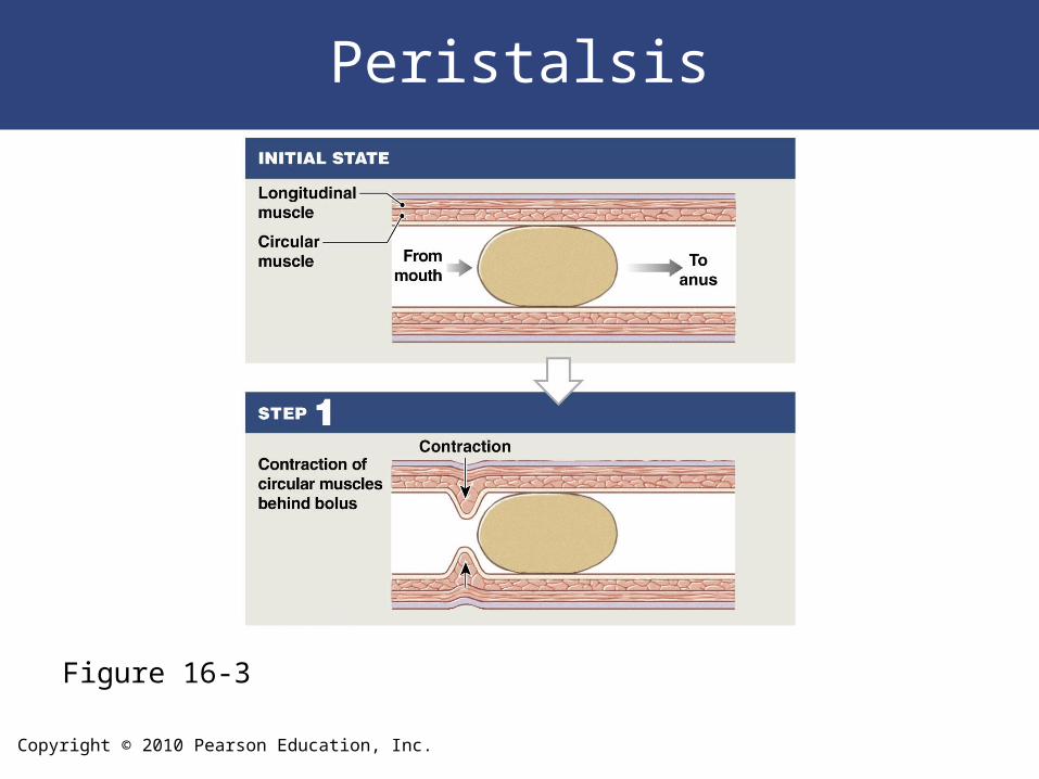
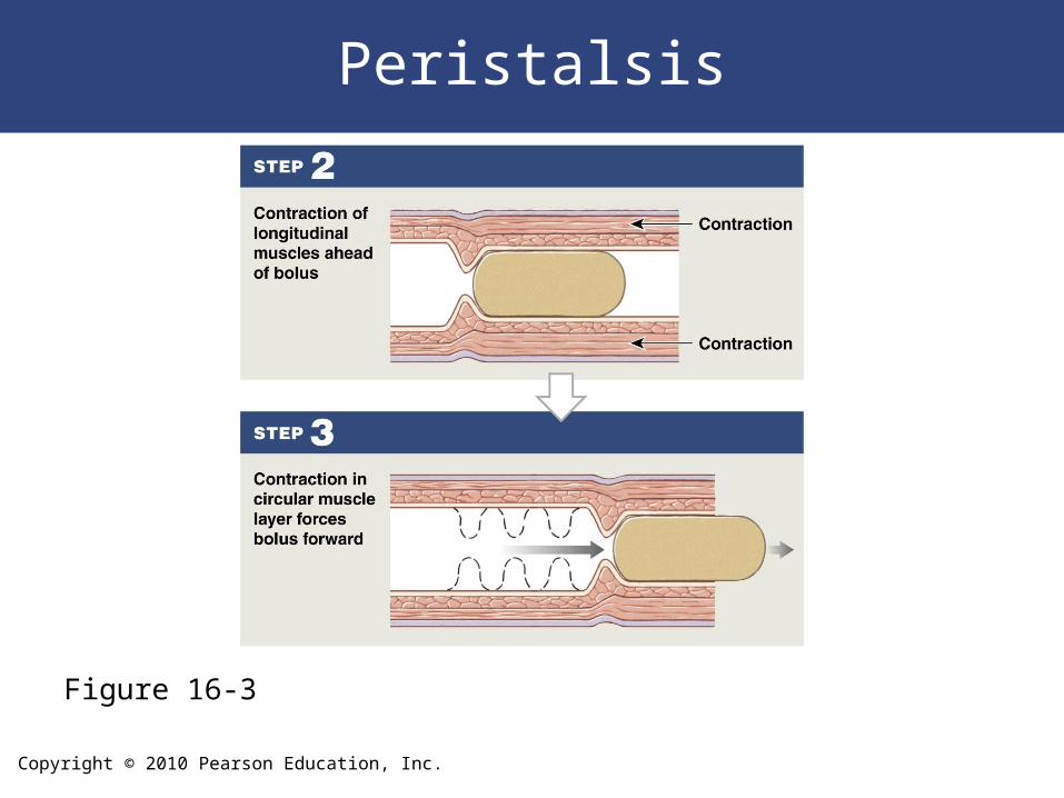
• Peristalsis
– Consists of waves of muscular contractions
– Moves a bolus along the length of the digestive tract
Copyright © 2010 Pearson Education, Inc.
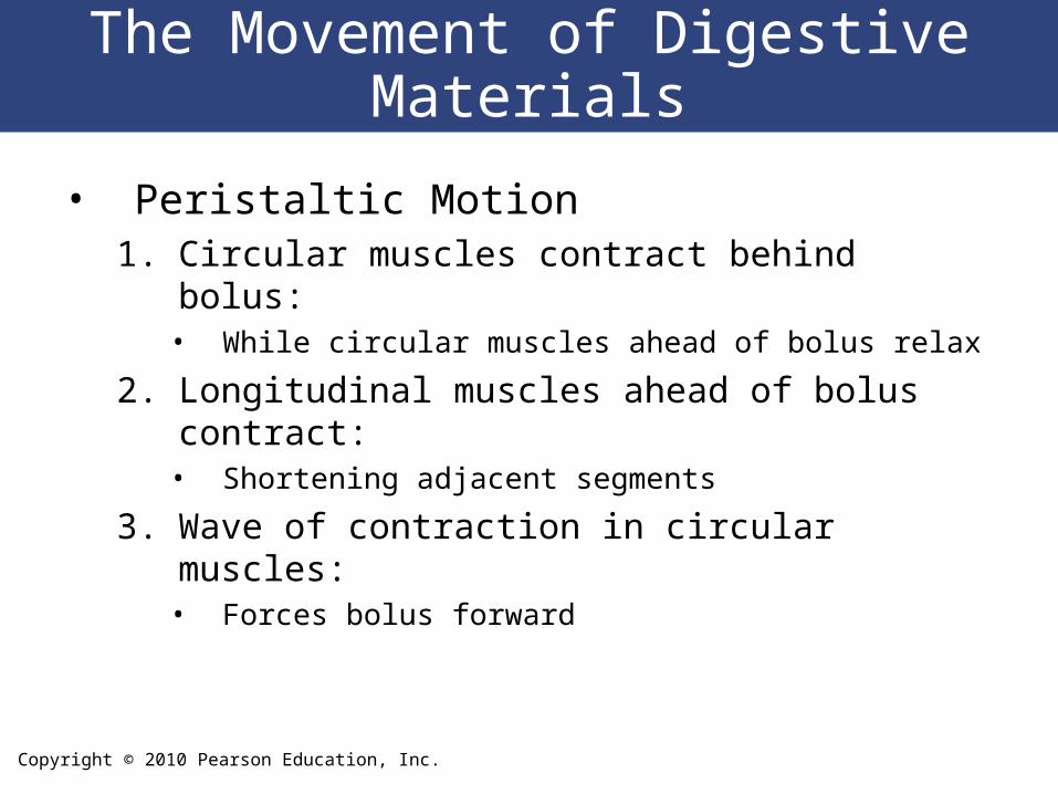
The Movement of Digestive Materials
• Peristaltic Motion1. Circular muscles contract behind bolus:
• While circular muscles ahead of bolus relax
2. Longitudinal muscles ahead of bolus contract:• Shortening adjacent segments
3. Wave of contraction in circular muscles:• Forces bolus forward
Copyright © 2010 Pearson Education, Inc.
Peristalsis
Figure 16-3
Copyright © 2010 Pearson Education, Inc.
Peristalsis
Figure 16-3
Copyright © 2010 Pearson Education, Inc.
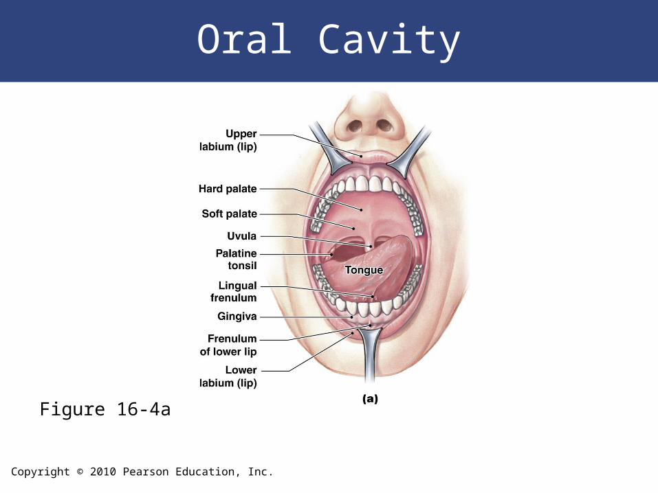
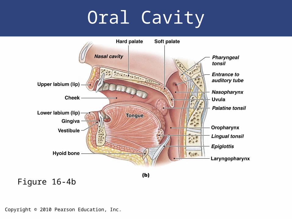
Functions of Oral Cavity
• Sensory analysis
– Of material before swallowing
• Mechanical processing
– Through actions of teeth, tongue, and palatal surfaces
• Lubrication
– Mixing with mucus and salivary gland secretions
• Limited digestion
– Of carbohydrates and lipids
Copyright © 2010 Pearson Education, Inc.
Oral Cavity
Figure 16-4a
Copyright © 2010 Pearson Education, Inc.
Oral Cavity
Figure 16-4b
Copyright © 2010 Pearson Education, Inc.
The Tongue
• Manipulates materials inside mouth
• Functions of the tongue
– Mechanical processing by compression, abrasion, and
distortion
– Manipulation to assist in chewing and to prepare material for
swallowing
– Sensory analysis by touch, temperature, and taste receptors
– Secretion of mucins and the enzyme lingual lipase
Copyright © 2010 Pearson Education, Inc.
Salivary Glands
• Three pairs secrete into oral cavity – Each pair has distinctive cellular organization:
• And produces saliva with different properties
Copyright © 2010 Pearson Education, Inc.
Oral Cavity
• Parotid Salivary Glands – Produce serous secretion:
• Enzyme salivary amylase (breaks down starches)
– Drained by parotid duct (Stensen duct):• Which empties into vestibule at second molar
Copyright © 2010 Pearson Education, Inc.
Oral Cavity
• Sublingual Salivary Glands – Covered by mucous membrane of floor of
mouth– Produce mucous secretion:
• Acts as a buffer and lubricant
– Sublingual ducts (Rivinus ducts):• Either side of lingual frenulum
Copyright © 2010 Pearson Education, Inc.
Oral Cavity
• Submandibular Salivary Glands
– In floor of mouth
– Within mandibular groove
– Secrete buffers, glycoproteins (mucins), and salivary
amylase
– Submandibular ducts (Wharton ducts):
• Open immediately posterior to teeth
• Either side of lingual frenulum
Copyright © 2010 Pearson Education, Inc.
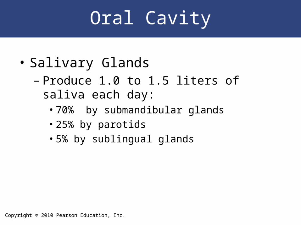
Oral Cavity
• Salivary Glands– Produce 1.0 to 1.5 liters of saliva each day:
• 70% by submandibular glands• 25% by parotids• 5% by sublingual glands
Copyright © 2010 Pearson Education, Inc.
Oral Cavity
• Saliva
– 99.4% water
– 0.6% includes:
• Electrolytes (Na+, Cl–, and HCO3–)
• Buffers
• Glycoproteins (mucins)
• Antibodies
• Enzymes
• Waste products
Copyright © 2010 Pearson Education, Inc.
The Teeth
• Tongue movements pass food across occlusal surfaces of teeth
• Chew (masticate) food• Tooth structure
– Dentin:• A mineralized matrix similar to that of bone• Does not contain cells
– Pulp cavity:• Receives blood vessels and nerves through the
root canal
Copyright © 2010 Pearson Education, Inc.
The Teeth
• Tooth Structure
– Root:
• Of each tooth sits in a bony socket (alveolus)
• A layer of cementum covers dentin of the root:
– providing protection and anchoring periodontal ligament
– Crown:
• Exposed portion of tooth
• Projects beyond soft tissue of gingiva
• Dentin covered by layer of enamel
Copyright © 2010 Pearson Education, Inc.
The Teeth
• Alveolar Processes– Of the maxillae:
• Form maxillary arcade (upper dental arch)
– Of the mandible:• Form mandibular arcade (lower dental arch)
Copyright © 2010 Pearson Education, Inc.
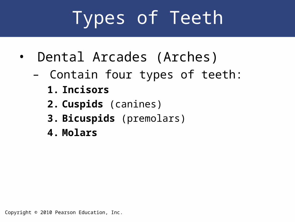
Types of Teeth
• Dental Arcades (Arches)– Contain four types of teeth:
1. Incisors
2. Cuspids (canines)
3. Bicuspids (premolars)
4. Molars
Copyright © 2010 Pearson Education, Inc.
Dental Succession
• During embryonic development, two sets of teeth form– Primary dentition, or deciduous teeth– Secondary dentition, or permanent
dentition
Copyright © 2010 Pearson Education, Inc.
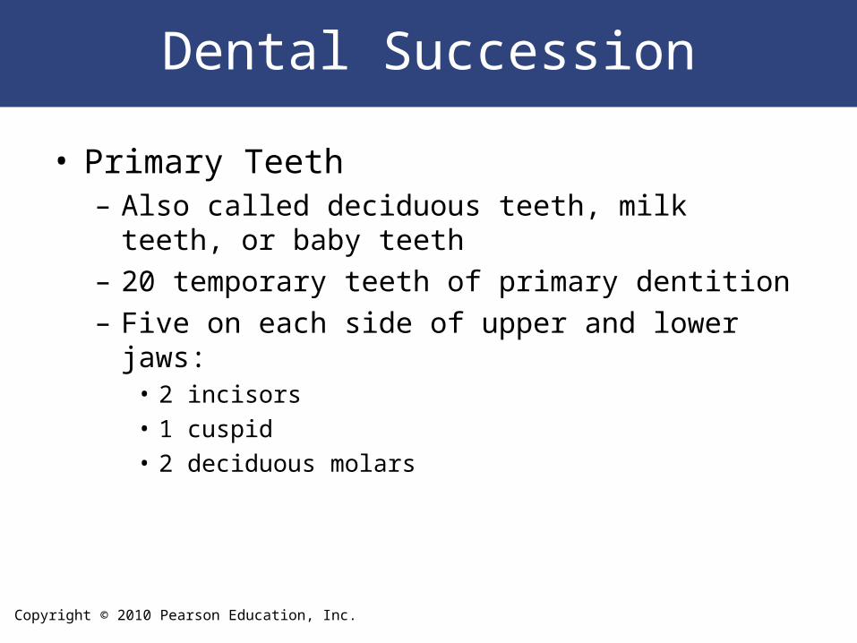
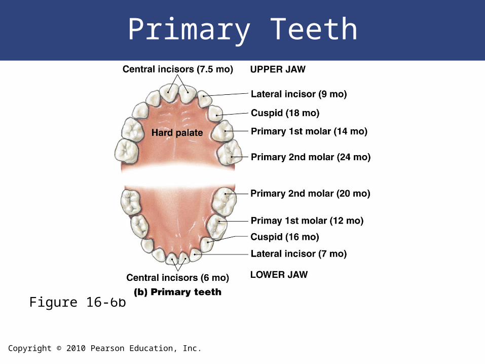
Dental Succession
• Primary Teeth– Also called deciduous teeth, milk teeth, or baby teeth– 20 temporary teeth of primary dentition – Five on each side of upper and lower jaws:
• 2 incisors• 1 cuspid• 2 deciduous molars
Copyright © 2010 Pearson Education, Inc.
Primary Teeth
Figure 16-6b
Copyright © 2010 Pearson Education, Inc.
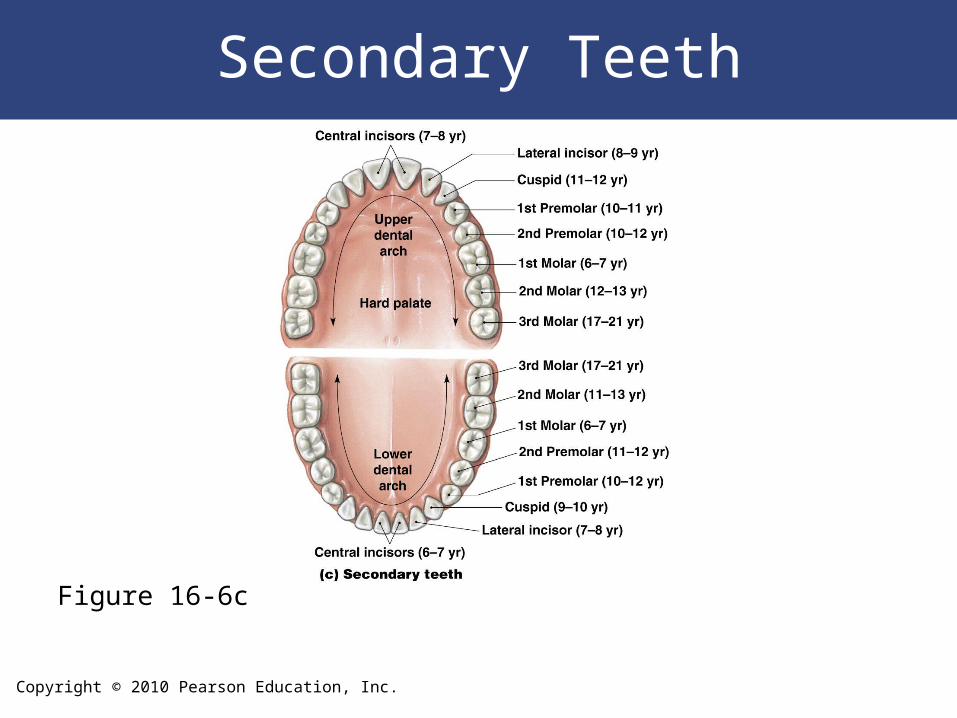
Dental Succession
• Secondary Dentition
– Also called permanent dentition
– Replaces deciduous teeth
– 32 permanent teeth
– Eight on each side, upper and lower:
• 2 incisors
• 1 cuspid
• 5 molars
Copyright © 2010 Pearson Education, Inc.
Secondary Teeth
Figure 16-6c
Copyright © 2010 Pearson Education, Inc.
The Pharynx
• A common passageway for solid food, liquids, and air
• Regions of the pharynx– Nasopharynx– Oropharynx– Laryngopharynx
Copyright © 2010 Pearson Education, Inc.
The Esophagus
• A hollow muscular tube
• About 25 cm (10 in.) long and 2 cm (0.80 in.)
wide
• Conveys solid food and liquids to the stomach
• Begins posterior to cricoid cartilage
• Is innervated by fibers from the esophageal
plexus
Copyright © 2010 Pearson Education, Inc.
The Esophagus
• Resting Muscle Tone – In the circular muscle layer in the superior
3 cm (1.2 in.) of esophagus, prevents air from entering
Copyright © 2010 Pearson Education, Inc.
Swallowing
• Also called deglutition
– Can be initiated voluntarily
– Proceeds automatically
– Is divided into three phases:
• Buccal phase
• Pharyngeal phase
• Esophageal phase
Copyright © 2010 Pearson Education, Inc.
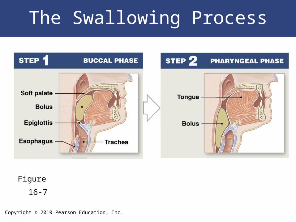
Figure 16-7
The Swallowing Process
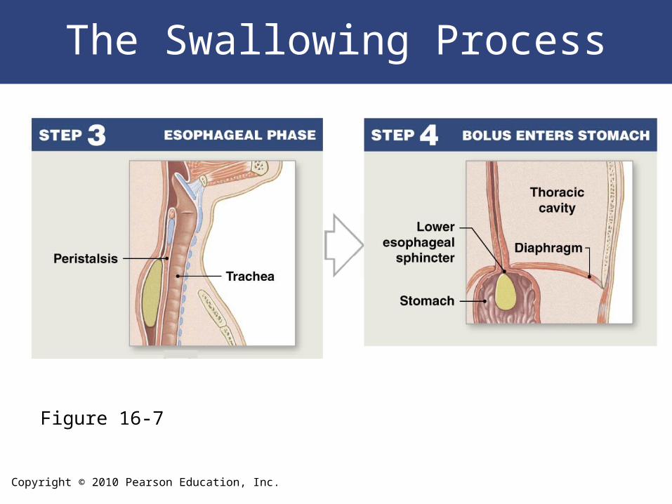
Copyright © 2010 Pearson Education, Inc.
The Swallowing Process
Figure 16-7
Copyright © 2010 Pearson Education, Inc.
The Stomach
• Major Functions of the Stomach
– Storage of ingested food
– Mechanical breakdown of ingested food
– Disruption of chemical bonds in food material by acid
and enzymes
– Production of intrinsic factor, a glycoprotein required
for absorption of vitamin B12 in small intestine
Copyright © 2010 Pearson Education, Inc.
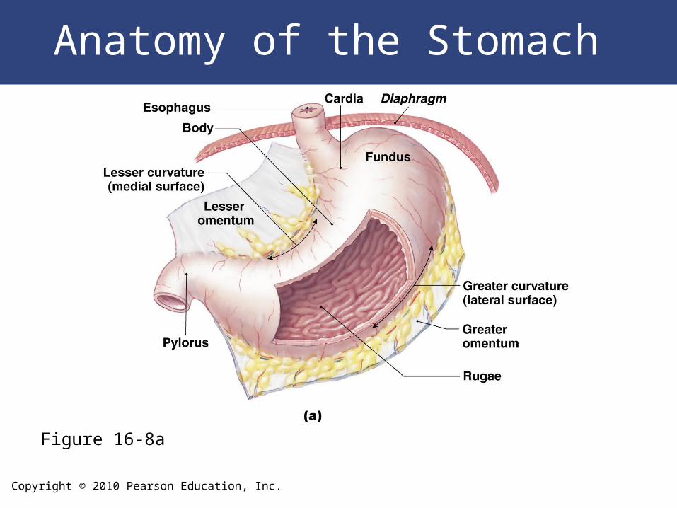
The Stomach
• Regions of the Stomach– Cardia– Fundus – Body– Pylorus
Copyright © 2010 Pearson Education, Inc.
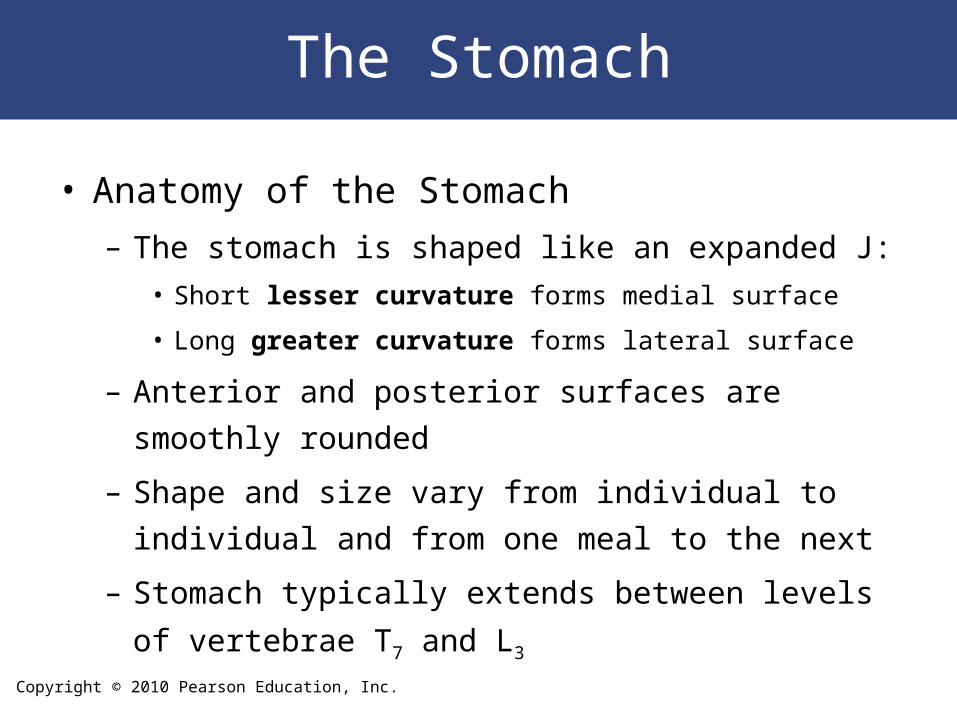
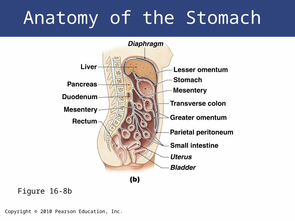
The Stomach
• Anatomy of the Stomach
– The stomach is shaped like an expanded J:
• Short lesser curvature forms medial surface
• Long greater curvature forms lateral surface
– Anterior and posterior surfaces are smoothly rounded
– Shape and size vary from individual to individual and
from one meal to the next
– Stomach typically extends between levels of
vertebrae T7 and L3
Copyright © 2010 Pearson Education, Inc.
Anatomy of the Stomach
Figure 16-8a
Copyright © 2010 Pearson Education, Inc.
Anatomy of the Stomach
Figure 16-8b
Copyright © 2010 Pearson Education, Inc.
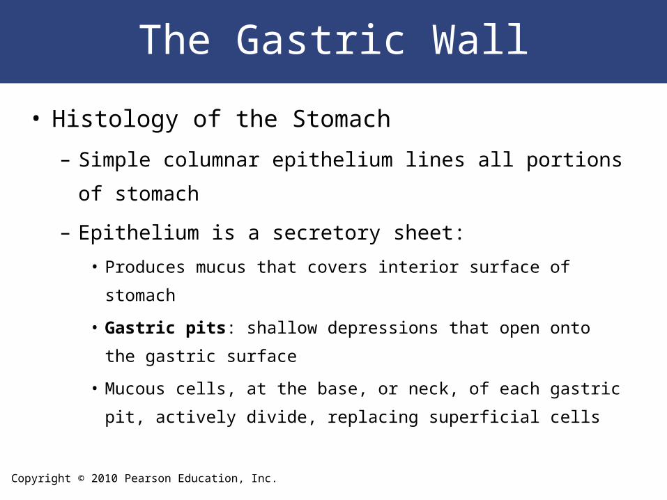
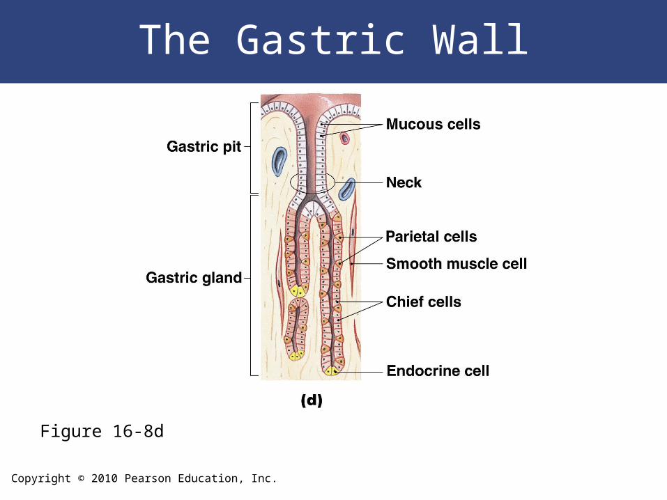
The Gastric Wall
• Histology of the Stomach
– Simple columnar epithelium lines all portions of
stomach
– Epithelium is a secretory sheet:
• Produces mucus that covers interior surface of stomach
• Gastric pits: shallow depressions that open onto the gastric
surface
• Mucous cells, at the base, or neck, of each gastric pit, actively
divide, replacing superficial cells
Copyright © 2010 Pearson Education, Inc.
The Gastric Wall
• Gastric Glands– In fundus and body of stomach:
• Extend deep into underlying lamina propria
– Each gastric pit communicates with several gastric glands:
• Parietal cells • Chief cells
Copyright © 2010 Pearson Education, Inc.
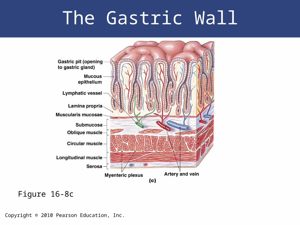
The Gastric Wall
Figure 16-8c
Copyright © 2010 Pearson Education, Inc.
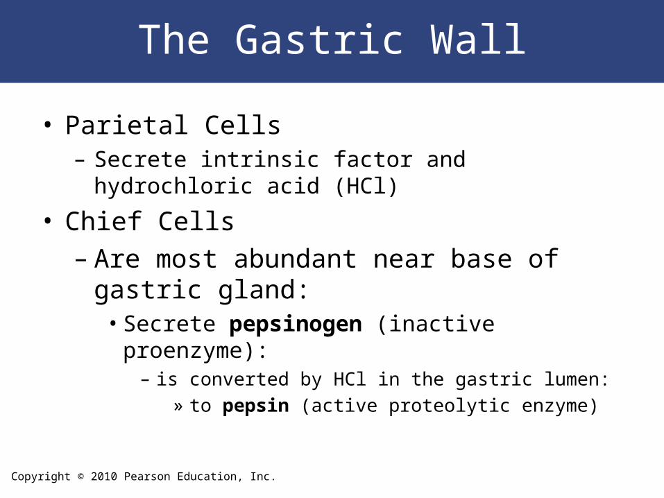
The Gastric Wall
• Parietal Cells– Secrete intrinsic factor and hydrochloric acid (HCl)
• Chief Cells– Are most abundant near base of gastric
gland:• Secrete pepsinogen (inactive proenzyme):
– is converted by HCl in the gastric lumen:» to pepsin (active proteolytic enzyme)
Copyright © 2010 Pearson Education, Inc.
The Gastric Wall
Figure 16-8d
Copyright © 2010 Pearson Education, Inc.
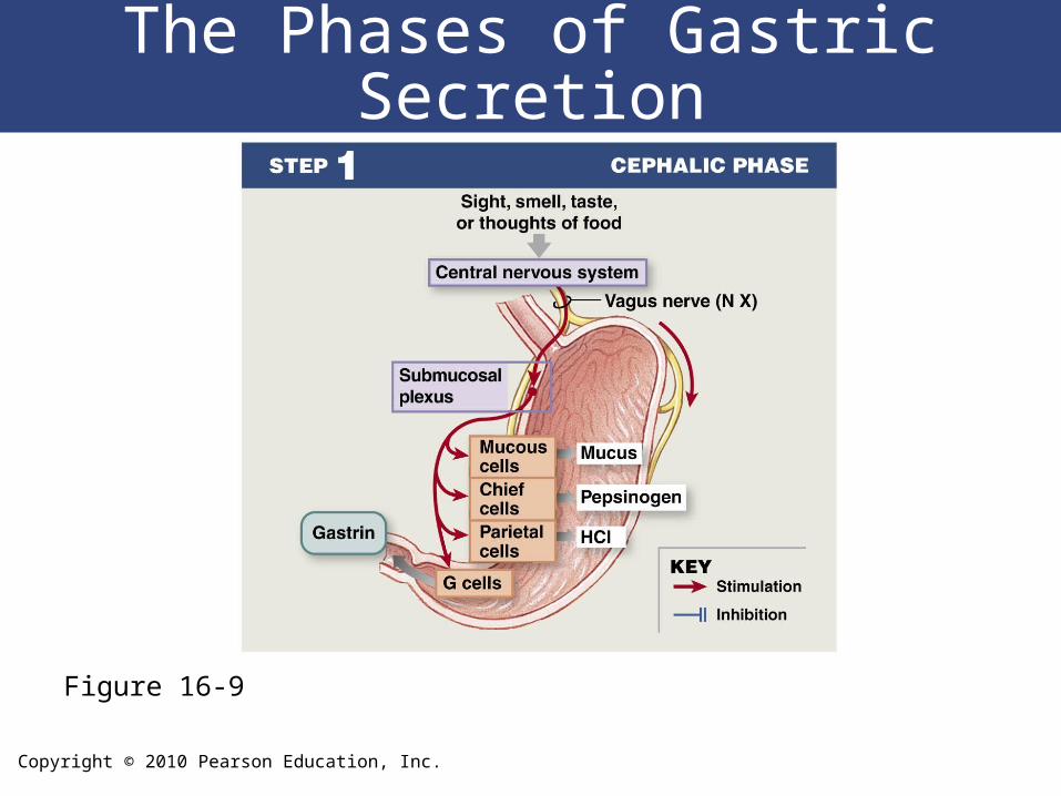
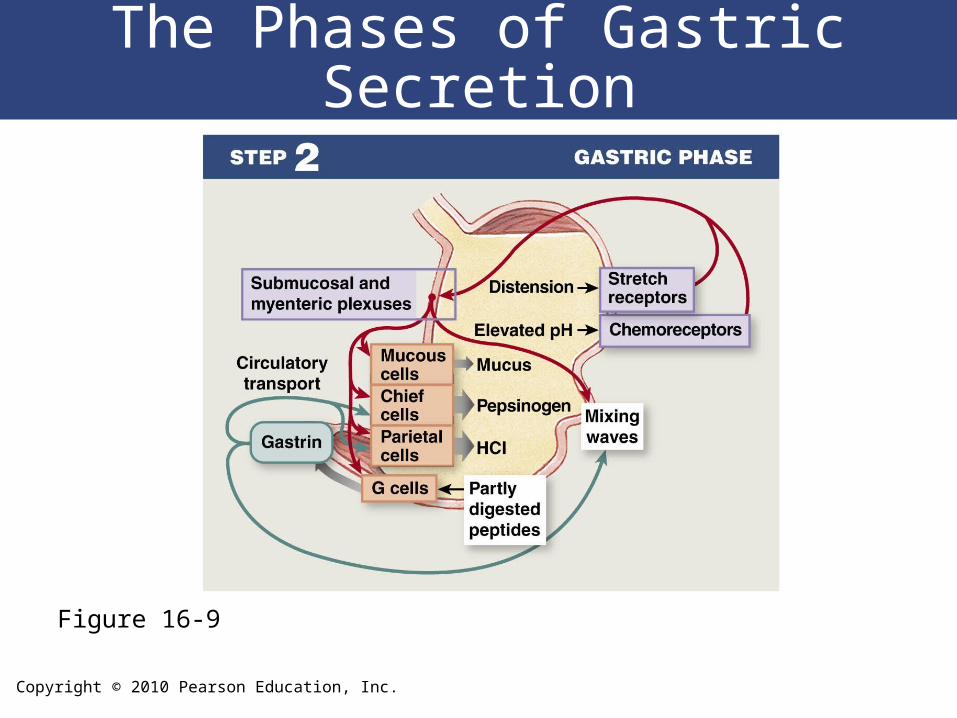
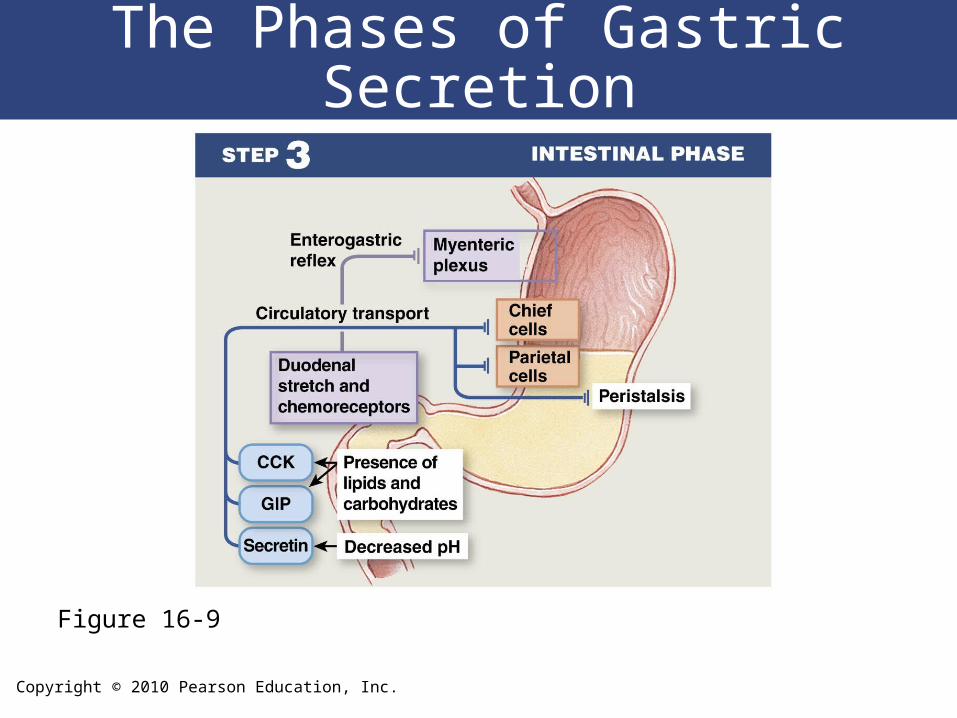
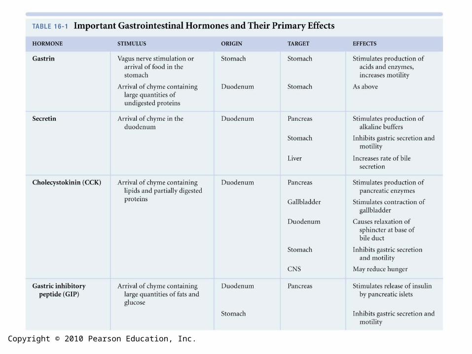
Regulation of Gastric Activity
• Production of acid and enzymes by the
gastric mucosa can be
– Controlled by the CNS
– Regulated by short reflexes of ENS
– Regulated by hormones of digestive tract
• Three phases: cephalic phase, gastric
phase, and intestinal phase
Copyright © 2010 Pearson Education, Inc.
The Phases of Gastric Secretion
Figure 16-9
Copyright © 2010 Pearson Education, Inc.
The Phases of Gastric Secretion
Figure 16-9
Copyright © 2010 Pearson Education, Inc.
The Phases of Gastric Secretion
Figure 16-9
Copyright © 2010 Pearson Education, Inc.
Digestion in the Stomach
• Stomach performs preliminary digestion of proteins by pepsin– Some digestion of carbohydrates (by salivary
amylase)– Lipids (by lingual lipase)
• Stomach contents– Become more fluid– pH approaches 2.0– Pepsin activity increases– Protein disassembly begins
• Although digestion occurs in the stomach, nutrients are not absorbed there
Copyright © 2010 Pearson Education, Inc.
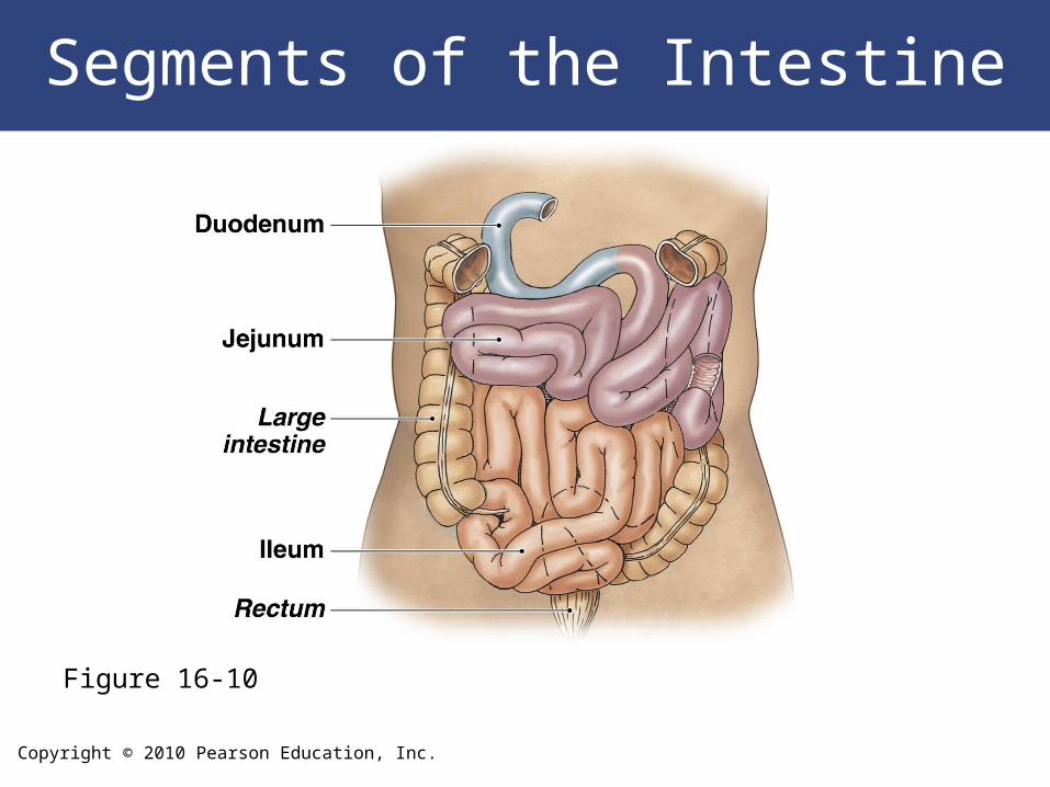
The Small Intestine
• Plays key role in digestion and absorption of nutrients
• 90% of nutrient absorption occurs in the small intestine
Copyright © 2010 Pearson Education, Inc.
The Small Intestine
• The Duodenum
– The segment of small intestine closest to the stomach
– 25 cm (10 in.) long
– “Mixing bowl” that receives chyme from stomach and
digestive secretions from pancreas and liver
– Functions of the duodenum:
• To receive chyme from stomach
• To neutralize acids before they can damage the absorptive
surfaces of the small intestine
Copyright © 2010 Pearson Education, Inc.
The Small Intestine
• The Jejunum – Is the middle segment of the small intestine– 2.5 meters (8.2 ft) long– Is the location of most:
• Chemical digestion• Nutrient absorption
– Has few plicae circulares– Small villi
Copyright © 2010 Pearson Education, Inc.
The Small Intestine
• The Ileum– The final segment of the small intestine– 3.5 meters (11.48 ft) long – Ends at the ileocecal valve, a sphincter that
controls flow of material from the ileum into the large intestine
Copyright © 2010 Pearson Education, Inc.
Segments of the Intestine
Figure 16-10
Copyright © 2010 Pearson Education, Inc.
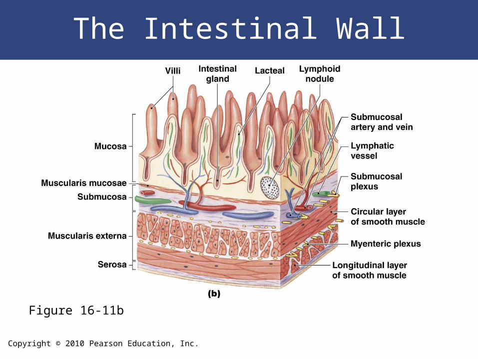
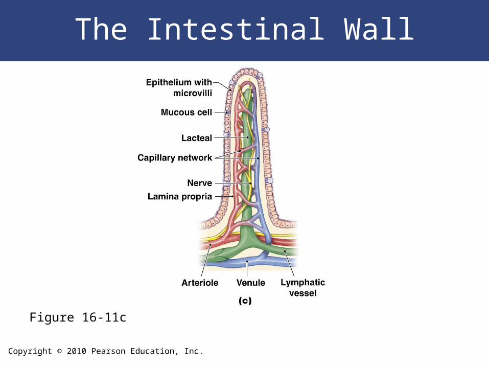
The Intestinal Wall
• Histology of the Small Intestine– Plicae circulares:
• Transverse folds in intestinal lining• Are permanent features:
– do not disappear when small intestine fills
– Intestinal villi: • A series of fingerlike projections:
– in mucosa of small intestine
• Covered by simple columnar epithelium:– covered with microvilli
Copyright © 2010 Pearson Education, Inc.
The Intestinal Wall
• Histology of the Small Intestine
– Intestinal glands: • Mucous cells between columnar epithelial cells
• Eject mucins onto intestinal surfaces
Copyright © 2010 Pearson Education, Inc.
The Intestinal Wall
Figure 16-11b
Copyright © 2010 Pearson Education, Inc.
The Intestinal Wall
Figure 16-11c
Copyright © 2010 Pearson Education, Inc.
The Small Intestine
• Duodenal Glands– Also called submucosal glands or Brunner
glands – Produce copious quantities of mucus:
• When chyme arrives from stomach
Copyright © 2010 Pearson Education, Inc.
Intestinal Movements
• Chyme arrives in duodenum
– Weak peristaltic contractions move it slowly
toward jejunum:
• Myenteric reflexes
• Not under CNS control
• Parasympathetic stimulation accelerates local
peristalsis and segmentation
Copyright © 2010 Pearson Education, Inc.
Intestinal Movements
• The Gastroenteric Reflex
– Stimulates motility and secretion:
• Along entire small intestine
• The Gastroileal Reflex
– Triggers relaxation of ileocecal valve
– Allows materials to pass from small intestine into
large intestine
Copyright © 2010 Pearson Education, Inc.
Intestinal Secretions
• Watery intestinal juice
– 1.8 liters per day enter intestinal lumen
– Moisten chyme
– Assist in buffering acids
– Keep digestive enzymes and products of
digestion in solution
Copyright © 2010 Pearson Education, Inc.
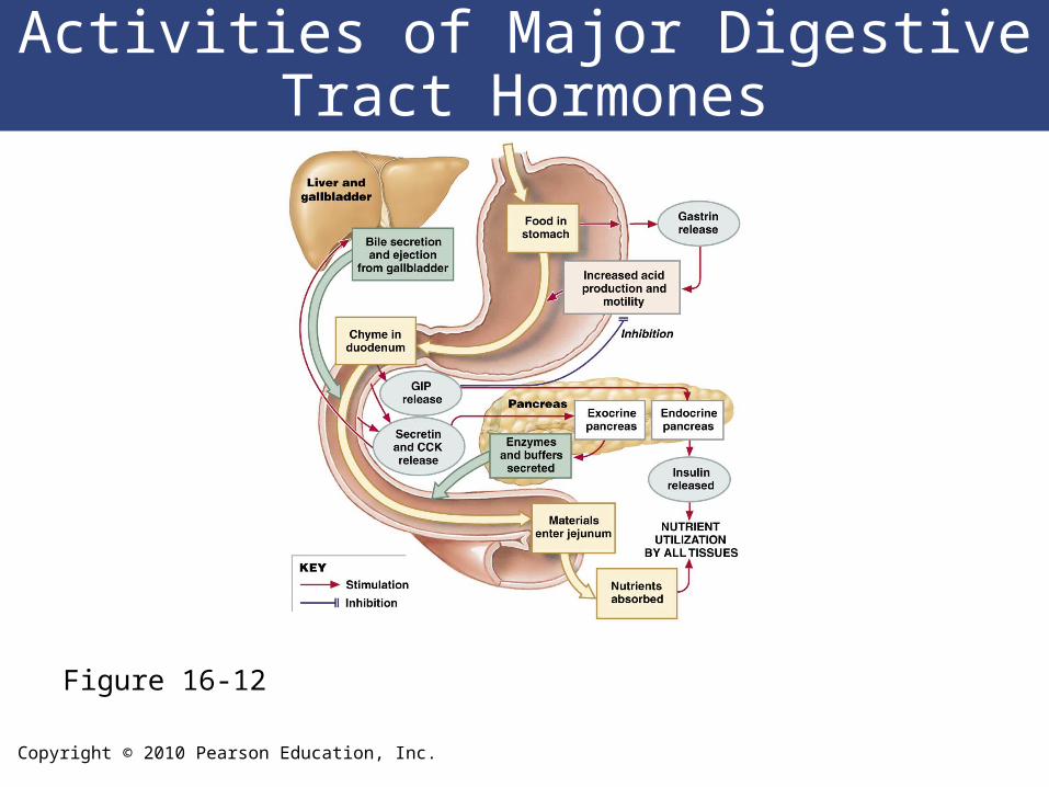
Copyright © 2010 Pearson Education, Inc.
Activities of Major Digestive Tract Hormones
Figure 16-12
Copyright © 2010 Pearson Education, Inc.
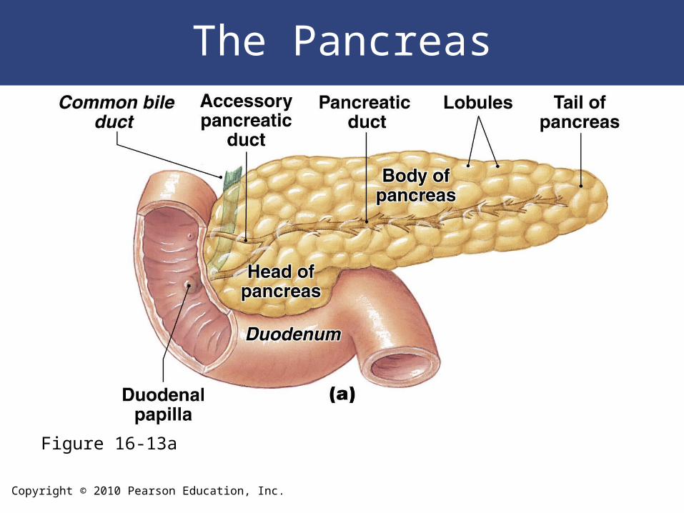
The Pancreas
• Lies posterior to the stomach– From duodenum toward spleen
• Is bound to posterior wall of abdominal cavity
• Is wrapped in thin, connective tissue capsule
Copyright © 2010 Pearson Education, Inc.
The Pancreas
• Histological Organization
– Lobules of the pancreas:
• Are separated by connective tissue partitions
(septa)
• Contain blood vessels and tributaries of pancreatic
ducts
• In each lobule:
– ducts branch repeatedly
– end in blind pockets (pancreatic acini)
Copyright © 2010 Pearson Education, Inc.
The Pancreas
• Pancreatic Acini– Blind pockets– Are lined with simple cuboidal epithelium – Contain scattered pancreatic islets
• Pancreatic Islets– Endocrine tissues of pancreas– Scattered (1% of pancreatic cells)
Copyright © 2010 Pearson Education, Inc.
The Pancreas
Figure 16-13a
Copyright © 2010 Pearson Education, Inc.
The Pancreas
• Pancreatic Secretions– 1000 mL (1 qt) pancreatic juice per day– Controlled by hormones from duodenum– Contain pancreatic enzymes
Copyright © 2010 Pearson Education, Inc.
The Pancreas
• Pancreatic Enzymes
– Pancreatic alpha-amylase:• A carbohydrase
• Breaks down starches
• Similar to salivary amylase
– Pancreatic lipase:• Breaks down complex lipids
• Releases products (e.g., fatty acids) that are easily absorbed
Copyright © 2010 Pearson Education, Inc.
The Pancreas
• Pancreatic Enzymes
– Nucleases:
• Break down nucleic acids
– Proteolytic enzymes:
• Break certain proteins apart
• Proteases break large protein complexes
• Peptidases break small peptides into amino acids
• 70% of all pancreatic enzyme production
• Secreted as inactive proenzymes
• Activated after reaching small intestine
Copyright © 2010 Pearson Education, Inc.
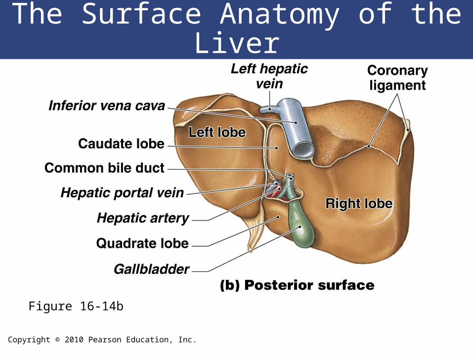
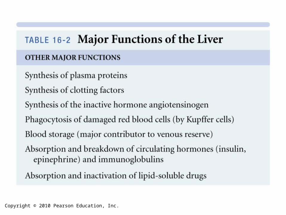
The Liver
• Is the largest visceral organ (1.5 kg; 3.3 lb)
• Lies in right hypochondriac and epigastric
regions
Copyright © 2010 Pearson Education, Inc.
The Liver
• Anatomy of the Liver– Is wrapped in tough fibrous capsule– Is covered by visceral peritoneum – Is divided into lobes
Copyright © 2010 Pearson Education, Inc.
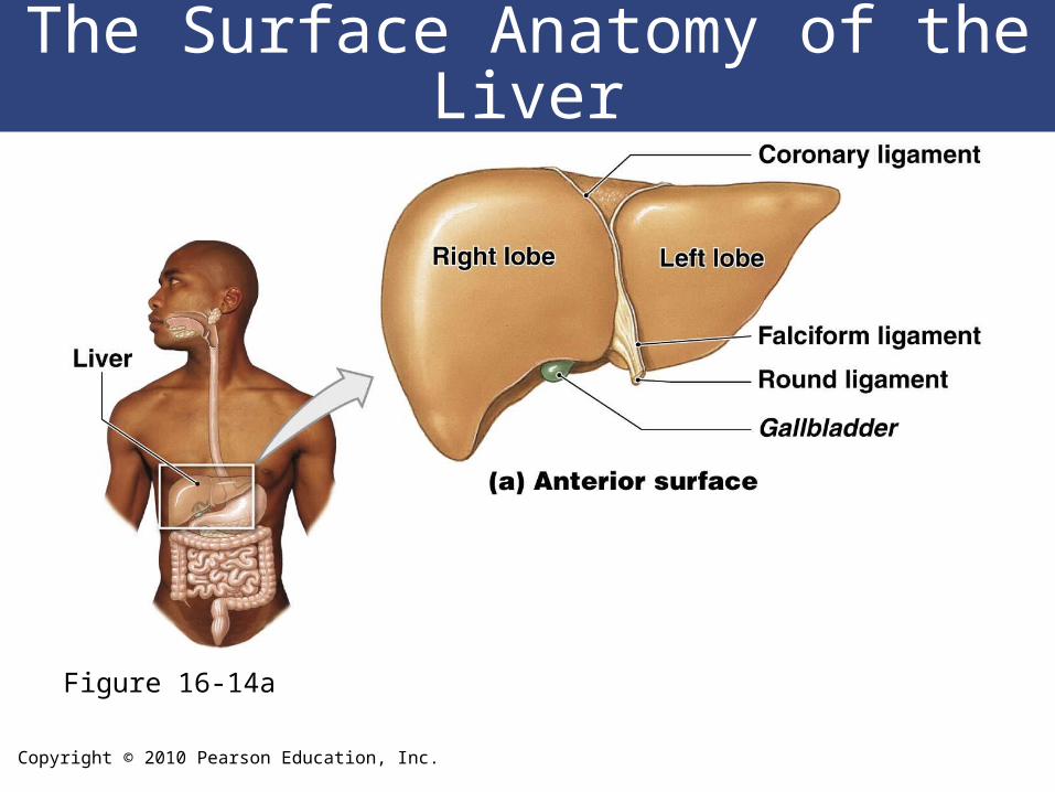
The Surface Anatomy of the Liver
Figure 16-14a
Copyright © 2010 Pearson Education, Inc.
The Surface Anatomy of the Liver
Figure 16-14b
Copyright © 2010 Pearson Education, Inc.
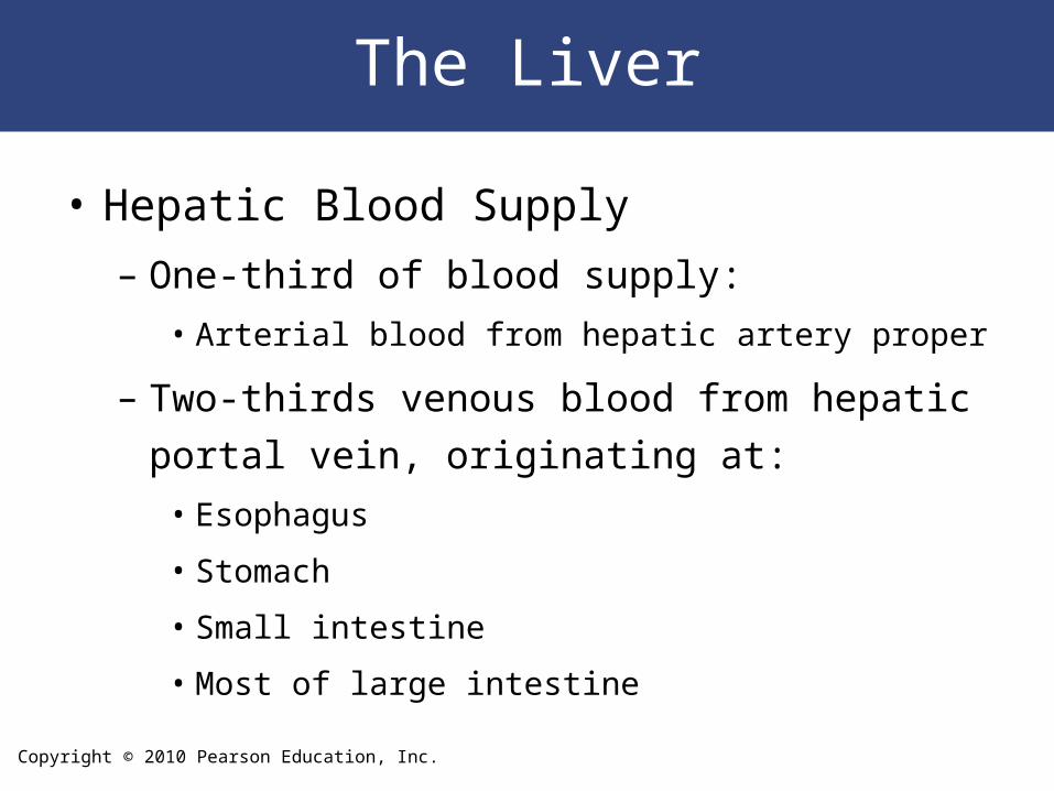
The Liver
• Hepatic Blood Supply
– One-third of blood supply:
• Arterial blood from hepatic artery proper
– Two-thirds venous blood from hepatic portal vein,
originating at:
• Esophagus
• Stomach
• Small intestine
• Most of large intestine
Copyright © 2010 Pearson Education, Inc.
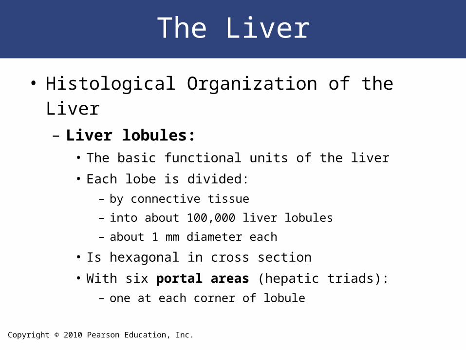
The Liver
• Histological Organization of the Liver– Liver lobules:
• The basic functional units of the liver
• Each lobe is divided:
– by connective tissue
– into about 100,000 liver lobules
– about 1 mm diameter each
• Is hexagonal in cross section
• With six portal areas (hepatic triads):
– one at each corner of lobule
Copyright © 2010 Pearson Education, Inc.
The Liver
• A Portal Area– Contains three structures:
• Branch of hepatic portal vein• Branch of hepatic artery proper• Small branch of bile duct
Copyright © 2010 Pearson Education, Inc.
The Liver
• Hepatocytes– Are liver cells
– Adjust circulating levels of nutrients:• Through selective absorption and secretion
– In a liver lobule form a series of irregular plates arranged like wheel spokes
– Many Kupffer cells (stellate reticuloendothelial cells) are located in sinusoidal lining
– As blood flows through sinusoids:• Hepatocytes absorb solutes from plasma
• And secrete materials such as plasma proteins

Copyright © 2010 Pearson Education, Inc.
Bile Ducts
Figure 16-16a
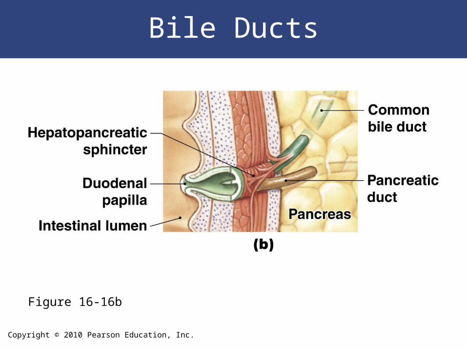
Copyright © 2010 Pearson Education, Inc.
Bile Ducts
Figure 16-16b
Copyright © 2010 Pearson Education, Inc.
The Liver
The Physiology of the Liver1. Metabolic regulation
2. Hematological regulation
3. Bile production
Copyright © 2010 Pearson Education, Inc.
Copyright © 2010 Pearson Education, Inc.
Copyright © 2010 Pearson Education, Inc.
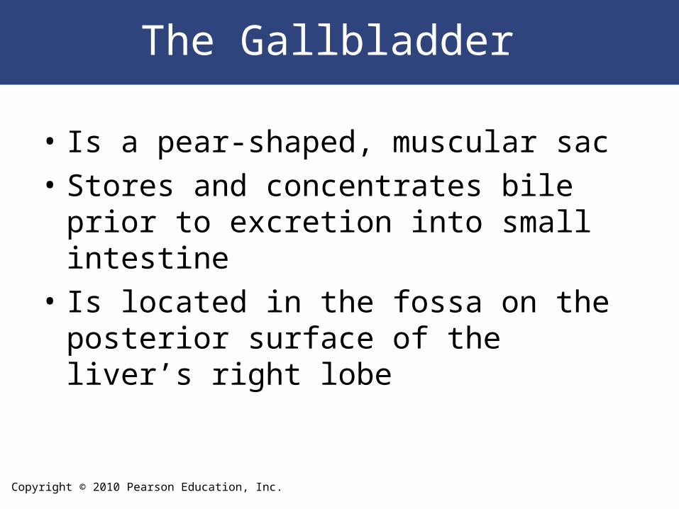
The Gallbladder
• Is a pear-shaped, muscular sac
• Stores and concentrates bile prior to excretion into small intestine
• Is located in the fossa on the posterior surface of the liver’s right lobe
Copyright © 2010 Pearson Education, Inc.
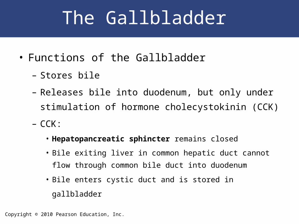
The Gallbladder
• Functions of the Gallbladder
– Stores bile
– Releases bile into duodenum, but only under
stimulation of hormone cholecystokinin (CCK)
– CCK:
• Hepatopancreatic sphincter remains closed
• Bile exiting liver in common hepatic duct cannot flow through
common bile duct into duodenum
• Bile enters cystic duct and is stored in gallbladder
Copyright © 2010 Pearson Education, Inc.
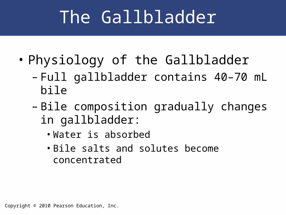
The Gallbladder
• Physiology of the Gallbladder– Full gallbladder contains 40–70 mL bile– Bile composition gradually changes in
gallbladder:• Water is absorbed• Bile salts and solutes become concentrated
Copyright © 2010 Pearson Education, Inc.
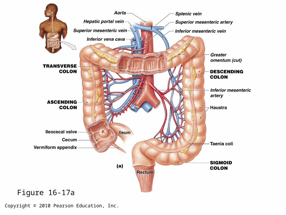
The Large Intestine
• Is horseshoe shaped
• Extends from end of ileum to anus
• Lies inferior to stomach and liver
• Frames the small intestine
• Also called large bowel
• Is about 1.5 meters (4.9 ft) long and 7.5 cm (3
in.) wide
Copyright © 2010 Pearson Education, Inc.
The Large Intestine
• Functions of the Large Intestine– Reabsorption of water – Compaction of intestinal contents into feces– Absorption of important vitamins produced by
bacteria– Storage of fecal material prior to defecation
Copyright © 2010 Pearson Education, Inc.
The Large Intestine
Parts of the Large Intestine1. Cecum:
• The pouchlike first portion
2. Colon: • The largest portion
3. Rectum: • The last 15 cm (6 in.) of digestive tract
Copyright © 2010 Pearson Education, Inc.
The Large Intestine
• The Cecum– Is an expanded pouch – Receives material arriving from the ileum– Stores materials and begins compaction
Copyright © 2010 Pearson Education, Inc.
The Large Intestine
• Appendix
– Also called vermiform appendix
– Is a slender, hollow appendage about 9 cm (3.6
in.) long
– Is dominated by lymphoid nodules (a lymphoid
organ)
– Is attached to posteromedial surface of cecum:
• Mesoappendix connects appendix to ileum and cecum
Copyright © 2010 Pearson Education, Inc.
The Large Intestine
• The Colon– Has a larger diameter and thinner wall than
small intestine – The wall of the colon:
• Forms a series of pouches (haustra)
– Haustra permit expansion and elongation of colon
Copyright © 2010 Pearson Education, Inc.
The Large Intestine
• Colon Muscles
– Three longitudinal bands of smooth muscle (taeniae
coli):
• Run along outer surfaces of colon
• Deep to the serosa
• Similar to outer layer of muscularis externa
– Muscle tone in taeniae coli creates the haustra
Copyright © 2010 Pearson Education, Inc.
The Large Intestine
• Ascending Colon – Begins at superior border of cecum
– Ascends along right lateral and posterior wall of peritoneal cavity to inferior surface of the liver and bends at right colic flexure (hepatic flexure)
• Transverse Colon– Crosses abdomen from right to left; turns at left colic
flexure (splenic flexure)
– Is supported by transverse mesocolon
– Is separated from anterior abdominal wall by greater omentum
Copyright © 2010 Pearson Education, Inc.
The Large Intestine
• The Descending Colon – Proceeds inferiorly along left side to the iliac fossa
(inner surface of left ilium)
– Is retroperitoneal, firmly attached to abdominal wall
• The Sigmoid Colon – Is an S-shaped segment, about 15 cm (6 in.) long
– Starts at sigmoid flexure
– Lies posterior to urinary bladder
– Is suspended from sigmoid mesocolon
– Empties into rectum
Copyright © 2010 Pearson Education, Inc.
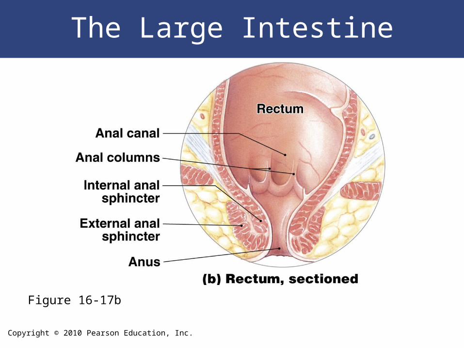
The Large Intestine
• The Rectum– Forms last 15 cm (6 in.) of digestive tract– Is an expandable organ for temporary storage of feces– Movement of fecal material into rectum triggers urge
to defecate
• The anal canal is the last portion of the rectum– Contains small longitudinal folds called anal columns
Copyright © 2010 Pearson Education, Inc.
The Large Intestine
• Anus– Also called anal orifice– Is exit of the anal canal– Has keratinized epidermis like skin
Copyright © 2010 Pearson Education, Inc.
The Large Intestine
• Anal Sphincters
– Internal anal sphincter:
• Circular muscle layer of muscularis externa
• Has smooth muscle cells, not under voluntary control
– External anal sphincter:
• Encircles distal portion of anal canal
• A ring of skeletal muscle fibers, under voluntary control
Copyright © 2010 Pearson Education, Inc.
Figure 16-17a
Copyright © 2010 Pearson Education, Inc.
The Large Intestine
Figure 16-17b
Copyright © 2010 Pearson Education, Inc.
The Large Intestine
• Histology of the Large Intestine
– Lack villi
– Abundance of mucous cells
– Presence of distinctive intestinal glands:
• Are deeper than glands of small intestine
• Are dominated by mucous cells
Copyright © 2010 Pearson Education, Inc.
The Functions of the Large Intestine
• Physiology of the Large Intestine– Less than 10% of nutrient absorption occurs
in large intestine– Prepares fecal material for ejection from the
body
Copyright © 2010 Pearson Education, Inc.
The Functions of the Large Intestine
• Absorption in the Large Intestine
– Reabsorption of water
– Reabsorption of bile salts:
• In the cecum
• Transported in blood to liver
– Absorption of vitamins produced by bacteria
– Absorption of organic wastes
Copyright © 2010 Pearson Education, Inc.
The Functions of the Large Intestine
• Vitamins – Are organic molecules – Are important as cofactors or coenzymes in
metabolism– Normal bacteria in colon make three vitamins
that supplement diet
Copyright © 2010 Pearson Education, Inc.
The Functions of the Large Intestine
Three Vitamins Produced in the Large Intestine
1. Vitamin K (fat soluble):
• Required by liver for synthesizing four clotting factors,
including prothrombin
2. Biotin (water soluble):
• Important in glucose metabolism
3. Pantothenic acid: B5 (water soluble):
• Required in manufacture of steroid hormones and some
neurotransmitters
Copyright © 2010 Pearson Education, Inc.
The Functions of the Large Intestine
• Organic Wastes
– Bacteria convert bilirubin to urobilinogens and
stercobilinogens:
• Urobilinogens absorbed into bloodstream are
excreted in urine
• Urobilinogens and stercobilinogens in colon
convert to urobilins and stercobilins by exposure
to oxygen
Copyright © 2010 Pearson Education, Inc.
The Functions of the Large Intestine
• Toxins
– Bacteria break down peptides in feces and
generate:
• Ammonia:
– as soluble ammonium ions
• Indole and skatole:
– nitrogen compounds responsible for odor of feces
• Hydrogen sulfide:
– gas that produces “rotten egg” odor
Copyright © 2010 Pearson Education, Inc.
The Functions of the Large Intestine
• Toxins– Bacteria feed on indigestible carbohydrates
(complex polysaccharides):• Produce flatus, or intestinal gas, in large intestine
Copyright © 2010 Pearson Education, Inc.
The Functions of the Large Intestine
• Movements of the Large Intestine
– Gastroileal and gastroenteric reflexes:
• Move materials into cecum while you eat
– Movement from cecum to transverse colon is very
slow, allowing hours for water absorption
– Peristaltic waves move material along length of colon
– Segmentation movements (haustral churning) mix
contents of adjacent haustra
Copyright © 2010 Pearson Education, Inc.
The Functions of the Large Intestine
• Movements of the Large Intestine
– Movement from transverse colon through rest of large intestine
results from powerful peristaltic contractions (mass
movements)
– Stimulus is distension of stomach and duodenum; relayed over
intestinal nerve plexuses
– Distension of the rectal wall triggers defecation reflex:
• Two positive feedback loops
• Both loops triggered by stretch receptors in rectum
Copyright © 2010 Pearson Education, Inc.
The Functions of the Large Intestine
• Elimination of Feces – Requires relaxation of internal and external
anal sphincters– Reflexes open internal sphincter and close
external sphincter– Opening external sphincter requires
conscious effort
Copyright © 2010 Pearson Education, Inc.
Digestion
• Essential Nutrients
– A typical meal contains:
• Carbohydrates
• Proteins
• Lipids
• Water
• Electrolytes
• Vitamins
Copyright © 2010 Pearson Education, Inc.
Digestion
• The Processing and Absorption of Nutrients– Breaks down physical structure of food– Disassembles component molecules – Molecules released into bloodstream are:
• Absorbed by cells
– Broken down to provide energy for ATP synthesis:• Or used to synthesize carbohydrates, proteins, and lipids
Copyright © 2010 Pearson Education, Inc.
Digestion
• Digestive Enzymes – Are secreted by:
• Salivary glands• Tongue• Stomach• Pancreas

Copyright © 2010 Pearson Education, Inc.
Digestion
• Digestive Enzymes – Break molecular bonds in large organic molecules:
• Carbohydrates, proteins, lipids, and nucleic acids• In a process called hydrolysis
– Are divided into classes by targets:• Carbohydrases break bonds between simple sugars• Proteases break bonds between amino acids• Lipases separate fatty acids from glycerides
Copyright © 2010 Pearson Education, Inc.
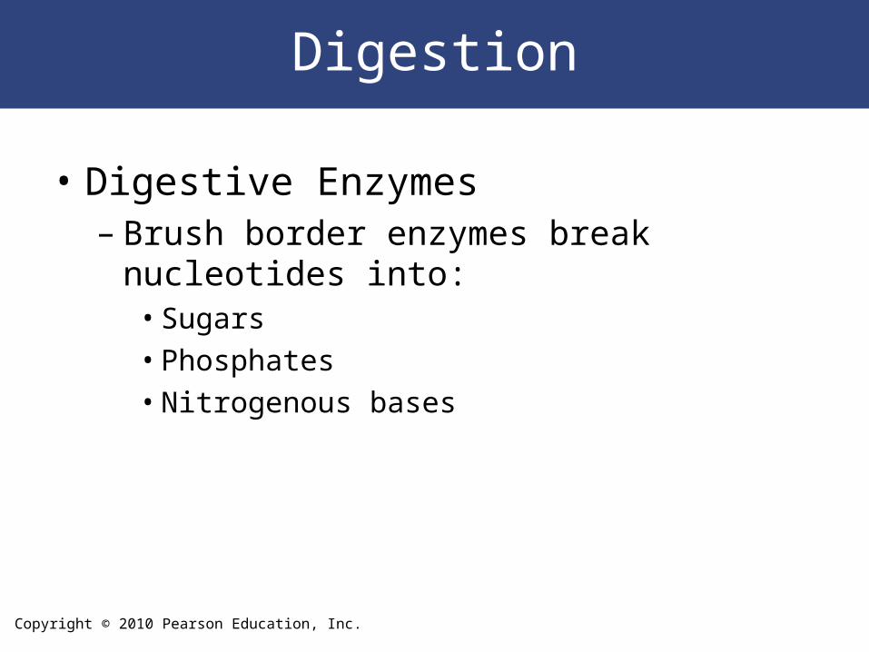
Digestion
• Digestive Enzymes – Brush border enzymes break nucleotides into:
• Sugars• Phosphates• Nitrogenous bases
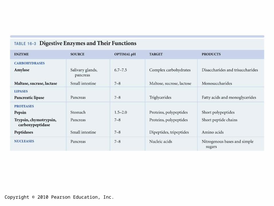
Copyright © 2010 Pearson Education, Inc.
Copyright © 2010 Pearson Education, Inc.
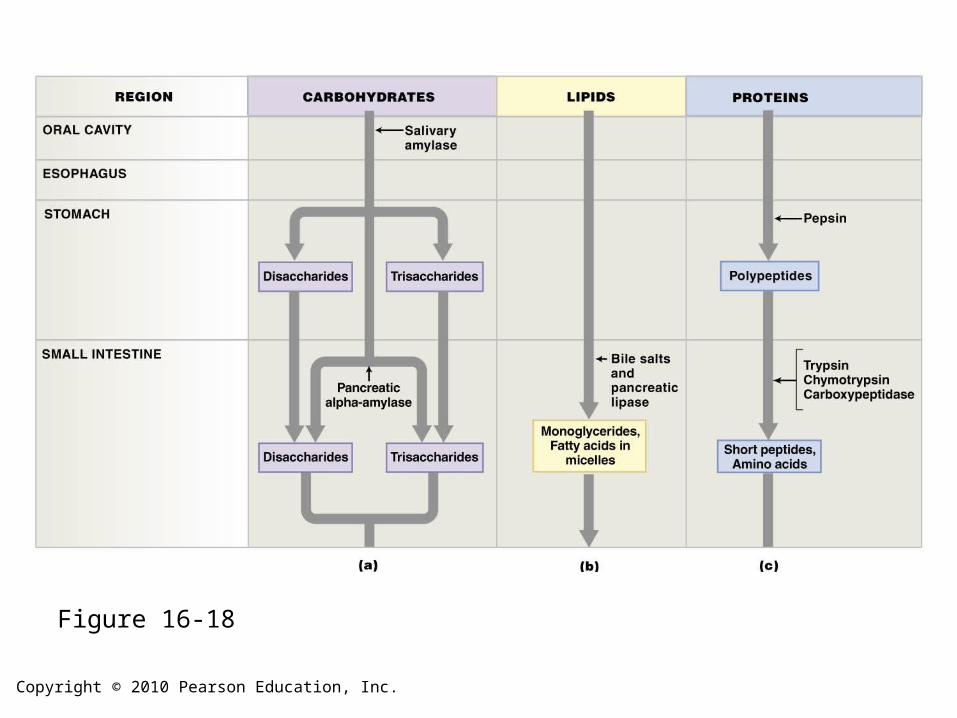
Figure 16-18
Copyright © 2010 Pearson Education, Inc.
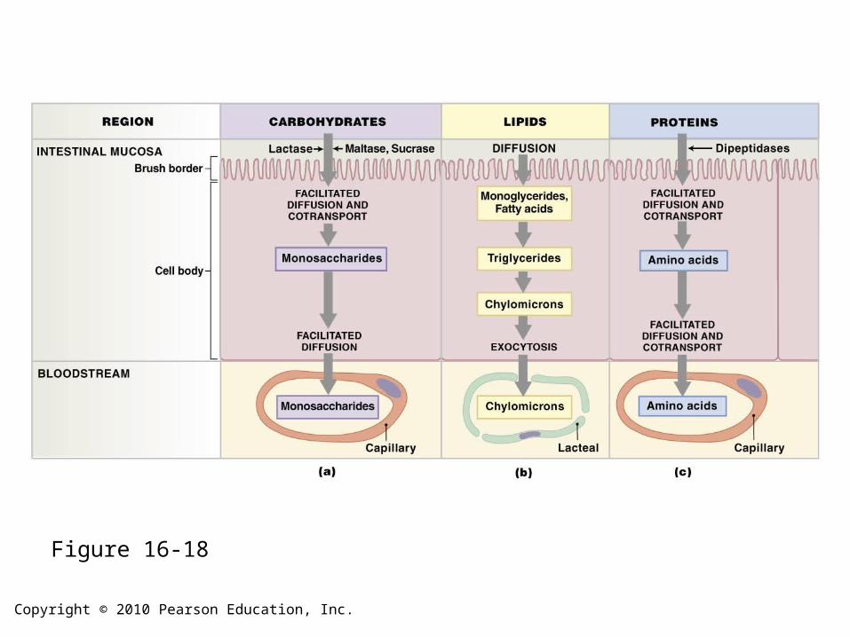
Figure 16-18
Copyright © 2010 Pearson Education, Inc.
Water and Electrolyte Absorption
• Water Absorption– Cells cannot actively absorb or secrete water– All movement of water across lining of
digestive tract:• Involves passive water flow down osmotic
gradients due to electrolyte movement
Copyright © 2010 Pearson Education, Inc.
The Absorption of Vitamins
• Vitamins are organic compounds required in very small quantities
• Are divided into two major groups:– Fat-soluble vitamins– Water-soluble vitamins
Copyright © 2010 Pearson Education, Inc.
Effects of Aging on the Digestive System
1. Division of epithelial stem cells declines
– Digestive epithelium becomes more
susceptible to damage by abrasion, acids, or
enzymes
2. Smooth muscle tone and general motility
decreases
– Peristaltic contractions become weaker
Copyright © 2010 Pearson Education, Inc.
Effects of Aging on the Digestive System
3. Cumulative damage from toxins (alcohol, other chemicals) absorbed by digestive tract and transported to liver for processing
Copyright © 2010 Pearson Education, Inc.
Effects of Aging on the Digestive System
4. Rates of colon cancer and stomach cancer
rise with age
– Oral and pharyngeal cancers common among
elderly smokers
5. Dehydration
6. Other systems changes (bone — calcium)