Embed Size (px)
Citation preview
Copyright © 2005 Pearson Education, Inc. publishing as Benjamin Cummings
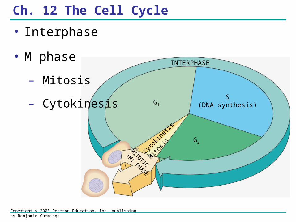
INTERPHASE
G1
S(DNA synthesis)
G2
Cytokin
esis
Mito
sisM
ITOTIC
(M) PHASE
Ch. 12 The Cell Cycle
• Interphase
• M phase
– Mitosis
– Cytokinesis
Copyright © 2005 Pearson Education, Inc. publishing as Benjamin Cummings
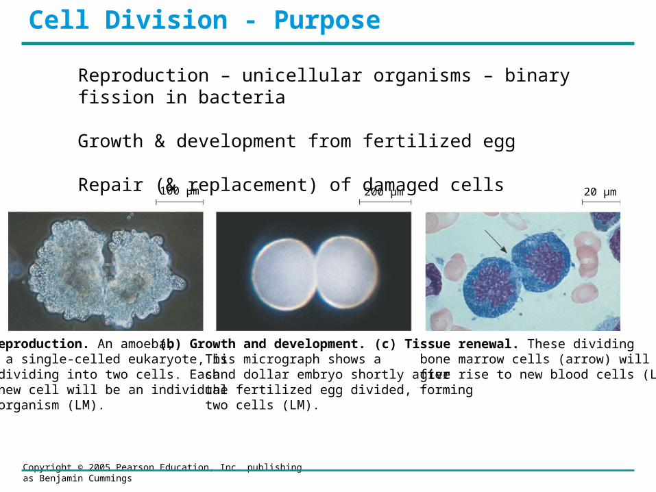
20 µm100 µm 200 µm
(a) Reproduction. An amoeba, a single-celled eukaryote, is dividing into two cells. Each new cell will be an individual organism (LM).
(b) Growth and development. This micrograph shows a sand dollar embryo shortly after the fertilized egg divided, forming two cells (LM).
(c) Tissue renewal. These dividing bone marrow cells (arrow) will give rise to new blood cells (LM).
Reproduction – unicellular organisms – binary fission in bacteria
Growth & development from fertilized egg
Repair (& replacement) of damaged cells
Cell Division - Purpose
Copyright © 2005 Pearson Education, Inc. publishing as Benjamin Cummings
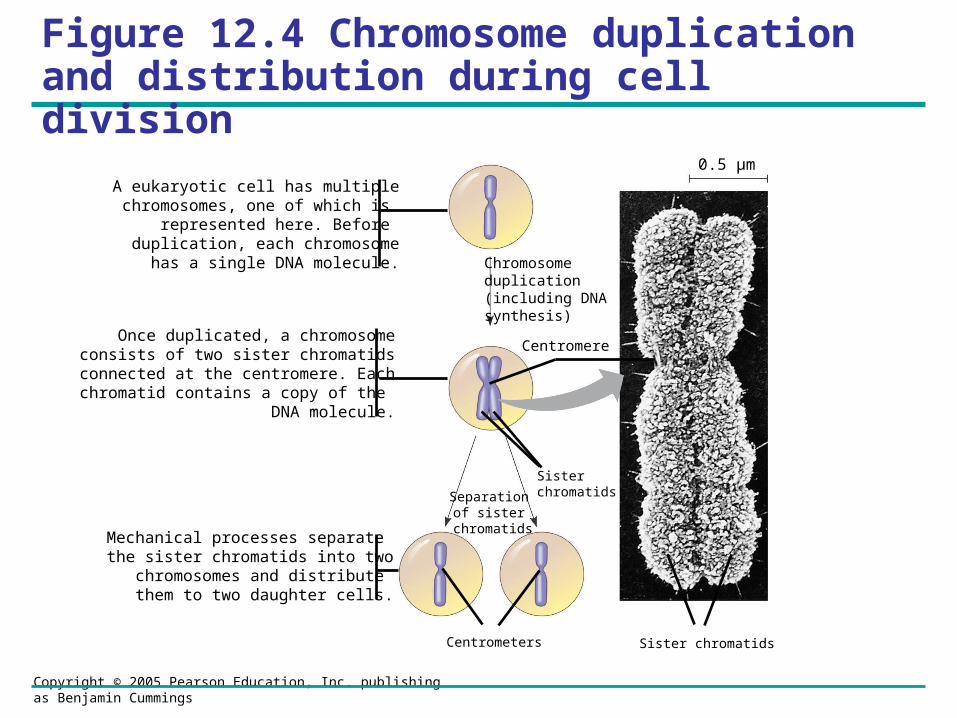
Figure 12.4 Chromosome duplication and distribution during cell division
0.5 µm
Chromosomeduplication(including DNA synthesis)
Centromere
Separation of sister
chromatids
Sisterchromatids
Centrometers Sister chromatids
A eukaryotic cell has multiplechromosomes, one of which is
represented here. Before duplication, each chromosome
has a single DNA molecule.
Once duplicated, a chromosomeconsists of two sister chromatids
connected at the centromere. Eachchromatid contains a copy of the
DNA molecule.
Mechanical processes separate the sister chromatids into two chromosomes and distribute
them to two daughter cells.
Copyright © 2005 Pearson Education, Inc. publishing as Benjamin Cummings
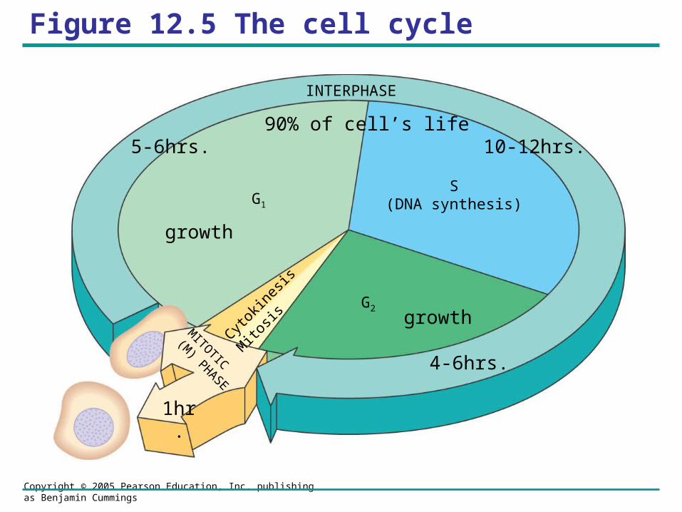
Figure 12.5 The cell cycle
INTERPHASE
G1
S(DNA synthesis)
G2
Cytokin
esis
Mito
sisM
ITOTIC
(M) PHASE
5-6hrs. 10-12hrs.
4-6hrs.
1hr.
growth
growth
90% of cell’s life
Copyright © 2005 Pearson Education, Inc. publishing as Benjamin Cummings
INTERPHASE
G1
S(DNA synthesis)
G2
Cytokin
esis
Mito
sisM
ITOTIC
(M) PHASE
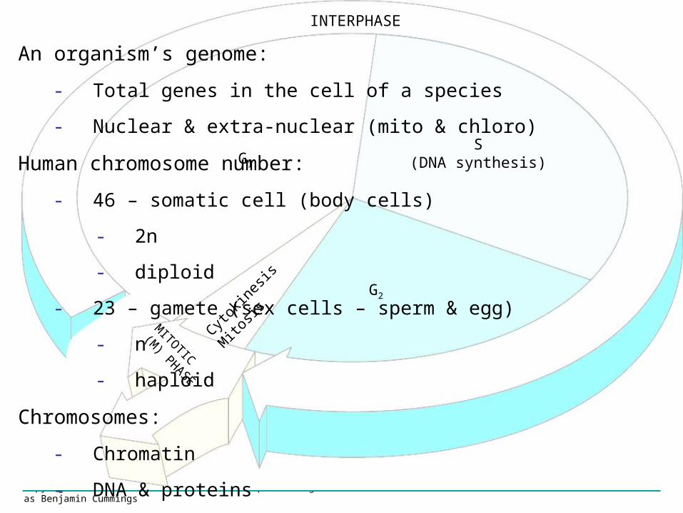
An organism’s genome:
- Total genes in the cell of a species
- Nuclear & extra-nuclear (mito & chloro)
Human chromosome number:
- 46 – somatic cell (body cells)
- 2n
- diploid
- 23 – gamete (sex cells – sperm & egg)
- n
- haploid
Chromosomes:
- Chromatin
- DNA & proteins
Copyright © 2005 Pearson Education, Inc. publishing as Benjamin Cummings
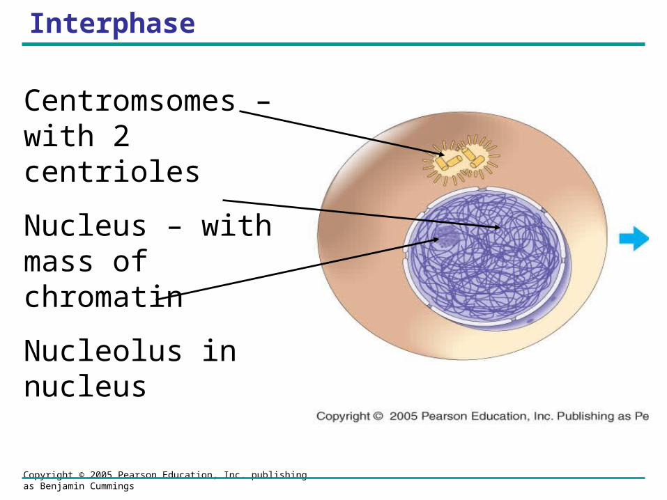
Interphase
Centromsomes – with 2 centrioles
Nucleus – with mass of chromatin
Nucleolus in nucleus
Copyright © 2005 Pearson Education, Inc. publishing as Benjamin Cummings
INTERPHASE
G1
S(DNA synthesis)
G2
Cytokin
esis
Mito
sisM
ITOTIC
(M) PHASE
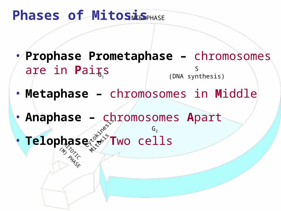
Phases of Mitosis
• Prophase Prometaphase – chromosomes are in Pairs
• Metaphase – chromosomes in Middle
• Anaphase – chromosomes Apart
• Telophase – Two cells

Copyright © 2005 Pearson Education, Inc. publishing as Benjamin Cummings
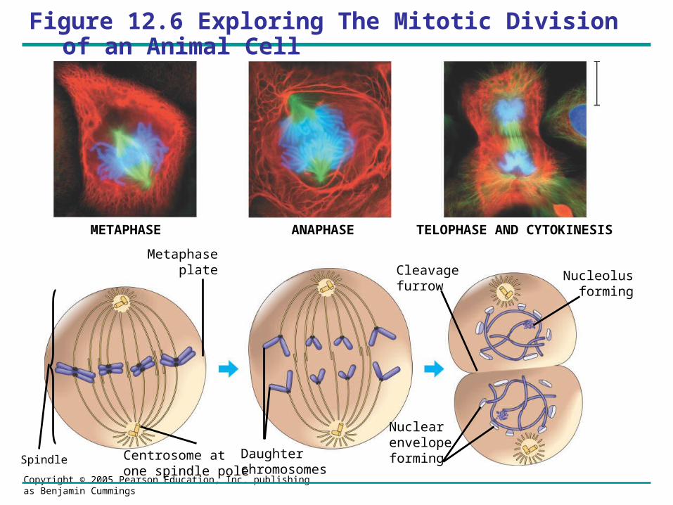
Figure 12.6 Exploring The Mitotic Division of an Animal Cell
G2 OF INTERPHASE PROPHASE PROMETAPHASE
Centrosomes(with centriole pairs) Chromatin
(duplicated)
Early mitoticspindle
Aster
CentromereFragmentsof nuclearenvelope
Kinetochore
Nucleolus Nuclearenvelope
Plasmamembrane
Chromosome, consistingof two sister chromatids
Kinetochore microtubule
Nonkinetochoremicrotubules
Copyright © 2005 Pearson Education, Inc. publishing as Benjamin Cummings
METAPHASE ANAPHASE TELOPHASE AND CYTOKINESIS
Spindle
Metaphaseplate Nucleolus
forming
Cleavagefurrow
Nuclear envelopeformingCentrosome at
one spindle poleDaughter chromosomes
Figure 12.6 Exploring The Mitotic Division of an Animal Cell
Copyright © 2005 Pearson Education, Inc. publishing as Benjamin Cummings
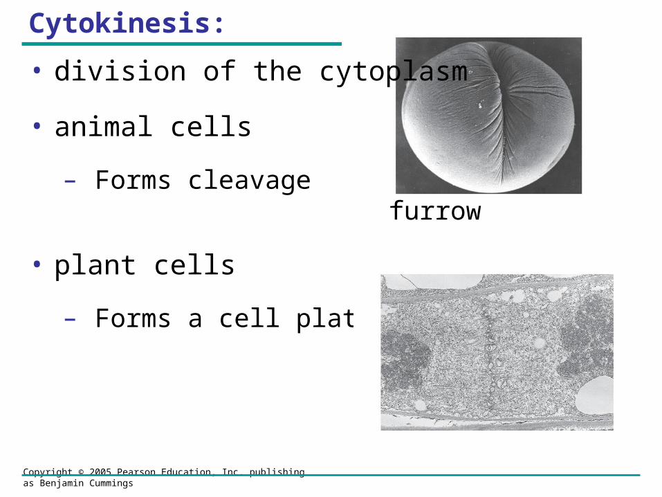
Cytokinesis:
• division of the cytoplasm
• animal cells
– Forms cleavage furrow
• plant cells
– Forms a cell plate
Copyright © 2005 Pearson Education, Inc. publishing as Benjamin Cummings
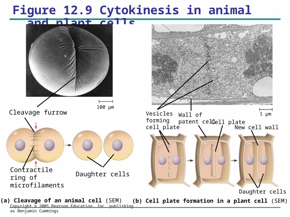
Figure 12.9 Cytokinesis in animal and plant cells
Daughter cells
Cleavage furrow
Contractile ring of microfilaments
Daughter cells
100 µm
1 µmVesiclesforming cell plate
Wall of patent cell Cell plate
New cell wall
(a) Cleavage of an animal cell (SEM) (b) Cell plate formation in a plant cell (SEM)
Copyright © 2005 Pearson Education, Inc. publishing as Benjamin Cummings
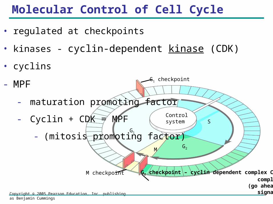
Control system
G2 checkpoint – cyclin dependent complex CDKcomplex
(go ahead signal)
M checkpoint
G1 checkpoint
G1
S
G2M
Molecular Control of Cell Cycle
• regulated at checkpoints
• kinases - cyclin-dependent kinase (CDK)
• cyclins
- MPF
- maturation promoting factor
- Cyclin + CDK = MPF
- (mitosis promoting factor)
Copyright © 2005 Pearson Education, Inc. publishing as Benjamin Cummings
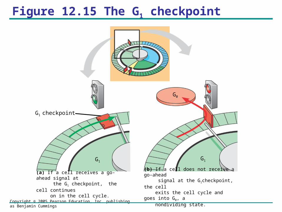
Figure 12.15 The G1 checkpoint
G1 checkpoint
G1G1
G0
(a) If a cell receives a go-ahead signal at the G1 checkpoint, the cell continues on in the cell cycle.
(b) If a cell does not receive a go-ahead signal at the G1checkpoint, the cell exits the cell cycle and goes into G0, a nondividing state.

Copyright © 2005 Pearson Education, Inc. publishing as Benjamin Cummings
Figure 12.16 Molecular control of the cell cycle at the G2 checkpoint
Accumulated cyclin moleculescombine with recycled Cdk mol-ecules, producing enough molecules of MPF to pass the G2 checkpoint and initiate the events of mitosis.
MPF promotes mitosis by phosphorylating various proteins. MPF‘s activity peaks during metaphase. (breaking down nuclear envelop, etc.)
3
During G1, conditions in the cell favor degradation of cyclin, and the Cdk component of MPF is recycled.
5
During anaphase, the cyclin component of MPF is degraded, terminating the M phase. The cell enters the G1 phase.
4
2
Synthesis of cyclin begins in late S phase and continues through G2. Because cyclin is protected from degradation during this stage, it accumulates.
1
Cdk
CdkG2
checkpoint
CyclinMPF
Cyclin is degraded
DegradedCyclin
G 1
G 2
S
M
G1G1 S G2 G2SM M
MPF activity
Cyclin
Time
(a) Fluctuation of MPF activity and cyclin concentration during the cell cycle
(b) Molecular mechanisms that help regulate the cell cycle
Rel
ativ
e C
on
cen
tra
tion
MPF – maturation
promoting factor
Copyright © 2005 Pearson Education, Inc. publishing as Benjamin Cummings
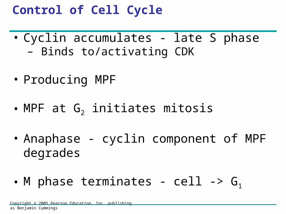
Control of Cell Cycle
• Cyclin accumulates - late S phase – Binds to/activating CDK
• Producing MPF
• MPF at G2 initiates mitosis
• Anaphase - cyclin component of MPF degrades
• M phase terminates - cell -> G1
Copyright © 2005 Pearson Education, Inc. publishing as Benjamin Cummings
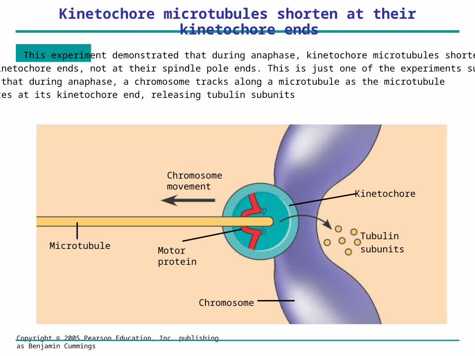
Kinetochore microtubules shorten at their kinetochore ends
CONCLUSION This experiment demonstrated that during anaphase, kinetochore microtubules shorten
at their kinetochore ends, not at their spindle pole ends. This is just one of the experiments supporting the
hypothesis that during anaphase, a chromosome tracks along a microtubule as the microtubule
depolymerizes at its kinetochore end, releasing tubulin subunits
Chromosomemovement
Microtubule Motorprotein
Chromosome
Kinetochore
Tubulin
subunits

Copyright © 2005 Pearson Education, Inc. publishing as Benjamin Cummings
Prophase:
• chromatin - condenses
• nucleoli disappear.
• sister chromatids appear
• mitotic spindle forms
• centrosomes move away from each other
Copyright © 2005 Pearson Education, Inc. publishing as Benjamin Cummings
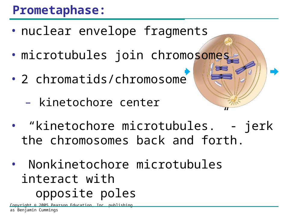
Prometaphase:
• nuclear envelope fragments
• microtubules join chromosomes
• 2 chromatids/chromosome
– kinetochore center
• “kinetochore microtubules.” - jerk the chromosomes back and forth.
• Nonkinetochore microtubules interact with opposite poles
Copyright © 2005 Pearson Education, Inc. publishing as Benjamin Cummings
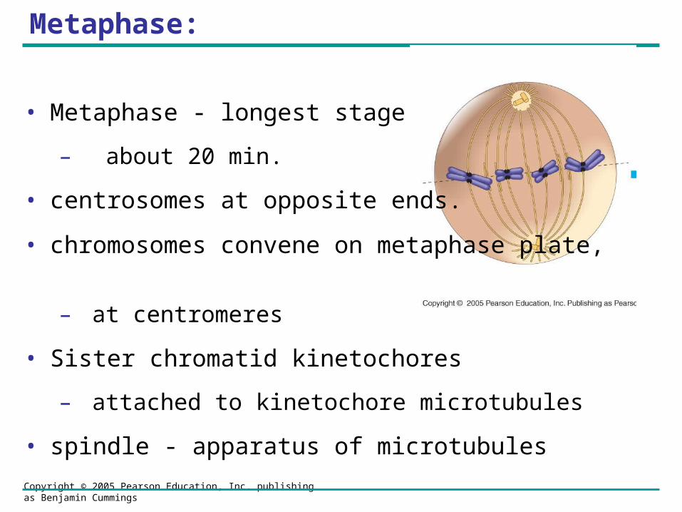
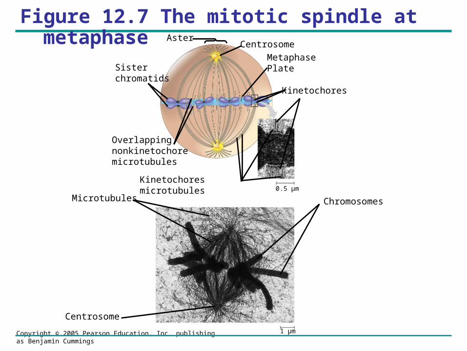
Metaphase:
• Metaphase - longest stage
– about 20 min.
• centrosomes at opposite ends.
• chromosomes convene on metaphase plate,
– at centromeres
• Sister chromatid kinetochores
– attached to kinetochore microtubules
• spindle - apparatus of microtubules
Copyright © 2005 Pearson Education, Inc. publishing as Benjamin Cummings
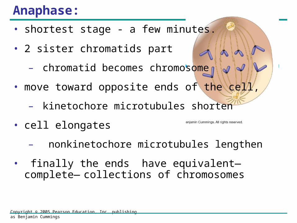
Anaphase:
• shortest stage - a few minutes.
• 2 sister chromatids part
– chromatid becomes chromosome
• move toward opposite ends of the cell,
– kinetochore microtubules shorten
• cell elongates
– nonkinetochore microtubules lengthen
• finally the ends have equivalent—complete—collections of chromosomes
Copyright © 2005 Pearson Education, Inc. publishing as Benjamin Cummings
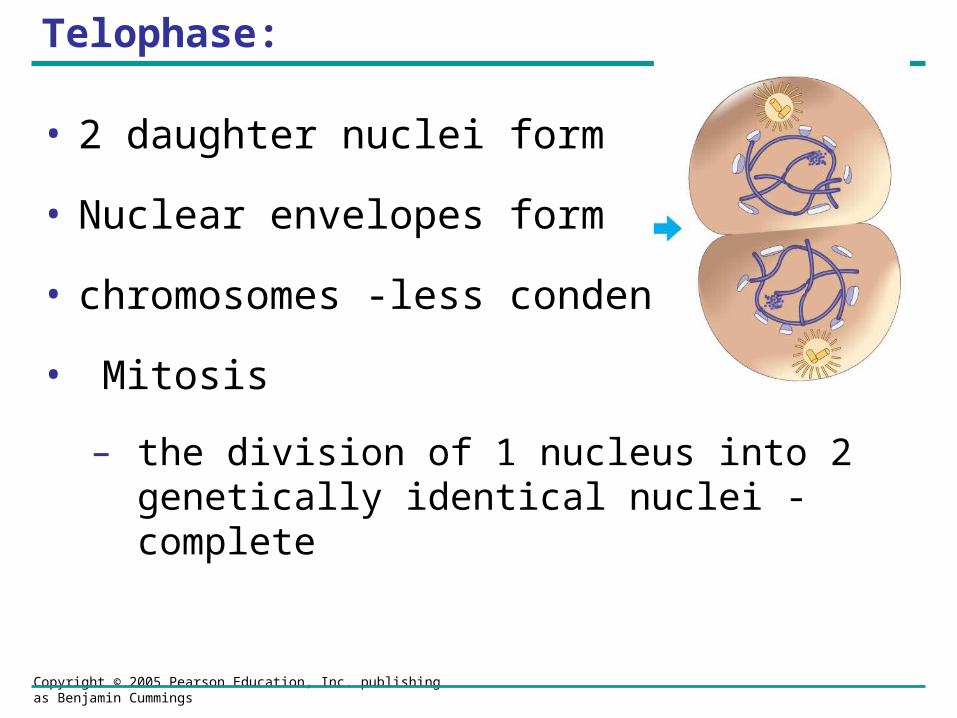
Telophase:
• 2 daughter nuclei form
• Nuclear envelopes form
• chromosomes -less condensed
• Mitosis
– the division of 1 nucleus into 2 genetically identical nuclei - complete
Copyright © 2005 Pearson Education, Inc. publishing as Benjamin Cummings
Figure 12.7 The mitotic spindle at metaphase
Sisterchromatids
MetaphasePlate
Kinetochores
Overlappingnonkinetochoremicrotubules
Kinetochores microtubules
Centrosome
ChromosomesMicrotubules0.5 µm
CentrosomeAster
1 µm
Copyright © 2005 Pearson Education, Inc. publishing as Benjamin Cummings
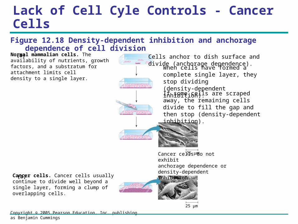
Lack of Cell Cyle Controls - Cancer Cells
25 µm
Cells anchor to dish surface anddivide (anchorage dependence).
When cells have formed a complete single layer, they stop dividing (density-dependent inhibition).
If some cells are scraped away, the remaining cells divide to fill the gap and then stop (density-dependent inhibition).
Cancer cells do not exhibitanchorage dependence or density-dependent inhibition.
25 µm
Cancer cells. Cancer cells usually continue to divide well beyond a single layer, forming a clump of overlapping cells.
Normal mammalian cells. The availability of nutrients, growth factors, and a substratum for attachment limits cell density to a single layer.
(a)
(b)
Figure 12.18 Density-dependent inhibition and anchorage dependence of cell division
Copyright © 2005 Pearson Education, Inc. publishing as Benjamin Cummings
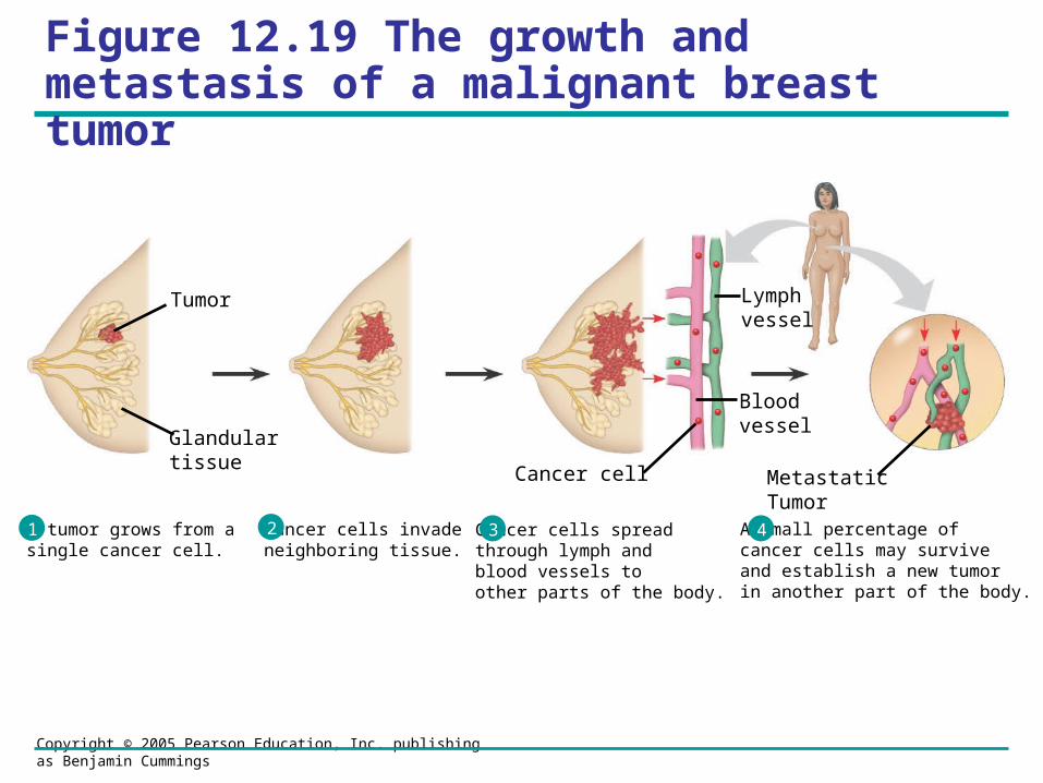
Figure 12.19 The growth and metastasis of a malignant breast tumor
A tumor grows from a single cancer cell.
Cancer cells invade neighboring tissue.
Cancer cells spread through lymph and blood vessels to other parts of the body.
A small percentage of cancer cells may survive and establish a new tumor in another part of the body.
2 431
Tumor
Glandulartissue
Cancer cell
Bloodvessel
Lymphvessel
MetastaticTumor
Copyright © 2005 Pearson Education, Inc. publishing as Benjamin Cummings
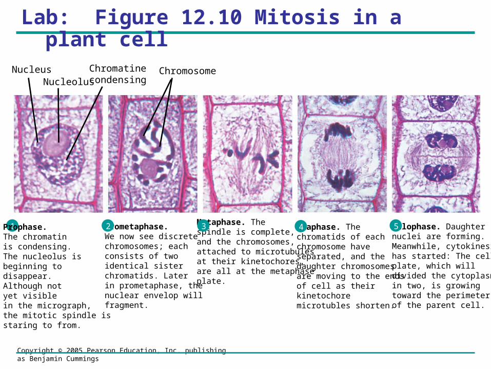
Lab: Figure 12.10 Mitosis in a plant cell
1 Prophase. The chromatinis condensing. The nucleolus is beginning to disappear.Although not yet visible in the micrograph, the mitotic spindle is staring to from.
Prometaphase.We now see discretechromosomes; each consists of two identical sister chromatids. Laterin prometaphase, the nuclear envelop will fragment.
Metaphase. The spindle is complete,and the chromosomes,attached to microtubulesat their kinetochores, are all at the metaphase plate.
Anaphase. Thechromatids of each chromosome have separated, and the daughter chromosomesare moving to the ends of cell as their kinetochoremicrotubles shorten.
Telophase. Daughternuclei are forming. Meanwhile, cytokinesishas started: The cellplate, which will divided the cytoplasm in two, is growing toward the perimeter of the parent cell.
2 3 4 5
NucleusNucleolus
ChromosomeChromatinecondensing
Copyright © 2005 Pearson Education, Inc. publishing as Benjamin Cummings
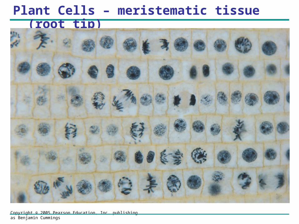
Plant Cells – meristematic tissue (root tip)
Copyright © 2005 Pearson Education, Inc. publishing as Benjamin Cummings
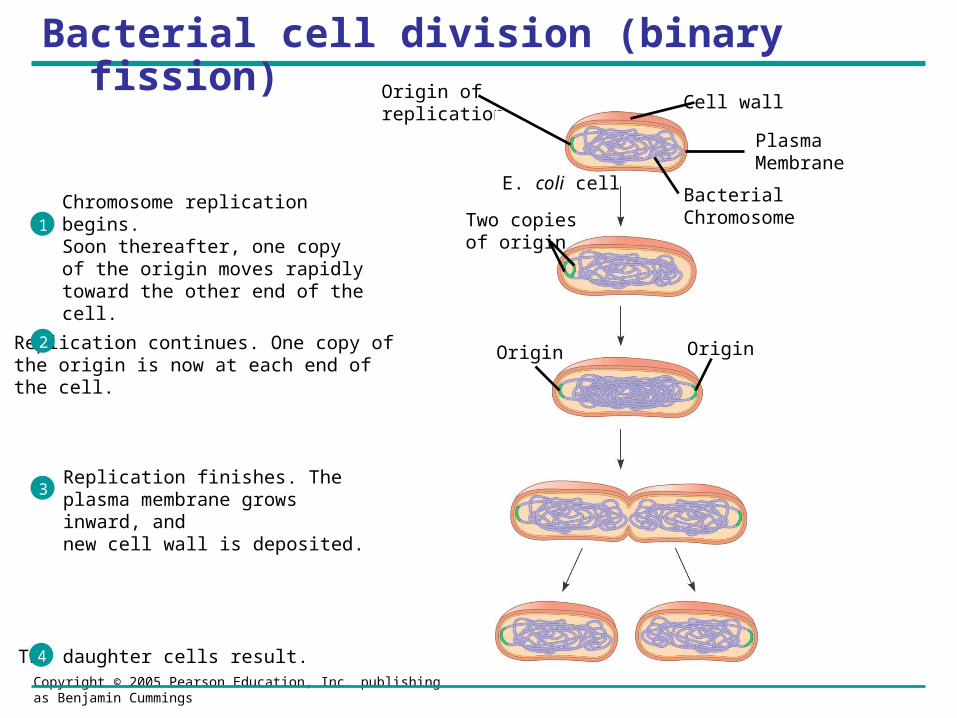
Bacterial cell division (binary fission)Origin ofreplication
E. coli cellBacterialChromosome
Cell wall
Plasma Membrane
Two copiesof origin
OriginOrigin
Chromosome replication begins.Soon thereafter, one copy of the origin moves rapidly toward the other end of the cell.
1
Replication continues. One copy ofthe origin is now at each end of the cell.
2
Replication finishes. The plasma membrane grows inward, andnew cell wall is deposited.
3
Two daughter cells result.4