Embed Size (px)
Citation preview

Chapter 12 The Cell Cycle

Fig. 12-UN1
Telophase andCytokinesis
Anaphase
Metaphase
Prometaphase
Prophase
MITOTIC (M) PHASE
Cytokinesis
Mitosis
SG1
G2

Fig. 12-8
EXPERIMENT
Kinetochore
RESULTS
CONCLUSION
Spindlepole
Mark
Chromosomemovement
Kinetochore
MicrotubuleMotorprotein
Chromosome
Tubulinsubunits

Binary Fission
• Prokaryotes (bacteria and archaea) reproduce by a type of cell division called binary fission
• In binary fission, the chromosome replicates (beginning at the origin of replication), and the two daughter chromosomes actively move apart
Copyright © 2008 Pearson Education, Inc., publishing as Pearson Benjamin Cummings

Fig. 12-11-4
Origin ofreplication
Two copiesof origin
E. coli cellBacterialchromosome
Plasmamembrane
Cell wall
Origin Origin

The Evolution of Mitosis• Since prokaryotes evolved before eukaryotes,
mitosis probably evolved from binary fission• Certain protists exhibit types of cell division
that seem intermediate between binary fission and mitosis
Copyright © 2008 Pearson Education, Inc., publishing as Pearson Benjamin Cummings

Fig. 12-12
(a) Bacteria
Bacterialchromosome
Chromosomes
Microtubules
Intact nuclearenvelope
(b) Dinoflagellates
Kinetochoremicrotubule
Intact nuclearenvelope
(c) Diatoms and yeasts
Kinetochoremicrotubule
Fragments ofnuclear envelope
(d) Most eukaryotes

Concept 12.3: The eukaryotic cell cycle is regulated by a molecular control
system
• The frequency of cell division varies with the type of cell
• These cell cycle differences result from regulation at the molecular level

Evidence for Cytoplasmic Signals
• The cell cycle appears to be driven by specific chemical signals present in the cytoplasm
• Some evidence for this hypothesis comes from experiments in which cultured mammalian cells at different phases of the cell cycle were fused to form a single cell with two nuclei

Masui and Markert’s study of oocyte maturation led to the identification of cyclin and cyclin-dependent kinase
• Frog oocytes are dormant in G2
• Progesterone makes oocytes progress to M• Progesterone must be affecting triggers to progres
s to M• 3 groups of donor oocytes
– Progesterone for 2 hours– Progesterone for 12 hours– No progesterone
• Inject donor oocyte cytosol into recipient oocytes• Only 12 hour donor caused progression• Maturation Promoting Factor (MPF) is mitotic cycli
n and cyclin-dependent kinase

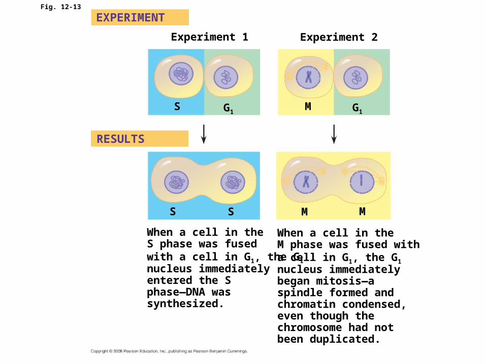
Fig. 12-13
Experiment 1 Experiment 2
EXPERIMENT
RESULTS
S G1M G1
M MSS
When a cell in theS phase was fused with a cell in G1, the G1 nucleus immediatelyentered the Sphase—DNA was synthesized.
When a cell in theM phase was fused with a cell in G1, the G1 nucleus immediatelybegan mitosis—aspindle formed andchromatin condensed,even though thechromosome had notbeen duplicated.

The Cell Cycle Control System
• The sequential events of the cell cycle are directed by a distinct cell cycle control system, which is similar to a clock
• The cell cycle control system is regulated by both internal and external controls
• The clock has specific checkpoints where the cell cycle stops until a go-ahead signal is received

Fig. 12-14
SG1
M checkpoint
G2M
Controlsystem
G1 checkpoint
G2 checkpoint

• For many cells, the G1 checkpoint seems to be the most important one
• If a cell receives a go-ahead signal at the G1 checkpoint, it will usually complete the S, G2, and M phases and divide
• If the cell does not receive the go-ahead signal, it will exit the cycle, switching into a nondividing state called the G0 phase
Copyright © 2008 Pearson Education, Inc., publishing as Pearson Benjamin Cummings
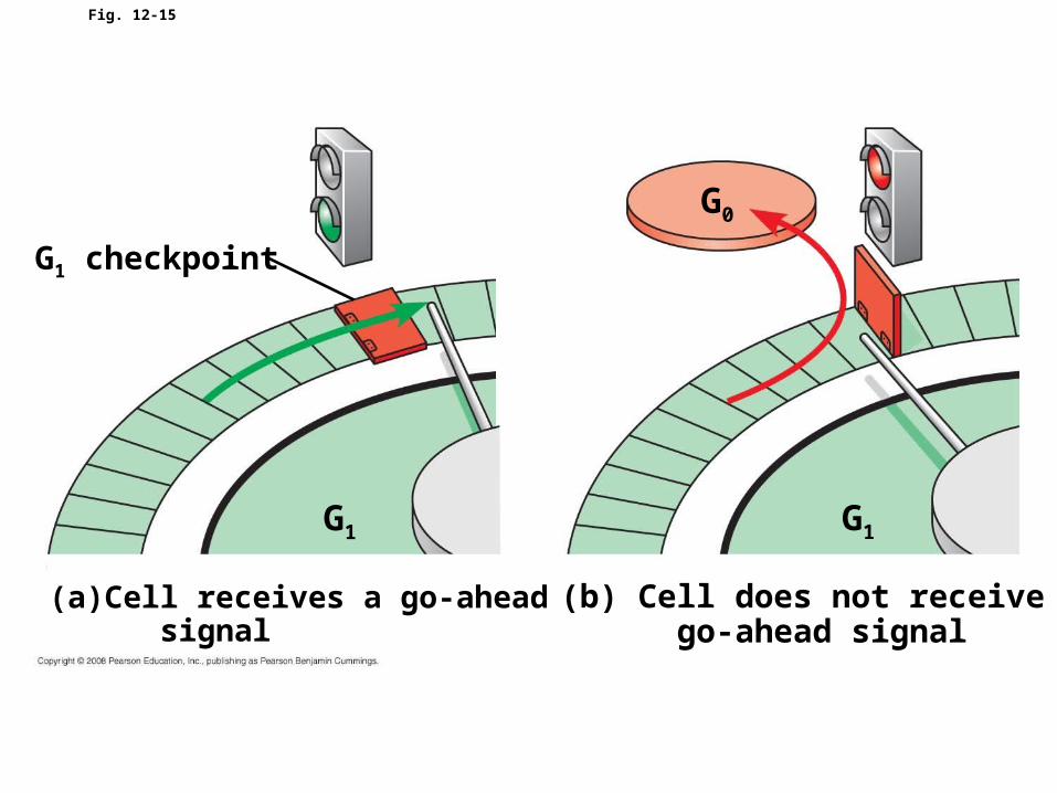
Fig. 12-15
G1
G0
G1 checkpoint
(a) Cell receives a go-ahead signal
G1
(b) Cell does not receive a go-ahead signal
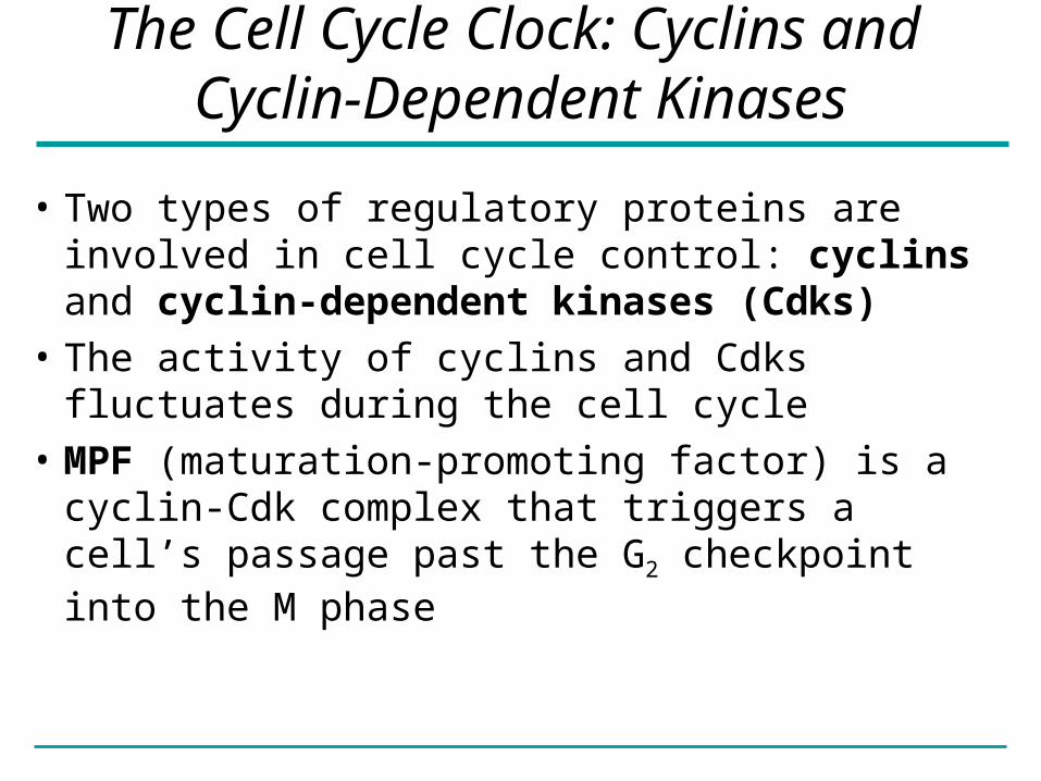
The Cell Cycle Clock: Cyclins and Cyclin-Dependent Kinases
• Two types of regulatory proteins are involved in cell cycle control: cyclins and cyclin-dependent kinases (Cdks)
• The activity of cyclins and Cdks fluctuates during the cell cycle
• MPF (maturation-promoting factor) is a cyclin-Cdk complex that triggers a cell’s passage past the G2 checkpoint into the M phase

Fig. 12-16
Pro
tein
kin
as
e a
cti
vit
y (
– )
% o
f d
ivid
ing
ce
lls (
– )
Time (min)300200 400100
0
1
2
3
4
5 30
500
0
20
10
RESULTS

Fig. 12-17
M G1S G2
M G1S G2
M G1
MPF activity
Cyclinconcentration
Time(a) Fluctuation of MPF activity and cyclin concentration during the cell cycle
Degradedcyclin
Cdk
G 1S
G 2
M
CdkG2
checkpointCyclin isdegraded
CyclinMPF
(b) Molecular mechanisms that help regulate the cell cycle
Cy
clin
ac
cu
mu
latio
n

Fig. 12-17a
Time(a) Fluctuation of MPF activity and cyclin concentration during the cell cycle
Cyclinconcentration
MPF activity
M M MSSG1 G1 G1G2 G2

Fig. 12-17b
Cyclin isdegraded
Cdk
MPF
Cdk
MS
G 1G2
checkpoint
Degradedcyclin
Cyclin
(b) Molecular mechanisms that help regulate the cell cycle
G2
Cyclin
accum
ulatio
n

Stop and Go Signs: Internal and External Signals at the Checkpoints
• An example of an internal signal is that kinetochores not attached to spindle microtubules send a molecular signal that delays anaphase
• Some external signals are growth factors, proteins released by certain cells that stimulate other cells to divide
• For example, platelet-derived growth factor (PDGF) stimulates the division of human fibroblast cells in culture
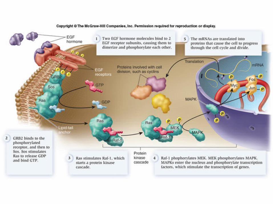

Fig. 12-18
Petriplate
Scalpels
Cultured fibroblasts
Without PDGFcells fail to divide
With PDGFcells prolifer-ate
10 µm
• Another example of external signals is density-dependent inhibition, in which crowded cells stop dividing
• Most animal cells also exhibit anchorage dependence, in which they must be attached to a substratum in order to divide

Fig. 12-19
Anchorage dependence
Density-dependent inhibition
Density-dependent inhibition
(a) Normal mammalian cells (b) Cancer cells25 µm25 µm

• Cancer cells exhibit neither density-dependent inhibition nor anchorage dependence
Copyright © 2008 Pearson Education, Inc., publishing as Pearson Benjamin Cummings

Loss of Cell Cycle Controls in Cancer Cells
• Cancer cells do not respond normally to the body’s control mechanisms
• Cancer cells may not need growth factors to grow and divide:– They may make their own growth factor– They may convey a growth factor’s signal
without the presence of the growth factor– They may have an abnormal cell cycle control
system

• A normal cell is converted to a cancerous cell by a process called transformation
• Cancer cells form tumors, masses of abnormal cells within otherwise normal tissue
• If abnormal cells remain at the original site, the lump is called a benign tumor
• Malignant tumors invade surrounding tissues and can metastasize, exporting cancer cells to other parts of the body, where they may form secondary tumors
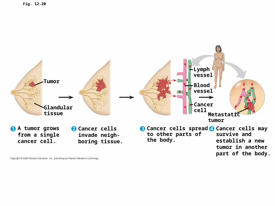
Fig. 12-20
Tumor
A tumor growsfrom a singlecancer cell.
Glandulartissue
Lymphvessel
Bloodvessel
Metastatictumor
Cancercell
Cancer cellsinvade neigh-boring tissue.
Cancer cells spreadto other parts ofthe body.
Cancer cells maysurvive andestablish a newtumor in anotherpart of the body.
1 2 3 4