Embed Size (px)
Citation preview
on July 12, 2018http://rstb.royalsocietypublishing.org/Downloaded from
rstb.royalsocietypublishing.org
ResearchCite this article: Cowan JR, Tariq M, Shaw C,
Rao M, Belmont JW, Lalani SR, Smolarek TA,
Ware SM. 2016 Copy number variation
as a genetic basis for heterotaxy and
heterotaxy-spectrum congenital heart defects.
Phil. Trans. R. Soc. B 371: 20150406.
http://dx.doi.org/10.1098/rstb.2015.0406
Accepted: 3 June 2016
One contribution of 17 to a theme issue
‘Provocative questions in left – right
asymmetry’.
Subject Areas:developmental biology, genetics, genomics,
molecular biology
Keywords:copy number variation, heterotaxy, left – right
patterning, congenital heart defects,
phosphofructokinase
Author for correspondence:Stephanie M. Ware
e-mail: [email protected]
& 2016 The Author(s) Published by the Royal Society. All rights reserved.
Electronic supplementary material is available
online at https://dx.doi.org/10.6084/m9.fig-
share.c.3515256.
Copy number variation as a genetic basisfor heterotaxy and heterotaxy-spectrumcongenital heart defects
Jason R. Cowan1,2, Muhammad Tariq2,3, Chad Shaw4, Mitchell Rao4, JohnW. Belmont4, Seema R. Lalani4, Teresa A. Smolarek5 and Stephanie M. Ware2
1Department of Pediatrics, University of Cincinnati College of Medicine, Cincinnati, OH 45229, USA2Department of Pediatrics and Medical and Molecular Genetics, Herman B Wells Center for Pediatric Research,Indiana University School of Medicine, Indianapolis, IN 46202, USA3Department of Clinical Biochemistry, University of Tabuk, Tabuk 71491, Kingdom of Saudi Arabia4Department of Molecular and Human Genetics, Baylor College of Medicine, Houston, TX 77030, USA5Cincinnati Children’s Hospital Medical Center, Division of Human Genetics, Cincinnati, OH 45229, USA
Genomic disorders and rare copy number abnormalities are identified in
15–25% of patients with syndromic conditions, but their prevalence in individ-
uals with isolated birth defects is less clear. A spectrum of congenital heart
defects (CHDs) is seen in heterotaxy, a highly heritable and genetically hetero-
geneous multiple congenital anomaly syndrome resulting from failure to
properly establish left–right (L-R) organ asymmetry during early embryonic
development. To identify novel genetic causes of heterotaxy, we analysed
copy number variants (CNVs) in 225 patients with heterotaxy and hetero-
taxy-spectrum CHDs using array-based genotyping methods. Clinically
relevant CNVs were identified in approximately 20% of patients and encom-
passed both known and putative heterotaxy genes. Patients were carefully
phenotyped, revealing a significant association of abdominal situs inversus
with pathogenic or likely pathogenic CNVs, while d-transposition of the
great arteries was more frequently associated with common CNVs. Identified
cytogenetic abnormalities ranged from large unbalanced translocations to
smaller, kilobase-scale CNVs, including a rare, single exon deletion in ZIC3,a gene known to cause X-linked heterotaxy. Morpholino loss-of-function exper-
iments in Xenopus support a role for one of these novel candidates, the platelet
isoform of phosphofructokinase-1 (PFKP) in heterotaxy. Collectively, our
results confirm a high CNV yield for array-based testing in patients with
heterotaxy, and support use of CNV analysis for identification of novel
biological processes relevant to human laterality.
This article is part of the themed issue ‘Provocative questions in left–
right asymmetry’.
1. IntroductionHeterotaxy is a relatively infrequent (approx. 1 : 10 000) multiple congenital
anomaly syndrome resulting from abnormal specification of the left–right
(L–R) body axis during early embryonic development [1]. In its classic form,
heterotaxy is characterized by combined occurrence of visceral situs abnormal-
ities (gut malrotation, stomach and liver situs anomalies, abnormalities of
spleen positioning or number) and congenital heart defects (CHDs) of varying
complexity, which account for the majority of associated morbidity and mortality.
Over 96% of patients with heterotaxy exhibit some form of CHD [2], often
requiring surgical intervention. Clinical outcomes are disproportionately poorer
than in patients without heterotaxy who have similar CHDs and are typified
by prolonged courses and significantly greater likelihood for post-surgical com-
plications [3–5]. This clinical picture firmly establishes heterotaxy as not only a
disease of significant phenotypic heterogeneity, but also one of considerable
medical and economic consequence.
rstb.royalsocietypublishing.orgPhil.Trans.R.Soc.B
371:20150406
2
on July 12, 2018http://rstb.royalsocietypublishing.org/Downloaded from
Although heterotaxy typically occurs sporadically and with
unknown cause, its relative risk is highest among all classes of
CHDs, supporting existence of a strong genetic component
[6]. Autosomal recessive, autosomal dominant and X-linked
inheritance patterns have all been described [7]. Mutations in
the zinc finger of the cerebellum 3 (ZIC3) gene are particularly
well documented and are considered to be causative in the
majority (approx. 75%) of familial X-linked pedigrees. Sur-
prisingly, however, ZIC3 mutations underlie only a minority
(3–5%) of sporadic heterotaxy cases [8,9]. Likewise, despite a
conserved and central role for Nodal signalling in establish-
ment of early molecular asymmetries, point mutations in
Nodal pathway components are also not routinely identified
and collectively explain only 5–10% of heterotaxy cases
[10–12]. Mutations in other causative genes are detected at simi-
lar or even lower frequencies, indicating significant genetic
heterogeneity. As a specific genetic etiology is currently identifi-
able in only a minority of patients, there remains enormous
potential for novel gene and pathway discovery.
Copy number variants (CNVs) in the form of complex
chromosomal rearrangements and submicroscopic dupli-
cations and deletions are increasingly recognized as
important causes of birth defects and neurodevelopmental
disease [13]. Depending on size and genomic position,
CNVs can encompass complete or partial intronic or exonic
regions, can include one or multiple genes, or can disrupt
regulatory regions such as promoters and enhancers. If a
CNV shifts the normal reading frame, this change may lead
to premature truncation and loss-of-function through non-
sense-mediated decay or to gain-of-function if the transcript
escapes decay [14]. Gene interruptions and fusions, positional
effects, recessive mutation unmasking and allelic transvection
events are all additional potential molecular consequences.
The pathogenic significance of a particular CNV is, therefore,
highly dependent on the location of its breakpoints, the geno-
mic content of the intervening deleted or duplicated segment,
and the genomic landscape in which the CNV is situated.
Studies of patients with CHD indicate that CNVs are a
major genetic cause of cardiovascular disease, occurring in
3–25% of patients with extra-cardiac abnormalities and in
3–10% with isolated heart defects [15]. To date, two studies
have employed genome-wide approaches to explicitly exam-
ine the role of CNVs in heterotaxy [16,17]. Analysing a cohort
of 262 patients with classic heterotaxy and/or isolated hetero-
taxy spectrum CHDs, Fakhro et al. [16] identified 45 rare
CNVs in 39 patients, representing a 15% CNV yield in their
phenotypically mixed population. A slightly higher rate of
26% (20 CNVs in 19/74 patients) was reported in a more
recent study that restricted analysis solely to patients with
classic heterotaxy [17]. Yields from both of these studies
approximated those seen in other CHD cohorts [15] and
together support CNVs as important contributors to human
laterality defects.
While copy number variation is now regarded as a major
cause of genetic disease, it is important to recognize that not
all CNVs are pathogenic. Indeed, CNVs are estimated to
encompass between 4.8 and 9.5% of the human genome
[18]. In order to systematically catalogue this genetic vari-
ation, online and clinical databases have been established as
repositories of commonly occurring CNVs. These databases
serve as vital reference points for clinical and research efforts
focused on identifying CNVs of pathogenic significance to
genetic disease.
The flexibility and versatility of array comparative genomic
hybridization (aCGH), and single-nucleotide polymorphism
(SNP) microarrays have made them excellent tools for both
gene discovery and clinical practice. In this study, we have
screened a large cohort of patients with heterotaxy spectrum
malformations using these complementary methods. In total,
we identified rare CNVs in 46/225 (20.44%) patients ranging
in size from large, megabase-scale translocations to smaller,
kilobase-scale CNVs. Excluding CNVs that were considered
to be clearly pathogenic owing to their overall size and com-
plexity, or their genomic content (encompassing regions or
genes previously associated with or identified as definitive
causes of heterotaxy/CHD), we reduced the number of
CNVs suitable for candidate gene analysis to 35 in 30/225
(13.33%) patients. Morpholino (MO)-based loss-of-function
screens in Xenopus laevis were then used to confirm roles for
gene candidates in L–R patterning. In this report, we describe
overall findings from these CNV screens while highlighting
functional testing results for platelet isoform of phosphofructo-
kinase 1 ( pfkp), a gene identified as a novel candidate.
Collectively, our analyses support CNVs as a significant contri-
butor to heterotaxy causation and reiterate the value of
genome-wide CNV screening as a tool for identifying novel
laterality genes and pathways.
2. Material and methods(a) Patient recruitment and phenotypic classificationDetailed phenotypic information was collected from patient his-
tories and chart review. Patients were classified as having situs
inversus (SI) totalis, heterotaxy, or isolated heterotaxy spectrum
CHD following previously defined criteria [9]. As some hetero-
taxy patients with ZIC3 mutations also exhibit VACTERL-like
(vertebral anomalies, anal atresia, cardiovascular malformations,
tracheo-esophageal fistula, renal anomalies, limb abnormalities)
phenotypes, patients with these features are noted. Disease was
considered to be ‘familial’ if (i) the pedigree demonstrated auto-
somal dominant, autosomal recessive or X-linked inheritance,
(ii) there was more than one family member with heterotaxy or
laterality disorder regardless of their degree of relationship to the
proband, (iii) there was heterotaxy in the proband and a first
degree relative with isolated CHD or (iv) there was heterotaxy in
the proband and a first degree relative with situs-related defects.
All other patients were considered to have sporadic disease.
Patients with previously identified cytogenetic findings or with
mutations in known heterotaxy genes were excluded from further
analysis. No patient had a previously identified CNV from prior
clinical screening. In total, the final cohort comprised 225 unrelated
patients with assorted situs and/or cardiac abnormalities, includ-
ing 139 males and 86 females. Full cohort demographics are
summarized in table 1.
(b) Chromosome microarray analysisGenomic DNA was prepared from blood samples following stan-
dard protocols. To evaluate for CNVs, samples were analysed
using genome-wide SNP analysis and aCGH, with platform
choice being dictated by sample availability at time of testing.
The majority of samples were analysed using both platforms,
which allowed cross-validation of CNVs. SNP genotyping was
performed using the Illumina Human370 DNA Analysis Bead-
Chip (42 patients) or Illumina HumanOmni1-Quad Beadchip
(106 patients) platforms (Illumina Inc., San Diego, CA, USA).
These chips encompassed approximately 370 000 SNP markers
(mean spacing 7.7 kb, median 5 kb) or 1 000 000 SNP markers
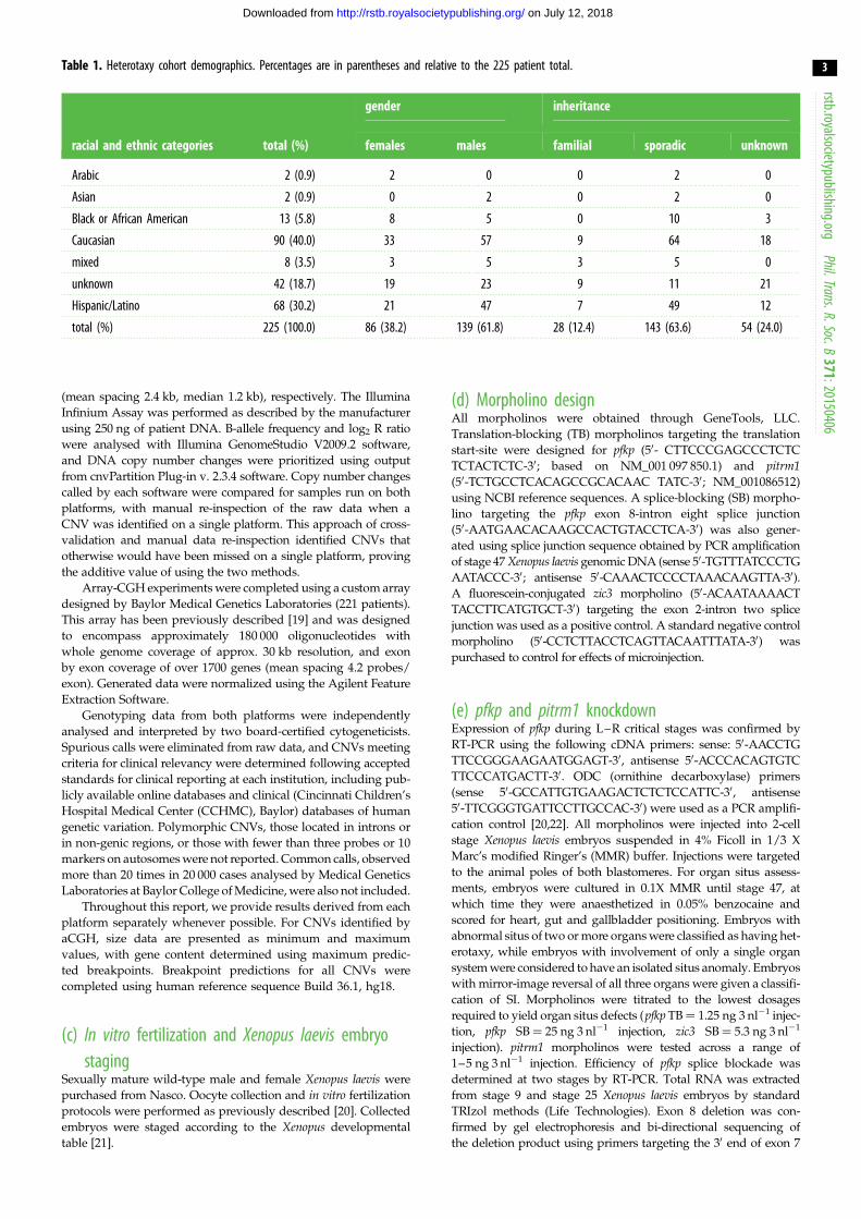
Table 1. Heterotaxy cohort demographics. Percentages are in parentheses and relative to the 225 patient total.
racial and ethnic categories total (%)
gender inheritance
females males familial sporadic unknown
Arabic 2 (0.9) 2 0 0 2 0
Asian 2 (0.9) 0 2 0 2 0
Black or African American 13 (5.8) 8 5 0 10 3
Caucasian 90 (40.0) 33 57 9 64 18
mixed 8 (3.5) 3 5 3 5 0
unknown 42 (18.7) 19 23 9 11 21
Hispanic/Latino 68 (30.2) 21 47 7 49 12
total (%) 225 (100.0) 86 (38.2) 139 (61.8) 28 (12.4) 143 (63.6) 54 (24.0)
rstb.royalsocietypublishing.orgPhil.Trans.R.Soc.B
371:20150406
3
on July 12, 2018http://rstb.royalsocietypublishing.org/Downloaded from
(mean spacing 2.4 kb, median 1.2 kb), respectively. The Illumina
Infinium Assay was performed as described by the manufacturer
using 250 ng of patient DNA. B-allele frequency and log2 R ratio
were analysed with Illumina GenomeStudio V2009.2 software,
and DNA copy number changes were prioritized using output
from cnvPartition Plug-in v. 2.3.4 software. Copy number changes
called by each software were compared for samples run on both
platforms, with manual re-inspection of the raw data when a
CNV was identified on a single platform. This approach of cross-
validation and manual data re-inspection identified CNVs that
otherwise would have been missed on a single platform, proving
the additive value of using the two methods.
Array-CGH experiments were completed using a custom array
designed by Baylor Medical Genetics Laboratories (221 patients).
This array has been previously described [19] and was designed
to encompass approximately 180 000 oligonucleotides with
whole genome coverage of approx. 30 kb resolution, and exon
by exon coverage of over 1700 genes (mean spacing 4.2 probes/
exon). Generated data were normalized using the Agilent Feature
Extraction Software.
Genotyping data from both platforms were independently
analysed and interpreted by two board-certified cytogeneticists.
Spurious calls were eliminated from raw data, and CNVs meeting
criteria for clinical relevancy were determined following accepted
standards for clinical reporting at each institution, including pub-
licly available online databases and clinical (Cincinnati Children’s
Hospital Medical Center (CCHMC), Baylor) databases of human
genetic variation. Polymorphic CNVs, those located in introns or
in non-genic regions, or those with fewer than three probes or 10
markers on autosomes were not reported. Common calls, observed
more than 20 times in 20 000 cases analysed by Medical Genetics
Laboratories at Baylor College of Medicine, were also not included.
Throughout this report, we provide results derived from each
platform separately whenever possible. For CNVs identified by
aCGH, size data are presented as minimum and maximum
values, with gene content determined using maximum predic-
ted breakpoints. Breakpoint predictions for all CNVs were
completed using human reference sequence Build 36.1, hg18.
(c) In vitro fertilization and Xenopus laevis embryostaging
Sexually mature wild-type male and female Xenopus laevis were
purchased from Nasco. Oocyte collection and in vitro fertilization
protocols were performed as previously described [20]. Collected
embryos were staged according to the Xenopus developmental
table [21].
(d) Morpholino designAll morpholinos were obtained through GeneTools, LLC.
Translation-blocking (TB) morpholinos targeting the translation
start-site were designed for pfkp (50- CTTCCCGAGCCCTCTC
TCTACTCTC-30; based on NM_001 097 850.1) and pitrm1(50-TCTGCCTCACAGCCGCACAAC TATC-30; NM_001086512)
using NCBI reference sequences. A splice-blocking (SB) morpho-
lino targeting the pfkp exon 8-intron eight splice junction
(50-AATGAACACAAGCCACTGTACCTCA-30) was also gener-
ated using splice junction sequence obtained by PCR amplification
of stage 47 Xenopus laevis genomic DNA (sense 50-TGTTTATCCCTG
AATACCC-30; antisense 50-CAAACTCCCCTAAACAAGTTA-30).
A fluorescein-conjugated zic3 morpholino (50-ACAATAAAACT
TACCTTCATGTGCT-30) targeting the exon 2-intron two splice
junction was used as a positive control. A standard negative control
morpholino (50-CCTCTTACCTCAGTTACAATTTATA-30) was
purchased to control for effects of microinjection.
(e) pfkp and pitrm1 knockdownExpression of pfkp during L–R critical stages was confirmed by
RT-PCR using the following cDNA primers: sense: 50-AACCTG
TTCCGGGAAGAATGGAGT-30, antisense 50-ACCCACAGTGTC
TTCCCATGACTT-30. ODC (ornithine decarboxylase) primers
(sense 50-GCCATTGTGAAGACTCTCTCCATTC-30, antisense
50-TTCGGGTGATTCCTTGCCAC-30) were used as a PCR amplifi-
cation control [20,22]. All morpholinos were injected into 2-cell
stage Xenopus laevis embryos suspended in 4% Ficoll in 1/3 X
Marc’s modified Ringer’s (MMR) buffer. Injections were targeted
to the animal poles of both blastomeres. For organ situs assess-
ments, embryos were cultured in 0.1X MMR until stage 47, at
which time they were anaesthetized in 0.05% benzocaine and
scored for heart, gut and gallbladder positioning. Embryos with
abnormal situs of two or more organs were classified as having het-
erotaxy, while embryos with involvement of only a single organ
system were considered to have an isolated situs anomaly. Embryos
with mirror-image reversal of all three organs were given a classifi-
cation of SI. Morpholinos were titrated to the lowest dosages
required to yield organ situs defects (pfkp TB¼ 1.25 ng 3 nl21 injec-
tion, pfkp SB ¼ 25 ng 3 nl21 injection, zic3 SB¼ 5.3 ng 3 nl21
injection). pitrm1 morpholinos were tested across a range of
1–5 ng 3 nl21 injection. Efficiency of pfkp splice blockade was
determined at two stages by RT-PCR. Total RNA was extracted
from stage 9 and stage 25 Xenopus laevis embryos by standard
TRIzol methods (Life Technologies). Exon 8 deletion was con-
firmed by gel electrophoresis and bi-directional sequencing of
the deletion product using primers targeting the 30 end of exon 7
rstb.royalsocietypublishing.orgPhil.Trans.R.Soc.B
371:20150406
4
on July 12, 2018http://rstb.royalsocietypublishing.org/Downloaded from
(50-CCAGAGGACCTTTGTGCTGGAAGTTATG-30) and the 50 end
of exon 9 (50-ACACAACACAGGCTGGCGTTTC-30). ODC primers
were again used for control of RNA quality and PCR amplification.
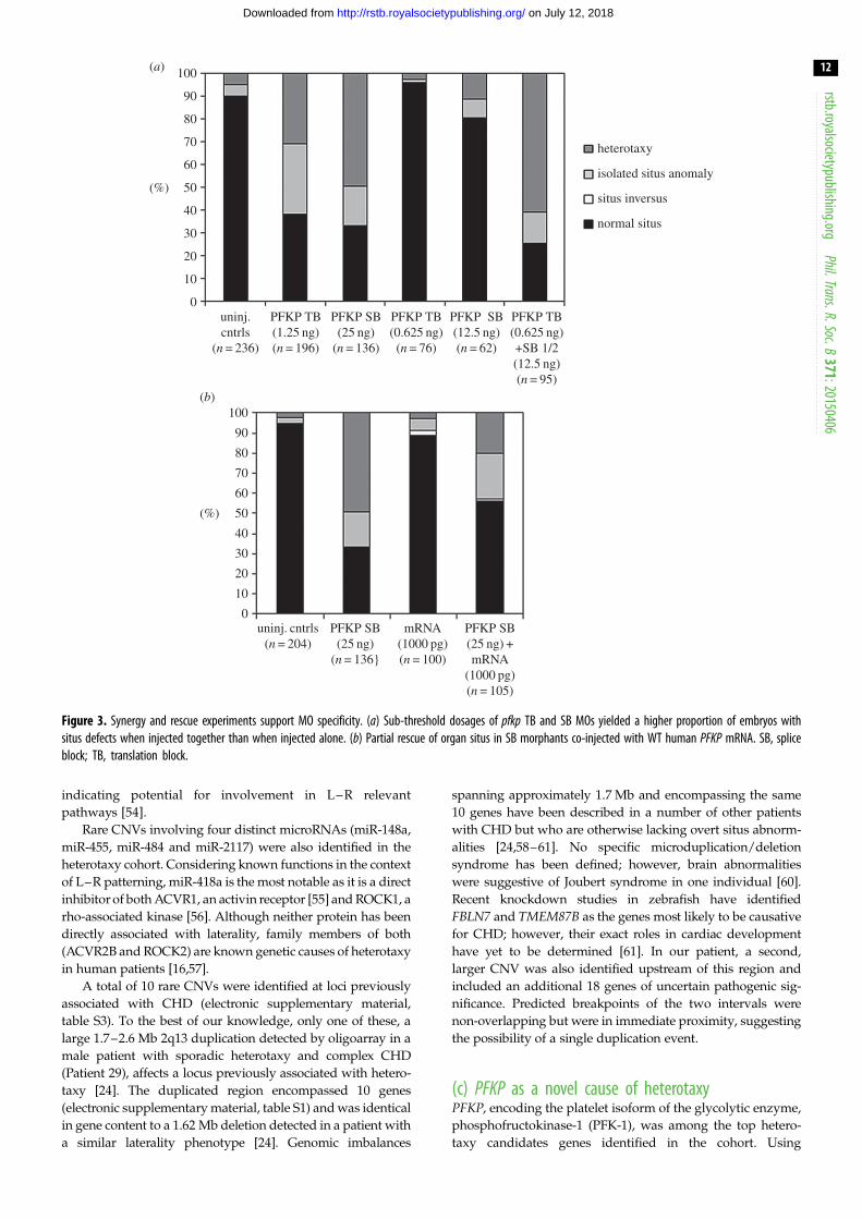
( f ) Synergy and rescueMorpholino synergy and mRNA rescue experiments were
performed to confirm specificity of targeting. For synergy exper-
iments, pfkp TB (0.83 ng) and SB (16.7 ng) morpholino doses that
did not independently cause phenotypic abnormalities were co-
or independently injected into both animal poles of 2-cell stage
embryos. Morpholino synergy was demonstrated by a significant
increase in organ situs defects in co-injected embryos relative to
independently injected embryos at stage 47. For mRNA rescue
experiments, in vitro transcribed full-length human PFKP mRNA
was co-injected with pfkp SB morpholino into both animal poles
of 2-cell stage embryos. Successful rescue was demonstrated by a
significant decrease in organ situs defects in mRNA-treated
embryos relative to untreated morphants. Full-length human
PFKP cDNA for mRNA synthesis was generated from Origene
cDNA clone SC118507 by PCR amplification (sense 50-GCAGA
GCTCGTTTAGTGAACCGC-30, antisense 50-GATGGGCACTCG
CCGATTAG-30). Identities of the cDNA restriction fragment and
PCR product were both confirmed by Sanger sequencing. PFKPmRNA was subsequently in vitro transcribed by T7 polymerase
using the mMessage mMachine kit (Ambion) following manufac-
turer’s protocols and stored at 2808C. For rescue experiments,
embryos were injected with either 500 or 1000 pg of transcribed
mRNA, either alone or in combination with pfkp MO.
(g) Left – right marker analysesXenopus laevis embryos were fixed overnight at 48C in MEMFA
(4% paraformaldehyde in MEM salts). Fixed embryos were sub-
sequently dehydrated through three consecutive 5 min 100%
methanol washes and stored at 2208C. An antisense RNA probe
for the L–R marker, coco was generated from a pCMV-SPORT6
plasmid (provided by Dr Mustafa Khokha) using a T7/SP6 DIG
RNA Labeling Kit (Roche) following manufacturer’s instructions.
Whole-mount in situ hybridizations (WISH) were performed as
previously described [23], with one modification: prior to hybrid-
ization, fixed stage 20–21 embryos were bisected along the
transverse plane into separate anterior and posterior halves, briefly
post-fixed (10 min in 4% MEMFA), and run through a graded
methanol series. Hybridization was performed on both halves,
which were then sorted and appropriately trimmed. Staining
was accomplished using BM Purple alkaline phosphatase chromo-
genic substrate (Roche). Images were captured using a Nikon
SMZ1500 stereomicroscope outfitted with a Nikon DXM1200F
digital camera and processed using Nikon Act-1 (v. 2.62) imaging
software. Coco expression was scored as left-sided biased,
right-sided biased or right/left unbiased by visual inspection.
Final embryo counts included only those embryos that could be
unequivocally classified.
3. Results(a) Genetic analyses of patients with heterotaxy
identify rare copy number variantsPatients were carefully phenotyped with regard to CHD and
extra-cardiac anomalies (table 2). In total, just under 20% of
patients had isolated CHD, with the remainder having situs
abnormalities of some kind. Of the 225 total patients tested,
148 (65.78%) were analysed by SNP array, 221 (98.22%) were
analysed by aCGH and 144 (64.00%) were analysed using
both platforms. To maximize our chances of identifying
novel causes of heterotaxy, we focused attention on rare
CNVs encompassing coding regions of one or more genes.
Novel pathogenic or likely pathogenic CNVs were identified
in 46/225 patients, representing an overall CNV yield of
20.4% (figure 1). We compared these 46 patients to the remain-
der of the heterotaxy cohort with common CNVs using two-
tailed Fisher’s exact tests (table 2). Notably, occurrence of
abdominal situs abnormalities was significantly greater in
this patient group compared with the remainder of the hetero-
taxy cohort. In addition, d-TGA was less likely to be associated
with pathogenic or likely pathogenic CNVs (table 2). Thirteen
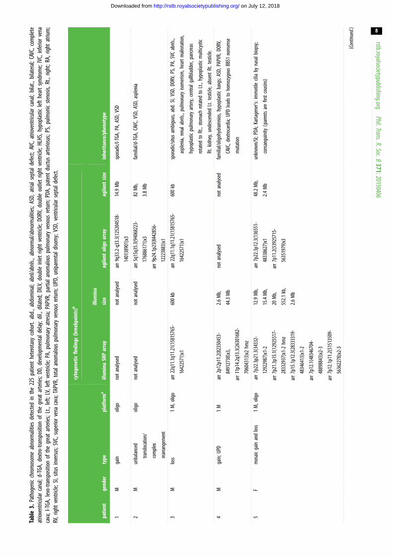
patients were found to carry large/complex chromosomal
abnormalities (table 3), including an approximate 3 Mb
22q11.2 critical region deletion consistent with a diagnosis
of 22q11.2 deletion syndrome (DiGeorge syndrome) in a
female patient with abdominal situs abnormalities and CHD
(Patient 6). Another three CNVs affected genes previously
associated with heterotaxy (table 4): a 553–686 kb 2p25.1
duplication encompassing ROCK2 in a male patient with
sporadic and complex CHD (Patient 15), a 1.47–1.61 Mb
3p24.1 deletion encompassing TGFBR2 in a female patient
with familial heterotaxy (Patient 16) and a 2.9–3.3 kb Xq26.2
deletion encompassing the third exon of ZIC3 in a male patient
with sporadic heterotaxy (Patient 14).
As it is important to rule out all potential heterotaxy
gene candidates in a CNV of interest, we prioritized CNVs con-
taining relatively fewer genes for further analysis. Twenty-two
(62.86%) of these 35 ‘CNVs of interest’ involved gains of genetic
material, while 13 (37.14%) involved genetic losses (electro-
nic supplementary material, table S1). As an estimation of
CNV gene content, we performed breakpoint predictions
using available marker/probe positioning. These analyses
were inherently limited by marker and probe spacing, but
suggested 15/22 (68%) gains and 5/13 (38.46%) losses encom-
passed the entire coding region of at least one gene. Only one of
the 22 identified gains (4.55%) and 4 of the 13 identified losses
(30.77%) were predicted to lie entirely within a single gene. The
remaining CNVs affected partial coding sequences or coding
sequences of more than one gene. There was no obvious
chromosome bias among detected CNVs, with CNVs of inter-
est detected on 21/23 chromosomes, the exceptions being the X
chromosome and chromosome 21 (data not shown).
Of the 35 CNVs of interest, seven were more than 1 Mb in
size (SNP size range 1.01–2.78 Mb, mean 1.96 Mb, median
2.06 Mb; aCGH size range 1.08–3.84 Mb, mean 2.02–
2.30 Mb, median 1.86–2.58 Mb), and encompassed between 1
and 18 genes (mean 7; median 7). The remaining 28 CNVs indi-
vidually affected less than 1 Mb of sequence (SNP size range
161.66–877.08 kb, mean 458.50 kb, median 472.45 kb; aCGH
size range 4.90–960.81 kb, mean 322.04–387.01 kb, median
205.73–279.31 kb) impacting between 1 and 16 genes each
(mean 3.96; median 2).
A total of 165 genes with diverse developmental and mol-
ecular functions were encompassed by the 35 CNVs of
interest (electronic supplementary material, table S2 and
figure S1). Among these genes were a subset with recognized
roles in developmental processes and pathways important for
L–R patterning, including ciliogenesis, transforming growth
factor-b (TGFb) signalling, and cell–cell communication.
Eleven of the 35 CNVs of interest encompassed loci pre-
viously associated with CHD (electronic supplementary
material, table S3), while one (a 1.70–2.59 Mb 2q13 deletion
identified in a patient with sporadic heterotaxy) disrupted a
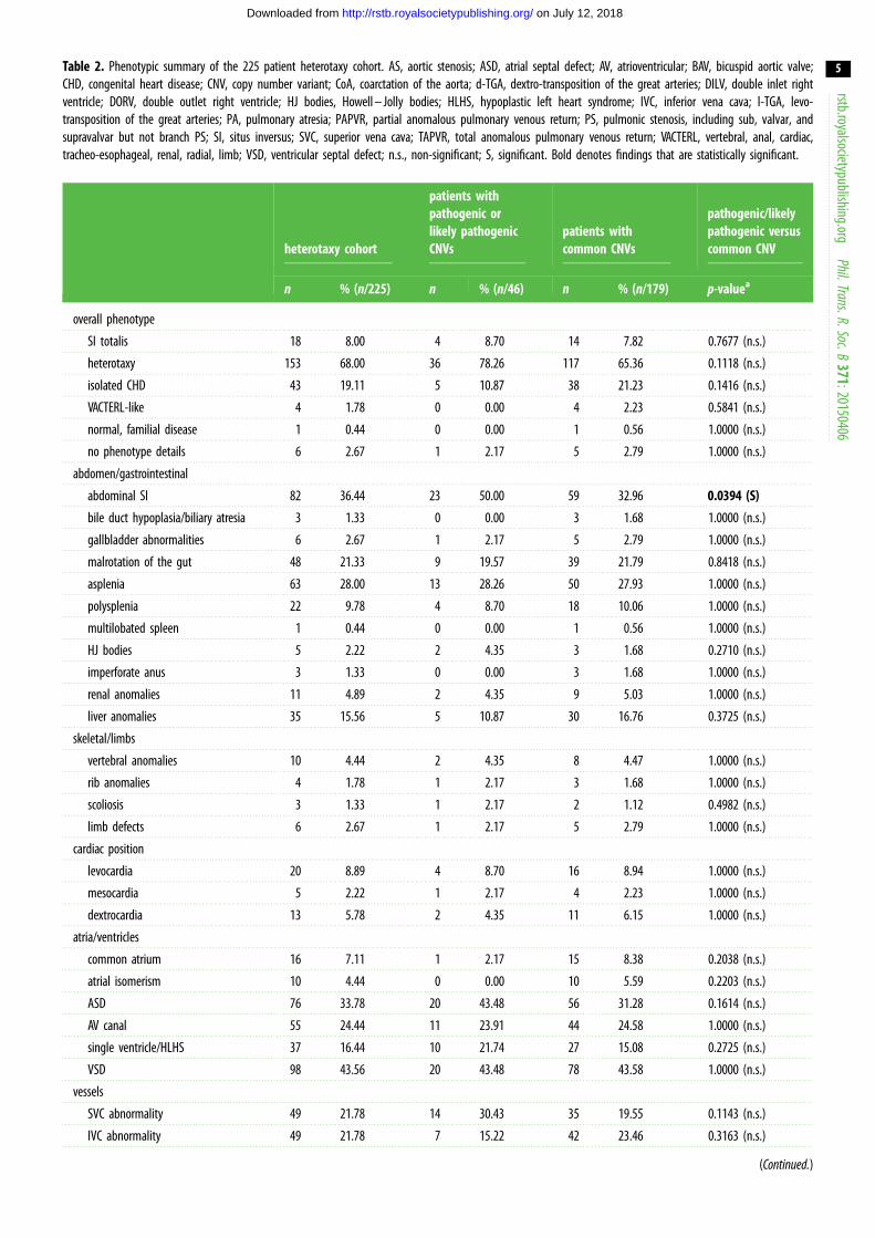
Table 2. Phenotypic summary of the 225 patient heterotaxy cohort. AS, aortic stenosis; ASD, atrial septal defect; AV, atrioventricular; BAV, bicuspid aortic valve;CHD, congenital heart disease; CNV, copy number variant; CoA, coarctation of the aorta; d-TGA, dextro-transposition of the great arteries; DILV, double inlet rightventricle; DORV, double outlet right ventricle; HJ bodies, Howell – Jolly bodies; HLHS, hypoplastic left heart syndrome; IVC, inferior vena cava; l-TGA, levo-transposition of the great arteries; PA, pulmonary atresia; PAPVR, partial anomalous pulmonary venous return; PS, pulmonic stenosis, including sub, valvar, andsupravalvar but not branch PS; SI, situs inversus; SVC, superior vena cava; TAPVR, total anomalous pulmonary venous return; VACTERL, vertebral, anal, cardiac,tracheo-esophageal, renal, radial, limb; VSD, ventricular septal defect; n.s., non-significant; S, significant. Bold denotes findings that are statistically significant.
heterotaxy cohort
patients withpathogenic orlikely pathogenicCNVs
patients withcommon CNVs
pathogenic/likelypathogenic versuscommon CNV
n % (n/225) n % (n/46) n % (n/179) p-valuea
overall phenotype
SI totalis 18 8.00 4 8.70 14 7.82 0.7677 (n.s.)
heterotaxy 153 68.00 36 78.26 117 65.36 0.1118 (n.s.)
isolated CHD 43 19.11 5 10.87 38 21.23 0.1416 (n.s.)
VACTERL-like 4 1.78 0 0.00 4 2.23 0.5841 (n.s.)
normal, familial disease 1 0.44 0 0.00 1 0.56 1.0000 (n.s.)
no phenotype details 6 2.67 1 2.17 5 2.79 1.0000 (n.s.)
abdomen/gastrointestinal
abdominal SI 82 36.44 23 50.00 59 32.96 0.0394 (S)
bile duct hypoplasia/biliary atresia 3 1.33 0 0.00 3 1.68 1.0000 (n.s.)
gallbladder abnormalities 6 2.67 1 2.17 5 2.79 1.0000 (n.s.)
malrotation of the gut 48 21.33 9 19.57 39 21.79 0.8418 (n.s.)
asplenia 63 28.00 13 28.26 50 27.93 1.0000 (n.s.)
polysplenia 22 9.78 4 8.70 18 10.06 1.0000 (n.s.)
multilobated spleen 1 0.44 0 0.00 1 0.56 1.0000 (n.s.)
HJ bodies 5 2.22 2 4.35 3 1.68 0.2710 (n.s.)
imperforate anus 3 1.33 0 0.00 3 1.68 1.0000 (n.s.)
renal anomalies 11 4.89 2 4.35 9 5.03 1.0000 (n.s.)
liver anomalies 35 15.56 5 10.87 30 16.76 0.3725 (n.s.)
skeletal/limbs
vertebral anomalies 10 4.44 2 4.35 8 4.47 1.0000 (n.s.)
rib anomalies 4 1.78 1 2.17 3 1.68 1.0000 (n.s.)
scoliosis 3 1.33 1 2.17 2 1.12 0.4982 (n.s.)
limb defects 6 2.67 1 2.17 5 2.79 1.0000 (n.s.)
cardiac position
levocardia 20 8.89 4 8.70 16 8.94 1.0000 (n.s.)
mesocardia 5 2.22 1 2.17 4 2.23 1.0000 (n.s.)
dextrocardia 13 5.78 2 4.35 11 6.15 1.0000 (n.s.)
atria/ventricles
common atrium 16 7.11 1 2.17 15 8.38 0.2038 (n.s.)
atrial isomerism 10 4.44 0 0.00 10 5.59 0.2203 (n.s.)
ASD 76 33.78 20 43.48 56 31.28 0.1614 (n.s.)
AV canal 55 24.44 11 23.91 44 24.58 1.0000 (n.s.)
single ventricle/HLHS 37 16.44 10 21.74 27 15.08 0.2725 (n.s.)
VSD 98 43.56 20 43.48 78 43.58 1.0000 (n.s.)
vessels
SVC abnormality 49 21.78 14 30.43 35 19.55 0.1143 (n.s.)
IVC abnormality 49 21.78 7 15.22 42 23.46 0.3163 (n.s.)
(Continued.)
rstb.royalsocietypublishing.orgPhil.Trans.R.Soc.B
371:20150406
5
on July 12, 2018http://rstb.royalsocietypublishing.org/Downloaded from
Table 2. (Continued.)
heterotaxy cohort
patients withpathogenic orlikely pathogenicCNVs
patients withcommon CNVs
pathogenic/likelypathogenic versuscommon CNV
n % (n/225) n % (n/46) n % (n/179) p-valuea
TAPVR 32 14.22 7 15.22 25 13.97 0.8150 (n.s.)
PAPVR 16 7.11 6 13.04 10 5.59 0.1038 (n.s.)
inflow/outflow
aortic arch abnormalities 57 25.33 15 32.61 42 23.46 0.2535 (n.s.)
d-TGA 81 36.00 9 19.57 72 40.22 0.0097 (S)
l-TGA 31 13.78 8 17.39 31 17.32 1.0000 (n.s.)
DILV 14 6.22 1 2.17 13 7.26 0.3107 (n.s.)
DORV 65 28.89 18 39.13 47 26.26 0.1013 (n.s.)
PA 49 21.78 12 26.09 37 20.67 0.4282 (n.s.)
PS 58 25.78 11 23.91 47 26.26 0.8511 (n.s.)
AS 4 1.78 2 4.35 2 1.12 0.1863 (n.s.)
CoA 22 9.78 3 6.52 19 10.61 0.5796 (n.s.)
BAV 2 0.89 1 2.17 1 0.56 0.3678 (n.s.)
other
arrhythmia 16 7.11 2 4.35 14 7.82 0.7466 (n.s.)ap-values for statistical significance ( p , 0.05) represent comparisons between patients with CNVs of clear or potential pathogenicity and patients withcommon CNVs by two-tailed Fischer’s Exact tests using Graphpad statistical software (http://www.graphpad.com/quickcalcs/).
rstb.royalsocietypublishing.orgPhil.Trans.R.Soc.B
371:20150406
6
on July 12, 2018http://rstb.royalsocietypublishing.org/Downloaded from
block of 10 genes previously deleted in a similarly affected
heterotaxy patient [24]. CNVs encompassing loci associated
with known microduplication/deletion syndromes but not
previously associated with heterotaxy or CHD are summar-
ized in the electronic supplementary material, table S4. For
candidate gene screens, we prioritized analysis of genes
with known expression during critical L–R developmental
stages in animal studies.
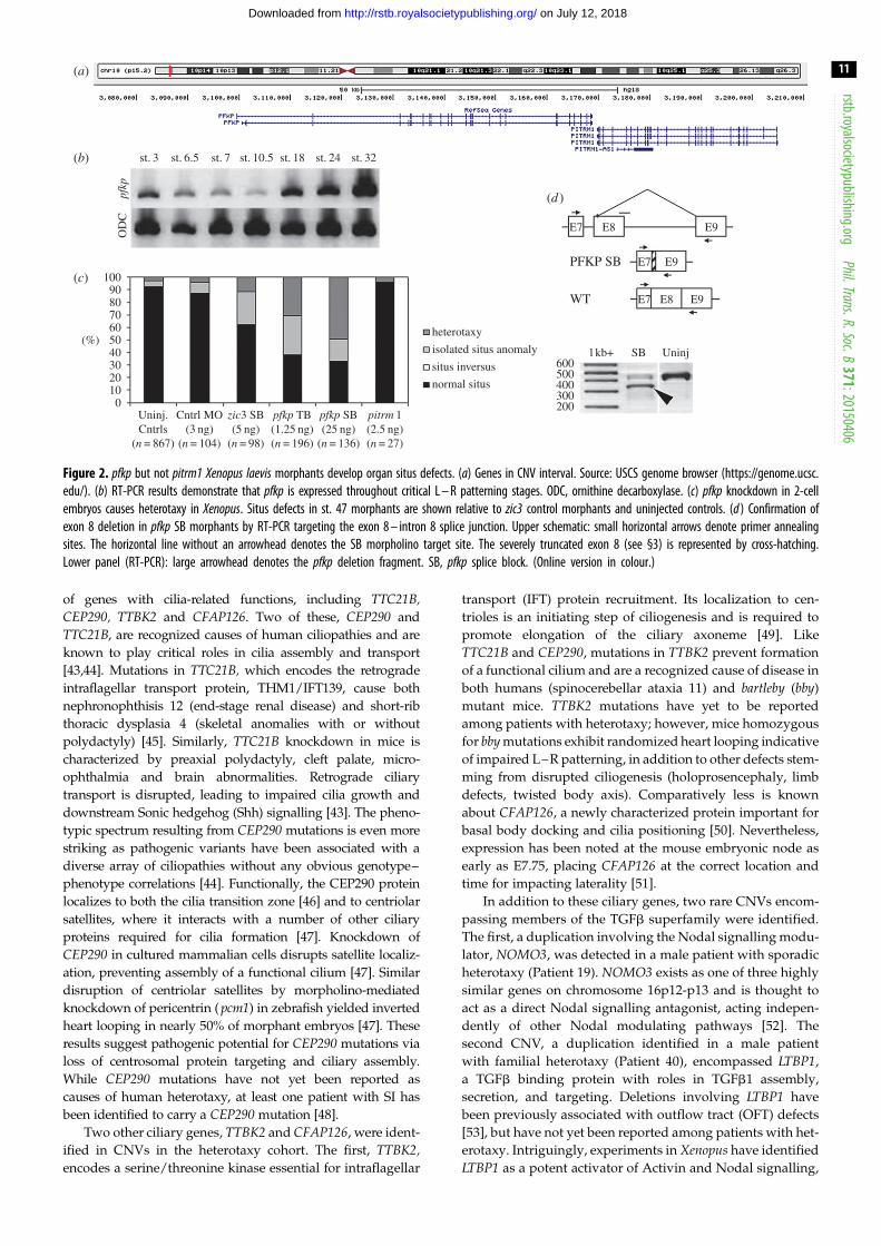
(b) Knockdown of pfkp results in left – right patterningdefects in Xenopus laevis
Because L–R patterning processes are highly conserved across
vertebrate species [25–29], significant knowledge regarding
human laterality can be gleaned from animal studies. We
undertook a Xenopus-based morpholino loss of function
approach to screen identified CNVs for potential heterotaxy
candidate genes. The merits of Xenopus as model for laterality
are extensive and have been reviewed in detail elsewhere [30].
A single patient (Patient 34) from our heterotaxy cohort was
found to carry a heterozygous 175 kb deletion encompassing
the PFKP and PITRM1 genes. These genes encode the platelet
isoform of phosphofructokinase-1 (PFK-1) and the protease
pitrilysin metallopeptidase 1, respectively (figure 2a). This
CNV was of interest as it was identified using both genotyping
platforms and contained an easily testable number of genes,
one of which (PFKP) is a recognized interaction partner of the
Hþ-V-ATPase proton pump [31,32], a known regulator of
left–right patterning [33]. Notably, the deletion was not pre-
viously described in either online or institutional cytogenetic
laboratory databases (see §2). To assess the effect of pfkp and
pitrm1 knockdown on L–R patterning, TB and SB morpholinos
(GeneTools, LLC.) were designed and injected into both blasto-
meres of 2-cell stage Xenopus laevis embryos. Gut, heart and
gallbladder were then scored for abnormal situs at stage 47
according to published criteria [34]. A significantly greater pro-
portion of pfkp, but not pitrm1, morphants developed organ
situs defects relative to uninjected or control morpholino-
injected embryos (figure 2c). Organ situs defects in pfkp mor-
phants were dose-dependent (data not shown). Consistent
with previous reports demonstrating PFK-1 expression in
early development [35–37], pfkp was expressed throughout
stages known to be important for L–R patterning in Xenopus(figure 2b). These results indicated that pfkp but not pitrm1was a suitable candidate for further study.
In order to confirm pfkp mRNA knockdown in pfkp SB mor-
phants, RT-PCR and gene-specific amplification of whole RNA
from st. 9 (not shown) and st. 25 embryos (figure 2d, lane 2) was
completed. Sequencing of the deletion product revealed acti-
vation of a cryptic splice site 13 bp downstream of the intron
7-exon 8 junction, but confirmed deletion of the majority of
exon 8 (96/109 bp). The resulting pfkp transcript is predicted
to encode a truncated, non-frameshifted 754aa (versus 786aa)
protein lacking residues predicted to contribute to ATP binding.
Synergy and rescue experiments using wild-type human PFKPmRNA- supported specificity of the SB morpholino effect
(figure 3).
In order to confirm an effect for pfkp knockdown on L–R
relevant signalling pathways, we examined expression of the
conserved L–R patterning marker, coco, at the gastrocoel roof
plate (GRP). Ciliated cells of the GRP generate a highly direc-
tional extracellular fluid flow that asymmetrically activates
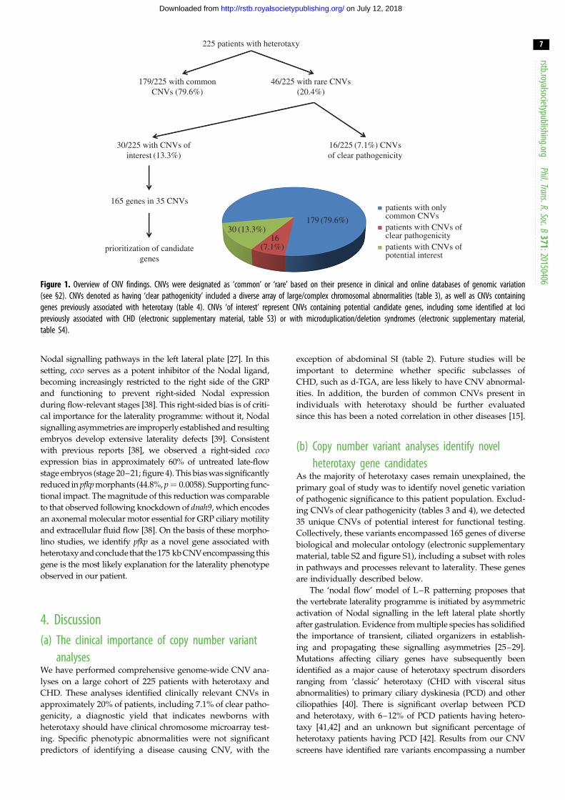
225 patients with heterotaxy
179/225 with commonCNVs (79.6%)
30/225 with CNVs ofinterest (13.3%)
16/225 (7.1%) CNVsof clear pathogenicity
165 genes in 35 CNVs
30 (13.3%)179 (79.6%)
16(7.1%)prioritization of candidate
genes
46/225 with rare CNVs(20.4%)
patients with onlycommon CNVspatients with CNVs ofclear pathogenicitypatients with CNVs ofpotential interest
Figure 1. Overview of CNV findings. CNVs were designated as ‘common’ or ‘rare’ based on their presence in clinical and online databases of genomic variation(see §2). CNVs denoted as having ‘clear pathogenicity’ included a diverse array of large/complex chromosomal abnormalities (table 3), as well as CNVs containinggenes previously associated with heterotaxy (table 4). CNVs ‘of interest’ represent CNVs containing potential candidate genes, including some identified at locipreviously associated with CHD (electronic supplementary material, table S3) or with microduplication/deletion syndromes (electronic supplementary material,table S4).
rstb.royalsocietypublishing.orgPhil.Trans.R.Soc.B
371:20150406
7
on July 12, 2018http://rstb.royalsocietypublishing.org/Downloaded from
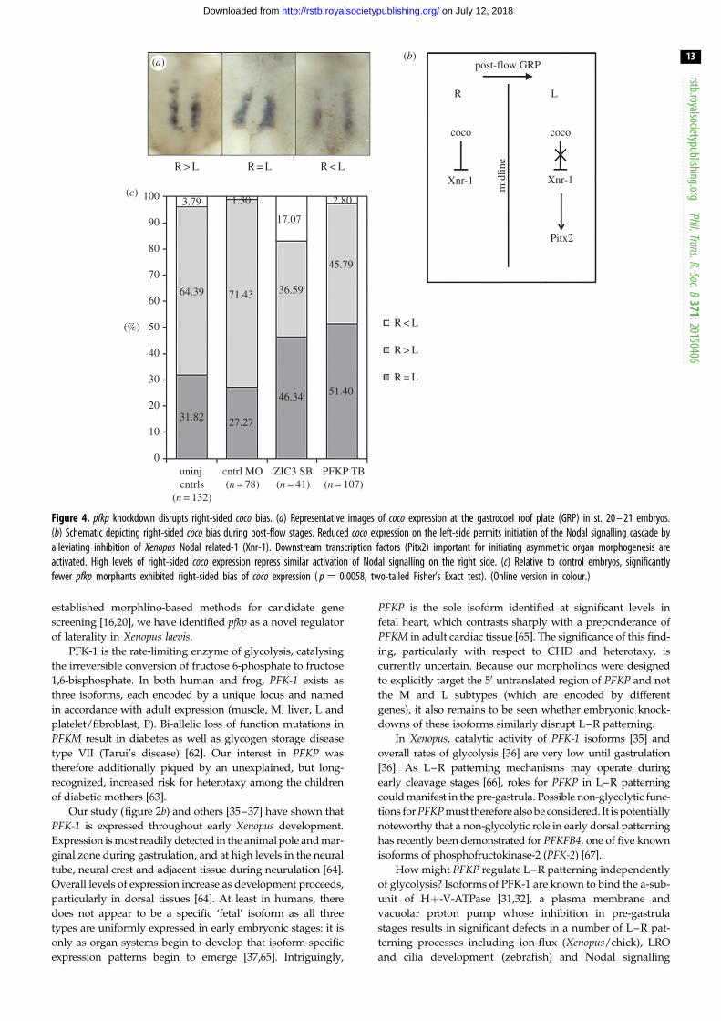
Nodal signalling pathways in the left lateral plate [27]. In this
setting, coco serves as a potent inhibitor of the Nodal ligand,
becoming increasingly restricted to the right side of the GRP
and functioning to prevent right-sided Nodal expression
during flow-relevant stages [38]. This right-sided bias is of criti-
cal importance for the laterality programme: without it, Nodal
signalling asymmetries are improperly established and resulting
embryos develop extensive laterality defects [39]. Consistent
with previous reports [38], we observed a right-sided cocoexpression bias in approximately 60% of untreated late-flow
stage embryos (stage 20–21; figure 4). This bias was significantly
reduced in pfkp morphants (44.8%, p ¼ 0.0058). Supporting func-
tional impact. The magnitude of this reduction was comparable
to that observed following knockdown of dnah9, which encodes
an axonemal molecular motor essential for GRP ciliary motility
and extracellular fluid flow [38]. On the basis of these morpho-
lino studies, we identify pfkp as a novel gene associated with
heterotaxy and conclude that the 175 kb CNV encompassing this
gene is the most likely explanation for the laterality phenotype
observed in our patient.
4. Discussion(a) The clinical importance of copy number variant
analysesWe have performed comprehensive genome-wide CNV ana-
lyses on a large cohort of 225 patients with heterotaxy and
CHD. These analyses identified clinically relevant CNVs in
approximately 20% of patients, including 7.1% of clear patho-
genicity, a diagnostic yield that indicates newborns with
heterotaxy should have clinical chromosome microarray test-
ing. Specific phenotypic abnormalities were not significant
predictors of identifying a disease causing CNV, with the
exception of abdominal SI (table 2). Future studies will be
important to determine whether specific subclasses of
CHD, such as d-TGA, are less likely to have CNV abnormal-
ities. In addition, the burden of common CNVs present in
individuals with heterotaxy should be further evaluated
since this has been a noted correlation in other diseases [15].
(b) Copy number variant analyses identify novelheterotaxy gene candidates
As the majority of heterotaxy cases remain unexplained, the
primary goal of study was to identify novel genetic variation
of pathogenic significance to this patient population. Exclud-
ing CNVs of clear pathogenicity (tables 3 and 4), we detected
35 unique CNVs of potential interest for functional testing.
Collectively, these variants encompassed 165 genes of diverse
biological and molecular ontology (electronic supplementary
material, table S2 and figure S1), including a subset with roles
in pathways and processes relevant to laterality. These genes
are individually described below.
The ‘nodal flow’ model of L–R patterning proposes that
the vertebrate laterality programme is initiated by asymmetric
activation of Nodal signalling in the left lateral plate shortly
after gastrulation. Evidence from multiple species has solidified
the importance of transient, ciliated organizers in establish-
ing and propagating these signalling asymmetries [25–29].
Mutations affecting ciliary genes have subsequently been
identified as a major cause of heterotaxy spectrum disorders
ranging from ‘classic’ heterotaxy (CHD with visceral situs
abnormalities) to primary ciliary dyskinesia (PCD) and other
ciliopathies [40]. There is significant overlap between PCD
and heterotaxy, with 6–12% of PCD patients having hetero-
taxy [41,42] and an unknown but significant percentage of
heterotaxy patients having PCD [42]. Results from our CNV
screens have identified rare variants encompassing a number
Tabl
e3.
Path
ogen
icch
rom
osom
eab
norm
alitie
sde
tecte
din
the
225
patie
nthe
tero
taxy
coho
rt.ab
d.,a
bdom
inal;
abnl
/abn
ls.,a
bnor
mal/
abno
rmali
ties;
ASD,
atria
lsep
tald
efec
t;AV
C,at
riove
ntric
ular
cana
l;bi
lat.,
bilat
eral;
CAVC
,com
plet
eat
riove
ntric
ular
cana
l;d-
TGA,
dext
ro-tr
ansp
ositi
onof
the
grea
tar
terie
s;DD
,dev
elopm
enta
ldela
y;di
l.,di
lated
;DILV
,dou
ble
inlet
right
vent
ricle;
DORV
,dou
ble
outle
trig
htve
ntric
le;HL
HS,h
ypop
lastic
lefth
eart
synd
rom
e;IV
C,in
ferio
rven
aca
va;l
-TGA
,lev
o-tra
nspo
sition
ofth
egr
eat
arte
ries;
Lt.,
left;
LV,l
eft
vent
ricle;
PA,p
ulm
onar
yat
resia
;PAP
VR,p
artia
lano
malo
uspu
lmon
ary
veno
usre
turn
;PDA
,pat
ent
ductu
sar
terio
sus;
PS,p
ulm
onic
steno
sis,R
t.,rig
ht;R
A,rig
htat
rium
;RV
,rig
htve
ntric
le;SI,
situs
inve
rsus;
SVC,
supe
riorv
ena
cava
;TAP
VR,t
otal
anom
alous
pulm
onar
yve
nous
retu
rn;U
PD,u
nipa
rent
aldi
som
y;VS
D,ve
ntric
ular
sept
alde
fect.
patie
ntge
nder
type
plat
form
a
cyto
gene
ticfin
ding
s(b
reak
poin
ts)b
inhe
ritan
ce/p
heno
type
illum
ina
SNP
arra
y
illum
ina
size
agile
ntol
igo
arra
yag
ilent
size
1M
gain
olig
ono
tana
lysed
nota
nalys
edar
r9q3
3.2-
q33.
3(12
5204
518-
1401
3890
1)x3
14.9
Mb
spor
adic/
l-TGA
,PA,
ASD,
VSD
2M
unba
lance
d
trans
loca
tion/
com
plex
rear
rang
emen
t
olig
ono
tana
lysed
nota
nalys
edar
r5q1
5q35
.3(9
4068
223-
1768
8617
1)x3
arr9
p24.
1p23
(844
2856
-
1222
3883
)x1
82M
b,
3.8
Mb
fam
ilial/d
-TGA
,CAV
C,VS
D,AS
D,as
plen
ia
3M
loss
1M
,olig
oar
r22q
11.1
q11.
21(1
5815
765-
1642
2571
)x1
600
kbar
r22q
11.1
q11.
21(1
5815
765-
1642
2571
)x1
600
kbsp
orad
ic/sit
usam
bigu
us,a
bd.S
I,VS
D,DO
RV,P
S,PA
,SVC
abnl
s.,
aspl
enia,
rena
labn
ls.,p
ulm
onar
yiso
mer
ism,h
eart
malr
otat
ion,
hypo
plas
ticpu
lmon
ary
arte
ry,c
entra
lgall
blad
der,
panc
reas
rota
ted
toRt
.,sto
mac
hro
tate
dto
Lt.,
hypo
plas
ticm
ultic
ystic
Rt.k
idne
y,un
desc
ende
dLt
.tes
ticle,
abse
ntRt
.tes
ticle.
4M
gain
;UPD
1M
arr2
p12p
11.2
(823
5045
3-
8491
2738
)x3,
arr1
1p14
.2q1
3.3(
2630
1682
-
7060
4313
)x2
hmz
2.6
Mb,
44.3
Mb
nota
nalys
edno
tana
lysed
fam
ilial/o
ligoh
ydra
mni
os,h
ypop
lastic
lung
s;AS
D,PA
PVR,
DORV
,
CAVC
,dex
troca
rdia;
UPD
leads
toho
moz
ygou
sBB
S1no
nsen
se
mut
ation
5F
mos
aicga
inan
dlo
ss1
M,o
ligo
arr7
p22.
3p21
.3(3
4332
-
1292
2987
)x1-
2
arr7
p21.
3p15
.1(1
2925
517-
2833
2937
)x1-
2hm
z
arr7
p15.
1p12
.3(2
8333
319-
4834
6413
)x1-
2
arr7
p12.
1(48
3467
04-
4889
8843
)x2-
3
arr7
p12.
1p11
.2(5
1513
509-
5636
2278
)x2-
3
12.9
Mb,
15.4
Mb,
20M
b,
552.
1kb
,
2.6
Mb
arr7
p22.
3p12
.3(1
3655
1-
4833
8627
)x1
arr7
p11.
2(53
9257
15-
5635
1979
)x3
48.2
Mb,
2.4
Mb
unkn
own/
SI;PD
A;Ka
rtage
ner’s
:im
mot
ilecil
iaby
nasa
lbiop
sy;
cons
angu
inity
(par
ents
are
first
cous
ins)
(Con
tinue
d.)
rstb.royalsocietypublishing.orgPhil.Trans.R.Soc.B
371:20150406
8
on July 12, 2018http://rstb.royalsocietypublishing.org/Downloaded from
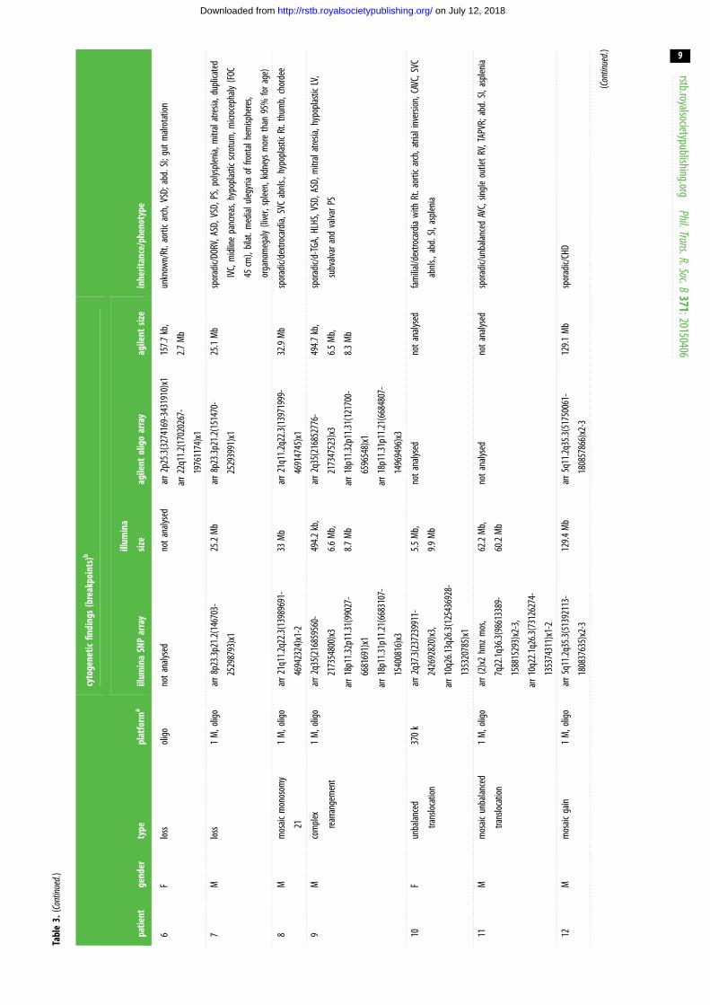
Tabl
e3.
(Con
tinue
d.)
patie
ntge
nder
type
plat
form
a
cyto
gene
ticfin
ding
s(b
reak
poin
ts)b
inhe
ritan
ce/p
heno
type
illum
ina
SNP
arra
y
illum
ina
size
agile
ntol
igo
arra
yag
ilent
size
6F
loss
olig
ono
tana
lysed
nota
nalys
edar
r2p2
5.3(
3274
169-
3431
910)
x1
arr2
2q11
.2(1
7020
267-
1976
1174
)x1
157.
7kb
,
2.7
Mb
unkn
own/
Rt.a
ortic
arch
,VSD
;abd
.SI;
gutm
alrot
ation
7M
loss
1M
,olig
oar
r8p2
3.3p
21.2
(146
703-
2529
8793
)x1
25.2
Mb
arr8
p23.
3p21
.2(1
5147
0-
2529
3991
)x1
25.1
Mb
spor
adic/
DORV
,ASD
,VSD
,PS,
polys
plen
ia,m
itral
atre
sia,d
uplic
ated
IVC,
mid
line
panc
reas
,hyp
oplas
ticsc
rotu
m,m
icroc
epha
ly(F
OC
45cm
),bi
lat.m
edial
uleg
yria
offro
ntal
hem
isphe
res,
orga
nom
egaly
(live
r,sp
leen,
kidne
ysm
ore
than
95%
fora
ge)
8M
mos
aicm
onos
omy
21
1M
,olig
oar
r21q
11.2
q22.
3(13
9896
91-
4694
2324
)x1-
2
33M
bar
r21q
11.2
q22.
3(13
9719
99-
4691
4745
)x1
32.9
Mb
spor
adic/
dext
roca
rdia,
SVC
abnl
s.,hy
popl
astic
Rt.t
hum
b,ch
orde
e
9M
com
plex
rear
rang
emen
t
1M
,olig
oar
r2q3
5(21
6859
560-
2173
5480
0)x3
arr1
8p11
.32p
11.3
1(99
027-
6681
691)
x1
arr1
8p11
.31p
11.2
1(66
8310
7-
1540
0816
)x3
494.
2kb
,
6.6
Mb,
8.7
Mb
arr2
q35(
2168
5277
6-
2173
4752
3)x3
arr1
8p11
.32p
11.3
1(12
1700
-
6596
548)
x1
arr1
8p11
.31p
11.2
1(66
8480
7-
1496
9496
)x3
494.
7kb
,
6.5
Mb,
8.3
Mb
spor
adic/
d-TG
A,HL
HS,V
SD,A
SD,m
itral
atre
sia,h
ypop
lastic
LV,
subv
alvar
and
valva
rPS
10F
unba
lance
d
trans
loca
tion
370
kar
r2q3
7.3(
2372
3991
1-
2426
9282
0)x3
,
arr1
0q26
.13q
26.3
(125
4369
28-
1353
2078
5)x1
5.5
Mb,
9.9
Mb
nota
nalys
edno
tana
lysed
fam
ilial/d
extro
card
iaw
ithRt
.aor
ticar
ch,a
trial
inve
rsion
,CAV
C,SV
C
abnl
s.,ab
d.SI,
aspl
enia
11M
mos
aicun
balan
ced
trans
loca
tion
1M
,olig
oar
r(2)
x2hm
zm
os,
7q22
.1q3
6.3(
9861
3389
-
1588
1529
3)x2
-3,
arr1
0q22
.1q2
6.3(
7312
6274
-
1353
7431
1)x1
-2
62.2
Mb,
60.2
Mb
nota
nalys
edno
tana
lysed
spor
adic/
unba
lance
dAV
C,sin
gle
outle
tRV,
TAPV
R;ab
d.SI,
aspl
enia
12M
mos
aicga
in1
M,o
ligo
arr5
q11.
2q35
.3(5
1392
113-
1808
3763
5)x2
-3
129.
4M
bar
r5q1
1.2q
35.3
(517
5006
1-
1808
5786
6)x2
-3
129.
1M
bsp
orad
ic/CH
D
(Con
tinue
d.)
rstb.royalsocietypublishing.orgPhil.Trans.R.Soc.B
371:20150406
9
on July 12, 2018http://rstb.royalsocietypublishing.org/Downloaded from
Tabl
e3.
(Con
tinue
d.)
patie
ntge
nder
type
plat
form
a
cyto
gene
ticfin
ding
s(b
reak
poin
ts)b
inhe
ritan
ce/p
heno
type
illum
ina
SNP
arra
y
illum
ina
size
agile
ntol
igo
arra
yag
ilent
size
13F
loss
1M
arr1
p34.
3p34
.1(3
6507
566-
4501
3872
)x2-
3
8.5
Mb
nota
nalys
edno
tana
lysed
spor
adic/
d-TG
A,VS
D,AS
D,DO
RV,m
od.d
il.RV
/RA,
mild
RV
hype
rtrop
hy,l
arge
tortu
ous
PDA,
exce
ssive
aorto
pulm
onar
y
colla
tera
ls,DD
(MRI
:2sm
allsit
esof
leuko
mala
ciain
whi
te
mat
tero
ffro
ntal
lobe
s)
a Fort
hepu
rpos
esof
this
and
allsu
bseq
uent
tabl
es,a
rray-
CGH
islis
ted
asol
igo
array
,whi
leSN
P-ar
rays
are
desig
nate
das
370
or1
Mde
pend
ing
onnu
mbe
rofi
nclu
ded
mar
kers
(370
000
versu
s1
000
000)
.b Lin
earp
ositi
ons
acco
rdin
gto
Build
36,h
g18.
Stan
dard
cyto
gene
ticno
men
clatu
reis
used
.
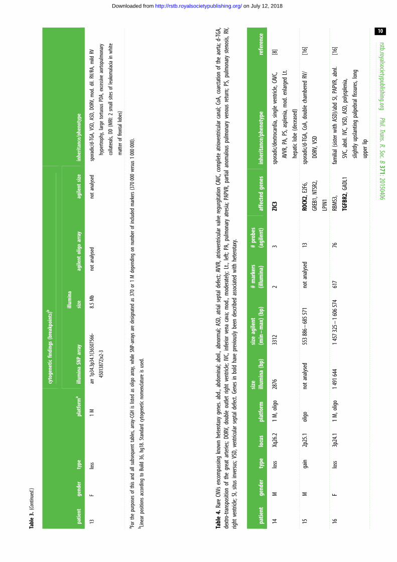
Tabl
e4.
Rare
CNVs
enco
mpa
ssin
gkn
own
hete
rota
xyge
nes.
abd.
,abd
omin
al;ab
nl.,
abno
rmal;
ASD,
atria
lsep
tald
efec
t;AV
VR,a
triov
entri
cular
valve
regu
rgita
tion
CAVC
,com
plet
eat
riove
ntric
ular
cana
l;Co
A,co
arcta
tion
ofth
eao
rta;d
-TGA
,de
xtro
-tran
spos
ition
ofth
egr
eat
arte
ries;
DORV
,dou
ble
outle
trig
htve
ntric
le;IV
C,in
ferio
rve
naca
va;m
od.,
mod
erat
ely;L
t.,lef
t;PA
,pul
mon
ary
atre
sia;P
APVR
,par
tiala
nom
alous
pulm
onar
yve
nous
retu
rn;P
S,pu
lmon
ary
steno
sis,R
V,rig
htve
ntric
le;SI,
situs
inve
rsus;
VSD,
vent
ricul
arse
ptal
defe
ct.Ge
nes
inbo
ldha
vepr
eviou
slybe
ende
scrib
edas
socia
ted
with
hete
rota
xy.
patie
ntge
nder
type
locu
spl
atfo
rmsiz
eill
umin
a(b
p)siz
eag
ilent
(min
–m
ax)(
bp)
#m
arke
rs(il
lum
ina)
#pr
obes
(agi
lent
)af
fect
edge
nes
inhe
ritan
ce/p
heno
type
refe
renc
e
14M
loss
Xq26
.21
M,o
ligo
2876
3312
23
ZIC3
spor
adic/
dext
roca
rdia,
singl
eve
ntric
le,CA
VC,
AVVR
,PA,
PS,a
splen
ia,m
od.e
nlar
ged
Lt.
hepa
ticlo
be(d
ecea
sed)
[8]
15M
gain
2p25
.1ol
igo
nota
nalys
ed55
388
6–68
557
1no
tana
lysed
13RO
CK2,
E2F6
,
GREB
1,NT
SR2,
LPIN
1
spor
adic/
d-TG
A,Co
A,do
uble
cham
bere
dRV
/
DORV
,VSD
[16]
16F
loss
3p24
.11
M,o
ligo
149
164
41
457
325–
160
657
461
776
RBM
S3,
TGFB
R2,G
ADL1
fam
ilial(
siste
rwith
ASD)
/abd
SI,PA
PVR,
abnl
.
SVC,
abnl
.IVC
,VSD
,ASD
,pol
yspl
enia,
sligh
tlyup
slant
ing
palp
ebra
lfiss
ures
,lon
g
uppe
rlip
[16]
rstb.royalsocietypublishing.orgPhil.Trans.R.Soc.B
371:20150406
10
on July 12, 2018http://rstb.royalsocietypublishing.org/Downloaded from
100
heterotaxy
isolated situs anomaly
situs inversus
normal situs
90807060
(%) 50403020100
st. 3
OD
Cpf
kp
st. 6.5 st. 7 st. 10.5 st. 18 st. 24 st. 32
Uninj.Cntrls
(n = 867)
Cntrl MO(3 ng)
(n = 104)
zic3 SB(5 ng)
(n = 98)
pfkp TB(1.25 ng)(n = 196)
pfkp SB(25 ng)
(n = 136)
pitrm 1(2.5 ng)(n = 27)
600l kb+
WT
PFKP SB
E7
E7
E7
E8 E9
E9
E9E8
SB Uninj
500400300200
(b)
(a)
(c)
(d)
Figure 2. pfkp but not pitrm1 Xenopus laevis morphants develop organ situs defects. (a) Genes in CNV interval. Source: USCS genome browser (https://genome.ucsc.edu/). (b) RT-PCR results demonstrate that pfkp is expressed throughout critical L – R patterning stages. ODC, ornithine decarboxylase. (c) pfkp knockdown in 2-cellembryos causes heterotaxy in Xenopus. Situs defects in st. 47 morphants are shown relative to zic3 control morphants and uninjected controls. (d ) Confirmation ofexon 8 deletion in pfkp SB morphants by RT-PCR targeting the exon 8 – intron 8 splice junction. Upper schematic: small horizontal arrows denote primer annealingsites. The horizontal line without an arrowhead denotes the SB morpholino target site. The severely truncated exon 8 (see §3) is represented by cross-hatching.Lower panel (RT-PCR): large arrowhead denotes the pfkp deletion fragment. SB, pfkp splice block. (Online version in colour.)
rstb.royalsocietypublishing.orgPhil.Trans.R.Soc.B
371:20150406
11
on July 12, 2018http://rstb.royalsocietypublishing.org/Downloaded from
of genes with cilia-related functions, including TTC21B,CEP290, TTBK2 and CFAP126. Two of these, CEP290 and
TTC21B, are recognized causes of human ciliopathies and are
known to play critical roles in cilia assembly and transport
[43,44]. Mutations in TTC21B, which encodes the retrograde
intraflagellar transport protein, THM1/IFT139, cause both
nephronophthisis 12 (end-stage renal disease) and short-rib
thoracic dysplasia 4 (skeletal anomalies with or without
polydactyly) [45]. Similarly, TTC21B knockdown in mice is
characterized by preaxial polydactyly, cleft palate, micro-
ophthalmia and brain abnormalities. Retrograde ciliary
transport is disrupted, leading to impaired cilia growth and
downstream Sonic hedgehog (Shh) signalling [43]. The pheno-
typic spectrum resulting from CEP290 mutations is even more
striking as pathogenic variants have been associated with a
diverse array of ciliopathies without any obvious genotype–
phenotype correlations [44]. Functionally, the CEP290 protein
localizes to both the cilia transition zone [46] and to centriolar
satellites, where it interacts with a number of other ciliary
proteins required for cilia formation [47]. Knockdown of
CEP290 in cultured mammalian cells disrupts satellite localiz-
ation, preventing assembly of a functional cilium [47]. Similar
disruption of centriolar satellites by morpholino-mediated
knockdown of pericentrin ( pcm1) in zebrafish yielded inverted
heart looping in nearly 50% of morphant embryos [47]. These
results suggest pathogenic potential for CEP290 mutations via
loss of centrosomal protein targeting and ciliary assembly.
While CEP290 mutations have not yet been reported as
causes of human heterotaxy, at least one patient with SI has
been identified to carry a CEP290 mutation [48].
Two other ciliary genes, TTBK2 and CFAP126, were ident-
ified in CNVs in the heterotaxy cohort. The first, TTBK2,encodes a serine/threonine kinase essential for intraflagellar
transport (IFT) protein recruitment. Its localization to cen-
trioles is an initiating step of ciliogenesis and is required to
promote elongation of the ciliary axoneme [49]. Like
TTC21B and CEP290, mutations in TTBK2 prevent formation
of a functional cilium and are a recognized cause of disease in
both humans (spinocerebellar ataxia 11) and bartleby (bby)
mutant mice. TTBK2 mutations have yet to be reported
among patients with heterotaxy; however, mice homozygous
for bby mutations exhibit randomized heart looping indicative
of impaired L–R patterning, in addition to other defects stem-
ming from disrupted ciliogenesis (holoprosencephaly, limb
defects, twisted body axis). Comparatively less is known
about CFAP126, a newly characterized protein important for
basal body docking and cilia positioning [50]. Nevertheless,
expression has been noted at the mouse embryonic node as
early as E7.75, placing CFAP126 at the correct location and
time for impacting laterality [51].
In addition to these ciliary genes, two rare CNVs encom-
passing members of the TGFb superfamily were identified.
The first, a duplication involving the Nodal signalling modu-
lator, NOMO3, was detected in a male patient with sporadic
heterotaxy (Patient 19). NOMO3 exists as one of three highly
similar genes on chromosome 16p12-p13 and is thought to
act as a direct Nodal signalling antagonist, acting indepen-
dently of other Nodal modulating pathways [52]. The
second CNV, a duplication identified in a male patient
with familial heterotaxy (Patient 40), encompassed LTBP1,
a TGFb binding protein with roles in TGFb1 assembly,
secretion, and targeting. Deletions involving LTBP1 have
been previously associated with outflow tract (OFT) defects
[53], but have not yet been reported among patients with het-
erotaxy. Intriguingly, experiments in Xenopus have identified
LTBP1 as a potent activator of Activin and Nodal signalling,
(%)
100
90
80
70
60
50
40
30
20
10
0
(%)
100
90
80
70
60
50
40
30
20
10
0
uninj.cntrls
(n = 236)
PFKP TB(1.25 ng)(n = 196)
PFKP SB(25 ng)
(n = 136)
PFKP TB(0.625 ng)
(n = 76)
PFKP SB(12.5 ng)(n = 62)
PFKP TB(0.625 ng)+SB 1/2(12.5 ng)(n = 95)
uninj. cntrls(n = 204)
PFKP SB(25 ng)
(n = 136}
mRNA(1000 pg)(n = 100)
PFKP SB(25 ng) +mRNA
(1000 pg)(n = 105)
heterotaxy
isolated situs anomaly
situs inversus
normal situs
(b)
(a)
Figure 3. Synergy and rescue experiments support MO specificity. (a) Sub-threshold dosages of pfkp TB and SB MOs yielded a higher proportion of embryos withsitus defects when injected together than when injected alone. (b) Partial rescue of organ situs in SB morphants co-injected with WT human PFKP mRNA. SB, spliceblock; TB, translation block.
rstb.royalsocietypublishing.orgPhil.Trans.R.Soc.B
371:20150406
12
on July 12, 2018http://rstb.royalsocietypublishing.org/Downloaded from
indicating potential for involvement in L–R relevant
pathways [54].
Rare CNVs involving four distinct microRNAs (miR-148a,
miR-455, miR-484 and miR-2117) were also identified in the
heterotaxy cohort. Considering known functions in the context
of L–R patterning, miR-418a is the most notable as it is a direct
inhibitor of both ACVR1, an activin receptor [55] and ROCK1, a
rho-associated kinase [56]. Although neither protein has been
directly associated with laterality, family members of both
(ACVR2B and ROCK2) are known genetic causes of heterotaxy
in human patients [16,57].
A total of 10 rare CNVs were identified at loci previously
associated with CHD (electronic supplementary material,
table S3). To the best of our knowledge, only one of these, a
large 1.7–2.6 Mb 2q13 duplication detected by oligoarray in a
male patient with sporadic heterotaxy and complex CHD
(Patient 29), affects a locus previously associated with hetero-
taxy [24]. The duplicated region encompassed 10 genes
(electronic supplementary material, table S1) and was identical
in gene content to a 1.62 Mb deletion detected in a patient with
a similar laterality phenotype [24]. Genomic imbalances
spanning approximately 1.7 Mb and encompassing the same
10 genes have been described in a number of other patients
with CHD but who are otherwise lacking overt situs abnorm-
alities [24,58–61]. No specific microduplication/deletion
syndrome has been defined; however, brain abnormalities
were suggestive of Joubert syndrome in one individual [60].
Recent knockdown studies in zebrafish have identified
FBLN7 and TMEM87B as the genes most likely to be causative
for CHD; however, their exact roles in cardiac development
have yet to be determined [61]. In our patient, a second,
larger CNV was also identified upstream of this region and
included an additional 18 genes of uncertain pathogenic sig-
nificance. Predicted breakpoints of the two intervals were
non-overlapping but were in immediate proximity, suggesting
the possibility of a single duplication event.
(c) PFKP as a novel cause of heterotaxyPFKP, encoding the platelet isoform of the glycolytic enzyme,
phosphofructokinase-1 (PFK-1), was among the top hetero-
taxy candidates genes identified in the cohort. Using
100
90
80
70
60
(%) 50
40
30
20
10
0uninj.cntrls
(n = 132)
31.82 27.27
46.34 51.40
36.59
R < L
R > L R = L R < L
R
post-flow GRP
coco
mid
line
Xnr-1 Xnr-1
Pitx2
coco
L
R > L
R = L
45.79
17.07
1.303.79
64.39 71.43
2.80
cntrl MO(n = 78)
ZIC3 SB(n = 41)
PFKP TB(n = 107)
(c)
(b)(a)
Figure 4. pfkp knockdown disrupts right-sided coco bias. (a) Representative images of coco expression at the gastrocoel roof plate (GRP) in st. 20 – 21 embryos.(b) Schematic depicting right-sided coco bias during post-flow stages. Reduced coco expression on the left-side permits initiation of the Nodal signalling cascade byalleviating inhibition of Xenopus Nodal related-1 (Xnr-1). Downstream transcription factors (Pitx2) important for initiating asymmetric organ morphogenesis areactivated. High levels of right-sided coco expression repress similar activation of Nodal signalling on the right side. (c) Relative to control embryos, significantlyfewer pfkp morphants exhibited right-sided bias of coco expression ( p ¼ 0.0058, two-tailed Fisher’s Exact test). (Online version in colour.)
rstb.royalsocietypublishing.orgPhil.Trans.R.Soc.B
371:20150406
13
on July 12, 2018http://rstb.royalsocietypublishing.org/Downloaded from
established morphlino-based methods for candidate gene
screening [16,20], we have identified pfkp as a novel regulator
of laterality in Xenopus laevis.
PFK-1 is the rate-limiting enzyme of glycolysis, catalysing
the irreversible conversion of fructose 6-phosphate to fructose
1,6-bisphosphate. In both human and frog, PFK-1 exists as
three isoforms, each encoded by a unique locus and named
in accordance with adult expression (muscle, M; liver, L and
platelet/fibroblast, P). Bi-allelic loss of function mutations in
PFKM result in diabetes as well as glycogen storage disease
type VII (Tarui’s disease) [62]. Our interest in PFKP was
therefore additionally piqued by an unexplained, but long-
recognized, increased risk for heterotaxy among the children
of diabetic mothers [63].
Our study (figure 2b) and others [35–37] have shown that
PFK-1 is expressed throughout early Xenopus development.
Expression is most readily detected in the animal pole and mar-
ginal zone during gastrulation, and at high levels in the neural
tube, neural crest and adjacent tissue during neurulation [64].
Overall levels of expression increase as development proceeds,
particularly in dorsal tissues [64]. At least in humans, there
does not appear to be a specific ‘fetal’ isoform as all three
types are uniformly expressed in early embryonic stages: it is
only as organ systems begin to develop that isoform-specific
expression patterns begin to emerge [37,65]. Intriguingly,
PFKP is the sole isoform identified at significant levels in
fetal heart, which contrasts sharply with a preponderance of
PFKM in adult cardiac tissue [65]. The significance of this find-
ing, particularly with respect to CHD and heterotaxy, is
currently uncertain. Because our morpholinos were designed
to explicitly target the 50 untranslated region of PFKP and not
the M and L subtypes (which are encoded by different
genes), it also remains to be seen whether embryonic knock-
downs of these isoforms similarly disrupt L–R patterning.
In Xenopus, catalytic activity of PFK-1 isoforms [35] and
overall rates of glycolysis [36] are very low until gastrulation
[36]. As L–R patterning mechanisms may operate during
early cleavage stages [66], roles for PFKP in L–R patterning
could manifest in the pre-gastrula. Possible non-glycolytic func-
tions for PFKP must therefore also be considered. It is potentially
noteworthy that a non-glycolytic role in early dorsal patterning
has recently been demonstrated for PFKFB4, one of five known
isoforms of phosphofructokinase-2 (PFK-2) [67].
How might PFKP regulate L–R patterning independently
of glycolysis? Isoforms of PFK-1 are known to bind the a-sub-
unit of Hþ-V-ATPase [31,32], a plasma membrane and
vacuolar proton pump whose inhibition in pre-gastrula
stages results in significant defects in a number of L–R pat-
terning processes including ion-flux (Xenopus/chick), LRO
and cilia development (zebrafish) and Nodal signalling
rstb.royalsocietypublishing.orgPhil.Trans.R.Soc.B
371:20150406
14
on July 12, 2018http://rstb.royalsocietypublishing.org/Downloaded from
(chick/zebrafish) [33,68]. Together with observations from
yeast that PFK-1 is required to maintain activity of the
HþV-ATPase proton pump and that even a catalytically inac-
tive PFK-1 is capable of completing this function [31], a
reasonable hypothesis is that PFKP knockdown may impact
L–R patterning through loss of a functionally critical inter-
action with the Hþ-V-ATPase a-subunit. As recent zebrafish
studies indicate a primary role for Hþ-V-ATPase in KV for-
mation [68], it is possible that PFKP may impact L–R
patterning in a similar manner. Interestingly, a rare duplication
involving the gene ATP6V1G1, which encodes the G-subunit
of Hþ-V-ATPAase, was also identified in a male heterotaxy
patient in our cohort (Patient 41). Like the a-subunit, the
G-subunit contributes to the peripheral stator stalk, the
non-rotating connection between the V1 and Vo halves of
the ATPase [69]. Two of three dimers composed of E- and G-
subunits attach to the a-subunit at its amino terminal domain,
a site thought to be of importance for a-subunit function [70].
(d) Study limitationsSample collection from trios (proband and parents) was not
feasible in many cases. As a result, determinations of de novoinheritance were not possible. Similarly, while a few instances
of copy number mosaicism were identified, direct comparisons
between mosaic and non-mosaic CNV carriers were prevented
by the rarity of individual CNVs. Cut-offs for CNV calling
were also set at a minimum coverage of three markers/
probes per CNV, restricting downstream genetic and func-
tional analyses to variants exceeding these minimum size
thresholds. While more difficult to detect reliably by microar-
ray approaches, small exonic CNVs of 1–30 kb have been
suggested to contribute to susceptibility of some genetic
diseases [71]. Finally, analyses were restricted to CNVs invol-
ving coding regions of at least one gene, ignoring variants
solely impacting intronic or regulatory sequences. Genetic vari-
ation in non-coding regions, while typically of less obvious
functional significance, has been linked to genetic disease
[72–74] but would not have been considered in our study.
Our analyses of PFKP as an L–R patterning candidate
were limited by an inability to definitively verify isoform-
specific knockdown by Western blotting as the M and L
isoforms are of similar size and commercially available
antibodies demonstrate high likelihood for subtype cross-
reactivity (data not shown). Nevertheless, confidence in the
specificity of TB knockdown was provided by replication of
the TB morphant phenotype in SB morphants and demon-
stration that sub-threshold dosages of both morpholinos
synergized to produce organ situs defects when used in
combination, but not in isolation.
5. SummaryIn summary, we have performed CNV analyses on a cohort of
225 patients with heterotaxy and heterotaxy-spectrum CHDs
and identified CNVs of potential pathogenic significance in a
large proportion (20.4%). Detected CNVs ranged in size and
complexity and collectively encompassed a number of genes
with known or suspected functions in L–R patterning develop-
mental programmes. Using rigorous morpholino-based
studies, a role for the platelet isoform of phosphofructoki-
nase-1, pfkp, in heterotaxy pathogenesis was confirmed in
Xenopus laevis. Future work using both animal and in vitro cul-
ture systems will help to tease out temporal requirements as
well as precise molecular and developmental functions for
pfkp in L–R patterning.
Ethics. All studies were approved by the Institutional Review Board atCincinnati Children’s Hospital Medical Center (CCHMC) andinformed consent was obtained from all study participants. Animalstudies were carried out in compliance with local and federalanimal use guidelines.
Authors’ contributions. J.R.C. participated in study design, carried out themolecular and functional laboratory work, completed data and stat-istical analyses, and drafted the manuscript. M.T. carried out samplegenotyping and participated in CNV analyses; C.S. and M.R. assistedwith CNV calling; J.W.B. assisted in patient recruitment and pheno-typing and provided DNA samples for genotyping, T.A.S. andS.R.L. reviewed and provided interpretation for all CNV data.S.M.W. conceived, designed and supervised the study. All authorsgave final approval for publication.
Competing interests. The authors have no competing interests.
Funding. This work was supported by funding from the BurroughsWelcome Fund Clinical Scientist Award in Translational Research(grant no. 1008496) and March of Dimes Research Foundation(grant nos. 1FY10-401 and 6-FY13-167) (to S.M.W.).
Acknowledgements. We thank the patients and families for their participationand John Wells for independently scoring coco hybridized embryos.
References
1. Lin AE, Ticho BS, Houde K, Westgate MN, Holmes LB.2000 Heterotaxy: associated conditions and hospital-based prevalence in newborns. Genet. Med. 2, 157 –172. (doi:10.109700125817-2000 05000-00002)
2. Lin AE et al. 2014 Laterality defects in the nationalbirth defects prevention study (1998 – 2007): birthprevalence and descriptive epidemiology.Am. J. Med. Genet. Part A 164A, 2581 – 2591.(doi:10.1002/ajmg.a.36695)
3. Kim SJ, Kim WH, Lim HG, Lee JY. 2008 Outcome of200 patients after an extracardiac Fontan procedure.J. Thorac. Cardiovasc. Surg. 136, 108 – 116. (doi:10.1016/j.jtcvs.2007.12.032)
4. Swisher M, Jonas R, Tian X, Lee ES, Lo CW,Leatherbury L. 2011 Increased postoperative and
respiratory complications in patients with congenitalheart disease associated with heterotaxy. J. Thorac.Cardiovasc. Surg. 141, 637 – 644. (doi:10.1016/j.jtcvs.2010.07.082)
5. Bartz PJ et al. 2006 Early and late results of themodified fontan operation for heterotaxy syndrome 30years of experience in 142 patients. J. Am. Coll. Cardiol.48, 2301 – 2305. (doi:10.1016/j.jacc.2006.07.053)
6. Oyen N, Poulsen G, Boyd HA, Wohlfahrt J, JensenPK, Melbye M. 2009 Recurrence of congenital heartdefects in families. Circulation 120, 295 – 301.(doi:10.1161/CIRCULATIONAHA.109.857987)
7. Zhu L, Belmont JW, Ware SM. 2006 Genetics ofhuman heterotaxias. Eur. J. Hum. Genet. 14,17 – 25. (doi:10.1038/sj.ejhg.5201506)
8. Cowan J, Tariq M, Ware SM. 2014 Genetic andfunctional analyses of ZIC3 variants in congenitalheart disease. Hum. Mutat. 35, 66 – 75. (doi:10.1002/humu.22457)
9. Ware SM, Peng J, Zhu L, Fernbach S, Colicos S,Casey B, Towbin J, Belmont JW. 2004 Identificationand functional analysis of ZIC3 mutations inheterotaxy and related congenital heart defects.Am. J. Hum. Genet. 74, 93 – 105. (doi:10.1086/380998)
10. Sutherland MJ, Wang S, Quinn ME, Haaning A, WareSM. 2013 Zic3 is required in the migrating primitivestreak for node morphogenesis and left – rightpatterning. Hum. Mol. Genet. 22, 1913 – 1923.(doi:10.1093/hmg/ddt001)
rstb.royalsocietypublishing.orgPhil.Trans.R.Soc.B
371:20150406
15
on July 12, 2018http://rstb.royalsocietypublishing.org/Downloaded from
11. Mohapatra B et al. 2009 Identification andfunctional characterization of NODAL rare variants inheterotaxy and isolated cardiovascularmalformations. Hum. Mol. Genet. 18, 861 – 871.(doi:10.1093/hmg/ddn411)
12. Roessler E et al. 2008 Reduced NODAL signalingstrength via mutation of several pathway membersincluding FOXH1 is linked to human heart defectsand holoprosencephaly. Am. J. Hum. Genet. 83,18 – 29. (doi:10.1016/j.ajhg.2008.05.012)
13. Girirajan S, Campbell CD, Eichler EE. 2011 Humancopy number variation and complex geneticdisease. Annu. Rev. Genet. 45, 203 – 226. (doi:10.1146/annurev-genet-102209-163544)
14. Zhang F, Gu W, Hurles ME, Lupski JR. 2009 Copynumber variation in human health, disease, andevolution. Annu. Rev. Genomics Hum. Genet.10, 451 – 481. (doi:10.1146/annurev.genom.9.081307.164217)
15. Lander J, Ware SM. 2014 Copy number variation incongenital heart defects. Curr. Genet. Med. Rep. 2,168 – 178. (doi:10.1007/s40142-014-0049-3)
16. Fakhro KA, Choi M, Ware SM, Belmont JW, TowbinJA, Lifton RP, Khokha MK, Brueckner M. 2011 Rarecopy number variations in congenital heart diseasepatients identify unique genes in left – rightpatterning. Proc. Natl Acad. Sci. USA 108,2915 – 2920. (doi:10.1073/pnas.1019645108)
17. Rigler SL et al. 2015 Novel copy-number variants ina population-based investigation of classicheterotaxy. Genet. Med. 17, 348 – 357. (doi:10.1038/gim.2014.112)
18. Zarrei M, MacDonald JR, Merico D, Scherer SW. 2015A copy number variation map of the humangenome. Nat. Rev. Genet. 16, 172 – 183. (doi:10.1038/nrg3871)
19. Boone PM et al. 2010 Detection of clinically relevantexonic copy-number changes by array CGH. Hum.Mutat. 31, 1326 – 1342. (doi:10.1002/humu.21360)
20. Cast AE, Gao C, Amack JD, Ware SM. 2012 Anessential and highly conserved role for Zic3 in left –right patterning, gastrulation and convergentextension morphogenesis. Dev. Biol. 364, 22 – 31.(doi:10.1016/j.ydbio.2012.01.011)
21. Nieuwkoop P, Faber J. 1994 Normal table ofXenopus laevis. New York, NY: Garland Publishing.
22. Heasman J, Kofron M, Wylie C. 2000 b-cateninsignaling activity dissected in the early Xenopusembryo: a novel antisense approach. Dev. Biol. 222,124 – 134. (doi:10.1006/dbio.2000.9720)
23. Sive HL, Grainger RM, Harland RM. 2000 Earlydevelopment of Xenopus laevis: a laboratorymanual. 2000 ed. Cold Spring Harbor, NY: ColdSpring Harbor Press.
24. Rudd MK, Keene J, Bunke B, Kaminsky EB, AdamMP, Mulle JG, Ledbetter DH, Martin CL. 2009Segmental duplications mediate novel, clinicallyrelevant chromosome rearrangements. Hum. Mol.Genet. 18, 2957 – 2962. (doi:10.1093/hmg/ddp233)
25. Essner JJ, Amack JD, Nyholm MK, Harris EB, Yost HJ.2005 Kupffer’s vesicle is a ciliated organ ofasymmetry in the zebrafish embryo that initiatesleft – right development of the brain, heart and
gut. Development 132, 1247 – 1260. (doi:10.1242/dev.01663)
26. Nonaka S, Tanaka Y, Okada Y, Takeda S, Harada A,Kanai Y, Kido M, Hirokawa N. 1998 Randomizationof left – right asymmetry due to loss of nodal ciliagenerating leftward flow of extraembryonic fluid inmice lacking KIF3B motor protein. Cell 95,829 – 837. (doi:10.1016/S0092-8674(00)81705-5)
27. Schweickert A, Weber T, Beyer T, Vick P, Bogusch S,Feistel K, Blum M. 2007 Cilia-driven leftward flowdetermines laterality in Xenopus. Curr. Biol. 17,60 – 66. (doi:10.1016/j.cub.2006.10.067)
28. Sulik K, Dehart DB, Iangaki T, Carson JL, Vrablic T,Gesteland K, Schoenwolf GC. 1994 Morphogenesisof the murine node and notochordal plate.Dev. Dyn. 201, 260 – 278. (doi:10.1002/aja.1002010309)
29. Vick P, Schweickert A, Weber T, Eberhardt M, MenclS, Shcherbakov D, Beyer T, Blum M. 2009 Flow onthe right side of the gastrocoel roof plate isdispensable for symmetry breakage in the frogXenopus laevis. Dev. Biol. 331, 281 – 291. (doi:10.1016/j.ydbio.2009.05.547)
30. Blum M, Beyer T, Weber T, Vick P, Andre P, Bitzer E,Schweickert A. 2009 Xenopus, an ideal modelsystem to study vertebrate left – right asymmetry.Dev. Dyn. 238, 1215 – 1225. (doi:10.1002/dvdy.21855)
31. Su Y, Blake-Palmer KG, Sorrell S, Javid B, Bowers K,Zhou A, Chang SH, Qamar S, Karet FE. 2008 HumanHþATPase a4 subunit mutations causing renaltubular acidosis reveal a role for interactionwith phosphofructokinase-1. Am. J. Physiol. RenalPhysiol. 295, F950 – F958. (doi:10.1152/ajprenal.90258.2008)
32. Su Y, Zhou A, Al-Lamki RS, Karet FE. 2003 The a-subunit of the V-type Hþ-ATPase interacts withphosphofructokinase-1 in humans. J. Biol. Chem.278, 20 013 – 20 018. (doi:10.1074/jbc.M210077200)
33. Adams DS, Robinson KR, Fukumoto T, Yuan S,Albertson RC, Yelick P, Kuo L, McSweeney M, LevinM. 2006 Early, Hþ-V-ATPase-dependent proton fluxis necessary for consistent left – right patterning ofnon-mammalian vertebrates. Development 133,1657 – 1671. (doi:10.1242/dev.02341)
34. Branford WW, Essner JJ, Yost HJ. 2000 Regulation ofgut and heart left – right asymmetry by context-dependent interactions between Xenopus lefty andBMP4 signaling. Dev. Biol. 223, 291 – 306. (doi:10.1006/dbio.2000.9739)
35. Raddatz E, Lovtrup-Rein H. 1986 Changes in activityof the regulatory glycolytic enzymes and of thepyruvate-dehydrogenase complex during thedevelopment of Xenopus laevis. Exp. Cell Biol. 54,53 – 60. (doi:10.1159/000163344)
36. Dworkin MB, Dworkin-Rastl E. 1991 Carbonmetabolism in early amphibian embryos. TrendsBiochem. Sci. 16, 229 – 234. (doi:10.1016/0968-0004(91)90091-9)
37. Davidson M, Collins M, Byrne J, Vora S. 1983Alterations in phosphofructokinase isoenzymesduring early human development. Establishment of
adult organ-specific patterns. Biochem. J. 214,703 – 710. (doi:10.1042/bj2140703)
38. Schweickert A, Vick P, Getwan M, Weber T,Schneider I, Eberhardt M, Beyer T, Pachur A, BlumM. 2010 The nodal inhibitor Coco is a critical targetof leftward flow in Xenopus. Curr. Biol. 20,738 – 743. (doi:10.1016/j.cub.2010.02.061)
39. Blum M, Schweickert A, Vick P, Wright CV, DanilchikMV. 2014 Symmetry breakage in the vertebrateembryo: when does it happen and how does itwork? Dev. Biol. 393, 109 – 123. (doi:10.1016/j.ydbio.2014.06.014)
40. Sutherland MJ, Ware SM. 2009 Disorders of left –right asymmetry: heterotaxy and situs inversus.Am. J. Med. Genet. 151C, 307 – 317. (doi:10.1002/ajmg.c.30228)
41. Kennedy MP et al. 2007 Congenital heart diseaseand other heterotaxic defects in a large cohort ofpatients with primary ciliary dyskinesia. Circulation115, 2814 – 2821. (doi:10.1161/CIRCULATIONAHA.106.649038)
42. Shapiro AJ et al. 2014 Laterality defects other thansitus inversus totalis in primary ciliary dyskinesia:insights into situs ambiguus and heterotaxy. Chest146, 1176 – 1186. (doi:10.1378/chest.13-1704)
43. Tran PV et al. 2008 THM1 negatively modulatesmouse sonic hedgehog signal transduction andaffects retrograde intraflagellar transport in cilia.Nat. Genet. 40, 403 – 410. (doi:10.1038/ng.105)
44. Coppieters F, Lefever S, Leroy BP, De Baere E. 2010CEP290, a gene with many faces: mutationoverview and presentation of CEP290base. Hum.Mutat. 31, 1097 – 1108. (doi:10.1002/humu.21337)
45. Davis EE et al. 2011 TTC21B contributes both causaland modifying alleles across the ciliopathyspectrum. Nat. Genet. 43, 189 – 196. (doi:10.1038/ng.756)
46. Craige B, Tsao CC, Diener DR, Hou Y, Lechtreck KF,Rosenbaum JL, Witman GB. 2010 CEP290 tethersflagellar transition zone microtubules to themembrane and regulates flagellar protein content.J. Cell Biol. 190, 927 – 940. (doi:10.1083/jcb.201006105)
47. Stowe TR, Wilkinson CJ, Iqbal A, Stearns T. 2012The centriolar satellite proteins Cep72 and Cep290interact and are required for recruitment of BBSproteins to the cilium. Mol. Biol. Cell. 23,3322 – 3335. (doi:10.1091/mbc.E12-02-0134)
48. Brancati F et al. 2007 CEP290 mutations arefrequently identified in the oculo-renal form ofJoubert syndrome-related disorders. Am. J. Hum.Genet. 81, 104 – 113. (doi:10.1086/519026)
49. Goetz SC, Liem Jr KF, Anderson KV. 2012 Thespinocerebellar ataxia-associated gene Tau tubulinkinase 2 controls the initiation of ciliogenesis. Cell151, 847 – 858. (doi:10.1016/j.cell.2012.10.010)
50. Gegg M, Bottcher A, Burtscher I, Hasenoeder S, VanCampenhout C, Aichler M, Walch A, Grant SG,Lickert H. 2014 Flattop regulates basal body dockingand positioning in mono- and multiciliated cells.eLife 3, e03842. (doi:10.7554/eLife.03842)
51. Lange A, Gegg M, Burtscher I, Bengel D, KremmerE, Lickert H. 2012 Fltp(T2AiCre): a new knock-in
rstb.royalsocietypublishing.orgPhil.Trans.R.Soc.B
371:20150406
16
on July 12, 2018http://rstb.royalsocietypublishing.org/Downloaded from
mouse line for conditional gene targeting indistinct mono- and multiciliated tissues.Differentiation. 83, S105 – S113. (doi:10.1016/j.diff.2011.11.003)
52. Haffner C, Frauli M, Topp S, Irmler M, Hofmann K,Regula JT, Bally-Cuif L, Haass C. 2004 Nicalin and itsbinding partner Nomo are novel Nodal signalingantagonists. EMBO J. 23, 3041 – 3050. (doi:10.1038/sj.emboj.7600307)
53. Todorovic V et al. 2007 Long form of latent TGF-bbinding protein 1 (Ltbp1 L) is essential for cardiacoutflow tract septation and remodeling.Development 134, 3723 – 3732. (doi:10.1242/dev.008599)
54. Altmann CR, Chang C, Munoz-Sanjuan I, Bell E,Heke M, Rifkin DB, Brivanlou AH. 2002 The latent-TGFb-binding-protein-1 (LTBP-1) is expressed inthe organizer and regulates nodal and activinsignaling. Dev. Biol. 248, 118 – 127. (doi:10.1006/dbio.2002.0716)
55. Song H, Wang Q, Wen J, Liu S, Gao X, Cheng J,Zhang D. 2012 ACVR1, a therapeutic target offibrodysplasia ossificans progressiva, is negativelyregulated by miR-148a. AAPG Bull. 13, 2063 – 2077.(doi:10.3390/ijms13022063)
56. Zhang J, Ying ZZ, Tang ZL, Long LQ, Li K. 2012MicroRNA-148a promotes myogenic differentiationby targeting the ROCK1 gene. J. Biol. Chem. 287, 21093 – 21 101. (doi:10.1074/jbc.M111.330381)
57. Kosaki R, Gebbia M, Kosaki K, Lewin M, Bowers P,Towbin JA, Casey B. 1999 Left – right axismalformations associated with mutations inACVR2B, the gene for human activin receptor typeIIB. Am. J. Med. Genet. 82, 70 – 76. (doi:10.1002/(SICI)1096-8628(19990101)82:1,70::AID-AJMG14.3.0.CO;2-Y)
58. Cooper GM et al. 2011 A copy number variationmorbidity map of developmental delay. Nat. Genet.43, 838 – 846. (doi:10.1038/ng.909)
59. Soemedi R et al. 2012 Contribution of global rarecopy-number variants to the risk of sporadiccongenital heart disease. Am. J. Hum. Genet. 91,489 – 501. (doi:10.1016/j.ajhg.2012.08.003)
60. Yu HE, Hawash K, Picker J, Stoler J, Urion D, Wu BL,Shen Y. 2012 A recurrent 1.71 Mb genomicimbalance at 2q13 increases the risk ofdevelopmental delay and dysmorphism. Clin. Genet.81, 257 – 264. (doi:10.1111/j.1399-0004.2011.01637.x)
61. Russell MW, Raeker MO, Geisler SB, Thomas PE,Simmons TA, Bernat JA, Thorsson T, Innis JW. 2014Functional analysis of candidate genes in 2q13deletion syndrome implicates FBLN7 and TMEM87Bdeficiency in congenital heart defects and FBLN7 incraniofacial malformations. Hum. Mol. Genet. 23,4272 – 4284. (doi:10.1093/hmg/ddu144)
62. Ristow M, Vorgerd M, Mohlig M, Schatz H, PfeifferA. 1997 Deficiency of phosphofructo-1-kinase/muscle subtype in humans impairs insulin secretionand causes insulin resistance. J. Clin. Invest. 100,2833 – 2841. (doi:10.1172/JCI119831)
63. Lisowski LA, Verheijen PM, Copel JA, Kleinman CS,Wassink S, Visser GH, Meijboom EJ. 2010 Congenitalheart disease in pregnancies complicated bymaternal diabetes mellitus. An internationalclinical collaboration, literature review, andmeta-analysis. Herz 35, 19 – 26. (doi:10.1007/s00059-010-3244-3)
64. Pegoraro C, Maczkowiak F, Monsoro-Burq AH. 2013Pfkfb (6-phosphofructo-2-kinase/fructose-2,6-bisphosphatase) isoforms display a tissue-specificand dynamic expression during Xenopus laevisdevelopment. Gene Expr. Patterns 13, 203 – 211.(doi:10.1016/j.gep.2013.04.002)
65. Kahn A, Cottreau D, Dreyfus JC. 1980Phosphofructokinase in human fetus. Pediatr. Res.14, 1162 – 1167. (doi:10.1203/00006450-198011000-00003)
66. Vandenberg LN, Levin M. 2010 Far from solved: aperspective on what we know about earlymechanisms of left – right asymmetry. Dev. Dyn.239, 3131 – 3146. (doi:10.1002/dvdy.22450)
67. Pegoraro C, Figueiredo AL, Maczkowiak F,Pouponnot C, Eychene A, Monsoro-Burq AH. 2015PFKFB4 controls embryonic patterning via Aktsignalling independently of glycolysis. Nat.Commun. 6, 5953. (doi:10.1038/ncomms6953)
68. Gokey JJ, Dasgupta A, Amack JD. 2015 The V-ATPaseaccessory protein Atp6ap1b mediates dorsalforerunner cell proliferation and left – rightasymmetry in zebrafish. Dev. Biol. 407, 115 – 130.(doi:10.1016/j.ydbio.2015.08.002)
69. Beyenbach KW, Wieczorek H. 2006 The V-type Hþ
ATPase: molecular structure and function,physiological roles and regulation. J. Exp. Biol. 209,577 – 589. (doi:10.1242/jeb.02014)
70. Holliday LS. 2014 Vacuolar Hþ-ATPase: an essentialmultitasking enzyme in physiology andpathophysiology. New J. Sci. 2014, 1 – 21.(doi:10.1155/2014/675430)
71. Poultney CS et al. 2013 Identification of smallexonic CNV from whole-exome sequence data andapplication to autism spectrum disorder.Am. J. Hum. Genet. 93, 607 – 619. (doi:10.1016/j.ajhg.2013.09.001)
72. Khurana E et al. 2013 Integrative annotation ofvariants from 1092 humans: application to cancergenomics. Science 342, 1235587. (doi:10.1126/science.1235587)
73. Maurano MT et al. 2012 Systematic localization ofcommon disease-associated variation in regulatoryDNA. Science 337, 1190 – 1195. (doi:10.1126/science.1222794)
74. Ward LD, Kellis M. 2012 Evidence of abundantpurifying selection in humans for recently acquiredregulatory functions. Science 337, 1675 – 1678.(doi:10.1126/science.1225057)


![MoCA 2.0 Specification for Device RF Characteristics · PDF fileMoCA 2.0 Specification for Device RF Characteristics 20150406 ... Extended Band D Channel Frequencies Frequency [MHz]](https://img.pdfslide.us/doc/110x75/5a7895597f8b9a8c428d9d0c/moca-20-specification-for-device-rf-characteristics-20-specification-for-device.jpg)