Embed Size (px)
Citation preview

Coherent and Incoherent imaging
revisited
• White light of low coherence imaging
• Coherent imaging
Coherent Two slits
Incoherent Two slits
Slit width =2 um
Slit separation=20 um
1 um wavelength
Convolution• Point Spread Function is convolved with the
object plane intensity distribution to give us the
image (Consider 2D PSF and focused case)

Convolution• Point Spread Function is convolved with the
object plane intensity distribution to give us the
image (Consider 2D PSF and focused case)
PSF
example
Deconvolution• Complicated image processing algorithms to
guess source from convoluted final image
See homework question about this method
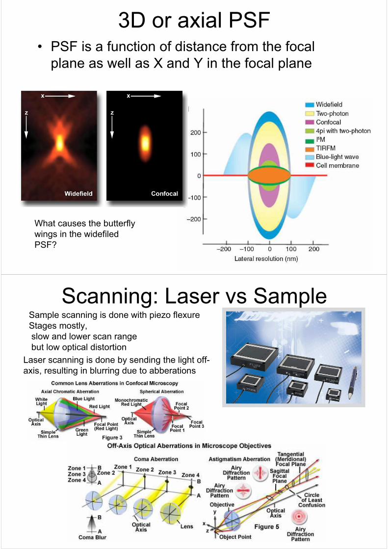
3D or axial PSF• PSF is a function of distance from the focal
plane as well as X and Y in the focal plane
What causes the butterfly
wings in the widefiled
PSF?
Scanning: Laser vs SampleSample scanning is done with piezo flexure
Stages mostly,
slow and lower scan range
but low optical distortion
Laser scanning is done by sending the light off-
axis, resulting in blurring due to abberations

Fluorescence
Bleaching is a problem
Fluorescence Resonance Energy Transfer (FRET)
Microscopy

Confocal Fluorescence microscope

Further Improvements
Example : Dual Axis Design

Beyond the Diffraction limit• Stimulated Emission Depletion of
Fluorescence
STED and 4Pi