Embed Size (px)
Citation preview

Convergent Multi-miRNA Targeting ofApoE Drives LRP1/LRP8-DependentMelanoma Metastasis and AngiogenesisNora Pencheva,1 Hien Tran,1,3 Colin Buss,1,3 Doowon Huh,1 Marija Drobnjak,2 Klaus Busam,2 and Sohail F. Tavazoie1,*1Laboratory of Systems Cancer Biology, Rockefeller University, New York, NY 10065, USA2Department of Pathology, Memorial Sloan-Kettering Cancer Center, New York, NY 10065, USA3These authors contributed equally to this work
*Correspondence: [email protected]://dx.doi.org/10.1016/j.cell.2012.10.028
SUMMARY
Through in vivo selection of human cancer cell pop-ulations, we uncover a convergent and cooperativemiRNA network that drives melanoma metastasis.We identify miR-1908, miR-199a-5p, and miR-199a-3p as endogenous promoters of metastaticinvasion, angiogenesis, and colonization in mela-noma. These miRNAs convergently target apolipo-protein E (ApoE) and the heat shock factor DNAJA4.Cancer-secreted ApoE suppresses invasion andmetastatic endothelial recruitment (MER) by en-gaging melanoma cell LRP1 and endothelial cellLRP8 receptors, respectively, while DNAJA4 pro-motes ApoE expression. Expression levels of thesemiRNAs and ApoE correlate with human metastaticprogression outcomes. Treatment of cells withlocked nucleic acids (LNAs) targeting these miRNAsinhibits metastasis to multiple organs, and thera-peutic delivery of these LNAs strongly suppressesmelanoma metastasis. We thus identify miRNAswith dual cell-intrinsic/cell-extrinsic roles in cancer,reveal convergent cooperativity in a metastaticmiRNA network, identify ApoE as an anti-angiogenicand metastasis-suppressive factor, and uncovermultiple prognostic miRNAs with synergistic combi-natorial therapeutic potential in melanoma.
INTRODUCTION
Metastatic progression requires that sets of effector proteins
involved in common cellular phenotypes be coherently ex-
pressed (Png et al., 2012; Gupta and Massague, 2006; Hana-
han and Weinberg, 2011; Talmadge and Fidler, 2010). Such
concerted expression states are apparent in gene expression
profiles of primary breast cancers that metastasize (Wang
et al., 2005) as well as in profiles of human cancer cell clones
that display enhanced metastatic activity (Kang et al., 2003;
Minn et al., 2005). In recent years, posttranscriptional regula-
1068 Cell 151, 1068–1082, November 21, 2012 ª2012 Elsevier Inc.
tion has emerged as a pervasive and robust mode of
concerted expression state and phenotype-level control. The
most studied class of posttranscriptional regulators with meta-
static regulatory activity is small noncoding RNAs (miRNAs)
(He and Hannon, 2004; Bartel, 2009; Filipowicz et al., 2008).
Metastasis suppressor miRNAs (Tavazoie et al., 2008) and
promoter miRNAs (Ma et al., 2007; Huang et al., 2008) were
originally discovered in breast cancer. Subsequent studies re-
vealed many more miRNAs with regulatory roles in the tumor-
igenesis and metastasis of other cancer types (Hurst et al.,
2009). In many cases, the expression levels of such miRNAs
in human cancer samples have supported their experimental
roles in metastasis. Thus, deregulated miRNA expression and
function (Calin and Croce, 2006; Lujambio and Lowe, 2012;
Poliseno et al., 2010) appear to be a pervasive feature of
human cancer. Clues regarding the robust control exerted by
specific miRNAs on metastatic progression emerged from
early work showing that concerted targeting of multiple metas-
tasis promoting genes by a single metastasis suppressor
miRNA was responsible for the dramatic metastasis suppres-
sion effects seen (Tavazoie et al., 2008). Such divergent gene
targeting by an individual miRNA has emerged as a defining
feature of these regulators.
By applying a systematic, in vivo selection-based approach,
we have identified a set of miRNAs that are deregulated in
independent metastatic lines derived from multiple patients
with melanoma—a highly prevalent cancer with increasing inci-
dence. These miRNAs convergently target the metabolic
protein apolipoprotein E (ApoE) and the heat shock factor
DNAJA4 and comprise a cooperative miRNA network that
maximally silences ApoE signaling. Cancer cell-secreted ApoE
inhibits metastatic invasion and endothelial recruitment through
its actions on distinct ApoE receptors on melanoma and
endothelial cells. These miRNAs are prognostic of clinical
metastasis, whereas their therapeutic inhibition displays in vivo
efficacy. The lack of effective therapies in melanoma for metas-
tasis prevention (Garbe et al., 2011) requires novel approaches.
Our unbiased and systematic approach has allowed us to
discover key noncoding and coding genes involved in mela-
noma progression and offers avenues for both identifying
patients at high risk for melanoma metastasis and rationally
treating them.

RESULTS
Endogenous miR-1908, miR-199a-3p, and miR-199a-5pPromote Human Melanoma MetastasisIn order to identify miRNA regulators of melanoma metastasis,
we utilized in vivo selection (Pollack and Fidler, 1982) with the
pigmented MeWo and nonpigmented A375 human melanoma
cell lines to generate multiple second- (LM2) and third-genera-
tion (LM3) lung metastatic derivatives that colonized the lung
more efficiently than their respective parental populations (Fig-
ure 1A and Figure S1A available online). Hybridization-based
small RNA profiling of 894 mature miRNAs followed by quanti-
tative stem-loop PCR (qRT-PCR) validation revealed four
miRNAs (miR-214, miR-199a-5p, miR-1908, and miR-199a-
3p) to be upregulated in multiple A375 and MeWo metastatic
derivatives relative to their respective parental lines (Figures
1B, 1C, and S1B). The significant induction of these miRNAs
suggested potential roles for them in metastatic progres-
sion. Indeed, overexpression of the precursor for miR-199a-
3p and miR-199a-5p (overexpressed concomitantly as the
miR-199a hairpin) or miR-1908 was sufficient to robustly
increase lung metastatic colonization by the poorly metastatic
MeWo parental cells (Figures 1D and S1C; 9.64-fold [miR-
1908]; 8.62-fold [miR-199a]), whereas miR-214 overexpression
did not significantly affect metastasis. We next asked whether
endogenous forms of these miRNAs promote metastasis. Indi-
vidual inhibition of miR-1908, miR-199a-3p, or miR-199a-5p in
the metastatic MeWo-LM2.3 cells (henceforth called MeWo-
LM2), as well as in the independent metastatic line A375-
LM3.2 (henceforth called A375-LM3), through a miR-Zip-based
stable silencing approach, significantly suppressed metastatic
colonization (Figures 1E and 1F), establishing these three
miRNAs as endogenous promoters of metastasis in melanoma.
Of note, both endogenous and exogenous miR-199a and miR-
1908 increased the number of metastatic nodules formed
(Figures S1D and S1E), consistent with a role for these miRNAs
in metastatic initiation.
Given the robust functional roles of miR-1908, miR-199a-
3p, and miR-199a-5p in promoting human melanoma
metastasis in a mouse model, we examined whether the
expression levels of these miRNAs correlate with the meta-
static relapse likelihood of human melanoma. To this end, 71
surgically resected primary melanoma skin lesions were
analyzed in a blinded manner for miRNA expression through
qRT-PCR. Consistent with our functional studies, all three miR-
NAs were significantly upregulated in primary melanomas that
had metastasized relative to those that had not (Figure 1G),
suggesting that induced expression of these miRNAs in pri-
mary lesions is an early event predictive of melanoma cancer
progression.
miR-1908, miR-199a-3p, and miR-199a-5p PromoteMetastatic Invasion, Endothelial Recruitment, andAngiogenesisWenext sought to determine the cellular mechanisms underlying
miRNA regulation of metastasis. Importantly, overexpression of
each miRNA reduced cell proliferation (Figure S2A) and did not
increase tumor growth (Figure 2A), indicating that the prometa-
C
static effects of miR-1908 and miR-199a are not secondary to
tumor growth promotion or enhanced proliferation. We next
asked whether these miRNAs regulate cell invasion, a key meta-
static phenotype. Overexpression of either miR-199a or miR-
1908 enhanced the ability of parental MeWo cells to invade
through matrigel (Figure 2B), whereas metastatic LM2 cells,
which express higher levels of these miRNAs, displayed sig-
nificantly greater matrigel invasion capacity (Figure S2B).
Conversely, individual inhibition of miR-199a-3p, miR-199a-5p,
or miR-1908 decreased the invasive capacity of MeWo-LM2
(Figure 2C) and A375-LM3 (Figure 2D) metastatic melanoma
cell derivatives.
Given the robust effects of these miRNAs on metastatic
progression, we examined whether they regulate additional
prometastatic phenotypes. Although overexpression of miR-
199a or miR-1908 did not modulate melanoma cell adhesion to
endothelial cells (Figure S2C), resistance to anoikis (Figure S2D),
survival during serum starvation (Figure S2E), or colony forma-
tion (Figure S2F), overexpression of each miRNA dramatically
enhanced the ability of parental MeWo cells to recruit endothelial
cells in trans-well endothelial recruitment assays (Figure 2E).
Consistent with this, metastatic MeWo-LM2 cells weremore effi-
cient at recruiting endothelial cells relative to their parental line
(Figure S2G). Furthermore, inhibition of miR-199a-3p, miR-
199a-5p, or miR-1908 in the metastatic MeWo-LM2 (Figure 2F),
as well as A375-LM3 (Figure 2G) cells, suppressed endothelial
recruitment.
To determine whether endogenous miR-199a-3p, miR-
199a-5p, and miR-1908 mediate endothelial recruitment by
metastatic cells in vivo, we examined metastatic endothelial
density in lung nodules formed by melanoma cells overex-
pressing or knocked down for each miRNA. Consistent with
the robust miRNA-dependent endothelial recruitment pheno-
type, each of the three miRNAs was both required and
sufficient for enhanced metastatic nodule endothelial content
(Figures 2H, S2H, and S2I). Remarkably, individual inhibition
of each miRNA robustly suppressed metastatic nodule
perfusion by roughly 5-fold following dextran injection (Figures
2I and S2J), indicating that each of these miRNAs also
promotes functional metastatic angiogenesis. Our findings
reveal miR-199a-3p, miR-199a-5p, and miR-1908 as neces-
sary and sufficient for enhanced invasion, metastatic endothe-
lial recruitment (MER), and angiogenesis during melanoma
progression.
miR-1908, miR-199a-3p, and miR-199a-5pConvergently and Cooperatively Target ApoE andDNAJA4We next employed a systematic and global approach to identify
the direct molecular targets of these miRNAs. Because miR-
1908, miR-199a-3p, and miR-199a-5p mediate the same sets
of in vitro and in vivo phenotypes and because miR-199a-5p
and miR-199a-3p arise from the same precursor hairpin,
we hypothesized that the prometastatic phenotypes of these
miRNAs may emerge through silencing of common target
genes. We thus performed transcriptomic profiling of melanoma
cells in the context of both loss and gain of function for each
miRNA (Figure S3A). This analysis, followed by qRT-PCR
ell 151, 1068–1082, November 21, 2012 ª2012 Elsevier Inc. 1069

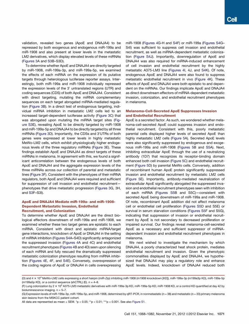
Figure 1. Identification of miR-1908, miR-199a-3p, and miR-199a-5p as Endogenous Promoters of Human Melanoma Metastasis
(A) Bioluminescence imaging plot of lung metastatic colonization by 4 3 104 parental MeWo or MeWo-LM2 cells. Lungs were extracted at day 72 and H&E
stained. n = 4-5.
(B and C) Expression levels of miR-214, miR-199a-5p, miR-1908, andmiR-199a-3p, determined by qRT-PCR, in multiple independent MeWo-LM2 (B) and A375-
LM2/LM3 (C) metastatic derivatives and their respective parental melanoma lines. n = 3.
(D and E) Bioluminescence quantification and representative H&E-stained lungs corresponding to lung colonization by 4 3 104 parental MeWo cells over-
expressing the precursor for miR-199a (giving rise to both miR-199a-3p and miR-199a-5p), miR-1908, or miR-214 or expressing a control hairpin (shCTRL)
1070 Cell 151, 1068–1082, November 21, 2012 ª2012 Elsevier Inc.
validation, revealed two genes (ApoE and DNAJA4) to be
repressed by both exogenous and endogenous miR-199a and
miR-1908 and also present at lower levels in the metastatic
LM2 derivatives, which display elevated levels of these miRNAs
(Figures 3A and S3B–S3D).
To determine whether ApoE and DNAJA4 are directly targeted
by miR-1908, miR-199a-3p, and miR-199a-5p, we examined
the effects of each miRNA on the expression of its putative
targets through heterologous luciferase reporter assays. Inter-
estingly, both miR-199a and miR-1908 individually repressed
the expression levels of the 30 untranslated regions (UTR) and
coding sequences (CDS) of both ApoE and DNAJA4. Consistent
with direct targeting, mutating the miRNA complementary
sequences on each target abrogated miRNA-mediated regula-
tion (Figure 3B). In a direct test of endogenous targeting, indi-
vidual miRNA inhibition in metastatic LM2 cells led to an
increased target-dependent luciferase activity (Figure 3C) that
was abrogated upon mutating the miRNA target sites (Fig-
ure S3E), revealing ApoE to be directly targeted by miR-1908
andmiR-199a-5p andDNAJA4 to be directly targeted by all three
miRNAs (Figure 3D). Importantly, the CDSs and 30UTRs of both
genes were expressed at lower levels in highly metastatic
MeWo-LM2 cells, which exhibit physiologically higher endoge-
nous levels of the three regulatory miRNAs (Figure 3E). These
findings establish ApoE and DNAJA4 as direct targets of these
miRNAs in melanoma. In agreement with this, we found a signif-
icant anticorrelation between the endogenous levels of both
ApoE and DNAJA4 and the aggregate expression levels of the
three miRNAs across our collection of parental and metastatic
lines (Figure 3F). Consistent with the phenotypes of their miRNA
regulators, both ApoE and DNAJA4 were required and sufficient
for suppression of cell invasion and endothelial recruitment—
phenotypes that drive metastatic progression (Figures 3G, 3H,
and S3F–S3I).
ApoE and DNAJA4 Mediate miR-199a- and miR-1908-Dependent Metastatic Invasion, EndothelialRecruitment, and ColonizationTo determine whether ApoE and DNAJA4 are the direct bio-
logical effectors downstream of miR-199a and miR-1908, we
examined whether these two target genes are epistatic to each
miRNA. Consistent with direct and epistatic miRNA/target
gene interactions, knockdown of ApoE or DNAJA4 in the setting
of miRNA inhibition (Figures S4A–S4D) significantly antagonized
the suppressed invasion (Figures 4A and 4C) and endothelial
recruitment phenotypes (Figures 4B and 4D) seen upon silencing
of each miRNA and fully rescued the dramatically suppressed
metastatic colonization phenotype resulting from miRNA inhibi-
tion (Figures 4E, 4F, and S4E). Conversely, overexpression of
the coding regions of ApoE or DNAJA4 in cells overexpressing
(D) and 43 104 MeWo-LM2 cells expressing a short hairpin (miR-Zip) inhibiting m
(m199a5p KD), or a control sequence (shCTRL) (E). n = 4–8.
(F) Lung colonization by 23 105 A375-LM3 metastatic derivatives with miR-199a-
bioluminescence imaging. n = 5–7.
(G) Expression levels ofmiR-199a-3p, miR-199a-5p, andmiR-1908, determined b
skin lesions from the MSKCC patient cohort.
All data are represented as mean ± SEM. *p < 0.05; **p < 0.01; ***p < 0.001. See
C
miR-1908 (Figures 4G-H and S4F) or miR-199a (Figures S4G–
S4I) was sufficient to suppress cell invasion and endothelial
recruitment, as well as miRNA-dependent metastatic coloniza-
tion (Figure S4J). Importantly, downregulation of ApoE and
DNAJA4 was also required for miRNA-induced enhancement
of cell invasion and endothelial recruitment by the highly
metastatic A375-LM3 line (Figures 4I, 4J, and S4K). Of note,
endogenous ApoE and DNAJA4 were also found to suppress
metastatic endothelial recruitment in vivo (Figure 4K). These
effects of ApoE and DNAJA4 were both epistatic to and depen-
dent on the miRNAs. Our findings implicate ApoE and DNAJA4
as direct downstream effectors of miRNA-dependent metastatic
invasion, colonization, and endothelial recruitment phenotypes
in melanoma.
Melanoma-Cell-Secreted ApoE Suppresses Invasionand Endothelial RecruitmentApoE is a secreted factor. As such, we wondered whether mela-
noma-cell-secreted ApoE could suppress invasion and endo-
thelial recruitment. Consistent with this, poorly metastatic
parental cells displayed higher levels of secreted ApoE than
highly metastatic LM2 cells (Figure 5A). Secreted ApoE levels
were also significantly suppressed by endogenous and exoge-
nous miR-199a and miR-1908 (Figures 5B and S5A). Next,
inhibiting extracellular ApoE through the use of a neutralizing
antibody (1D7) that recognizes its receptor-binding domain
enhanced both cell invasion (Figure 5C) and endothelial recruit-
ment (Figure 5D) by parental MeWo cells. Conversely, addition
of recombinant human ApoE protein significantly suppressed
invasion and endothelial recruitment by metastatic LM2 cells
(Figure 5E). Importantly, antibody-mediated neutralization of
extracellular ApoE significantly abrogated the suppressed inva-
sion and endothelial recruitment phenotypes seen with inhibition
of each miRNA (Figures S5B and S5C)—consistent with
secreted ApoE being downstream of miR-199a and miR-1908.
Of note, recombinant ApoE addition did not affect melanoma
cell or endothelial cell proliferation (Figures S5D and S5E) or
survival in serum starvation conditions (Figures S5F and S5G),
indicating that suppression of invasion or endothelial recruit-
ment by ApoE is not secondary to decreased proliferation or
impaired survival. Our findings reveal melanoma-cell-secreted
ApoE as a necessary and sufficient suppressor of miRNA-
dependent invasion and endothelial recruitment phenotypes in
melanoma.
We next wished to investigate the mechanism by which
DNAJA4, a poorly characterized heat shock protein, mediates
endothelial recruitment and invasion. Given the phenotypic
commonalities displayed by ApoE and DNAJA4, we hypothe-
sized that DNAJA4 may play a regulatory role and enhance
ApoE levels. Indeed, knockdown of DNAJA4 reduced both
iR-1908 (m1908 knockdown [KD]), miR-199a-3p (m199a3p KD), miR-199a-5p
3p KD, miR-199a-5p KD, miR-1908 KD, or a control KD quantified at day 42 by
y qRT-PCR, in nonmetastatic (n = 38) andmetastatic (n = 33) primarymelanoma
also Figure S1.
ell 151, 1068–1082, November 21, 2012 ª2012 Elsevier Inc. 1071

Figure 2. miR-1908, miR-199a-3p, and miR-199a-5p Display Cell-Autonomous/Non-Cell-Autonomous Prometastatic Roles in Melanoma
(A) Primary tumor growth by 13 106 MeWo cells overexpressing the miR-199a or miR-1908 precursor hairpins or expressing an shCTRL following subcutaneous
injection into mice. n = 4–10.
(B) Matrigel invasion by 13 105 MeWo cells overexpressing miR-199a or miR-1908 or expressing an shCTRL was quantified by counting the number of cells that
invaded into the basal side of matrigel-coated trans-well inserts after 24 hr. n = 3–7.
(C and D) Matrigel invasion by MeWo-LM2 (C) and A375-LM3 (D) cells with miR-199a-3p KD, miR-199a-5p KD, miR-1908 KD, or a control KD. n = 3–7.
(E) Trans-well recruitment of 1 3 105 human umbilical vein endothelial cells (HUVECs) by 5 3 104 MeWo cells overexpressing miR-199a or miR-1908 or ex-
pressing a control hairpin. Endothelial recruitment capacity was measured by counting the number of HUVECs that migrated to the basal side of each trans-well
insert after 16 hr. n = 7–9.
1072 Cell 151, 1068–1082, November 21, 2012 ª2012 Elsevier Inc.

Figure 3. Identification of ApoE and
DNAJA4 as Direct Target Genes of miR-
199a and miR-1908
(A) Heatmap depicting the mRNA expression
levels of ApoE and DNAJA4, measured by qRT-
PCR, in MeWo parental cells expressing an
shCTRL or overexpressing miR-199a or miR-1908
and in MeWo-LM2 cells. Color map illustrates the
change in standard deviations from the mean for
each gene’s expression values across the four cell
line samples.
(B and C) Heterologous luciferase reporter assays
measuring the expression of wild-type or miRNA
target site mutant ApoE and DNAJA4 CDSs and
30UTRs inMeWo cells overexpressingmiR-199a or
miR-1908 or in control MeWo cells (B) and in
MeWo-LM2cellswithmiR-199a-3pKD,miR-199a-
5p KD, miR-1908 KD, or a control KD (C). n = 4–6.
(D) Schematic of an experimentally derived model
depicting the convergent targeting of the CDSs
and 30UTRs of ApoE and DNAJA4 by miR-199a-
3p, miR-199a-5p, and miR-1908.
(E) Luciferase activity of wild-type and miRNA
target site mutant ApoE and DNAJA4 30UTR/CDSluciferase constructs transfected into MeWo
parental and MeWo-LM2 cells. n = 3–6.
(F) Linear regression analyses of the expression
levels of each gene (ApoE and DNAJA4) and the
aggregate expression levels of the three miRNAs
(average of the individual miRNA expression
values) in multiple MeWo and A375 metastatic
derivatives and their parental lines. n = 12.
(G and H) Trans-well matrigel invasion (G) and
endothelial recruitment (H) by parental MeWo
cells expressing short hairpin RNAs (shRNAs)
targeting ApoE (shAPOE), DNAJA4 (shDNAJA4),
or a control sequence (shCTRL). n = 4–8.
All data are represented as mean ± SEM. Scale
bar, 100 mm. See also Figure S3.
ApoE transcript levels (Figure S5H) and secreted ApoE levels
(Figure 5F), whereas DNAJA4 overexpression substantially
elevated ApoE transcript expression (Figure S5I). Consistent
with DNAJA4 acting upstream of ApoE, addition of recombinant
ApoE abrogated the enhanced cell invasion and endothelial
recruitment phenotypes resulting from DNAJA4 knockdown
(Figures 5G and 5H), whereas antibody-mediated neutralization
of ApoE significantly antagonized the suppression of invasion
and endothelial recruitment resulting from DNAJA4 overexpres-
sion (Figures S5J and S5K). These findings reveal DNAJA4 to
(F and G) Endothelial recruitment by MeWo-LM2 (F) and A375-LM3 (G) cells inhibited for miR-199a-3p, miR-19
(H and I) Cumulative fraction plots depicting the endothelial cell (H) or dextran perfusion (I) density distribution
LM2 cells with miR-199a-3p KD, miR-199a-5p KD, miR-1908 KD, or a control KD. Lung sections were double
and intravenously injected dextran (I). n = 211, 30, 138, and 39 nodules for control KD, m199a3p KD, m199a5p
and 40 nodules for control KD, m199a3p KD, m199a5p KD, and m1908 KD, respectively (I).
All data are represented as mean ± SEM. Scale bar, 100 mm. See also Figure S2.
Cell 151, 1068–1082, No
suppress melanoma invasion and endo-
thelial recruitment through positive regu-
lation of ApoE expression, leading to its
increased extracellular levels.
The regulatory convergence of three metastasis-promoting
miRNAs and the DNAJA4 protein onto ApoE motivated us to
determine whether ApoE expression correlates with human
melanoma progression. To this end, we analyzed published
array-based expression data for ApoE (Haqq et al., 2005) in
nevi, primary, and metastatic lesions. Consistent with a metas-
tasis-suppressive role, ApoE transcript levels were significantly
lower in distal organ metastases relative to primary melanoma
and nevi lesions (Figure 5I). We also examined ApoE pro-
tein expression in a large (n = 168) human tissue-microarray
9a-5p, miR-1908, or a control sequence. n = 6–10.
s for metastatic nodules formed by 23 105 MeWo-
stained for vimentin and MECA-32 (H) or vimentin
KD, andm1908 KD, respectively (H); n = 30, 18, 38,
vember 21, 2012 ª2012 Elsevier Inc. 1073

Figure 4. ApoE and DNAJA4 Epistatically Interact with miR-199a and miR-1908 in Regulating Metastatic Invasion, Endothelial Recruitment,
and Colonization
(A–D) MeWo-LM2 cells expressing a control shRNA or shRNAs targeting ApoE or DNAJA4 in the context of miR-1908 KD (A and B) or miR-199a-5p KD (C and D)
were subjected to the trans-well invasion (A and C) and endothelial recruitment (B and D) assays. n = 6–11.
(E and F) Bioluminescence imaging plots and H&E-stained lungs corresponding to lung metastasis by 1 3 105 MeWo-LM2 cells expressing a control shRNA or
shRNAs targeting ApoE or DNAJA4 in the setting of miR-1908 KD (E) or miR-199a-5p KD (F). n = 5.
(G and H) MeWo cells overexpressing the coding regions of ApoE or DNAJA4 or expressing a control vector in the context of miR-1908 overexpression were
analyzed for the invasion (G) and endothelial recruitment (H) phenotypes. n = 7–12.
1074 Cell 151, 1068–1082, November 21, 2012 ª2012 Elsevier Inc.
melanoma progression panel. Blinded immunohistochemical
analysis revealed that ApoE protein levels were significantly
lower in lymph node metastases (p = 0.035) and even further
downregulated in distal melanoma metastases (p < 0.0001) rela-
tive to primary melanomas (Figure 5J).
Genetic Inactivation of ApoE Promotes Metastasis,whereas ApoE Pretreatment Blocks MetastaticInitiationGiven the robust suppression of metastatic phenotypes ex-
erted by ApoE and its clinical association with melanoma
progression, we next investigated the impact of genetic
deletion of systemic ApoE on melanoma progression in an
immunocompetent mouse model of melanoma metastasis.
Consistent with a major suppressive role for extracellular
ApoE in metastasis, B16F10 mouse melanoma cells injected
into the circulation exhibited a 10-fold increase in metastatic
colonization in ApoE genetically null mice compared to their
wild-type littermates. Importantly, the metastatic capacity of
B16F10 cells was dramatically abrogated by ApoE cell pre-
treatment in both wild-type (60-fold inhibition) and ApoE null
(460-fold inhibition) genetic contexts (Figure 5K). These find-
ings establish both systemic and cancer-secreted ApoE as a
robust suppressor of metastasis by human and mouse mela-
noma cells.
We next wondered whether enhanced ApoE signaling in
melanoma cells could have therapeutic efficacy in preventing
melanoma metastasis. We thus preincubated melanoma cells
with recombinant ApoE or BSA for 24 hr prior to injection into
mice. Of note, ApoE pretreatment robustly suppressed meta-
static colonization by the highly aggressive MeWo-LM2 cells
by >300-fold (Figure 5L). Remarkably, ApoE preincubation abro-
gated the metastatic capacity of four additional human mela-
noma cell lines: A375-LM3 (145-fold, Figure 5M), WM-266-4
(80-fold, Figure 5N), HT-144 (119-fold, Figure S5L), and A2058
(85-fold, Figure S5M).
Extracellular ApoE Divergently Targets Melanoma CellLRP1 and Endothelial Cell LRP8 ReceptorsWe next investigated the molecular mechanisms by which ApoE
suppresses metastasis. In order to identify the ApoE receptor(s)
that mediate(s) invasion, we knocked down all four known ApoE
receptors (VLDLR, LRP1, LRP8, and LDLR) in melanoma cells.
Interestingly, knockdown of LRP1, but not the other ApoE
receptors, abolished ApoE-induced suppression of invasion
(Figures 6A and S6A), whereas LRP1 knockdown in the setting
of miRNA silencing rescued the suppressed invasion phenotype
resulting from miRNA inhibition (Figures 6B and S6B) and signif-
icantly enhanced in vivo metastatic colonization by MeWo-LM2
cells depleted for miR-1908 (Figures 6C and S6C). These find-
ings reveal LRP1 to be a downstream mediator of miRNA/
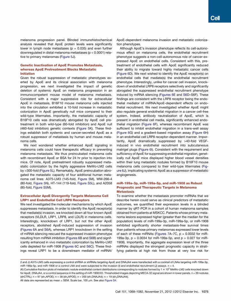
(I and J) A375-LM3 cells expressing a control shRNA or shRNAs targeting ApoE
miR-199a-5p, and miR-1908 or a control LNA and were subjected to the invasio
(K) Cumulative fraction plots of metastatic nodule endothelial content distributions
for ApoE, DNAJA4, or a control sequence in the setting ofmiR-1908 KD. Threshold
(shCTRL); n = 97 (sh1APOE); n = 38 (sh2APOE); n = 200 (sh1DNAJA4); n = 19 (sh
All data are represented as mean ± SEM. Scale bar, 100 mm. See also Figure S4
C
ApoE-dependent melanoma invasion and metastatic coloniza-
tion phenotypes.
Although ApoE’s invasion phenotype reflects its cell-autono-
mous effect on melanoma cells, the endothelial recruitment
phenotype suggests a non-cell-autonomous role of cancer-ex-
pressed ApoE on endothelial cells. Consistent with this, pre-
treatment of endothelial cells with ApoE significantly reduced
their ability to migrate toward highly metastatic cancer cells
(Figure 6D). We next wished to identify the ApoE receptor(s) on
endothelial cells that mediate(s) the endothelial recruitment
phenotype. Interestingly, unlike for cancer cell invasion, knock-
down of endothelial LRP8 receptors selectively and significantly
abrogated the suppressed endothelial recruitment phenotype
induced by miRNA silencing (Figures 6E and S6D–S6F). These
findings are consistent with the LRP8 receptor being the endo-
thelial mediator of miRNA/ApoE-dependent effects on endo-
thelial recruitment. We next investigated whether ApoE might
also regulate general endothelial migration in a cancer cell-free
system. Indeed, antibody neutralization of ApoE, which is
present in endothelial cell media, significantly enhanced endo-
thelial migration (Figure 6F), whereas recombinant ApoE was
sufficient to inhibit endothelial migration in a trans-well assay
(Figure 6G) and a gradient-based migration assay (Figure 6H)
in an endothelial cell LRP8 receptor-dependent manner. Impor-
tantly, ApoE dramatically suppressed by >40-fold VEGF-
induced in vivo endothelial recruitment into subcutaneous
matrigel plugs (Figure 6I). Consistent with the requirement and
sufficiency of ApoE for suppressing endothelial migration, genet-
ically null ApoE mice displayed higher blood vessel densities
within their lung metastatic nodules formed by B16F10 mouse
melanoma cells compared to their wild-type littermates (Fig-
ure 6J), implicating systemic ApoE as a suppressor of metastatic
angiogenesis.
miR-199a-3p, miR-199a-5p, and miR-1908 as RobustPrognostic and Therapeutic Targets in MelanomaMetastasisTo examine whether the metastasis promoter miRNAs that we
describe herein could serve as clinical predictors of metastatic
outcomes, we quantified their expression levels in a blinded
manner by qRT-PCR in a cohort of human melanoma samples
obtained frompatients atMSKCC. Patientswhose primarymela-
noma lesions expressed higher (greater than the median for the
population) levels of miR-199a-3p, miR-199a-5p, or miR-1908
exhibited significantly shorter metastasis-free survival times
than patients whose primary melanomas expressed lower levels
of each of these miRNAs (Figures 7A–7C, p = 0.0032 for miR-
199a-3p, p = 0.0034 for miR-199a-5p, and p = 0.027 for miR-
1908). Importantly, the aggregate expression level of the three
miRNAs displayed the strongest prognostic capacity in strati-
fying patients at high risk from those at very low risk for
and DNAJA4 were transfected with a cocktail of LNAs targeting miR-199a-3p,
n (I) and endothelial recruitment (J) assays. n = 4.
corresponding to nodules formed by 13 105 MeWo-LM2 cells knocked down
ed images depictingMECA-32 signal are shown in lower panels. n = 39 nodules
2DNAJA4).
.
ell 151, 1068–1082, November 21, 2012 ª2012 Elsevier Inc. 1075

Figure 5. Extracellular ApoE Inhibits Invasion, Endothelial Recruitment, and Metastasis, whereas Genetic Deletion of ApoE Enhances
Metastasis
(A and B) Extracellular ApoE levels, quantified by ELISA, in conditioned media from MeWo parental and LM2 cells (A) and in MeWo-LM2 cells silenced for miR-
199a-5p, miR-1908, or a control sequence (B). n = 3.
(C) Matrigel invasion by MeWo cells in response to the ApoE-neutralizing antibody 1D7 (5, 10, 20, or 40 mg/ml) or IgG control (40 mg/ml). n = 4–6.
(D) Endothelial recruitment by MeWo cells in the presence of 1D7 or a control IgG antibody at 40 mg/ml. n = 4.
(E) The invasion and endothelial recruitment phenotypes were assessed in MeWo-LM2 cells in response to bovine serum albumin (BSA) or recombinant ApoE3
added to the cell media at 100 mM. n = 7–10.
1076 Cell 151, 1068–1082, November 21, 2012 ª2012 Elsevier Inc.

metastatic relapse (Figure 7D, p < 0.001). These clinical findings
are indicative of functional cooperativity between the three miR-
NAs in the regulation of cancer progression and suggest utility
for these molecules as clinical prognostic biomarkers of mela-
noma metastasis. Our identification of miR-199a in melanoma
is consistent with a previous clinical association study that re-
vealed increased miR-199a levels to correlate with uveal mela-
noma progression as well (Worley et al., 2008).
In light of the current lack of effective treatment options for the
prevention of melanoma metastasis after surgical resection of
the primary lesion (Garbe et al., 2011) and the strong prognostic
value of these regulatory miRNAs in melanoma metastasis, we
decided to therapeutically target them using antisense locked
nucleic acids (LNAs) (Elmen et al., 2008). Highly metastatic
MeWo-LM2 cells pretreated with LNA oligonucleotides anti-
sense to each mature miRNA (miR-199a-3p, miR-199a-5p, or
miR-1908) exhibited a roughly 4-fold decrease in metastatic
activity (Figure 7E). Given our clinical evidence for cooperativity
among these miRNAs, as well as their convergent targeting of
ApoE andDNAJA4, we examined the impact of silencing all three
miRNAs on metastatic progression. Cotransfection of LNAs tar-
geting all three miRNAs suppressed metastatic colonization
by >70-fold, revealing dramatic synergy and cooperativity
between endogenous miR-199a-3p, miR-199a-5p, and miR-
1908 (Figure 7E). Importantly, inhibition of these miRNAs with
triple LNA pretreatment did not decrease in vitro proliferation
(Figure S7A), indicating that the metastasis suppression pheno-
type is not secondary to impaired proliferation. Remarkably,
combinatorial LNA-mediated miRNA inhibition robustly inhibited
lung colonization by the independent SK-Mel-2 (24-fold,
Figure 7F), WM-266-4 (77-fold, Figure 7G), HT-144 (15-fold, Fig-
ure 7H), A2058 (17-fold, Figure 7I), and A375-LM3 (3-fold, Fig-
ure S7B) human melanoma cell lines, as well as micrometastasis
formation by the SK-Mel-28 human melanoma cell line (Figures
S7C and S7D). These findings indicate that combinatorial target-
ing of the three miRNAs exhibits robust therapeutic potential
across a large variety of melanotic and amelanotic human mela-
noma lines of diverse BRAF and NRAS mutational statuses.
Additionally, intracardiac injection of highly metastatic MeWo-
LM2 cells pretreatedwith a cocktail of LNAs targeting thesemiR-
NAs suppressed systemic metastasis to multiple distal organs
(Figures 7J-K) such as the brain (Figure 7L) and bone (Fig-
ure S7E). We next examined the therapeutic efficacy of system-
ically administered in-vivo-optimized LNAs in melanoma metas-
tasis prevention. To this end, highly metastatic MeWo-LM2 cells
(F) Extracellular ApoE levels, quantified by ELISA, in conditioned media from Me
(G and H) MeWo cells with shRNA-induced silencing of DNAJA4 were analyzed
presence of either BSA or recombinant ApoE3 at 100 mM. n = 4.
(I) Array-based ApoE transcript expression levels in nevi (n = 9), primary melanom
(J) Box and whisker plot of ApoE protein expression determined by blinded immu
set (NIH) comprised of primary melanoma lesions (n = 66), nodal (n = 36), and d
whereas box extent corresponds to 25th–75th percentile of the sample populati
(K) Bioluminescence quantification and representative ex vivo lung images corre
melanoma cells pretreated with recombinant ApoE3 (100 mg/ml) or control for 24 h
22 (control) and n = 5–7 (ApoE pretreatment).
(L–N) Bioluminescence imaging plots of lung colonization by 4 3 104 MeWo-LM
pretreated with ApoE3 or BSA at 100 mg/ml for 24 hr prior to injection. n = 5–8.
All data are represented as mean ± SEM except for Figure 4J. Scale bar, 100 mm
C
were injected into mice. The following day, mice were intrave-
nously treated with LNAs targeting miR-199a-3p, miR-199a-
5p, and miR-1908 at a low dose (12.5 mg/kg total) on a biweekly
basis for 4 weeks followed by weekly dosing for 7 weeks. Of
note, combinatorial LNA treatment reduced lung colonization
by >10-fold (Figure 7M, p = 0.039) without causing weight loss
(Figure S7F). Importantly, therapeutic LNA-mediated miRNA tar-
geting was confirmed by qRT-PCR analysis of lung metastatic
nodules (Figure S7G) and mouse cardiac and liver tissues
(Figures S7H and S7I). Taken together, our findings reveal a
miRNA-dependent regulatory network that converges onto
ApoE signaling to control cell-autonomous and non-cell-autono-
mous features of melanoma metastatic initiation and progres-
sion (Figure 7N).
DISCUSSION
The complexity of cancer requires the application of systems-
level and integrative approaches. Using a systematic global
approach, we have uncovered a cooperative network of miRNAs
that are: (1) upregulated in highly metastatic human melanoma
cells, (2) required and sufficient for optimal metastatic coloniza-
tion and angiogenesis in melanoma, (3) pathologic predictors of
human melanoma metastatic relapse, and (4) potential thera-
peutic targets across a variety of human melanoma cell lines.
Through a transcriptomic-based and biologically guided target
identification approach, we have found miR-1908, miR-199a-
3p, and miR-199a-5p to convergently target the heat shock
factor DNAJA4 and the metabolic gene ApoE. Our identification
of ApoE as a gene negatively regulated by three metastasis pro-
moter miRNAs, positively regulated by a metastasis suppressor
gene (DNAJA4) and silenced in clinical metastasis samples,
highlights the significance of this gene as a key suppressor of
melanoma metastatic progression. ApoE mediates its effects
by targeting two distinct yet homologous receptors on two
diverse cell types. ApoE acting on melanoma cell LRP1 recep-
tors inhibits melanoma invasion, whereas its action on endothe-
lial cell LRP8 receptors suppresses endothelial migration. Our
findings also reveal LRP1 and LRP8 to be endogenous suppres-
sors of prometastatic phenotypes in melanoma. Our results from
loss-of-function, gain-of-function, epistasis, clinical correlation,
and in vivo selection-based expression analyses give rise to
a model wherein three miRNAs convergently target a metastasis
suppressor network to limit ApoE secretion, thus suppressing
ApoE engagement of melanoma LRP1 receptors and endothelial
Wo cells expressing shRNAs targeting DNAJA4 or an shCTRL. n = 3.
for the matrigel invasion (G) and endothelial recruitment (H) phenotypes in the
as (n = 6), and distal melanoma metastases (n = 19).
nohistochemical analysis in a tissue microarray (TMA) melanoma progression
istal (n = 66) melanoma metastases. Whiskers represent 5th–95th percentile,
on.
sponding to lung metastasis 19 days postinjection of 5 3 104 B16F10 mouse
r and intravenously injected into ApoE genetically null or wild-typemice. n = 12–
2 cells (L), 2 3 105 A375-LM3 cells (M), and 1.5 3 105 WM-266-4 cells (N)
. See also Figure S5.
ell 151, 1068–1082, November 21, 2012 ª2012 Elsevier Inc. 1077

Figure 6. Distinct Melanoma and Endothelial Cell Receptors Mediate the Effects of ApoE on Invasion and Endothelial Recruitment
(A) Trans-well invasion by MeWo-LM2 cells transfected with short interfering RNAs (siRNAs) targeting LDLR, VLDLR, LRP8, LRP1, or a control siRNA (siCTRL) in
the presence of either BSA (100 mM) or recombinant ApoE3 (100 mM). n = 3–7.
(B) MeWo-LM2 cells with miR-1908 KD or a control KD were transfected with siRNAs targeting LRP1 or siCTRL and subjected to the matrigel invasion assay.
n = 4.
(C) Lung colonization by 1 3 105 MeWo-LM2 cells transfected with siRNAs targeting LRP1 or siCTRL in the setting of miR-1908 KD. n = 4–8.
(D) Trans-well recruitment of endothelial cells preincubated with BSA or recombinant ApoE3 at 100 mM for 24 hr by MeWo-LM2 cells. n = 3–4.
1078 Cell 151, 1068–1082, November 21, 2012 ª2012 Elsevier Inc.

LRP8 receptors (Figure 7N). The cooperative suppression of
these inhibitory pathways by these miRNAs provides melanoma
cells withmaximal invasive and endothelial recruitment capacity.
Though we have identified LRP1 as a mediator of the ApoE
response on melanoma cells, it remains possible that additional
ApoE receptorsmaymediate ApoE responses inmelanoma cells
from distinct patients.
We reveal combined molecular, genetic, and in vivo evidence
for a required and sufficient role for melanoma-cell-secreted and
systemic ApoE in the suppression of melanoma metastatic
progression. The ability of recombinant ApoE to inhibit these
prometastatic phenotypes, as well as the enhanced melanoma
invasion and endothelial recruitment phenotypes resulting from
ApoE neutralization by an antibody that targets the ApoE protein,
suggest that the ApoE molecule itself, rather than ApoE-associ-
ated lipids in lipoprotein particles, is the key mediator of these
phenotypes.
Our molecular and in vivo studies reveal a role for endogenous
cancer-derived ApoE in the modulation of endothelial migration
and cancer angiogenesis through engagement of the endo-
thelial LRP8 receptor. This non-cell-autonomous endothelial
recruitment phenotype mediated by ApoE suggests that ApoE
may also modulate metastatic angiogenesis and tumor angio-
genesis in other cancer types. ApoE is a polymorphic molecule
with well-established roles in cardiovascular and neurodegener-
ative disorders. Its three major variants—ApoE2, ApoE3, and
ApoE4—display varying prevalences in the human population,
with ApoE3 being the most common variant. The three isoforms
differ at residues 112 and 158 in the N-terminal region, which
contains the receptor-bindingdomain (Hatters et al., 2006).Given
that the MeWo and A375 cell lines analyzed in our study are
homozygous for the ApoE3 allele and given the ability of
recombinant ApoE3 to inhibit melanomametastatic phenotypes,
our findings are consistent with ApoE3 being sufficient and re-
quired for the suppression of melanoma metastatic progression.
Despite recent promising advances such as the BRAF V600E
inhibitor Vemurafenib (Chapman et al., 2011; Sosman et al.,
2012) and the immunomodulatory antibody ipilimumab (Hodi
et al., 2010; Sharma et al., 2011) that have extended the median
survival times for subsets of patients diagnosed with metastatic
(stage IV) melanoma, there are currently no effective therapies
for the prevention of melanoma metastasis in the adjuvant
setting, with interferon therapy increasing overall survival rates
at 5 years by ameager 3%based onmeta-analyses, while phase
III trial data demonstration of a significant survival benefit is still
outstanding (Garbe et al., 2011). The substantial enhancement
of melanoma metastatic progression in the context of genetic
(E) Recruitment of HUVECs transfected with siRNAs targeting LDLR, VLDLR, L
n = 4–12.
(F) Trans-well migration by HUVECs in the presence of IgG or 1D7 antibody at 4
(G) Trans-well migration by endothelial cells transfected with siRNAs targeting LR
(H) Trans-well migration along an ApoE matrigel gradient by endothelial cells tra
(I) Endothelial recruitment into subcutaneously implanted matrigel plugs containi
ApoE3 (10 mg/ml). The number of MECA-32-positive endothelial cells was quant
(J) Endothelial cell content, determined by staining for MECA-32, within lung met
wild-type or ApoE genetically null mice. n = 17–20.
All data are represented as mean ± SEM. Scale bar, 100 mm. See also Figure S6
C
ablation of systemic ApoE, as well as the dramatic abrogation
of metastatic capacity by ApoE pretreatment, suggest that
modulating ApoE levels may have significant therapeutic impli-
cations for melanoma—a disease that claims �48,000 lives a
year globally (Lucas et al., 2006). Therapeutic approaches aimed
at pharmacological induction of endogenous ApoE levels could
potentially reduce melanoma mortality rates by decreasing
metastatic relapse incidence.
Our establishment of in vivo selectionmodels of melanotic and
amelanotic melanoma metastasis has allowed us to identify the
cellular phenotypes displayed by highly metastatic melanoma
cells. Our work reveals that, in addition to enhanced invasive-
ness, the capacity of melanoma cells to recruit endothelial cells
in vitro and in vivo is significantly enhanced in highly metastatic
melanoma cells. Additionally, we find that three major posttran-
scriptional regulators of metastasis strongly regulate endothelial
recruitment. We further show that the downstream signaling
pathway modulated by these miRNAs also regulates endothelial
recruitment. These findings reveal endothelial recruitment to
be a defining feature of metastatic melanoma cells. Metastatic
breast cancer derivatives obtained from multiple patients were
also recently reported to recruit endothelial cells more efficiently
than their parental lines by silencing the metastasis suppressor
miRNA miR-126, thereby enhancing metastatic endothelial re-
cruitment (MER) (Png et al., 2012). These findings as a whole
highlight enhancedMER capacity as a key attribute ofmetastatic
cells from multiple epithelial cancer types and suggest a sig-
nificant role for cancer-endothelial interactions in metastatic
initiation and progression that complements their established
conventional roles in primary tumor growth and perfusion (Car-
meliet and Jain, 2000).
The ability of miR-199a-3p, miR-199a-5p, and miR-1908
to individually predict metastasis-free survival in melanoma
patients indicates the significance of each miRNA as a clinical
predictor of melanoma cancer progression. Importantly, the
robust capacity of the three-miRNA aggregate signature to
separate patients at high risk from those at very low risk for
metastatic relapse reveals both the cooperativity of these
miRNAs and their clinical potential as melanoma biomarkers
(Sawyers, 2008) for identifying the subset of patients that
might benefit from experimental therapies such as miRNA
inhibition therapy. Therapeutic miRNA targeting has gained
momentum through the use of in vivo LNAs that have been
shown to antagonize miRNAs in mice and primates (Elmen
et al., 2008) and are currently being tested in human clinical
trials. The powerful prognostic capacity of the three miRNAs,
the proof-of-principle demonstration of robust metastasis
RP1, LRP8, or siCTRL by MeWo-LM2 cells with miR-1908 KD or control KD.
0 mg/ml. n = 6–8.
P8 or siCTRL in the presence of BSA or recombinant ApoE3 at 100 mM. n = 6–7.
nsfected with siRNAs targeting LRP8 or siCTRL. n = 4–8.
ng BSA (10 mg/ml), VEGF (400 ng/ml) + BSA (10 mg/ml), or VEGF (400 ng/ml) +
ified after 3 days by immunohistochemical detection. n = 3–6.
astatic nodules formed 19 days following injection of 53 104 B16F10 cells into
.
ell 151, 1068–1082, November 21, 2012 ª2012 Elsevier Inc. 1079

Figure 7. Clinical and Therapeutic Cooperativity among miR-199a-3p, miR-199a-5p, and miR-1908 in Melanoma Metastasis
(A–D) Kaplan-Meier curves for the MSKCC cohort (n = 71) representing metastasis-free survival of patients as a function of their primary melanoma lesion’s miR-
199a-3p (A), miR-199a-5p (B), or miR-1908 (C) expression levels and the aggregate three miRNA expression (sum of the expression values of the individual
miRNAs) (D). Patients whose primary tumors’ miRNA expression levels were greater or lower than the median for the population were classified as miRNA
expression positive (red) or negative (blue), respectively.
(E) Lung metastasis by 1 3 105 MeWo-LM2 cells transfected with LNAs individually targeting miR-1908, miR-199a-3p, miR-199a-5p, a combination of LNAs
targeting all three miRNAs (LNA-3 miRNAs), or a control LNA (LNA-CTRL) 48 hr prior to injection. n = 5–6.
1080 Cell 151, 1068–1082, November 21, 2012 ª2012 Elsevier Inc.

suppression by combinatorial LNA-mediated miRNA targeting
across multiple independent human melanoma cell lines, as
well as the metastasis inhibitory effect of therapeutically deliv-
ered in-vivo-optimized LNAs targeting these miRNAs motivate
future clinical studies aimed at determining the therapeutic
potential of combinatorially targeting these prometastatic and
proangiogenic miRNAs in patients at high risk for melanoma
metastasis—an outcome currently lacking effective chemother-
apeutic options.
EXPERIMENTAL PROCEDURES
Animal Studies
All mouse experiments were conducted in agreement with a protocol
approved by the Institutional Animal Care and Use Committee (IACUC) at
The Rockefeller University. Six- to eight-week-old sex-matched NOD scid,
NOD scid gamma, athymic nu/nu, and C57Bl6 mice (Jackson Labs) were
used. In vivo selection and metastasis assays were performed as previously
described (Pollack and Fidler, 1982; Tavazoie et al., 2008). For ApoE preincu-
bationmetastasis experiments, cells were pretreated with recombinant human
ApoE3 (BioVision) or BSA (Sigma-Aldrich) at 100 mg/ml for 24 hr prior to intra-
venous injection into mice. For LNA pretreatment metastasis assays, cells
were transfected with LNAs (Exiqon) at a final concentration of 50 nM and in-
jected into mice 48 hr later. In the LNA therapy experiment, MeWo-LM2 cells
were injected tail-vein into mice, which were intravenously administered in-
vivo-optimized LNAs (Exiqon) antisense to miR-199a-3p, miR-199a-5p, and
miR-1908 at a total dose of 12.5 mg/kg delivered in 0.1 ml of PBS biweekly
for 4 weeks and then once weekly for 7 weeks. See Extended Experimental
Procedures.
Cell Culture
Various cell lines were obtained from ATCC andmaintained in standard condi-
tions. Cell line information, miRNA and gene knockdown/overexpression
studies in cell lines, and in vitro functional assays are described in Extended
Experimental Procedures.
Microarray Hybridization
To identify miRNAs deregulated across highly metastatic derivatives, we en-
riched small RNAs from total RNA derived from MeWo and A375 cell lines
and profiled by LC Sciences. For identification of miRNA gene targets, total
RNA from MeWo cell lines was labeled and hybridized onto Illumina HT-12
v3 Expression BeadChip arrays by The Rockefeller University genomics core
facility. Microarray-based expression findings were validated by qRT-PCR
using TaqMan miRNA expression assays (Applied Biosystems) or SYBR
Green-based detection of gene expression (Invitrogen). See Extended Exper-
imental Procedures.
Analysis of miRNA Expression in Human Melanoma Skin Lesions
All human clinical samples were obtained, processed, and analyzed in accor-
dance with IRB guidelines. Total RNA was extracted from paraffin-embedded
tissue sections of primary melanoma skin lesions previously resected from
patients at MSKCC, and specific miRNA expression levels were analyzed in
(F–I) Melanoma cells were pretreated with LNA-CTRL or LNA-3 miRNAs. After 48 h
intravenously injected into mice, and lung colonization was monitored by biolum
(J) Systemic metastasis by 1 3 105 MeWo-LM2 cells transfected with LNA-CTR
(K and L) Number of systemic metastatic foci (K) and bioluminescence signal qua
LNAs targeting the three miRNAs (LNA-3 miRNAs) or a control LNA (LNA-CTRL)
(M) One day following tail-vein injection of 4 3 104 MeWo-LM2 cells, mice were
1908, miR-199a-3p, and miR-199a-5p (12.5 mg/kg total dose) or a mock PBS
nescence imaging, and representative H&E-stained lungs extracted at day 80 ar
(N) Emerging model of miRNA-dependent regulation of metastatic invasion, endot
of ApoE-mediated LRP1/LRP8 receptor binding.
All data are represented as mean ± SEM. See also Figure S7.
C
a blinded manner using TaqMan miRNA expression assays. See Extended
Experimental Procedures.
ApoE ELISA
Conditioned cancer cell media was prepared by incubating 70%-confluent
cells in 0.2% FBS serum starvation DMEM-based media for 24 hr. ApoE levels
in conditioned media were quantified using the APOE ELISA kit (Innovative
Research).
Histochemistry
For gross macroscopic metastatic nodule visualization, 5 mm thick lung tissue
sections were hematoxylin and eosin (H&E) stained. For metastatic endothelial
content and perfusion analyses, lung sections were double stained with anti-
bodies against MECA-32 (Developmental Studies Hybridoma Bank, University
of Iowa) and human vimentin (Vector Laboratories) or biotinylated dextran
(Vector Laboratories) and vimentin, respectively. ApoE protein expression in
the melanoma tissue microarray progression set (NIH) was detected by stain-
ing with D6E10 anti-ApoE antibody (Abcam). See Extended Experimental
Procedures.
Data Analysis
The Kolmogorov-Smirnov test was used to determine significance of differ-
ences in metastatic nodule endothelial content and dextran perfusion cumula-
tive distributions. The prognostic power of the miRNAs to predict metastatic
outcomes was tested for significance using the Mantel-Cox log-rank test.
The one-way Mann-Whitney t test was used to determine significance of
non-Gaussian bioluminescence measurements. For all other comparisons,
the one-tailed Student’s t test was used. The following designations apply to
all figures: *p < 0.05, **p < 0.01, ***p < 0.001; p % 0.05 were deemed statisti-
cally significant.
ACCESSION NUMBERS
The data from the miRNA gain- and loss-of-function microarray experiments
are deposited at GEO under the accession number GSE35668.
SUPPLEMENTAL INFORMATION
Supplemental Information includes Extended Experimental Procedures and
seven figures and can be found with this article online at http://dx.doi.org/
10.1016/j.cell.2012.10.028.
ACKNOWLEDGMENTS
We thank the members of our laboratory for thoughtful comments on previous
versions of this manuscript and Masoud Tavazoie for insightful discussions
regarding melanoma. ApoE null mice were generously provided by Jan
Breslow. We thank Jordana Ray-Kirton for tremendous help with clinical
sample procurement and Yves Marcel for kindly providing ApoE neutralization
antibody. S.F.T. was supported by the Rita Allen and the Elizabeth and Vincent
Mayer foundations and a DOD Era of Hope Scholar award. H.T. was partially
supported by an NIH/NCRR clinical scholar and CTSA awards 5KL2
RR024142-04. This work was also generously supported by the Melanoma
Research Alliance and the Melanoma Research Foundation. S.F.T. is a
r, 53 105 SK-Mel-2 (F), WM-266-4 (G), HT-144 cells (H), or A2058 (I) cells were
inescence imaging. n = 5–6.
L or LNA-3 miRNAs 48 hr prior to intracardiac injection into mice. n = 5.
ntification of brain metastasis (L) arising from MeWo-LM2 cells pretreated with
at day 28 postintracardiac injection. n = 5.
intravenously treated with a cocktail of in-vivo-optimized LNAs targeting miR-
control treatment as indicated. Lung colonization was quantified by biolumi-
e shown. n = 5–6.
helial recruitment, and colonization in melanoma through cooperative targeting
ell 151, 1068–1082, November 21, 2012 ª2012 Elsevier Inc. 1081

cofounder and shareholder of Rgenix and a member of its scientific advisory
board.
Received: February 29, 2012
Revised: July 9, 2012
Accepted: September 27, 2012
Published online: November 8, 2012
REFERENCES
Bartel, D.P. (2009). MicroRNAs: target recognition and regulatory functions.
Cell 136, 215–233.
Calin, G.A., and Croce, C.M. (2006). MicroRNA signatures in human cancers.
Nat. Rev. Cancer 6, 857–866.
Carmeliet, P., and Jain, R.K. (2000). Angiogenesis in cancer and other
diseases. Nature 407, 249–257.
Chapman, P.B., Hauschild, A., Robert, C., Haanen, J.B., Ascierto, P., Larkin,
J., Dummer, R., Garbe, C., Testori, A., Maio, M., et al.; BRIM-3 Study Group.
(2011). Improved survival with vemurafenib in melanoma with BRAF V600E
mutation. N. Engl. J. Med. 364, 2507–2516.
Elmen, J., Lindow, M., Schutz, S., Lawrence, M., Petri, A., Obad, S., Lindholm,
M., Hedtjarn, M., Hansen, H.F., Berger, U., et al. (2008). LNA-mediated micro-
RNA silencing in non-human primates. Nature 452, 896–899.
Filipowicz,W., Bhattacharyya, S.N., and Sonenberg, N. (2008). Mechanisms of
post-transcriptional regulation by microRNAs: are the answers in sight? Nat.
Rev. Genet. 9, 102–114.
Garbe, C., Eigentler, T.K., Keilholz, U., Hauschild, A., and Kirkwood, J.M.
(2011). Systematic review of medical treatment in melanoma: current status
and future prospects. Oncologist 16, 5–24.
Gupta, G.P., and Massague, J. (2006). Cancer metastasis: building a frame-
work. Cell 127, 679–695.
Hanahan, D., and Weinberg, R.A. (2011). Hallmarks of cancer: the next gener-
ation. Cell 144, 646–674.
Haqq, C., Nosrati, M., Sudilovsky, D., Crothers, J., Khodabakhsh, D., Pulliam,
B.L., Federman, S., Miller, J.R., III, Allen, R.E., Singer, M.I., et al. (2005). The
gene expression signatures of melanoma progression. Proc. Natl. Acad. Sci.
USA 102, 6092–6097.
Hatters, D.M., Peters-Libeu, C.A., andWeisgraber, K.H. (2006). Apolipoprotein
E structure: insights into function. Trends Biochem. Sci. 31, 445–454.
He, L., and Hannon, G.J. (2004). MicroRNAs: small RNAswith a big role in gene
regulation. Nat. Rev. Genet. 5, 522–531.
Hodi, F.S., O’Day, S.J., McDermott, D.F.,Weber, R.W., Sosman, J.A., Haanen,
J.B., Gonzalez, R., Robert, C., Schadendorf, D., Hassel, J.C., et al. (2010).
Improved survival with ipilimumab in patients with metastatic melanoma. N.
Engl. J. Med. 363, 711–723.
Huang, Q., Gumireddy, K., Schrier, M., le Sage, C., Nagel, R., Nair, S., Egan,
D.A., Li, A., Huang, G., Klein-Szanto, A.J., et al. (2008). The microRNAs miR-
1082 Cell 151, 1068–1082, November 21, 2012 ª2012 Elsevier Inc.
373 and miR-520c promote tumour invasion and metastasis. Nat. Cell Biol.
10, 202–210.
Hurst, D.R., Edmonds, M.D., and Welch, D.R. (2009). Metastamir: the field of
metastasis-regulatory microRNA is spreading. Cancer Res. 69, 7495–7498.
Kang, Y., Siegel, P.M., Shu, W., Drobnjak, M., Kakonen, S.M., Cordon-Cardo,
C., Guise, T.A., and Massague, J. (2003). A multigenic program mediating
breast cancer metastasis to bone. Cancer Cell 3, 537–549.
Lucas, R., McMichael, T., Wayne, S., and Armstrong, B. (2006). Solar ultravi-
olet radiation: global burden of disease from solar ultraviolet radiation. Envi-
ronmental Burden of Disease Series. 13 (Geneva, Switzerland: World Health
Organization, Public Health and the Environment).
Lujambio, A., and Lowe, S.W. (2012). Themicrocosmos of cancer. Nature 482,
347–355.
Ma, L., Teruya-Feldstein, J., and Weinberg, R.A. (2007). Tumour invasion and
metastasis initiated by microRNA-10b in breast cancer. Nature 449, 682–688.
Minn, A.J., Gupta, G.P., Siegel, P.M., Bos, P.D., Shu, W., Giri, D.D., Viale, A.,
Olshen, A.B., Gerald, W.L., and Massague, J. (2005). Genes that mediate
breast cancer metastasis to lung. Nature 436, 518–524.
Png, K.J., Halberg, N., Yoshida, M., and Tavazoie, S.F. (2012). A microRNA
regulon that mediates endothelial recruitment and metastasis by cancer cells.
Nature 481, 190–194.
Poliseno, L., Salmena, L., Zhang, J., Carver, B., Haveman, W.J., and Pandolfi,
P.P. (2010). A coding-independent function of gene and pseudogene mRNAs
regulates tumour biology. Nature 465, 1033–1038.
Pollack, V.A., and Fidler, I.J. (1982). Use of young nude mice for selection of
subpopulations of cells with increasedmetastatic potential from nonsyngeneic
neoplasms. J. Natl. Cancer Inst. 69, 137–141.
Sawyers, C.L. (2008). The cancer biomarker problem. Nature 452, 548–552.
Sharma, P., Wagner, K., Wolchok, J.D., and Allison, J.P. (2011). Novel cancer
immunotherapy agents with survival benefit: recent successes and next steps.
Nat. Rev. Cancer 11, 805–812.
Sosman, J.A., Kim, K.B., Schuchter, L., Gonzalez, R., Pavlick, A.C., Weber,
J.S., McArthur, G.A., Hutson, T.E., Moschos, S.J., Flaherty, K.T., et al.
(2012). Survival in BRAF V600-mutant advanced melanoma treated with
vemurafenib. N. Engl. J. Med. 366, 707–714.
Talmadge, J.E., and Fidler, I.J. (2010). AACR centennial series: the biology of
cancer metastasis: historical perspective. Cancer Res. 70, 5649–5669.
Tavazoie, S.F., Alarcon, C., Oskarsson, T., Padua, D., Wang, Q., Bos, P.D.,
Gerald, W.L., and Massague, J. (2008). Endogenous human microRNAs that
suppress breast cancer metastasis. Nature 451, 147–152.
Wang, Y., Klijn, J.G., Zhang, Y., Sieuwerts, A.M., Look, M.P., Yang, F., Talan-
tov, D., Timmermans, M., Meijer-van Gelder, M.E., Yu, J., et al. (2005). Gene-
expression profiles to predict distant metastasis of lymph-node-negative
primary breast cancer. Lancet 365, 671–679.
Worley, L.A., Long, M.D., Onken, M.D., and Harbour, J.W. (2008). Micro-RNAs
associated withmetastasis in uveal melanoma identified bymultiplexedmicro-
array profiling. Melanoma Res. 18, 184–190.