Embed Size (px)
Citation preview
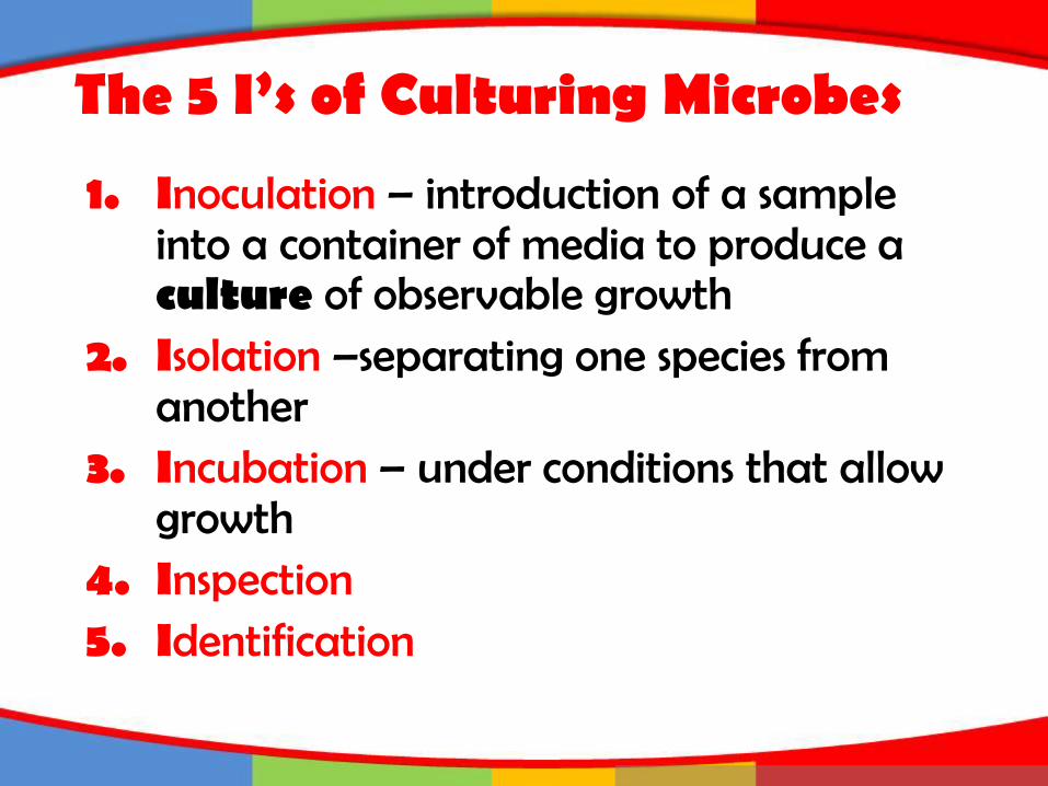
The 5 I’s of Culturing Microbes
1. Inoculation – introduction of a sample into a container of media to produce a culture of observable growth
2. Isolation –separating one species from another
3. Incubation – under conditions that allow growth
4. Inspection5. Identification

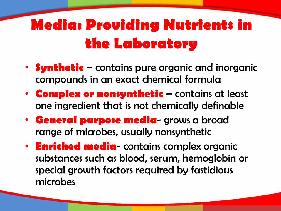
Media: Providing Nutrients in the Laboratory
Media can be classified according to threeproperties:1. Physical state – liquid, semisolid and solid
2. Chemical composition – synthetic (chemically defined) and nonsynthetic(complex)
3. Functional type – general purpose, enriched, selective, differential, anaerobic, transport, assay, enumeration

Media: Providing Nutrients in the Laboratory
• Most commonly used media:–nutrient broth – liquid medium
containing beef extract and peptone–nutrient agar – solid media
containing beef extract, peptone and agar
Media: Providing Nutrients in the Laboratory
• Synthetic – contains pure organic and inorganic compounds in an exact chemical formula
• Complex or nonsynthetic – contains at least one ingredient that is not chemically definable
• General purpose media- grows a broad range of microbes, usually nonsynthetic
• Enriched media- contains complex organic substances such as blood, serum, hemoglobin or special growth factors required by fastidious microbes
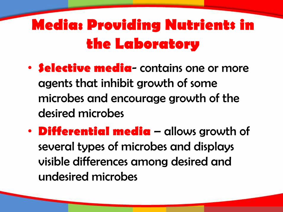
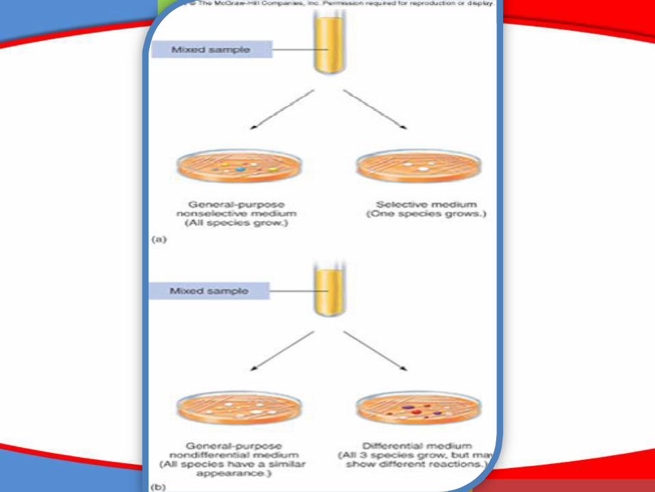
• Selective media- contains one or more agents that inhibit growth of some microbes and encourage growth of the desired microbes
• Differential media – allows growth of several types of microbes and displays visible differences among desired and undesired microbes
Media: Providing Nutrients in the Laboratory
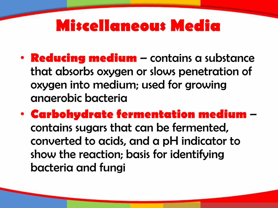
Miscellaneous Media
• Reducing medium – contains a substance that absorbs oxygen or slows penetration of oxygen into medium; used for growing anaerobic bacteria
• Carbohydrate fermentation medium –contains sugars that can be fermented, converted to acids, and a pH indicator to show the reaction; basis for identifying bacteria and fungi
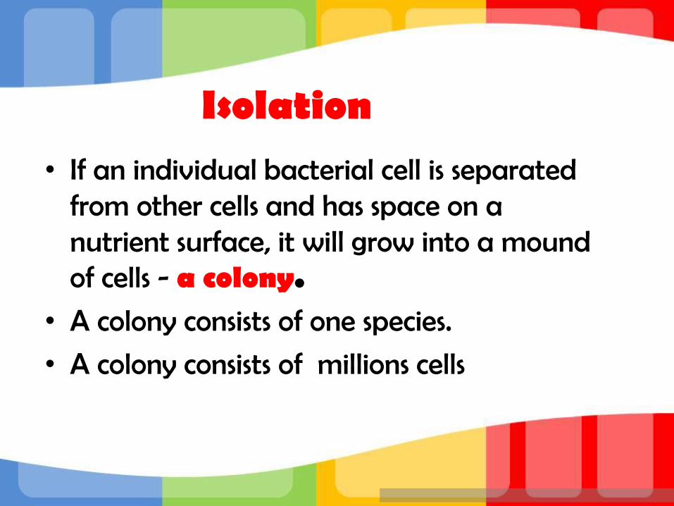
Isolation• If an individual bacterial cell is separated
from other cells and has space on a nutrient surface, it will grow into a mound of cells - a colony.
• A colony consists of one species.
• A colony consists of millions cells

Insert figure 3.2Isolation technique

Streak Plate
Spread Plate
Pour Plate
• Isolation techniques include:

Insert figure 3.3Isolation methods
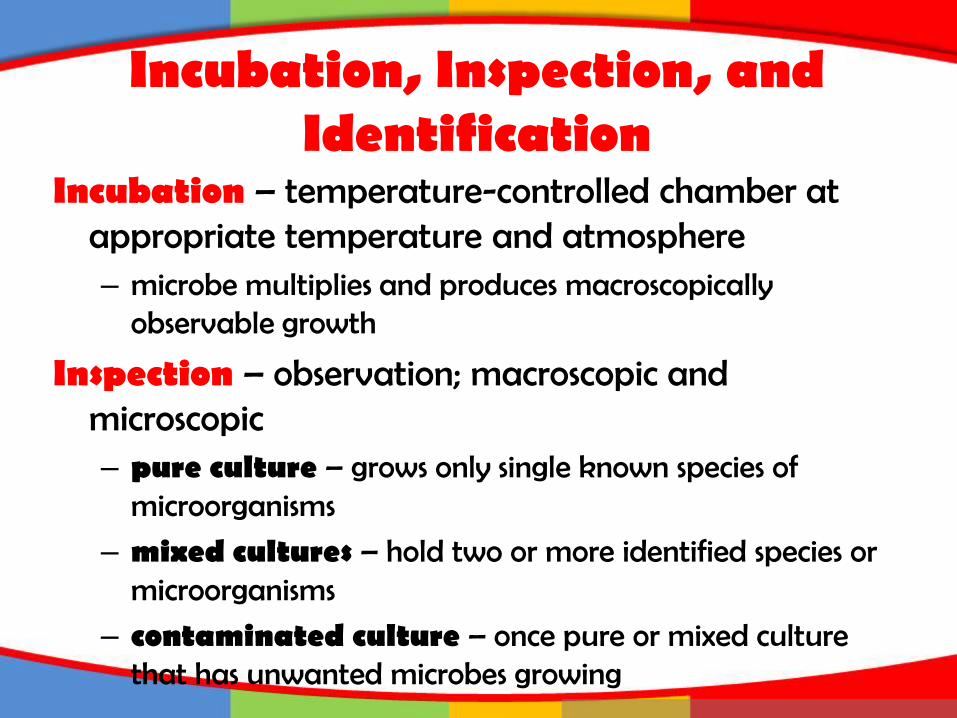
Incubation, Inspection, and Identification
Incubation – temperature-controlled chamber at appropriate temperature and atmosphere– microbe multiplies and produces macroscopically
observable growth
Inspection – observation; macroscopic and microscopic– pure culture – grows only single known species of
microorganisms
– mixed cultures – hold two or more identified species or microorganisms
– contaminated culture – once pure or mixed culture that has unwanted microbes growing
Incubation, Inspection, and Identification
Identification – macroscopic and microscopic appearance, biochemical tests, genetic characteristics, immunological testing
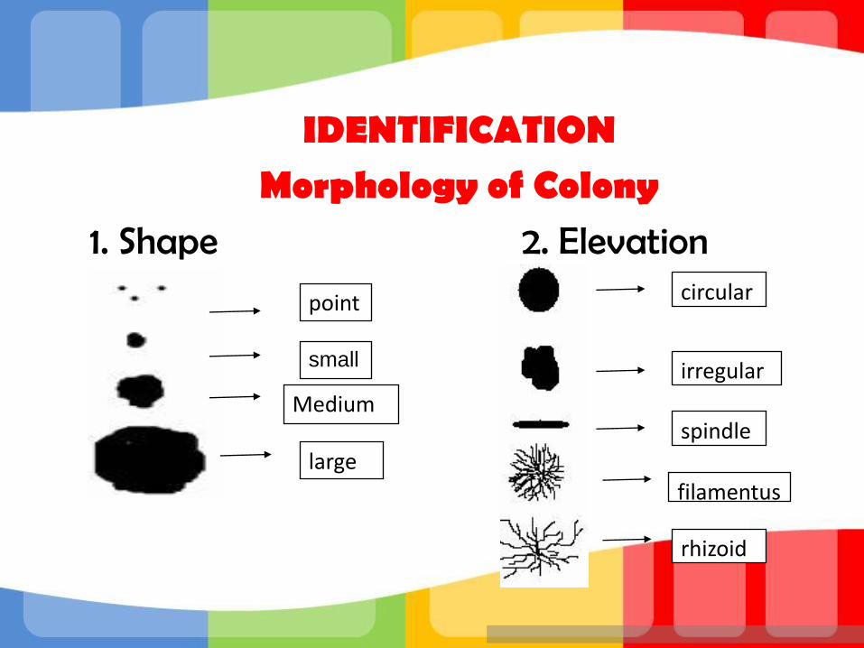
IDENTIFICATION Morphology of Colony
1. Shape 2. Elevation
point
small
Medium
large
circular
irregular
spindle
filamentus
rhizoid
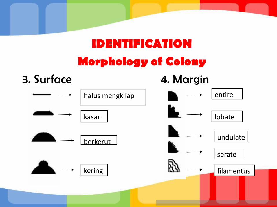
IDENTIFICATION Morphology of Colony
3. Surface 4. Marginhalus mengkilap
kasar
berkerut
kering
entire
lobate
undulate
serate
filamentus
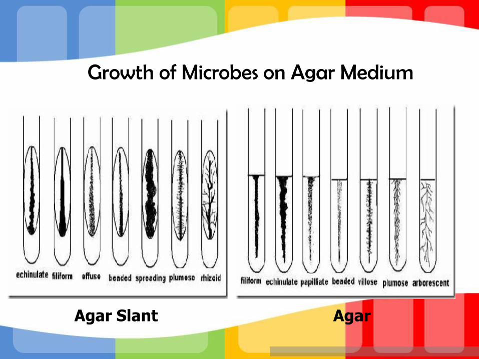
Growth of Microbes on Agar Medium
Agar Slant Agar

Specimen Preparation for Optical Microscopes
• Wet mounts and hanging drop mounts – allow examination of characteristics of live cells: motility, shape, and arrangement
• Fixed mounts are made by drying and heating a film of specimen. This smear is stained using dyes to permit visualization of cells or cell parts.
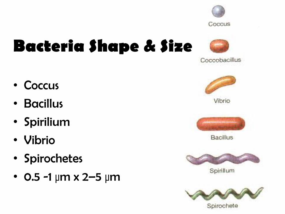
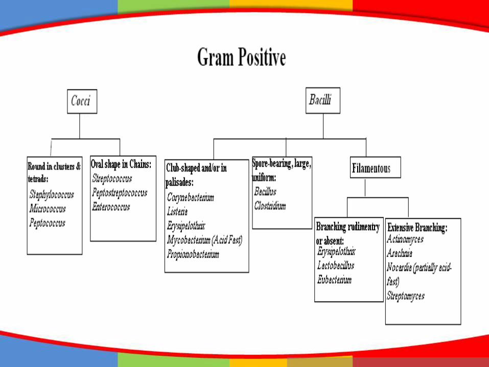
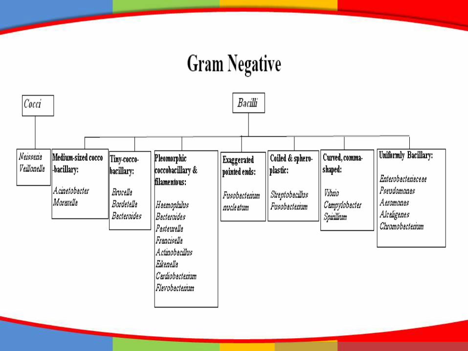
Bacteria Shape & Size
• Coccus
• Bacillus
• Spirilium
• Vibrio
• Spirochetes
• 0.5 -1 µm x 2–5 µm
Staining
Dyes create contrast by imparting a color to cells or cell parts.
• Basic dyes - cationic, with positive charges on the chromophore
• Acidic dyes - anionic, with negative charges on the chromophore
• Positive staining – surfaces of microbes are negatively charged and attract basic dyes
• Negative staining – microbe repels dye, the dye stains the background
Staining
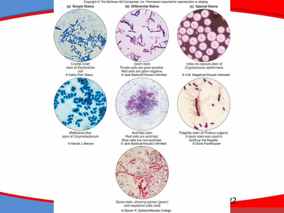
• Simple stains – one dye is used; reveals shape, size, and arrangement
• Differential stains – use a primary stain and a counterstain to distinguish cell types or parts (examples: gram stain, acid-fast stain and endospore stain)
• Special stains – reveal certain cell parts not revealed by conventional methods: capsule and flagellar stains
22
Insert figure 3.27Types of stains

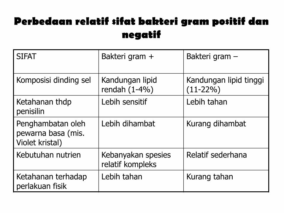
Perbedaan relatif sifat bakteri gram positif dannegatif
SIFAT Bakteri gram + Bakteri gram –
Komposisi dinding sel Kandungan lipid rendah (1-4%)
Kandungan lipid tinggi (11-22%)
Ketahanan thdp penisilin
Lebih sensitif Lebih tahan
Penghambatan oleh pewarna basa (mis. Violet kristal)
Lebih dihambat Kurang dihambat
Kebutuhan nutrien Kebanyakan spesies relatif kompleks
Relatif sederhana
Ketahanan terhadap perlakuan fisik
Lebih tahan Kurang tahan
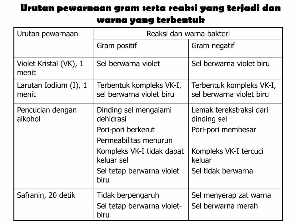
Urutan pewarnaan gram serta reaksi yang terjadi danwarna yang terbentuk
Urutan pewarnaan Reaksi dan warna bakteri
Gram positif Gram negatif
Violet Kristal (VK), 1 menit
Sel berwarna violet Sel berwarna violet biru
Larutan Iodium (I), 1 menit
Terbentuk kompleks VK-I, sel berwarna violet biru
Terbentuk kompleks VK-I, sel berwarna violet biru
Pencucian dengan alkohol
Dinding sel mengalami dehidrasi
Pori-pori berkerut
Permeabilitas menurun
Kompleks VK-I tidak dapat keluar sel
Sel tetap berwarna violet biru
Lemak terekstraksi dari dinding sel
Pori-pori membesar
Kompleks VK-I tercuci keluar
Sel tidak berwarna
Safranin, 20 detik Tidak berpengaruh
Sel tetap berwarna violet-biru
Sel menyerap zat warna
Sel berwarna merah
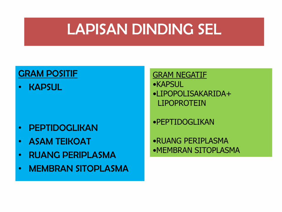
LAPISAN DINDING SEL
GRAM POSITIF
• KAPSUL
• PEPTIDOGLIKAN
• ASAM TEIKOAT
• RUANG PERIPLASMA
• MEMBRAN SITOPLASMA
GRAM NEGATIF•KAPSUL•LIPOPOLISAKARIDA+ LIPOPROTEIN
•PEPTIDOGLIKAN
•RUANG PERIPLASMA•MEMBRAN SITOPLASMA
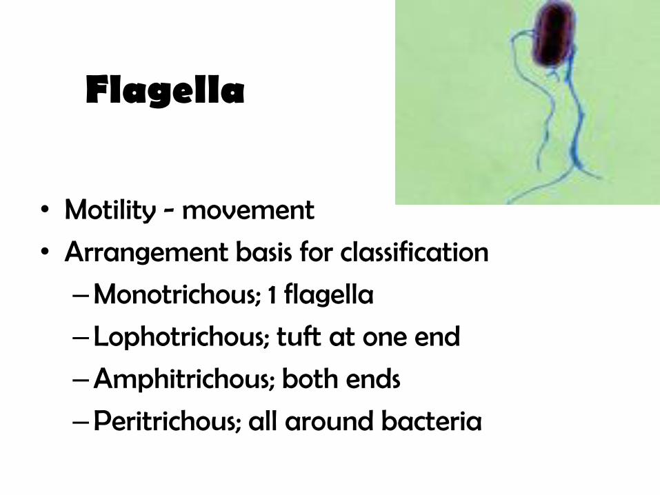
• Motility - movement
• Arrangement basis for classification
–Monotrichous; 1 flagella
–Lophotrichous; tuft at one end
–Amphitrichous; both ends
–Peritrichous; all around bacteria
Flagella

Flagella Type of Bacteria
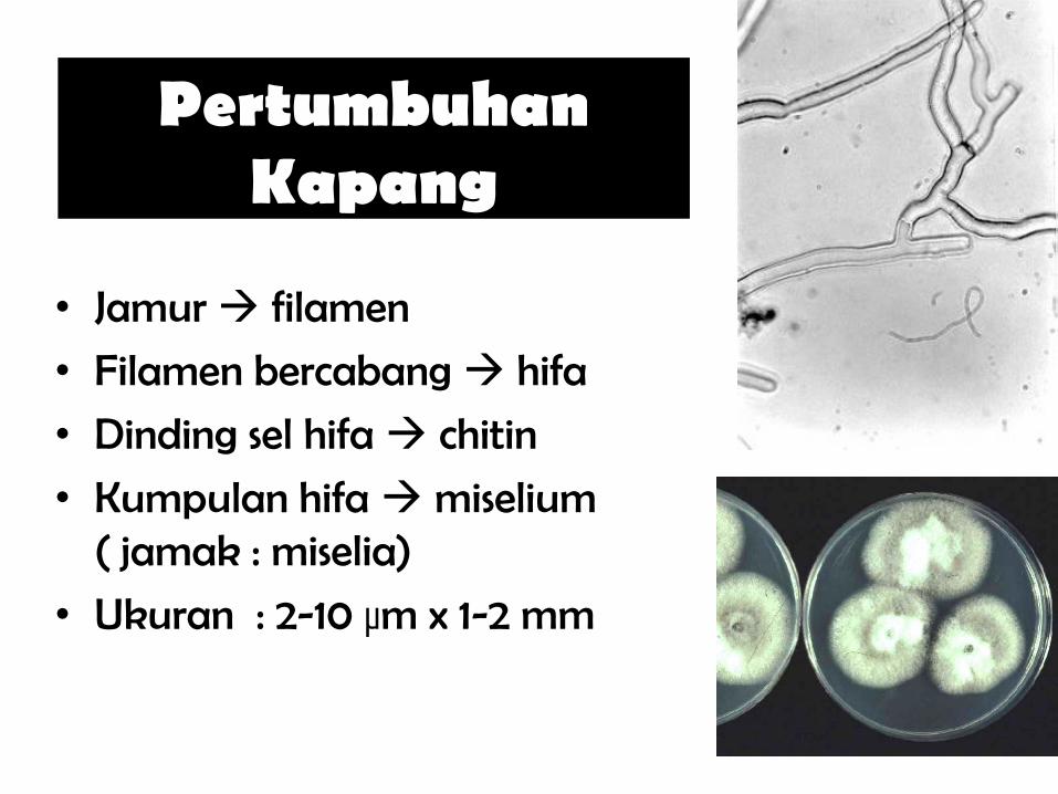
PertumbuhanKapang
• Jamur filamen
• Filamen bercabang hifa
• Dinding sel hifa chitin
• Kumpulan hifamiselium( jamak : miselia)
• Ukuran : 2-10 µm x 1-2 mm

Pertumbuhan Hifa
mycelium
germinating
spore
hifa
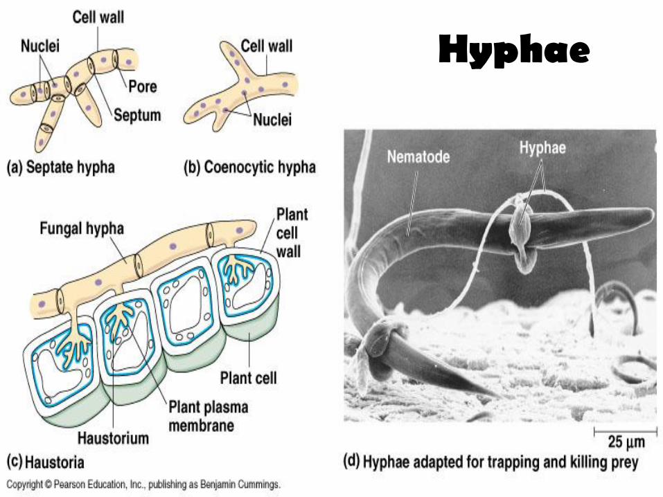
Hyphae
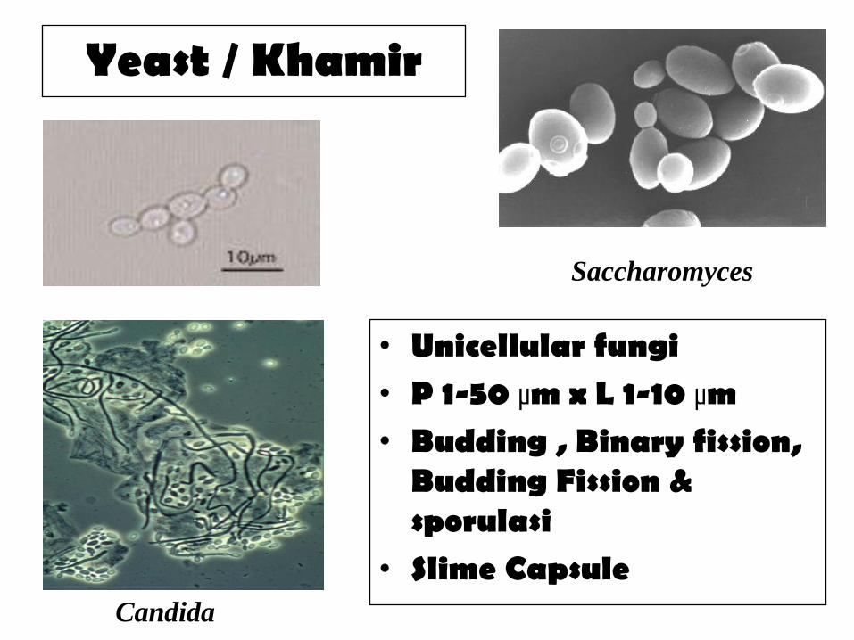
Yeast / Khamir
• Unicellular fungi• P 1-50 µm x L 1-10 µm • Budding , Binary fission,
Budding Fission & sporulasi
• Slime CapsuleCandida
Saccharomyces

IDENTIFICATION(Metabolic Testing)
Catalase Test :• To detect the presence of the enzyme catalase
• Catalase enzyme is found in most bacteria
• Breakdown of 3% hydrogen peroxide (H2O2) with the release of free Oxygen
• Catalase (+) bubbles formation
• False (+) using catalase containing medium
(ex : blood agar medium)
• False (-) using old culture
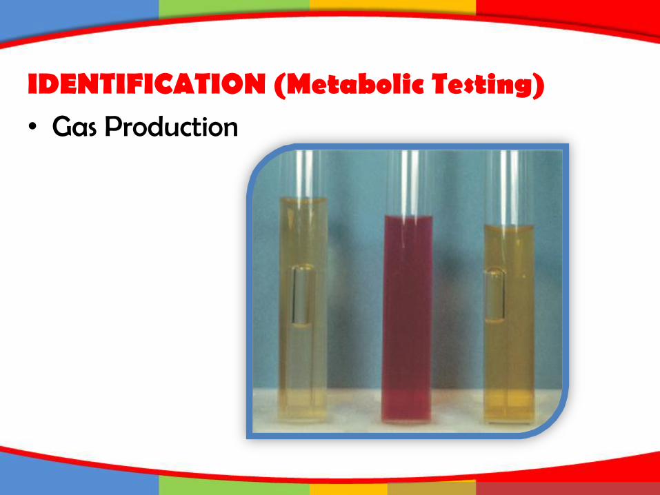
IDENTIFICATION (Metabolic Testing)• Gas Production
IDENTIFICATION(Metabolic Testing)
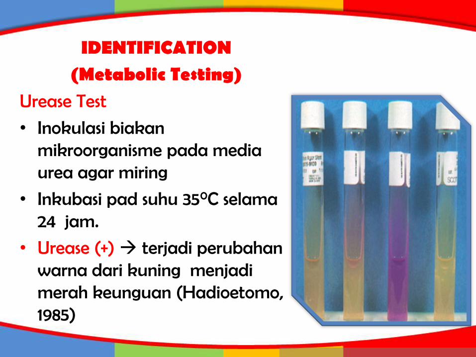
Urease Test
• Inokulasi biakan mikroorganisme pada media urea agar miring
• Inkubasi pad suhu 350C selama 24 jam.
• Urease (+) terjadi perubahan warna dari kuning menjadi merah keunguan (Hadioetomo, 1985)
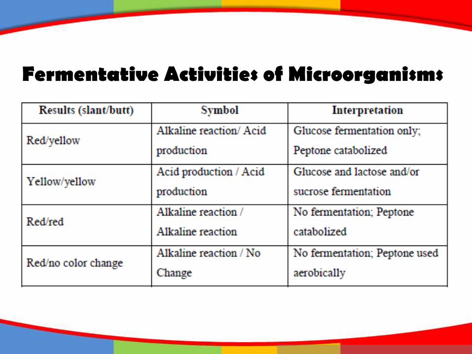
Fermentative Activities of Microorganisms
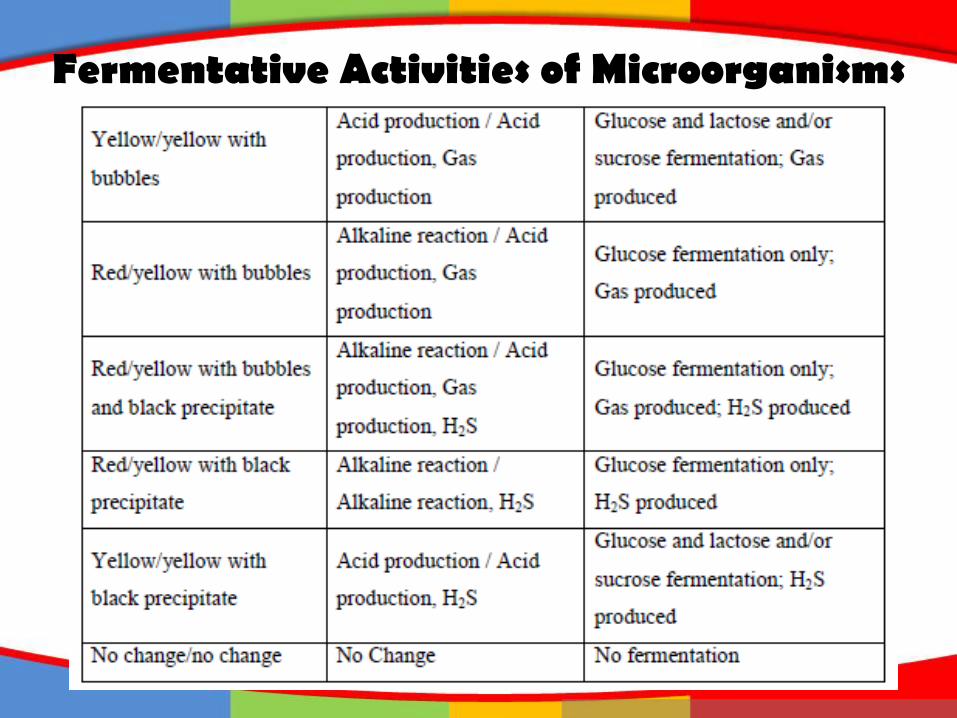
Fermentative Activities of Microorganisms

IDENTIFICATION(Metabolic Testing)
Triple Sugar Iron (TSI) :• Identifying Gram negative enteric Bacili based on
fermentation glucose, lactose, sucrose, and H2S production.
• Alkaline slant & alkaline butt (K/K) : nonfermenter
• Alkaline slant and acid butt (K/A) : glucose fermentation
• Acid slant & acid butt (A/A) : glucose, sucrose, lactose fermenter
• K/A/gas + H2S = glucose fermentation, gas n H2S production

IDENTIFICATION(Metabolic Testing)
Lysine Iron Agar (LIA) :• Determine wheteher a bacterium decarboxylates or
deaminates lysine and form H2S.
• Alkaline slant & acid butt (K/A) = glucose fermentation
• Alkaline slant & alkaline butt (K/K) =lysine decarboxylation or no fermentation
• Red slant and acid butt (R/A) = lysine deamination and glucose fermentation
IDENTIFICATION(Metabolic Testing)
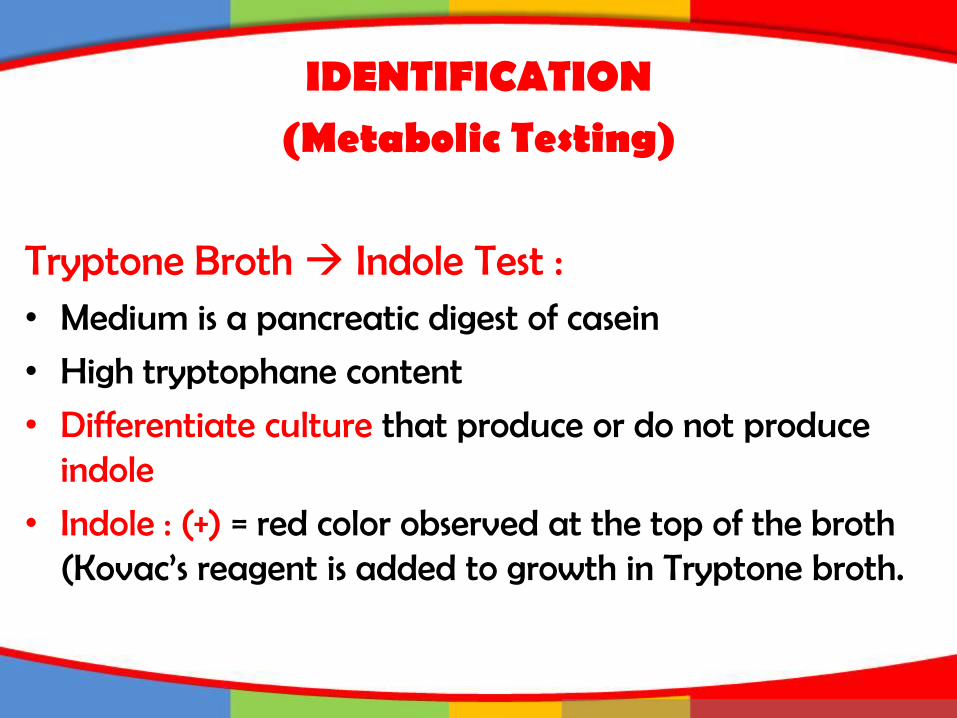
Tryptone Broth Indole Test :• Medium is a pancreatic digest of casein
• High tryptophane content
• Differentiate culture that produce or do not produce indole
• Indole : (+) = red color observed at the top of the broth (Kovac’s reagent is added to growth in Tryptone broth.
IDENTIFICATION(Metabolic Testing)
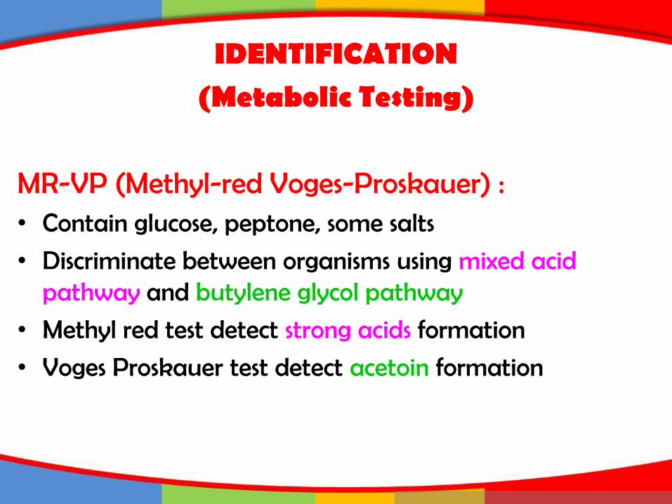
MR-VP (Methyl-red Voges-Proskauer) :• Contain glucose, peptone, some salts
• Discriminate between organisms using mixed acid pathway and butylene glycol pathway
• Methyl red test detect strong acids formation
• Voges Proskauer test detect acetoin formation

Karakteristik Escherichia coli
Karakteristik Salmonella spp.
Karakteristik Staphylococcus aureus

Escherichia coli
Karakteristik :Enterobacteriaceae
Gram (-)Flagellated rod-shaped
Facultative anaerobOxidase (-)Indole (+)Citrate (-)
Ferments glucoseProducing acid & gas
Optimum 35-37oC
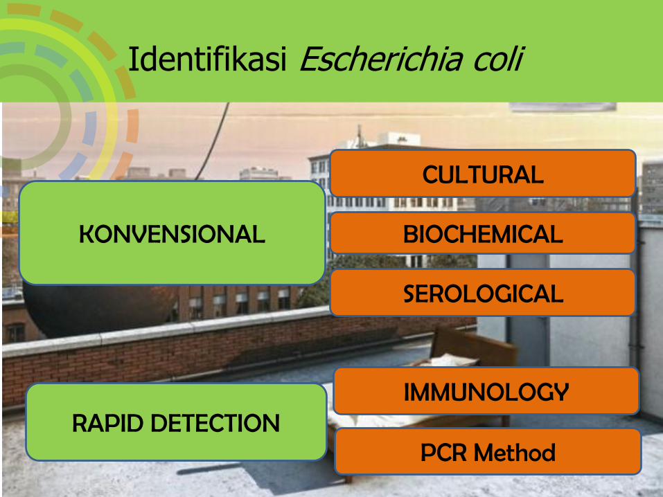
Identifikasi Escherichia coli
KONVENSIONAL
RAPID DETECTION
CULTURAL
BIOCHEMICAL
SEROLOGICAL
IMMUNOLOGY
PCR Method
Karakteristik Escherichia coli
Karakteristik Salmonella spp.
Karkateristik Staphylococcus aureus
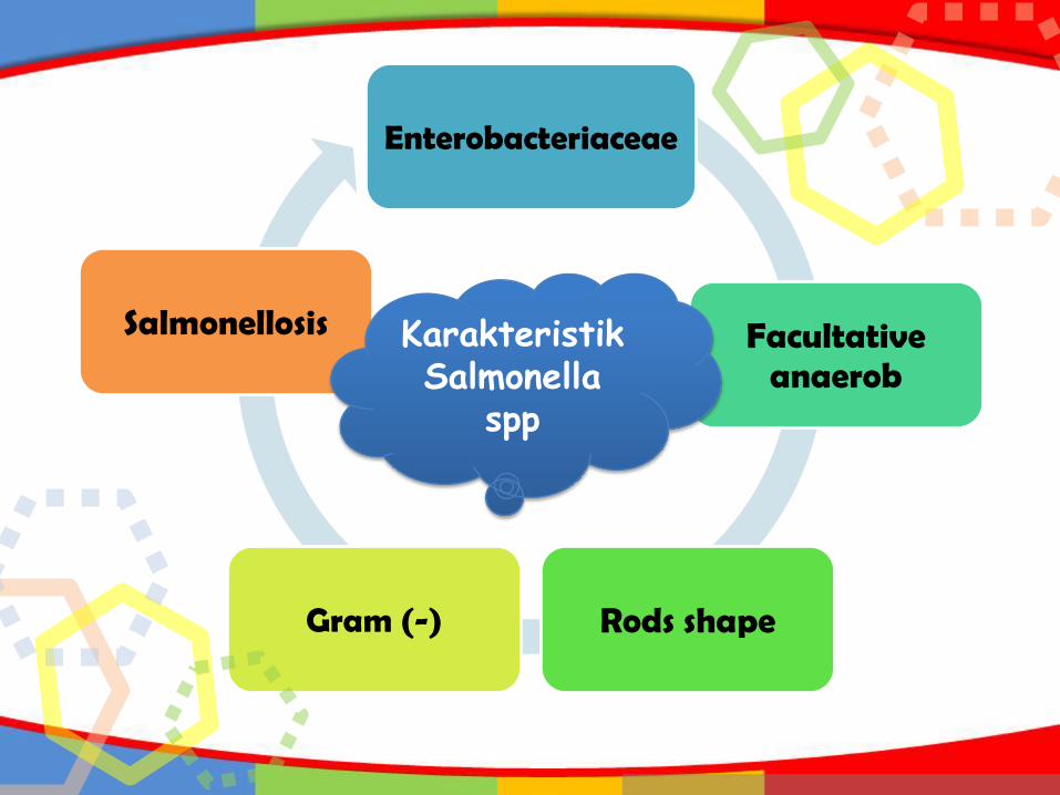
Enterobacteriaceae
Facultative anaerob
Rods shapeGram (-)
Salmonellosis Karakteristik Salmonella
spp
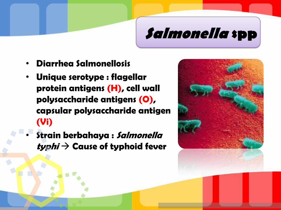
• Diarrhea Salmonellosis
• Unique serotype : flagellar protein antigens (H), cell wall polysaccharide antigens (O), capsular polysaccharide antigen (Vi)
• Strain berbahaya : Salmonella typhi Cause of typhoid fever
Salmonella spp

Biochemical Reactions
OrganismsTSI*
(Slant)
LIA**
(Slant)H2S Gas Indole MR VP Citrate
Citrobacter A/A K/A +/- + +/- + - +
E. coli A/A K/K - + + + - -
Edwardsiella K/A K/K +/- + + + - -
Enterobacter A/A K/A - + - - + +
Klebsiella A/A K/K - + - - + +
Morganella K/A R/A - + + + - -
Proteus A/A R/A +/- + +/- + - +/-
Providencia K/A R/A - - + + - +
Salmonella K/A K/K + + - + - +
Serratia A/A K/A - + - - + -
Shigella K/A K/A - - +/- + - -
*K=Alkaline (red), A=Acid (yellow or black [acid+H2S])** K=Alkaline (purple), no fermentation or lysine decarboxylation, A=Acid (yellow), R=deamination rx
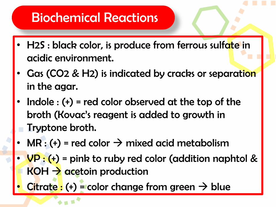
Biochemical Reactions
• H2S : black color, is produce from ferrous sulfate in acidic environment.
• Gas (CO2 & H2) is indicated by cracks or separation in the agar.
• Indole : (+) = red color observed at the top of the broth (Kovac’s reagent is added to growth in Tryptone broth.
• MR : (+) = red color mixed acid metabolism
• VP : (+) = pink to ruby red color (addition naphtol & KOH acetoin production
• Citrate : (+) = color change from green blue
Karakteristik Escherichia coli
Karakteristik Salmonella spp.
Karakteristik Staphylococcus aureus
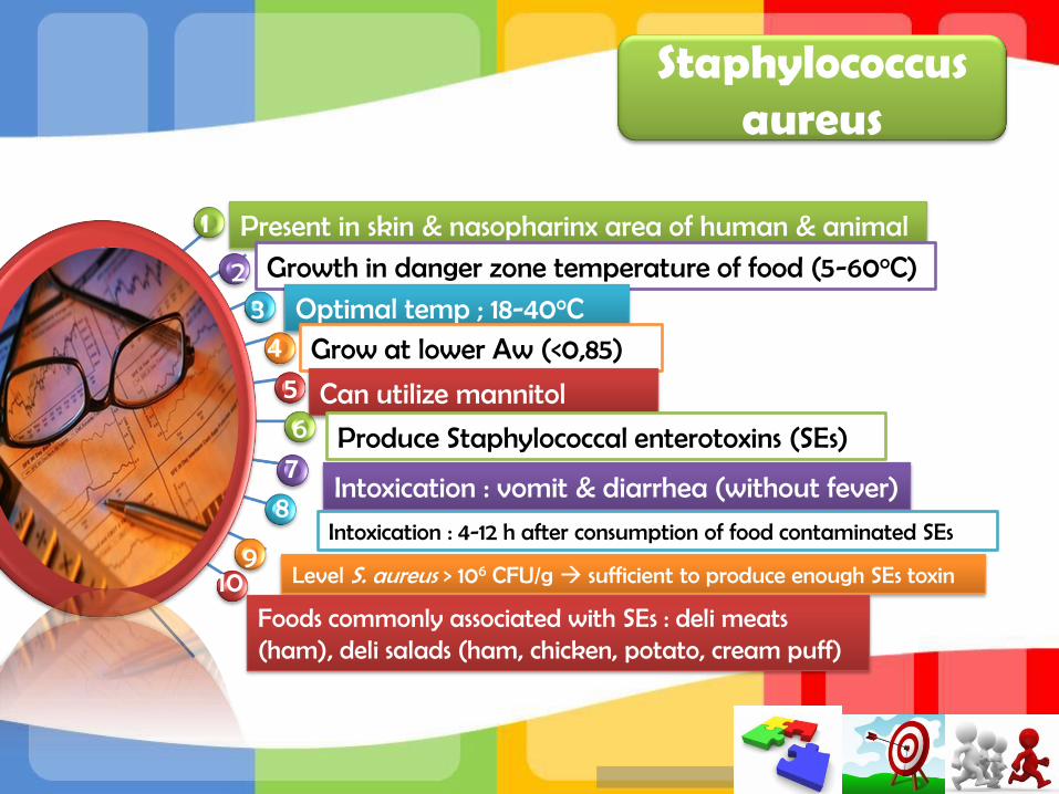
Staphylococcus aureus
Present in skin & nasopharinx area of human & animal
Growth in danger zone temperature of food (5-60oC)
Optimal temp ; 18-40oCGrow at lower Aw (<0,85)
Can utilize mannitol
Produce Staphylococcal enterotoxins (SEs)
Intoxication : vomit & diarrhea (without fever)
Intoxication : 4-12 h after consumption of food contaminated SEs
Level S. aureus > 106 CFU/g sufficient to produce enough SEs toxin
1
23
4
567
8
910
Foods commonly associated with SEs : deli meats (ham), deli salads (ham, chicken, potato, cream puff)


REFERENCES• Talaro KP. 2012. Foundation in Microbiology
sixth edition. McGraw-Hill Company
• Yousef AE and Carlstrom C. 2003. Food Microbiology: A Laboratory Manual. John Wiley and Sons, Inc. New Jersey
• McLandsborough L. 2005. Food Microbiology Laboratory. CRC Press LLC. Boca Raton

Terima kasih