Embed Size (px)
Citation preview
Proc. Natl. Acad. Sci. USAVol. 95, pp. 15781–15786, December 1998Neurobiology
Conserved structural features of the synaptic fusion complex:SNARE proteins reclassified as Q- and R-SNAREs
(membrane fusionyneurotransmissionyclostridial neurotoxins)
DIRK FASSHAUER*, R. BRYAN SUTTON†, AXEL T. BRUNGER†‡, AND REINHARD JAHN**Department of Neurobiology, Max Planck Institute for Biophysical Chemistry, D-37077 Gottingen, Germany; and †Howard Hughes Medical Institute andDepartment of Molecular Biophysics and Biochemistry, Yale University, New Haven, CT 06520
Communicated by Peter B. Moore, Yale University, New Haven, CT, October 27, 1998 (received for review September 22, 1998)
ABSTRACT SNARE [soluble NSF (N-ethylmaleimide-sensitive fusion protein) attachment protein receptor] proteinsare essential for membrane fusion and are conserved from yeastto humans. Sequence alignments of the most conserved regionswere mapped onto the recently solved crystal structure of theheterotrimeric synaptic fusion complex. The association of thefour a-helices in the synaptic fusion complex structure produceshighly conserved layers of interacting amino acid side chains inthe center of the four-helix bundle. Mutations in these layersreduce complex stability and cause defects in membrane trafficeven in distantly related SNAREs. When syntaxin-4 is modeledinto the synaptic fusion complex as a replacement of syntaxin-1A,no major steric clashes arise and the most variable amino acidslocalize to the outer surface of the complex. We conclude that themain structural features of the neuronal complex are highlyconserved during evolution. On the basis of these features wehave reclassified SNARE proteins into Q-SNAREs and R-SNAREs, and we propose that fusion-competent SNARE com-plexes generally consist of four-helix bundles composed of threeQ-SNAREs and one R-SNARE.
Intracellular membrane fusion involves conserved sets of mem-brane proteins that are commonly referred to as SNARE proteins[soluble NSF (N-ethylmaleimide-sensitive fusion protein) attach-ment protein receptor proteins] (1–4). SNARE proteins can begrouped into several small protein families with a growingnumber of members. The similarity between distant members ofthese protein families is rather limited, but it is thought that theyall operate by means of a common mechanism. The variantsfunctioning in neuronal exocytosis are among the best charac-terized; they include the synaptic vesicle protein synaptobrevin(also referred to as VAMP) and the synaptic plasma membraneproteins SNAP-25 and syntaxin-1A. These proteins readily as-semble into a stable ternary complex whose core structure hasbeen recently solved by x-ray crystallography (5). The SNAREcomplex can be reversibly disassembled by the ATPase NSF inconjunction with soluble cofactors termed SNAPs (soluble NSFattachment proteins) (6, 7). The formation of the assembledcomplex is now believed to be a critical step leading to membranefusion. Assembly proceeds spontaneously from less structuredmonomers and results in a stoichiometric and elongated complexwith all membrane anchor domains located at one side of therod-shaped particle (2, 5, 8, 9). These findings led to a model thatassembly juxtaposes membranes, thus overcoming the free energybarrier for fusion (2, 5, 9, 10). However, it remains to beestablished whether more distantly related SNARE proteinsform similar complexes and which of the structural features of theneuronal complex are generally relevant for SNARE proteinfunction.
METHODSSequence Analysis. Sequences were aligned by using the
CLUSTALW software available at http:yywww2.ebi.ac.ukyclustalw (11). A nearest-neighbor dendrogram of the SNAP-25, syntaxin, and synaptobrevin families was obtained and usedto define subgroups consisting of syntaxin-1 through syn-taxin-4, all known homologues of SNAP-25 excluding thedistant yeast Sec9p and Sto20p homologues, and synaptobre-vin-I, synaptobrevin-II and cellubrevin. The sequence varia-tion was computed as follows: The amino acids were separatedinto five classes, hydrophobic (Ala, Val, Phe, Ile, Leu, Pro,Met), positively charged (Lys, Arg), negatively charged (Asp,Glu), hydrophilic (Ser, Thr, Tyr, Cys, Asn, Gln, His, Trp), andglycine. The sequence variation was defined as
E 5 2Oi51
5
f~i , t!log2 f~i , t!,
where f(i, t) is the frequency of the particular amino acid classat position i (12).
Modeling. The accessible surface area of the synaptic fusioncomplex was computed by the method of Lee and Richards(13). The syntaxin-4 substitution was modeled, based on thesynaptic fusion complex crystal structure (5), by substitutingthe residues that differ between syntaxin-4 and syntaxin-1,using the programs O and SOD (14). The side-chain rotamerwas transferred from the original residue to the substitutedresidue where possible; otherwise the most favorable sidechain rotamer was picked. The model was regularized by usingenergy minimization as implemented in the Crystallographyand NMR System (CNS) (15).
RESULTS AND DISCUSSIONFunctional Significance and Evolutionary Conservation of the
Central Layer Domain. Primary sequence comparison withclosely and distantly related homologues shows that the aminoacids in the ionic layer (designated as ‘‘0’’ layer, ref. 5 and Fig. 1B)at the center of the synaptic fusion complex are the most highlyconserved residues in all SNARE proteins (16) (Fig. 1A). Thislayer is composed of Arg-56 of synaptobrevin-II, Gln-226 ofsyntaxin-1A, and Gln-53 and Gln-174 of SNAP-25B (5). Aminoacid substitutions in this layer severely disrupt membrane traffic.For example, Sec22p, a distantly related yeast synaptobrevinhomologue, participates in the fusion of transport vesicles withthe cis face of the Golgi apparatus (17). Replacing the conservedarginine with glycine results in defects that are comparable to adeletion of the gene (18, 19). Trafficking is also perturbed by adouble mutation in the yeast SNARE Vti1p that affects both the
The publication costs of this article were defrayed in part by page chargepayment. This article must therefore be hereby marked ‘‘advertisement’’ inaccordance with 18 U.S.C. §1734 solely to indicate this fact.
© 1998 by The National Academy of Sciences 0027-8424y98y9515781-6$2.00y0PNAS is available online at www.pnas.org.
Abbreviations: NSF, N-ethylmaleimide-sensitive fusion protein; SNARE,soluble NSF attachment protein receptor; SNAP, soluble NSF attach-ment protein; BoNT, botulinum neurotoxin; TeNT, tetanus neurotoxin.‡To whom reprint requests should be addressed. e-mail: [email protected].
15781
Dow
nloa
ded
by g
uest
on
Dec
embe
r 4,
202
1

0 layer and the 25 layer, Gln-158 3 Arg and Ala-141 3 Ser,respectively (20) (Fig. 1A).
The layers flanking the ionic 0 layer are maintained pri-marily by hydrophobic interactions (5). The functional rele-vance of these layers is highlighted by phenotypes in differentspecies that are caused by mutations resulting from single
amino acid substitutions. Interactions in the 11 layer aredisrupted by a loss-of-function mutation in the sec9–7 allele,Leu-627 3 His (Ile-178 in SNAP-25B) (21, 22). Anotherexample of a mutation in the 11 layer is provided by the bos1–1allele of a yeast homologue of the SNARE family (Fig. 1 A).In this case, a Leu-1903 Ser mutation results in lower stability
B
A
FIG. 1. (A) Sequence alignment (16) of the four-helix bundle region of the synaptic fusion complex (5) for a representative subset of the entire SNAREfamily. The sequence analysis was restricted to 16 layers (blue) of the four-helix bundle in the synaptic fusion complex (5), including 7 layers upstream(layers 21 to 27) and 8 layers downstream (layers 11 to 18) of the ionic layer (layer 0). Conserved residues are shaded in gray. The conserved glutamineand arginine residues forming the ionic 0 layer are indicated in red and green, respectively. The mutations discussed in the text are indicated by blackshaded boxes. There are two alignment tables for the SNAP-25 family that correspond to the two SNAP-25 a-helices in the synaptic fusion complex (5).GenBank accession numbers for the syntaxin family are as follows: sx1a, RN, P32851; unc-64b, CE, AF047885; sx3, RN, Q08849; sx4, RN, Q08850; Sso1,SC, P32867; sed5, SC, Q01590; sx5, RN, Q08851; vam3, SC, Q12241. Accession numbers for the SNAP-25 family are SNAP-25B, HS, P13795; syndet,MM, U73143; SNAP-25, DM, U81153; Y22F5A.5, CE, AL021479; sec9, SC, L34336; and spo20, SC, Z49211. Accession numbers for the bos-group arebet1, SC, P22804; mbet1, MM, AF007552; vti1, HS, AF035824; vti1, SC, AF006074; membrin, RN, U91539; bos1, SC, P25385; and vam7, SC, P32912.Accession numbers for the synaptobrevinyvamp family (R-SNAREs) are sb2, RN, M24105; cbycellubrevin, RN, S63830; sb1 CE AF003281; sb, HM,U85805; Snc1, SC, M91157; sb5y6, HS, AA222692; sb7, MM, X96737; sec22 (Sly2), SC, L8479; sec22b, MM, U91538; nyv1, SC, Z73265; and tomosyn,RN, U92072. The two-letter species abbreviations after the protein name are as follows: HS, Homo sapiens; MM, Mus musculus; RN, Rattus norvegicus;SC, Saccharomyces cerevisiae; DM, Drosophila melanogaster; TM, Torpedo marmorata; CE, Caenorhabditis elegans; and HM, Hirudo medicinalis. (B) Layersof the synaptic fusion complex crystal structure (5). Indicated are Ca traces (gray), local helical axes (blue, red, and green for synaptobrevin-II, syntaxin-1A,and SNAP-25b, respectively), and layers (black) by virtual bonds between corresponding Ca positions.
15782 Neurobiology: Fasshauer et al. Proc. Natl. Acad. Sci. USA 95 (1998)
Dow
nloa
ded
by g
uest
on
Dec
embe
r 4,
202
1
of the SNARE complex in vitro and in a disruption ofmembrane traffic in vivo. In terms of the synaptic fusioncomplex, this hydrophilic substitution could destabilize thehydrophobic leucine zipper interactions in the 11 layer. Thereare also known single-site mutations that map to the outerlayers of the fusion complex: C. elegans unc-64 Ala-2413 Val(Ala-240 in syntaxin-1A) and Ala-248 3 Val (Ala-247 insyntaxin-1A), and C. elegans synaptobrevin Leu-62 3 Phe(Leu-70 in synaptobrevin-II), and Ala-66 3 Gly (Ala-74 insynaptobrevin-II). These single-site mutations in the 14, 15,and 16 layers affect synaptic transmission, resulting in lethar-gic animals with locomotory abnormalities. Interestingly, dou-ble mutants involving neighboring layers produced synergisticeffects with severe defects in neurotransmission (23). Thesemore severe phenotypes may be explained by disruption ofpacking interactions between the layers. Another example ofa single amino acid substitution is the exchange of Thr-254 forIle-254 in layer 17 of Drosophila syntaxin-1A (Thr-251 in ratsyntaxin-1A). This mutation is characterized by a temperature-induced block of synaptic transmission, the lack of detectableSNARE complexes in neuronal extracts, and reduced bindingto synaptobrevin in vitro (24).
The complementary nature of the ionic 0 layer reflects itsability to form strong hydrogen bonds between the three glu-tamine residues and the guanidino group of the arginine sidechain. A different type of asymmetric ‘‘complementarity’’ isfound in layers 23, 22, and 16 (5), where bulky side chains arepacked together with smaller ones. These layers may enforce thecorrect register between the a-helical components of the fusioncomplex. Some of these layers could also enforce the correct
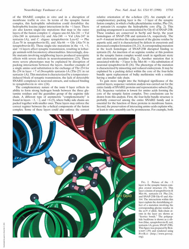
relative orientation of the a-helices (25). An example of acomplementary packing layer is the 23 layer of the synapticfusion complex, in which a bulky phenylalanine residue, Phe-216,of syntaxin-1A occupies the hydrophobic core (Fig. 2). Thispacking arrangement is accommodated by Gly-43 of SNAP-25B.These residues are conserved in Sec9p and Sso1p, the yeasthomologues of SNAP-25B and syntaxin-1A, respectively. Thesec9–4 mutant involves the replacement of the glycine residue byaspartic acid, and it is characterized by defects in exocytosis anddecreased complex formation (18, 21). A corresponding mutationin the leech homologue of SNAP-25B disrupted binding tosyntaxin (8). An insertion of an arginine residue at this positionin the synaptic fusion complex would result in significant stericand electrostatic penalties (Fig. 2). Another mutation that isassociated with the 23 layer is the Met-463 Ala substitution ofneuronal synaptobrevin-II (26). The phenotype of the mutationis characterized by missorting and reduced endocytosis. It may beexplained by a packing defect within the core of the four-helixbundle upon replacement of bulky methionine with a residuehaving a smaller side chain.
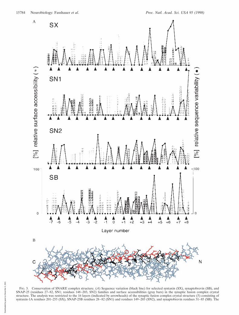
To gain more insight into the biological significance of thecentral layers, sequence variation analysis was carried out for theentire family of SNARE proteins and representative subsets (Fig.3A). Sequence variation is lowest for amino acids forming thecore of the synaptic fusion complex. Two conclusions can bedrawn from this analysis. First, the four-helix bundle structure isprobably conserved among the entire SNARE family and isessential for the function of these proteins in membrane fusion.Second, the preservation of interacting amino acids explains why,at least in vitro, assembly can be promiscuous. For example, both
FIG. 2. Picture of the 23layer in the synaptic fusion com-plex crystal structure (5). Thislayer consists of synaptobrevin-IIMet-46, syntaxin-1A Phe-216,and SNAP-25B Gly-43 and Ala-164. The interactions within thislayer explain the destabilizing ef-fect of a mutation involving thecorresponding glycine residue inthe sec9–4 allele. Side-chain at-oms in the layer are shown as‘‘licorice bonds.’’ The polypep-tide backbone is shown as a rib-bon (blue, synaptobrevin-II; red,syntaxin-1A; green, SNAP-25B).This figure was prepared by BOB-SCRIPT (39) and rendered usingPOVRAY (http:yywww.povray.org).
Neurobiology: Fasshauer et al. Proc. Natl. Acad. Sci. USA 95 (1998) 15783
Dow
nloa
ded
by g
uest
on
Dec
embe
r 4,
202
1
B
A
FIG. 3. Conservation of SNARE complex structure. (A) Sequence variation (black line) for selected syntaxin (SX), synaptobrevin (SB), andSNAP-25 (residues 27–82, SN1; residues 148–203, SN2) families and surface accessibilities (gray bars) in the synaptic fusion complex crystalstructure. The analysis was restricted to the 16 layers (indicated by arrowheads) of the synaptic fusion complex crystal structure (5) consisting ofsyntaxin-1A residues 201–255 (SX), SNAP-25B residues 28–82 (SN1) and residues 149–203 (SN2), and synaptobrevin residues 31–85 (SB). The
15784 Neurobiology: Fasshauer et al. Proc. Natl. Acad. Sci. USA 95 (1998)
Dow
nloa
ded
by g
uest
on
Dec
embe
r 4,
202
1

syntaxin-1A and syntaxin-4 can form a complex with synapto-brevin-II and SNAP-25B in vitro (27). Modeling of this complexbased on the crystal structure of the syntaxin-1AzSNAP-25Bzsynaptobrevin-II complex (5) required no major rearrange-ments because most substitutions occur on the surface of thecomplex (Fig. 3B). Furthermore, several of the yeast homologuesinvolved in trafficking to and from the Golgi apparatus appear tofunction in complexes with different SNARE partners (28).Therefore, in contrast to one of the postulates of the ‘‘SNARE-hypothesis’’ (3), other factors may determine SNARE bindingspecificity. It should be mentioned, however, that there arenonconserved core residues in the carboxyl-terminal region ofsyntaxin and synaptobrevin (Fig. 3A). These residues may affectmembrane fusion activity by reducing structural integrity in thecarboxyl-terminal region when mismatched SNAREs interact.
Conservation of Surface Features and the Potential Role ofDivalent Cation Binding Sites. While most surface residues of thesynaptic fusion complex are highly variable (Fig. 3A), somesurface features are conserved among subgroups of SNAREhomologues. For instance, the acidic residues forming a charac-teristic acidic patch in the middle of the synaptic fusion complexstructure (5) are conserved among several homologues, but arenot found in the yeast family members. Distinct acidic, basic, andhydrophobic patches on the four-helix bundle surface may de-
termine the ability of the SNARE complex to interact withdifferent effector proteins, such as synaptotagmin, complexin,a-SNAP, and NSF. The binding sites of these proteins remain tobe clarified.
The SNARE proteins are the target for the clostridialneurotoxins, including botulinum (BoNT) and tetanus (TeNT)neurotoxins. As outlined before, the parallel orientation ofboth SNAP-25B a-helices in the crystal structure places allneurotoxin cleavage sites (10) into two distinct regions be-tween the carboxyl-terminal membrane anchors and the ionic0 layer (5). The crystal structure of synaptic fusion complexrevealed a number of potential divalent cation binding sites onthe surface (5). One such site is close to the cleavage sites ofTeNT and BoNTyA (Fig. 4). It is possible that this potentialcation binding site is involved in Ca21-mediated triggering ofexocytosis. Indeed, shortening of SNAP-25B by BoNTyAresults in a partial block of exocytosis that can be overcome byincreased Ca21 levels, suggesting a possible link between Ca21
binding and fusion complex function (29–32).Structure-Based Reclassification of SNARE Proteins into
‘‘Q-SNAREs’’ and ‘‘R-SNAREs.’’ The conserved properties ofSNARE complexes provide a structural basis for a regrouping ofSNARE proteins. Previously, SNARE proteins were divided into‘‘v-SNAREs’’ (including proteins homologous to synaptobrevin)
FIG. 4. A binding site forSr21 is close to the TeNT andBoNTyA toxin cleavage sites ofthe synaptic fusion complex (5).This binding site for Sr21 isfound in two of the three com-plexes in the asymmetric unit ofthe crystal structure of the syn-aptic fusion complex (5). Thedivalent cation is coordinated bythe conserved (among the syn-aptobrevin family selected in Fig.3A) synaptobrevin-II Ser-75,Glu-78, and partially conservedThr-79 residues. The proteinbackbone is represented as a rib-bon drawing. Blue, synaptobre-vin-II; red, syntaxin-1A; green,SNAP-25B. The indicated toxincleavage sites are located at thepeptide bonds between Gln-76and Phe-77 (TeNT and BoNTyB), and between Ala-81 andAla-82 (BoNTyG) for synapto-brevin-II, between Lys-253 andAla-254 (BoNTyC) for syntaxin-1A, and between Gln-197 andArg-198 (BoNTyA) for SNAP-25B. The residues adjacent to thescissile bond are shown as lico-rice bonds. This figure preparedby BOBSCRIPT (36) and renderedby using POVRAY.
layer numbers refer to Fig. 1B. The GenBank accession numbers used for the syntaxin-1 through -4 family are as follows: sx-1b, RN, P32853; sx1b, BS,P41414; sx-1a, RN, P32851; sx-1a, HS, L37792; sx-1a, MM, D45208; sx-A, HS, U12918; sx1a, BS, P32850; sx1, HM, U85807; sx, AC, U03123; sx1a, DM,L37732; unc-64b, CE, AF047885; sx, SP, AF014122; sx2, RN, P50279; sx3, RN, Q08849; sx3a, HS, AJ002076; sx4, RN, Q08850; sx4, HS, X85784; and sx4,MM, U76832. The accession numbers used for the SNAP-25 family are SNAP-25B, HS, P13795; SNAP-25, TM, P36976; syndet, MM, U73143; SNAP-25,SP, AF036902; SNAP-25, DM, U81153; SNAP-25, HM, U85806; SNAP-25, GG, L09253; SNAP-25A, CA, L22973; SNAP-25D, CA, L22976; andY22F5A.5, CE, AL021479. The accession numbers used for the synaptobrevin family members in the second branch of the dendrogram (not shown) areSb1, RN, M24104; Sb1B, RN, U74621; Sb1, MM, AF007167; Sb1, TM, P13701; Syb-B, DM, L14270; Syb-A, DM, L14270; N-Syb, DM, S66686; Sb, AC,P35589; cellubrevin (Sb3), MM U61751; cellubrevin (Sb3), RN S63830; Sb, SP, AF014119; Sb2, RN, M24105; Sb2, HS, AJ5044; Sb2, MM, AF007168;Sb, HM, U85805; Sb, FR, AF016494; Sb, LP, X74748; XSybI, XL, AF035016; Syb2, XL, U16801; Sb, BT, X76199; Snb1, CE, AF003281; and Sb, SM,U30182. The following two-letter species abbreviations are used: CA, Carrassius auratus; HS, Homo sapiens; MM, Mus musculus; RN, Rattus norvegicus;SC, Saccharomyces cerevisiae; DM, Drosophila melanogaster; TM, Torpedo marmorata; CE, Caenorhabditis elegans; SP, Strongylocentrotus purpuratus; HM,Hirudo medicinalis; GG, Gallus gallus; BT, Bos taurus; AC, Aplysia californica; SM, Schistosoma mansoni; FR, Fugu rubripes; XL, Xenopus laevis; and LP,Loligo pealei. (B) Model of a synaptic fusion complex consisting of syntaxin-4, SNAP-25B, and synaptobrevin-II, based on the crystal structure of thesynaptic fusion complex (5), which contains syntaxin-1A. Syntaxin-4 residues are shown in black and red for matching and different residue types betweensyntaxin-1A and syntaxin-4, respectively. Most substituted residues occur on the surface of the complex.
Neurobiology: Fasshauer et al. Proc. Natl. Acad. Sci. USA 95 (1998) 15785
Dow
nloa
ded
by g
uest
on
Dec
embe
r 4,
202
1
and ‘‘t-SNAREs’’ (including proteins homologous to syntaxin-1and SNAP-25), based on their preferred localization on either thetrafficking vesicle (v) or the target membrane (t), respectively (3,6). As outlined below, this classification scheme may not beaccurate for all vesicular transport steps. Furthermore, it does notcover homotypic fusion events—i.e., fusion between vesicles thatare functionally and structurally equivalent. Here we propose areclassification of the SNARE proteins based on their contribu-tions to the ionic 0 layer. R-SNAREs would provide an arginine(R) to this ionic layer and Q-SNAREs would provide comple-mentary glutamines (Q) (Fig. 1A). Presently, there are only twoexceptions to this convention. Assuming correct primary se-quence alignment, yeast Bet1p would provide a serine to thelayer. Leech synaptobrevin would provide a lysine to the layerinstead of an arginine, resulting in a modified layer geometry.Although the R-SNAREs include most of the proteins previouslyclassified as v-SNAREs, there are no structural reasons why thea-helices provided by the trafficking vesicles need to be derivedonly from R-SNAREs, as in the case of the synaptic fusioncomplex.
At present, only few SNARE complexes have been character-ized in which all partners have been identified and localized, suchas the SNARE complex involved in yeast exocytosis (21, 22). Forother trafficking steps, relevant SNARE proteins are known, butit is unclear which of them interact in a particular fusion step. Awell studied example is the vesicular traffic between the endo-plasmic reticulum and the Golgi apparatus. Despite the assign-ment of several SNARE proteins it remains to be establishedwhich of them function in anterograde vs. retrograde traffic andwhether intermediate fusion steps are involved (33). Antero-gradely transported vesicles contain both an R-SNARE (Sec22p)and a Q-SNARE (Bos1p), which appear to form a complex withother SNAREs, probably involving Sed5p, a Q-SNARE. If theneuronal SNARE structure is conserved, the complex wouldconsist of Sec22p (R-SNARE)yBos1p (Q-SNARE)ySed5p (QSNARE) and a fourth Q-SNARE helix which may be contrib-uted either by an additional SNARE or by a second copy of Bos1por Sed5p. When applied to a SNARE complex that functions in‘‘homotypic’’ vacuolar fusion, consisting of Nyv1p (R-SNARE),Vam3p (Q-SNARE), Vti1p (Q-SNARE), and Vam7p (Q-SNARE) (34), our convention predicts the formation of atetrameric complex. Each Q-SNARE would contribute only asingle a-helix to the four-helix bundle of this putative complex.
Although all characterized SNARE complexes involved inmembrane fusion are of the 3 Q-SNAREy1 (R-SNARE) type,defined complexes have been reported that consist only ofQ-SNAREs (9, 35, 36). It remains to be established whetherthese complexes are also four helix bundles and whether theyplay any role in membrane fusion events.
The recently discovered syntaxin-binding protein tomosyn (37,38) exhibits similarity to R-SNAREs at its carboxyl terminus. Inparticular, tomosyn contains an arginine residue flanked byhydrophobic layers as found in all other R-SNAREs (Fig. 1A).Tomosyn binds the H3 domain of syntaxin-1 and immunopre-cipitates with syntaxin-1A and syntaxin-1B, SNAP-25, and syn-aptotagmin. We predict that the R-SNARE domain in tomosynforms a similar four-helix bundle with the Q-SNAREs syntaxinand SNAP-25. Tomosyn does not possess a transmembranedomain and is therefore unlikely to participate in membranefusion. It is more likely that the R-SNARE domain enablestomosyn to bind to Q-SNAREs, thereby playing a regulatory rolein SNARE complex assembly.
Our results support a model in which complex formationpromotes membrane fusion (2, 5, 9, 36). Indeed, the a-helicesare closely packed in the region directly adjacent to thetransmembrane domains, and this region is characterized by aconserved group of basic and aromatic residues (5). Further-more, this region is sensitive to neurotoxin cleavage. Suchpacking may be completed only after the fusion reaction when
the membrane anchors are aligned in parallel in the samemembrane. Consequently, partial assembly states may occurduring membrane docking in which the membrane anchorspoint away from each other (figure 5 in ref. 5). It remains tobe established whether the free energy released by theseintermediate assembly states suffices to induce lipid mixing.Perhaps this process is assisted or regulated by accessoryproteins, such as synaptotagmin, which could link Ca21-dependent exocytosis and the synaptic fusion complex.
The authors thank Mark Gerstein and Paul D. Adams for expertadvice; Wolfram Antonin for drawing our attention to the putativeR-SNARE tomosyn; Klaus M. Fiebig, Christian Ostermeier, Gabi Fi-scher von Mollard, Rainer Ossig, and Martin Margittai for stimulatingdiscussions; and Lothar Esser for assistance in figure preparation. Sup-port by the National Institutes of Health to A.T.B. and by the DeutscheForschungsgemeinschaft to R.J. (SFB 523) is gratefully acknowledged.
1. Ferro-Novick, S. & Jahn, R. (1994) Nature (London) 370, 191–193.2. Hanson, P. I., Heuser, J. E. & Jahn, R. (1997) Curr. Opin. Neurobiol. 7,
310–315.3. Rothman, J. E. (1994) Nature (London) 372, 55–63.4. Sudhof, T. C. (1995) Nature (London) 375, 645–653.5. Sutton, R. B., Fasshauer, D., Jahn, R. & Brunger, A. T. (1998) Nature
(London) 395, 347–353.6. Sollner, T., Whiteheart, S. W., Brunner, M., Erdjument-Bromage, H.,
Geromanos, S., Tempst, P. & Rothman, J. E. (1993) Nature (London) 362,318–324.
7. Hayashi, T., McMahon, H., Yamasaki, S., Binz, T., Hata, Y., Sudhof, T. &Niemann, H. (1994) EMBO J. 13, 5051–5061.
8. Fasshauer, D., Bruns, D., Shen, B., Jahn, R. & Brunger, A. T. (1997) J. Biol.Chem. 272, 4582–4590.
9. Fasshauer, D., Otto, H., Eliason, W. K., Jahn, R. & Brunger, A. T. (1997)J. Biol. Chem. 272, 28036–28041.
10. Jahn, R. & Niemann, H. (1994) Ann. N.Y. Acad. Sci. 733, 245–255.11. Thompson, J. D., Higgins, D. G. & Gibson, T. J. (1994) Nucleic Acids Res.
22, 4673–4680.12. Gerstein, M. & Altman, R. B. (1995) J. Mol. Biol. 251, 161–175.13. Lee, B. & Richards, F. M. (1971) J. Mol. Biol. 55, 379–400.14. Jones, T. A., Zou, J. Y., Cowan, S. W. & Kjeldgaard (1991) Acta Crystallogr.
A 47, 110–119.15. Brunger, A. T., Adams, P. D., Clore, G. M., Gros, P., Grosse-Kunstleve,
R. W., Jiang, J.-S., Kuszewski, J., Nilges, M., Pannu, N. S., Read, R. J., etal. (1998) Acta Crystallogr. D 54, 905–921.
16. Weimbs, T., Mostov, K. E., Low, S. H. & Hofmann, K. (1998) Trends CellBiol. 8, 260–262.
17. Lian, J. P. & Ferro-Novick, S. (1993) Cell 73, 735–745.18. Novick, P., Field, C. & Schekman, R. (1980) Cell 21, 205–215.19. Ossig, R., Dascher, C., Trepte, H. H., Schmitt, H. D. & Gallwitz, D. (1991)
Mol. Cell Biol. 11, 2980–2993.20. Fischer von Mollard, G. & Stevens, T. H. (1998) J. Biol. Chem. 273,
2624–2630.21. Brennwald, P., Kearns, B., Champion, K., Keranen, S., Bankaitis, V. &
Novick, P. (1994) Cell 79, 245–258.22. Rossi, G., Salminen, A., Rice, L. M., Brunger, A. T. & Brennwald, P. (1997)
J. Biol. Chem. 272, 16610–16617.23. Saifee, O., Wei, L. & Nonet, M. L. (1998) Mol. Biol. Cell 9, 1235–1239.24. Littleton, J. T., Chapman, E. R., Kreber, R., Garment, M. B., Carlson, S. D.
& Ganetzky, B. (1998) Neuron 21, 401–413.25. Oakley, M. G. & Kim, P. S. (1998) Biochemistry 37, 12603–12610.26. Hao, J. C., Salem, N., Peng, X. R., Kelly, R. B. & Bennett, M. K. (1997)
J. Neurosci. 17, 1596–1603.27. Pevsner, J., Hsu, S. C., Braun, J. E., Calakos, N., Ting, A. E., Bennett, M. K.
& Scheller, R. H. (1994) Neuron 13, 353–361.28. Gotte, M. & Fischer von Mollard, G. (1998) Trends Cell Biol. 8, 215–218.29. Xu, T., Binz, T., Niemann, H. & Neher, E. (1998) Nat. Neurosci. 1, 192–200.30. Capogna, M., McKinney, R. A., O’Connor, V., Gahwiler, B. H. & Thomp-
son, S. M. (1997) J. Neurosci. 17, 7190–7202.31. Banerjee, A., Barry, V. A., DasGupta, B. R. & Martin, T. F. J. (1996) J. Biol.
Chem. 271, 20223–20226.32. Trudeau, L. E., Fang, Y. & Haydon, P. G. (1998) Proc. Natl. Acad. Sci. USA
95, 7163–7168.33. Nichols, B. J. & Pelham, H. R. B. (1998) Biochim. Biophys. Acta 1404, 9–31.34. Ungermann, C. & Wickner, W. (1998) EMBO J. 17, 3269–3276.35. Fasshauer, D., Eliason, W. K., Brunger, A. T. & Jahn, R. (1998) Biochem-
istry 37, 10354–10362.36. Fiebig, K., Rice, L. M., Pollock, E. & Brunger, A. T. Nature Structural
Biology, in press.37. Fujita, Y., Shirataki, H., Sakisaka, T., Asakura, T., Ohya, T., Kotani, H.,
Yokoyama, S., Nishioka, H., Matsuura, Y., Mizoguchi, A., Scheller, R. H.& Takai, Y. (1998) Neuron 20, 905–915.
38. Masuda, E. S., Huang, B. C. B., Fisher, J. M., Luo, Y. & Scheller, R. H.(1998) Neuron 21 479–480.
39. Esnouf, R. M. (1997) J. Mol. Graph. Model. 15, 132–134, 112–113.
15786 Neurobiology: Fasshauer et al. Proc. Natl. Acad. Sci. USA 95 (1998)
Dow
nloa
ded
by g
uest
on
Dec
embe
r 4,
202
1