Embed Size (px)
Citation preview
The Open Dentistry Journal, 2012, 6, 1-4 1
1874-2106/12 2012 Bentham Open
Open Access
Conservative Approach to Unilateral Condylar Fracture in a Growing Patient: A 2.5-Year Follow Up
Elif Bahar Tuna1,
*, Aysun Dündar1, Abdülkadir Burak Çankaya
2 and Koray Gençay
1
1Department of Pedodontics, Istanbul University Faculty of Dentistry, Istanbul, Turkey
2Department of Oral and Maxilofacial Surgery, Istanbul University Faculty of Dentistry, Istanbul, Turkey
Abstract: Condylar fractures in children are especially important because of the risk of a mandibular growth-center being
affected in the condylar head, which can lead to growth retardation and facial asymmetry. The purpose of this article is to
follow up the two and half year clinical and radiological evaluation of the conservative treatment of a 10 year-old patient,
who had a unilateral green-stick type fracture. The patient presented with painful facial swelling localized over the left
condylar region, limited mouth-opening and mandibular deviation to the left. Panoramic radiography and computed to-
mography confirmed the diagnosis of incomplete fracture on the left condyle with one side of the bone fractured and the
other bent. Closed reduction was chosen to allow for initial fibrous union of the fracture segments and remodeling with a
normal functional stimulus. A non-rigid mandibular splint was applied in order to remove the direct pressure on the frac-
ture side of the mandible. Clinical and radiologic examination after 30 months revealed uneventful healing with reduction
of the condylar head and remodeling of the condylar process following conservative treatment.
Keywords: Condylar fracture, trauma, growing child, closed management.
INTRODUCTION
It is well documented that mandibular fracture is the commonest craniofacial injury [1, 2] and 19-52% of them involve the condyle [3]. The anatomical level of the condylar fracture is divided into three sites: the condylar head (intra-capsular), the condylar neck (extracapsular) and the subcon-dylar region [4]. Although the condyle is well protected in the glenoid fossa, its neck is a relatively fragile area [5]. The subcondylar fracture, which was associated with a green-stick fracture, is usually seen at the age of less than 6 years due to the fact that a child’s bone is more flexible, so that it can be more likely bent rather than a complete breakage [4].
The etiology of condylar fractures cited includes motor vehicle accidents, falls, work-related fractures, and fractures caused by sporting activities, and personal violence. The most common causes of trauma in children are falls from bicycle, on steps and sports. Most fractures are caused by indirect forces transmitted to the condyle from a blow else-where while others result from direct trauma [2]. Trauma are extrinsic factors that can cause severe growth disturbances [6].
Condylar fractures must be focused not only as a cause
of direct damage to osseous structures, but also of future
disturbances of dentofacial development. Condylar fractures
in children are especially important due to the risk of a
mandibular growth-center in the cartilage of the condylar
*Address correspondence to this author at the Istanbul University Faculty of
Dentistry Department of Pedodontics 34093, Capa, Istanbul, Turkey;
Tel: +90 212 414 2020; Fax: +90 212 531 0515;
E-mail: [email protected]
head. It can also retard growth and/or cause facial asymme-try [2, 7, 8]. Other complications of the condylar fracture should include pain, restricted mandibular movement, mus-cle spasm and deviation [3, 8].
Some studies demonstrated that, after fracture of the mandibular condyle in children, there is an excellent chance that the condylar process would regenerate to approximately its original size and a small chance that it would overgrow after the injury if an adequate function can be obtained [4, 6]. It was reported that the presence of the articular disc and capsule seems to play an important role in this process [2, 6].
The present case report aims to present the consequences of a conservative approach to condylar fractures in a growing child.
CASE REPORT
Diagnosis
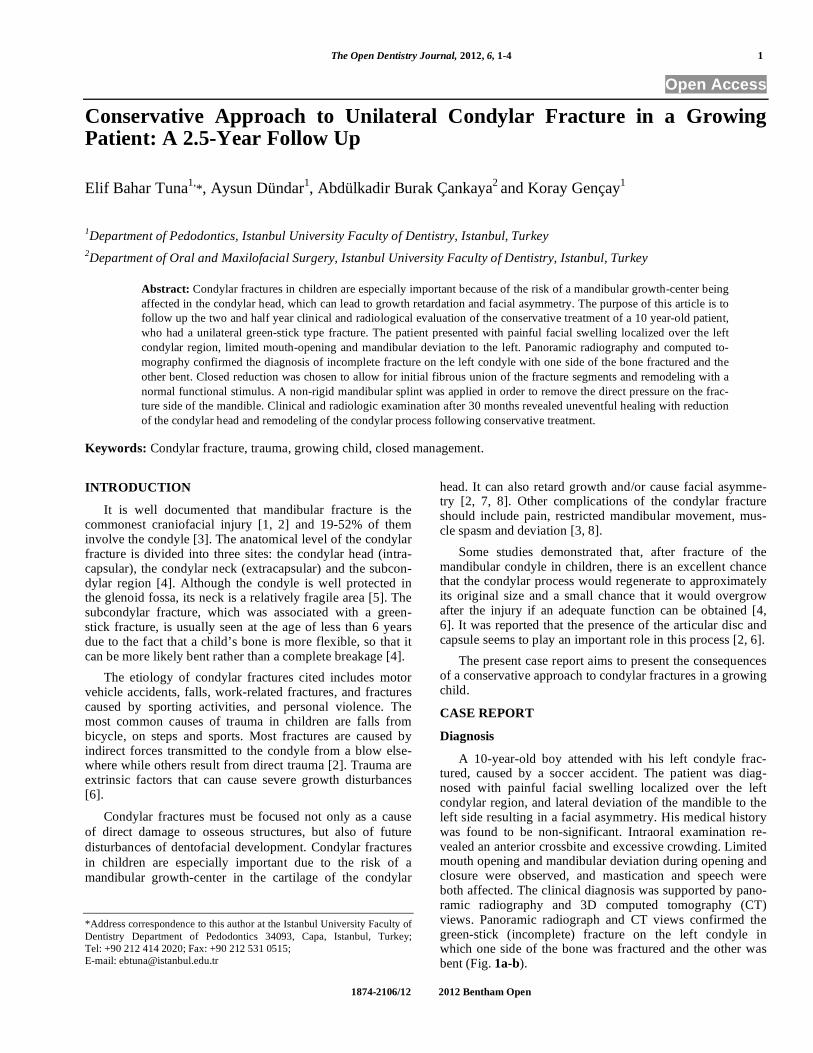
A 10-year-old boy attended with his left condyle frac-tured, caused by a soccer accident. The patient was diag-nosed with painful facial swelling localized over the left condylar region, and lateral deviation of the mandible to the left side resulting in a facial asymmetry. His medical history was found to be non-significant. Intraoral examination re-vealed an anterior crossbite and excessive crowding. Limited mouth opening and mandibular deviation during opening and closure were observed, and mastication and speech were both affected. The clinical diagnosis was supported by pano-ramic radiography and 3D computed tomography (CT) views. Panoramic radiograph and CT views confirmed the green-stick (incomplete) fracture on the left condyle in which one side of the bone was fractured and the other was bent (Fig. 1a-b).
2 The Open Dentistry Journal, 2012, Volume 6 Tuna et al.
Treatment
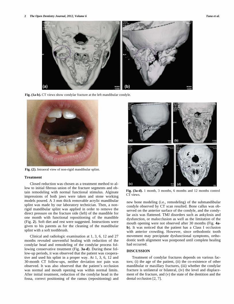
Closed reduction was chosen as a treatment method to al-low to initial fibrous union of the fracture segments and ob-tain remodeling with normal functional stimulus. Alginate impressions of both jaws were taken and stone working models poured. A 3 mm thick removable acrylic mandibular splint was made by our laboratory technician. Then, a non-rigid mandibular splint was applied in order to remove the direct pressure on the fracture side (left) of the mandible for one month with functional repositioning of the mandible (Fig. 2). Soft diet and rest were suggested. Instructions were given to his parents as for the cleaning of the mandibular splint with a soft toothbrush.
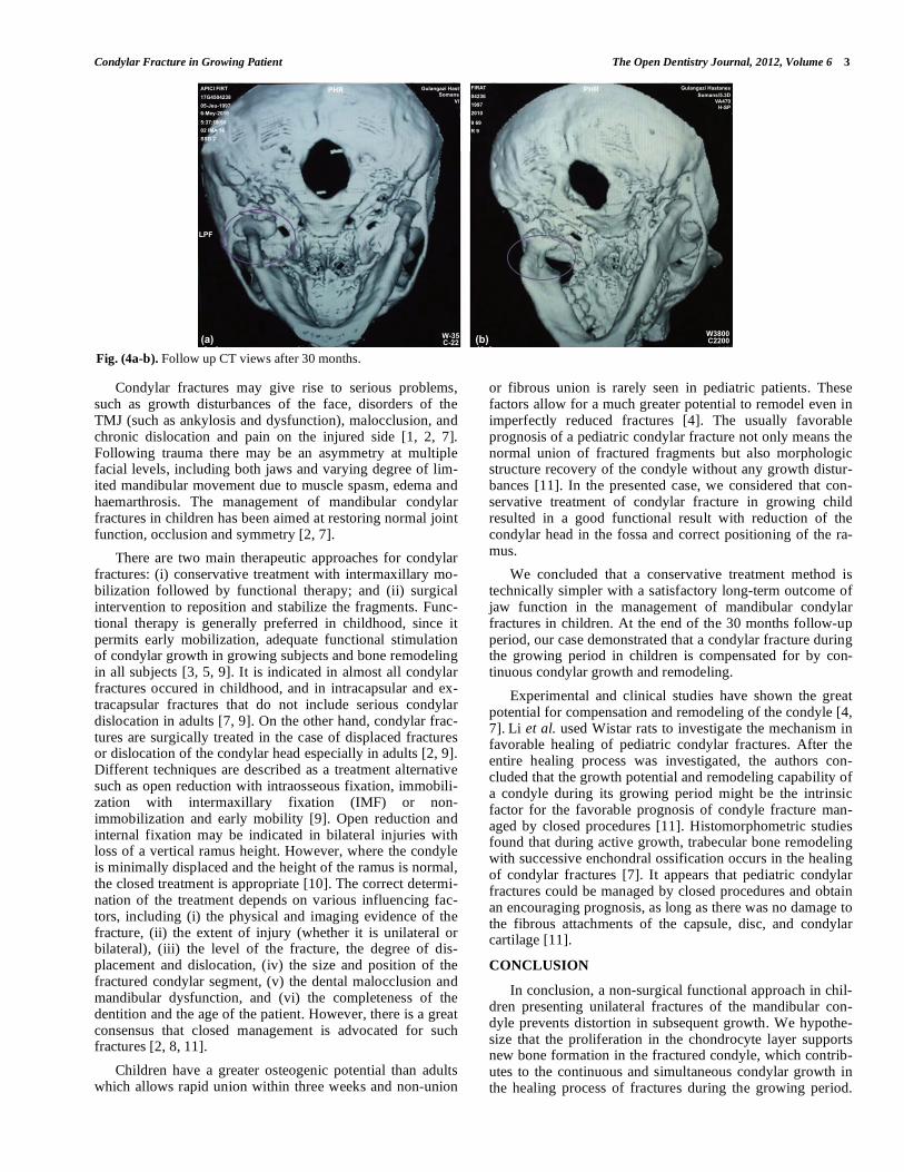
Clinical and radiologic examination at 1, 3, 6, 12 and 27 months revealed uneventful healing with reduction of the condylar head and remodeling of the condylar process fol-lowing conservative treatment (Fig. 3a-d). During these fol-low-up periods, it was observed that the patient was coopera-tive and used his splint in a proper way. At 1, 3, 6, 12 and 30-month CT follow-ups, neither deviation nor pain was observed. It was also observed that the patient’s occlusion was normal and mouth opening was within normal limits. After initial treatment, reduction of the condylar head in the fossa, correct positioning of the ramus (repositioning) and
new bone modeling (i.e., remodeling) of the submandibular condyle observed by CT scan resulted. Bone callus was ob-served on the anterior surface of the condyle, and the condy-lar axis was flattened. TMJ disorders such as ankylosis and dysfunction, or malocclusion as well as the limitation of the mouth opening were not observed after 30 months (Fig. 4a-
b). It was noticed that the patient has a Class I occlusion with anterior crowding. However, since orthodontic tooth movement may precipitate dysfunctional symptoms, ortho-dontic teeth alignment was postponed until complete healing had occurred.
DISCUSSION
Treatment of condylar fractures depends on various fac-tors; (i) the age of the patient, (ii) the co-existence of other mandibular or maxillary fractures, (iii) whether the condylar fracture is unilateral or bilateral, (iv) the level and displace-ment of the fracture, and (v) the state of the dentition and the dental occlusion [2, 7].
Fig. (2). Intraoral view of non-rigid mandibular splint.
Fig. (3a-d). 1 month, 3 months, 6 months and 12 months control
CT views.
Fig. (1a-b). CT views show condylar fracture at the left mandibular condyle.
����������������� ������� �������������������� ����
��� �������������������� ���!���� ��������������"#$�%����
&����'��(
)���*+�,��'����#��*�
(-
.�/ .�/0 ��%����
0 ��%����
��'
1�2�3�(4%4�������56����� ��� ��� ���&��� ���
)���*+�,��'����#��*�
�(' �('
7�2�2�(%2������������������������� �����!�� ������������
�#�8�
.�/ .�/0 �%���
0� ��%�����
.6/ .9/
�23�(4%4�����:�*��
%5� �"�����5��
)���*+�,��'����#��*�
;1�3�3�(%% �� ����������
���������%55�&�������
)���*+�,��'����#��*�
�('
�('
0� �%��� 0 �
%���
:�*���� ���������
(5����
Condylar Fracture in Growing Patient The Open Dentistry Journal, 2012, Volume 6 3
Condylar fractures may give rise to serious problems, such as growth disturbances of the face, disorders of the TMJ (such as ankylosis and dysfunction), malocclusion, and chronic dislocation and pain on the injured side [1, 2, 7]. Following trauma there may be an asymmetry at multiple facial levels, including both jaws and varying degree of lim-ited mandibular movement due to muscle spasm, edema and haemarthrosis. The management of mandibular condylar fractures in children has been aimed at restoring normal joint function, occlusion and symmetry [2, 7].
There are two main therapeutic approaches for condylar fractures: (i) conservative treatment with intermaxillary mo-bilization followed by functional therapy; and (ii) surgical intervention to reposition and stabilize the fragments. Func-tional therapy is generally preferred in childhood, since it permits early mobilization, adequate functional stimulation of condylar growth in growing subjects and bone remodeling in all subjects [3, 5, 9]. It is indicated in almost all condylar fractures occured in childhood, and in intracapsular and ex-tracapsular fractures that do not include serious condylar dislocation in adults [7, 9]. On the other hand, condylar frac-tures are surgically treated in the case of displaced fractures or dislocation of the condylar head especially in adults [2, 9]. Different techniques are described as a treatment alternative such as open reduction with intraosseous fixation, immobili-zation with intermaxillary fixation (IMF) or non-immobilization and early mobility [9]. Open reduction and internal fixation may be indicated in bilateral injuries with loss of a vertical ramus height. However, where the condyle is minimally displaced and the height of the ramus is normal, the closed treatment is appropriate [10]. The correct determi-nation of the treatment depends on various influencing fac-tors, including (i) the physical and imaging evidence of the fracture, (ii) the extent of injury (whether it is unilateral or bilateral), (iii) the level of the fracture, the degree of dis-placement and dislocation, (iv) the size and position of the fractured condylar segment, (v) the dental malocclusion and mandibular dysfunction, and (vi) the completeness of the dentition and the age of the patient. However, there is a great consensus that closed management is advocated for such fractures [2, 8, 11].
Children have a greater osteogenic potential than adults which allows rapid union within three weeks and non-union
or fibrous union is rarely seen in pediatric patients. These factors allow for a much greater potential to remodel even in imperfectly reduced fractures [4]. The usually favorable prognosis of a pediatric condylar fracture not only means the normal union of fractured fragments but also morphologic structure recovery of the condyle without any growth distur-bances [11]. In the presented case, we considered that con-servative treatment of condylar fracture in growing child resulted in a good functional result with reduction of the condylar head in the fossa and correct positioning of the ra-mus.
We concluded that a conservative treatment method is technically simpler with a satisfactory long-term outcome of jaw function in the management of mandibular condylar fractures in children. At the end of the 30 months follow-up period, our case demonstrated that a condylar fracture during the growing period in children is compensated for by con-tinuous condylar growth and remodeling.
Experimental and clinical studies have shown the great potential for compensation and remodeling of the condyle [4, 7].
Li et al. used Wistar rats to investigate the mechanism in
favorable healing of pediatric condylar fractures. After the entire healing process was investigated, the authors con-cluded that the growth potential and remodeling capability of a condyle during its growing period might be the intrinsic factor for the favorable prognosis of condyle fracture man-aged by closed procedures [11]. Histomorphometric studies found that during active growth, trabecular bone remodeling with successive enchondral ossification occurs in the healing of condylar fractures [7]. It appears that pediatric condylar fractures could be managed by closed procedures and obtain an encouraging prognosis, as long as there was no damage to the fibrous attachments of the capsule, disc, and condylar cartilage [11].
CONCLUSION
In conclusion, a non-surgical functional approach in chil-dren presenting unilateral fractures of the mandibular con-dyle prevents distortion in subsequent growth. We hypothe-size that the proliferation in the chondrocyte layer supports new bone formation in the fractured condyle, which contrib-utes to the continuous and simultaneous condylar growth in the healing process of fractures during the growing period.
Fig. (4a-b). Follow up CT views after 30 months.
�(4%4�-412
�)�����
���:��������<�����
�� ����
���4�����
�����
('1 )���*+�,��'����#��*�
&�
�(-
.�/ .�/0� �%���
0 ��%����
('1-41�2
��� �
�
����
��
1�
)���*+�,��'����*���#��*�!�� �
&���'��(
4 The Open Dentistry Journal, 2012, Volume 6 Tuna et al.
The results obtained in this case demonstrate that functional therapy resulted in remodeling with functional adaptation of the condyle to the fossa.
REFERENCES
[1] Silvennoinen U, Lindqvist C, Oikarinen K. Dental injuries in asso-ciation with mandibular condyle fracture. Endod Dent Traumatol
1993; 9: 254-9. [2] Zachariades N, Mezitis M, Mourouzis C, Papadakis D, Spanou A.
Fractures of the mandibular condyle: a review of 466 cases. Litera-ture review, reflections on treatment and proposals. J Craniomaxil-
lofac Surg 2006; 34: 421-32. [3] Valiati R, Ibrahim D, Abreu ME, et al. The treatment of condylar
fractures: to open or not to open? A critical review of this contro-versy. Int J Med Sci 2008; 5: 313-8.
[4] Kalia V, Singh AP. Greenstick fracture of the mandible: A case report. J Indian Soc Pedod Prev Dent 2008; 26: 32-5.
[5] Proffit WR. Contemporary orthodontics. 2nd ed. St Louis: Mosby, Mosby Year Book 1993.
[6] Giannı` E, Bruno E, Farronato G, Giannı` AB. The temporoman-
dibular joint: diagnosis-functional aspects. Front Oral Physiol 1990; 7: 38-45.
[7] Farronato G, Grillo ME, Giannini L, et al. Long-term results of early condylar fracture correction: case report. Dent Traumatol
2009; 25: e37-42. [8] Hackett JF, Sleeman DJ. Vertical-split fracture of mandibular con-
dyle and its sequelae. Br Dent J 2001; 191: 557-8. [9] De Riu G, Gamba U, Anghinoni M, Sesenna E. A comparison of
open and closed treatment of condylar fractures: a change in phi-losophy. Int J Oral Maxillofac Surg 2001; 30: 384-9.
[10] Lloyd T, Nightingale C, Edler R. The use of vacuum-formed splints for temporary intermaxillary fixation in the management of
unilateral condylar fractures. Br J Oral Maxillofac Surg 2001; 39: 301-3.
[11] Li Z, Zhang W, Li ZB, Li JR. Mechanism in favorable prognosis of pediatric condylar fractures managed by closed procedures: an ex-
perimental study in growing rats. Dent Traumatol 2010; 26(3): 228-35.
Received: September 28, 2011 Revised: November 11, 2011 Accepted: November 22, 2011
© Tuna et al.; Licensee Bentham Open.
This is an open access article licensed under the terms of the Creative Commons Attribution Non-Commercial License
(http://creativecommons.org/licenses/by-nc/3.0/) which permits unrestricted, non-commercial use, distribution and reproduction in any medium, provided the
work is properly cited.